14 minute read
Evaluation of Pre-Treatment and Post-Treatment Changes in Lower Anterior Facial Height in Extraction and Non-Extraction Cases: A Digital Cephalometric Study


by

Advertisement
Abstract:
Objective
The purpose of this present study was to evaluate pretreatment and post-treatment cephalometric changes in the lower anterior facial height (LAFH) in class I all first premolar extraction cases and compare them with class I non-extraction cases.


Materials and Methods:
Pre-treatment and post-treatment cephalometric radiographs of 40 Class I normo-divergent patientswere selected and divided into two groups i.e extraction and non-extraction groups. The extraction group included 20 patients (12 females and 08 males; pre-treatment age, 17.4 +/- 3.89 years, treatment duration 24 +/5 months) and non-extraction group also included 20 patients (11 females and 09 males; pre-treatment age, 18.3+/- 3.61 years, treatment duration 19 +/- 5 months). The linear and angular measurements were selected and measured with the help of NEMOCEPH software to see the changes in LAFH.
Result
The results obtained revealed that there was a significant increase in LAFH in non-extraction group and no significant
*This article has been peer reviewed change in LAFH in extraction group, although there was no significant difference in facial height when both the groups were compared.
Conclusion
Thus it can be concluded that both treatment strategies when used judiciously with good anchorage and vertical control do not lead to any significant increase in vertical facial dimension.
Keywords
Lower anterior facial height, NEMOCEPH, Normo-divergent, Extraction, Cephalometric radiographs.
Introduction
Extraction of teeth has always been a controversial topic right from the angle era to modern times. Among the various reasons for the removal of permanent teeth, the two major reasons are to reduce bi-maxillary protrusion and for the correction of tooth size/arch length discrepancy. Apart from these two reasons, a third and underappreciated reason for permanent tooth extraction is in order to control the vertical dimension.1
The vertical disturbances can be of two types. One is the anterior teeth fail to contact each other, or open bite; the other condition in there is an excessive vertical overlap of the anterior teeth, or deep bite.2 In order to correct the above-mentioned vertical disturbances, be it open bite or deep bite, it is suggested by several authors that movement of the posterior teeth can be used to treat vertical disturbances. The rationale for such treatment is often based on the “occlusal wedge hypothesis.”
According to this hypothesis, the dentoalveolar apparatus can be presumed to be an occlusal wedge, so when bicuspids and molars are distalized or extruded, there is an opening of bite and, on the contrary, when molars are mesialized after extraction of bicuspids, there is deepening of bite. This hypothesis seems to be logical from a biomechanical point of view but orthodontic treatment is performed in an oral biological environment, which has its own complexities. So, it doesn’t come with a surprise that there is a difference in opinion among various authors when it comes to the validity of this hypothesis.
One group of authors suggest that extraction and subsequent protraction in order to close space causes anti-clockwise rotation of mandible leading to correction of open bite and a resulting decrease in vertical facial height, more specifically, the lower anterior facial height (LAFH).3,4,5 On the other hand, another group states that there is no change in the LAFH and on the contrary may sometimes cause temporomandibular joint (TMJ) problems due to overclosure of the mandible.6-12
The LAFH is the vertical distance between the anterior nasal spine (ANS) and the menton (Me) points. The two main factors which can affect its height are the increase or decrease in the bone deposition in the baso-alveolar bone through the process of remodelling and other being the variation in the eruption of teeth. It has been proposed by some authors that orthodontic treatment causes an alteration in the lower facial height by extraction or non-extraction treatment philosophy, on the contrary other authors claim that orthodontic treatment doesn’t cause any change in LAFH. Thus, in order to accurately evaluate vertical problems involving skeletal and dental components cephalometric assessment acts as an important tool.2
Standardized cephalometric radiographs help us to diagnose and record pre-treatment skeletal and dental relationships and compare them with post treatment radiographs and evaluate the changes due to orthodontic treatment.13,14 Although this method of cephalometric tracing is widely used, it has several drawbacks ranging from being time consuming to increased chances of errors in landmark identification, tracing, and measurement. Thus, with the advent of computers in orthodontics many of the problems encountered by the traditional methods have been solved as digital cephalometric tracing reduces the frequency of inaccuracies due to operator fatigue, provides standardized, fast, and effective evaluation of lateral cephalograms. There are several advantages of using digital cephalometric software notably - several analyses can be performed simultaneously, helps in generating treatment predictions, allows superimposition of images, digital record keeping which overcomes the problem of film deterioration and lastly it is easy to use.15
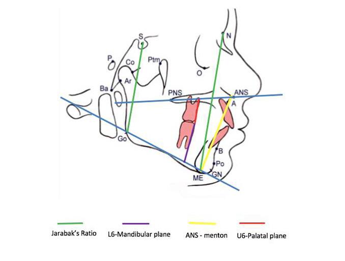
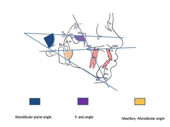
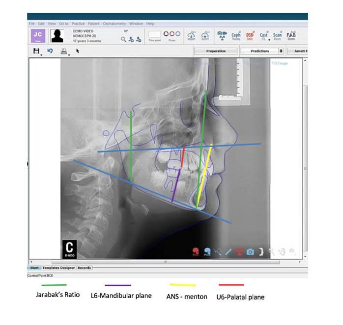
The linear parameters are much more affected by magnification error in cephalogram in comparison to angular parameters like y-axis angle, MM angle, MP angle (Fig 1); that’s why we use angular parameters along with linear parameter (ANS – Me) and U6-PP,
L6-MP to assess changes in pre-treatment and posttreatment LAFH (Fig 2).
Hence, the purpose of this present study was to evaluate pre-treatment and post-treatment cephalometric changes in the lower anterior facial height in class I all first premolar extraction cases and compare them with class I non – extraction cases.
Material and Methods
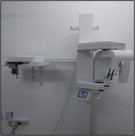
Materials (Fig 3,4)
1. Cephalostat (PlanmecaProline XC)
2. NEMOCEPH ORTHODONTIC
Software
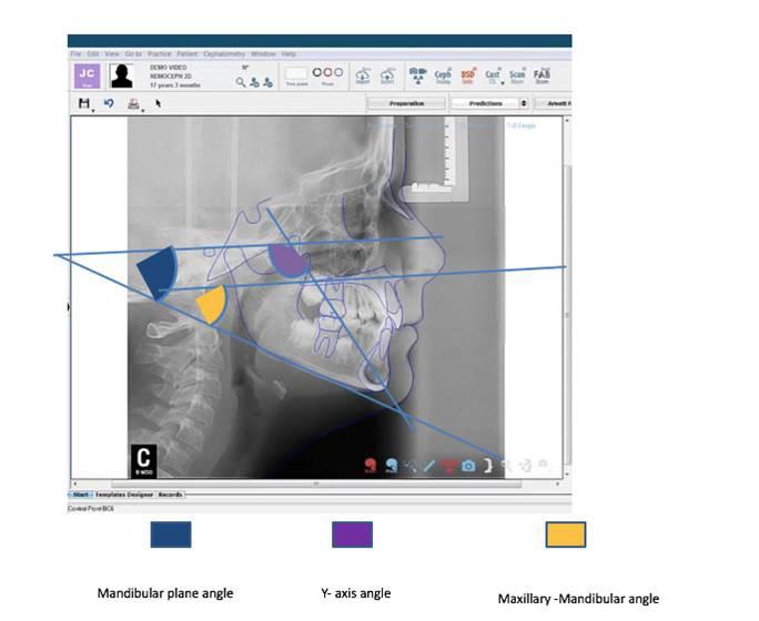
In this retrospective study, pre-treatment and post-treatment cephalometric radiographs of 40 Class I normo-divergent patientswere selected from the available records of 84 patients which met the selection criteria. Master 2.0 software was used for sample size calculation and the power of the study was taken to be 80% and Confidence Interval (C.I.) was taken to be 95%. The sample size calculation was done as per the article by Hans et al.1 The sample size was estimated to be a minimum of 20 patients. The obtained records were divided into two groups (i.e., extraction and non-extraction groups). The extraction group included 20 patients (12 females and 8 males; pre-treatment age, 17.4 +/- 3.89 years, treatment duration 24 +/- 5 months) and nonextraction group also included 20 patients (11 females and 9 males; pre-treatment age, 18.3+/- 3.61 years, treatment duration 19 +/- 5 months with minimal crowding). All subjects were treated by using Damon
Q metal with banding till second molars in order to increase anchorage along with the use of transpalatal arch and consolidation of posterior segments.In all the extraction cases, Class I (intra-maxillary force) force with the help of Class I elastics was used to close the extraction spaces. After the initial selection, all X-rays were traced by the same investigator and landmarks were identified & all cephalometric measurements(three angular and four linear) used in this study; Y-axis angle, MM angle, MP angle, ANS – Me, U6-PP, L6-MP and Jarabak’s ratio were made with the NEMOCEPH software on a computer and the mean values of those measurements were calculated and statistically compared with post treatment values (Fig 5,6).16 In order to rule out bias, intra class correlation coefficient has been done for both groups that were traced at the same time by the same operator and pre-treatment and post-treatment cephalograms of 10 randomly selected patients were traced by the same operator to eliminate memory bias within a duration of a month.
Selection criteria
Inclusion criteria:
1. Patient with age between 15 to 25 years.
2. The subjects with full complement of teeth up to first molars in maxillary and mandibular arches without any craniofacial abnormality.
3. Class I normo-divergent malocclusion patients (SN-GoGn, 32°+/-1°).
4. Do not have severe antero-posterior discrepancy (0° <ANB <5°)
5. Vertical discrepancy (0 mm <overbite < 6 mm)
Exclusion criteria:
Patients were excluded from the study if they presented with
1. Previous history of orthodontic treatment.
2. Congenitally missing teeth/supernumerary teeth.
3. Patient with history of trauma.
4. Facial asymmetry.
5. Patients with craniofacial syndromes.
6. Patients treated with fixed functional appliances.
7. Patients treated with myofunctional appliances.
8. Patients treated with head gear.
9. Use of Temporary anchorage devices.
Results
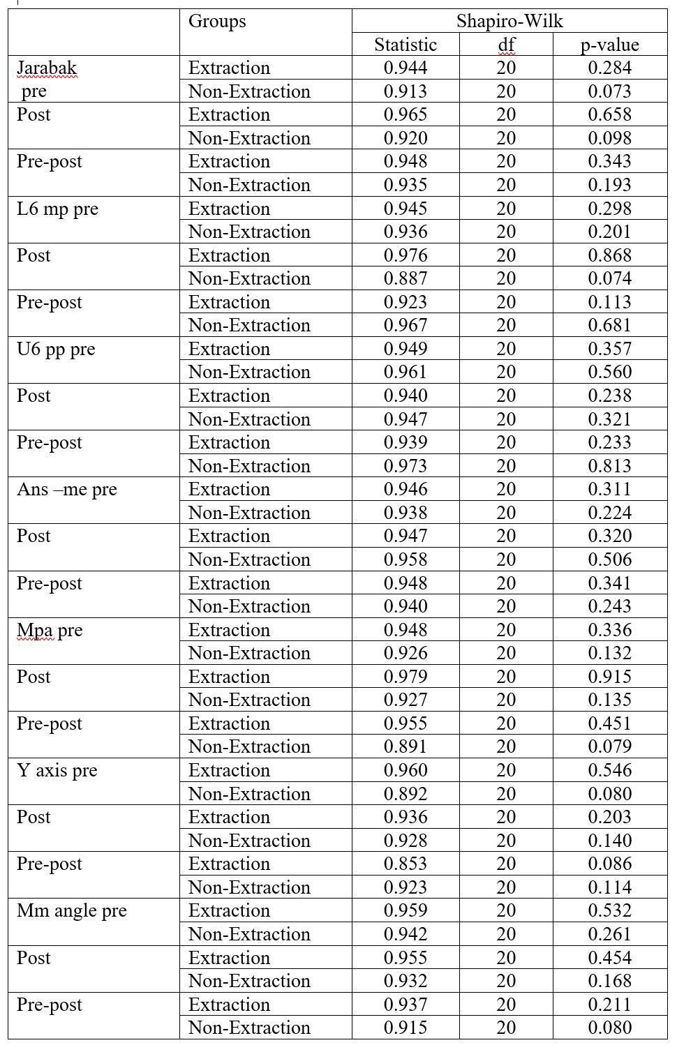
There was a normal distribution of all the variables for both Extraction and Non-Extraction groups in the study (Table 1).
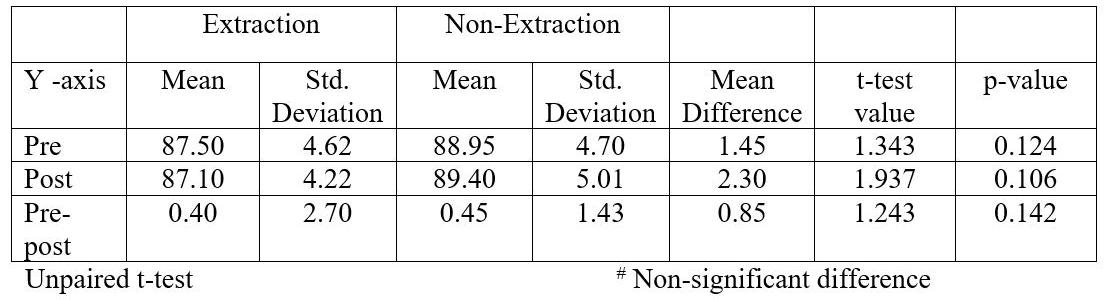
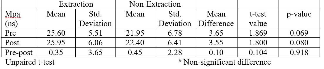
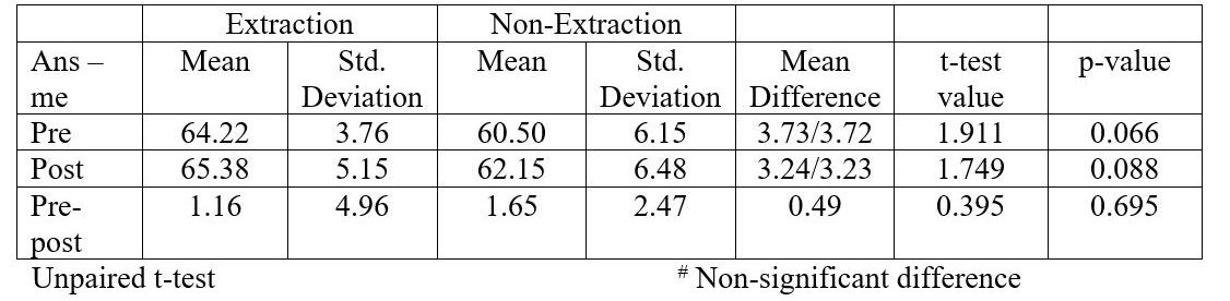
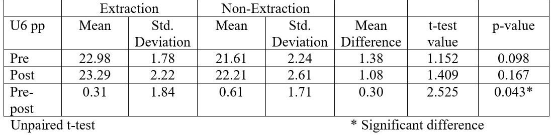
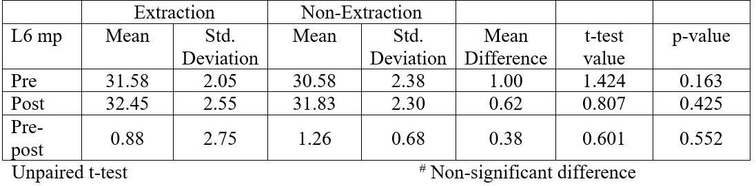
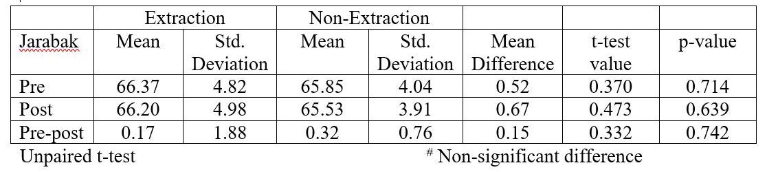
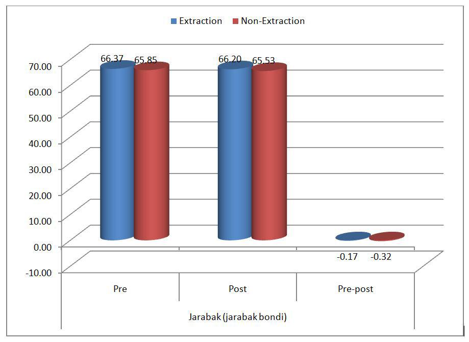
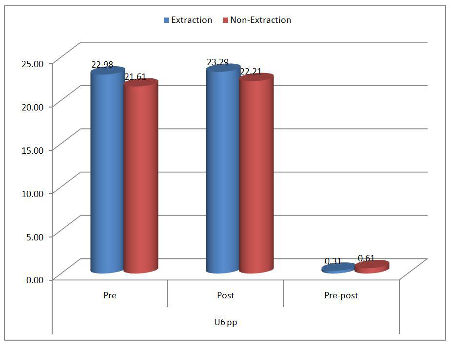
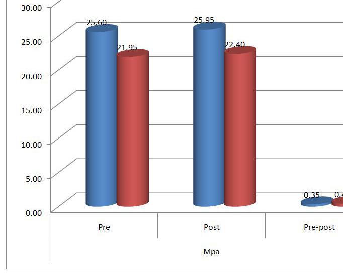
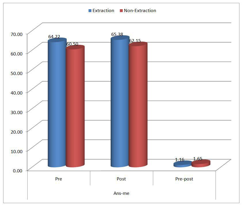
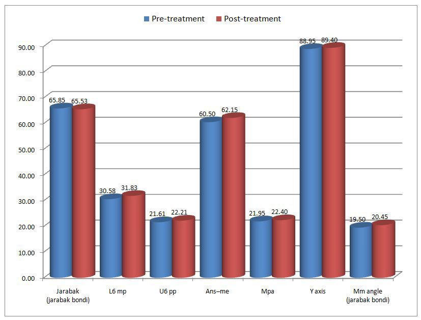
The mean of pre-treatment, post-treatment and difference form pre to post treatment was compared between extraction and non-extraction cases using the unpaired t-test for all the parameters i.e: Jarabak ratio, L6-mp, U6-pp, Ans –me, Mpa, Y-axis and Mm angle. (Table 2-8 and figure 7-13 ).There was no significant difference in any of the parameters, except the mean difference from Pre to post treatment was found to be significantly more among NonExtraction group (Table 4 and figure 9).
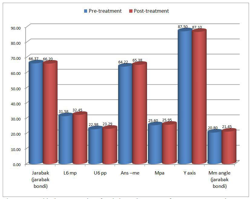
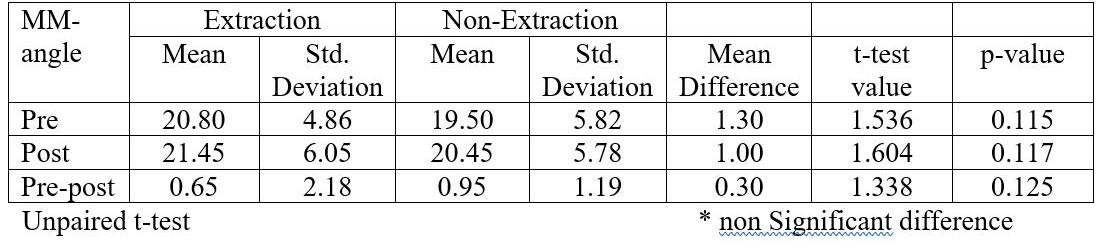
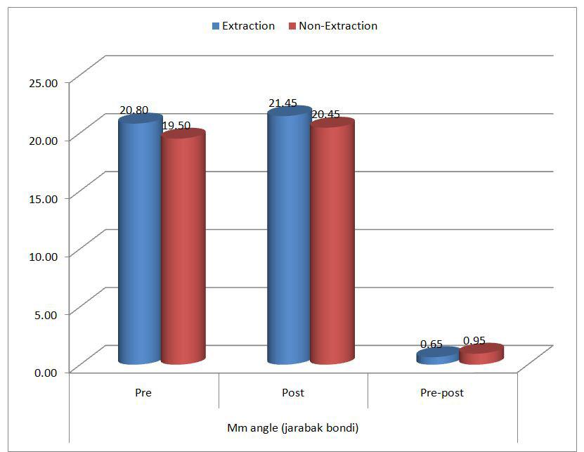
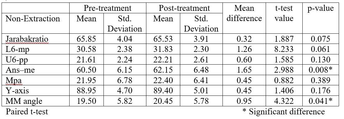
The mean values of all the parameters were compared between pre and post-treatment using the paired t-test in both extraction and non-extraction groups. No significant difference was found in both the groups except the Ans–me and MM angle values which increased significantly from pre to posttreatment in the non- extraction group (Table 9-10 and figure 14-15).
Discussion
There has always been a lot of debate regarding the role of premolar extraction in change in the facial height. The purpose of this study was to investigate the effect of extraction of first premolars on LAFH as there is a lot of controversy when it comes to the role of premolar extraction and change in vertical height. There is a general consensus among orthodontists that pre molar extraction according to the occlusal wedge hypothesis causes an anti-clockwise rotation of the mandible and hence reduces the lower anterior facial height whereas a non-extraction treatment protocol on the contrary causes a clockwise rotation of the mandible and as a result increase in LAFH .The results obtained in this study tend to differ from the above mentioned effect of premolar extraction on facial height as no reduction in LAFH was to be found in the extraction group but there was a significant increase in LAFH in the nonextraction treatment group.
Chua et al devised a study to see whether is there any effect of the type of treatment plan on the patient’s LAFH and for this 174 Class I and II patients were selected who were further divided into extraction and non-extraction patient groups. The outcome of the study came out that there was no notable change in LAFH in extraction group but on the contrary there was notable increase in LAFH in non-extraction group which corroborates with our findings.17
Kocadereli et al devised a study to see whether the extraction of 1st bicuspids had any effect on the facial height. For this, two groups with forty patients each were created, one with 1st bicuspids extraction line of treatment and second with non-extraction and their cephalometric readings were recorded and compared. The results indicated that there was no significant difference in vertical height between extraction and non-extraction cases.6 Staggers et al tested the hypothesis that removal of first bicuspids may result in a decrease of LAFH for which pretreatment and post-treatment cephalographs of forty five Class I patients who were treated by nonextraction treatment plan were compared with thirty eight Class I, first bicuspids removal cases. The cephalogram were digitized and angular along with linear cephalometric parameters were evaluated and results obtained stated that there was a significant increase in LAFH in both the groups.18
Kim T.K et al tested the occlusal wedge hypothesis which states that extraction of second bicuspids and mesial movement of molars leads to decrease in facial height in Class I malocclusion and hypo-divergent facial type. The results obtained revealed that there was no change in vertical height in either of the groups and hence declined the validity of the occlusal wedge hypothesis.7 Hayasaki et al evaluated the changes in facial height in class II and class I malocclusion and found that Jaraback’s ratio between extraction and non-extraction treatment in both Class I and Class II patients was similar and no significant changes between the groups were found.19
Al nimri studied the effects of mandibular first and second premolar extraction cases on vertical facial height and concluded that although there was a mesialization of molar in the second premolar extraction group there was no significant change in the MM angle and facial height in any of the group.20 Similarly, Hans et al devised a study see whether there was any change in LAFH after extraction of four fist molars and compared them with all four first premolar cases and found that no significant cephalometric changes were found in either of the group.21
All of the above mentioned studies indicate similar results that premolar extraction does not have a significant impact on the vertical facial dimensions. This can be explained by the fact that most of the extractions done in class I malocclusion are done to relieve crowding and in bi-max cases rest of the space is utilised for the retraction of anterior teeth and during this retraction if proper anchorage is maintained, there is no or very little mesialization of molars which do not produce any significant change in facial height. In the present study, all the cases selected were skeletally and dentally class I and the extraction spaces were used to relieve crowding and retraction of anterior teeth along with anchorage preservation and vertical position maintenance of posterior teeth which led to insignificant change in facial height which was confirmed by no significant changes in angular and linear cephalometric values (table 8).
Another important factor to take into account is seeing the effect the premolar extraction on facial height is growth. Kim et al and Harris et al advocated that change in facial height was due to the presence of residual growth in late teens.7,22 In another study done by Taner-Sarisoy it was put forward that there was no change in the LAFH after extraction of premolars as any mesialization and reduction in facial height by the wedging effect was compensated by the growth potential in the patient. The above mentioned reason doesn’t holds true in our study as the patients selected in our study were well beyond their growth potential with cervical vertebrae in stage 5 (Hassel and Farman) and hence disapproves the theory of wedging effect as there was no significant change in LAFH after extraction of premolars.23
Aras et al evaluated the changes that occurred in patients with open bite after orthodontic treatment and concluded that extraction of second bicuspids and first molars led to an anti-clockwise or closing rotation in AOB patients.3 Beit P et al evaluated the changes in LAFH in extraction of all 4 premolar extraction cases and compared them with non extraction cases and found that LAFH increased in non extraction cases but decreased in extraction group which may be due to difference in characteristics between the study sample, hence direct comparisons between these studies is impossible.5 Similar results were obtained when a systematic review was carried out by Kouvelis et al to see the effects of all 4 first premolar extraction on vertical height of face and was concluded that there was no evidence to claim that extraction of premolar had any effect on the facial height.24 In a meta-analysis by Jain et al to evaluate LAFH in extraction v/s non –extraction cases it was found that there is no statistically significant effect of extraction of four first premolars on lower anterior facial height.25
In the present study digital Cephalometry software, NEMOCEPH, was used to avoid the disadvantages associated with conventional methods namely being more time consuming, magnification error to high-risk of errors in tracing, landmark identification, reproducibility and measurement. Moreover, angular parameters were included in addition to linear parameters to measure the changes the LAFH.16
The results of our study revealed that extraction of maxillary and mandibular first premolars in class I patients did not have any significant change in the facial height which is in accordance to several studies mentioned above.
Conclusions
The purpose of this study was to test the hypothesis that extraction of premolars leads to reduction in LAFH due to “wedging effect” which states that in extraction cases there is mesialization of molars which leads to anti-clockwise rotation of mandible and consequently a reduction in lower anterior facial height. Thus, the following conclusion can be drawn from this study.
1. There is no significant change in LAFH when all four first premolar extraction protocol was followed, hence disapproving the occlusal wedge hypothesis.
2. No significant change in vertical facial height was seen when all four first premolar extraction cases were compared with non-extraction group, concluding that the type of protocol used had a little impact on LAFH.
Data Availability
References
1. Hans MG, Kishiyama C, Parker S, Wolf GR, Noachtar R. Cephalometric evaluation of two treatment strategies for deep overbite correction. Angle Orthod. 1994;64:265-76.
2. Al–Zubaidi SA, Obaidi HA. The variation of the lower anterior facial height and its component parameters among the three over bite relationships (Cephalometric study). Al–Rafidain Dent J. 2006; 6(2):106-113.
3. Aras A. vertical changes following orthodontic extraction treatment in skeletal open bite subjects. European journal of orthodontics 24(2002) 407-416.
4. Ramesh GC, Pradeep MC, Kumar GA, Girish KS, Suresh BS. Over-bite and vertical changes following first premolar extraction in high angle cases. J Contemp Dent Pract. 2012;13:812–8.
5. Beit P, Konstantonis D, Papagiannis A, Eliades T. Vertical skeletal changes after extraction and non-extraction treatment in matched class I patients identified by a discriminant analysis: cephalometric appraisal and Procrustes superimposition. Progress in Orthodontics. 2017;18(44):1-10.
6. Kocadereli I. The effect of first premolar extraction on vertical dimension. Am J OrthodDentofacialOrthop1999;116:41-5.
7. Kim TK, Kim JT, Mah J, Yang WS, Baek SH. First or second premolar extraction effects on facial vertical dimension. Angle Orthod. 2005; 75:177-82.
8. Tulley WJ. The role of extractions in orthodontic treatment. Br Dent J 1959;107:199- 205.
9. Wyatt NE. Preventing adverse effects on the temporomandibular joint through ortho- dontic treatment. Am J Orthod. 1987;91:4939.
10. Bowbeer GR. The sixth key to facial beauty and TMJ health. Funct Orthod. 1987;4:4-22.
11. Witzig JW, Spahl TJ. The clinical management of basic maxillofacial orthopedic appliances. Littleton (MA): PSG Publishing; 1987. p. 161-216.
12. Farrar WB, Mc Carty WL. A clinical outline of temporomandibular joint diagnosis and treatment. Montgomery (AL): Walker; 1983. p. 84-5.
13. Proffit W.R., Field H.W., Ackerman J.L., Bailey L.T., Tulloch J.F.C. Contemporary orthodontics 4th, C.V.Mosby Co; 2000.
14. Graber TM, Vanarsdall R. Orthodontics. Current principles and techniques 4thEd.St.Louis. Mosby yearbook; 1994.
15. Silva MBG, Sant’Anna EF. The evolution of cephalomet- ric diagnosis in Orthodontics. Dental Press J Orthod. 2013;18(3):63-71.
16. Tikku T, Khanna R, Maurya RP, Srivastava K, Bhushan R. Comparative evaluation of cephalometric measurements of monitor-displayed images by Nemoceph software and its hard copy by manual tracing. Journal of Oral Biology and Craniofacial Research. 2014;4(1):35–41.
17. Chua AL, Lim JY, Lubit EC. The effects of extraction versus nonextraction orthodontic treatment on the growth of lower anterior face height. Am J Orthod DentofacialOrthop. 1993;104:361-8.
18. Staggers JA. Vertical changes following first premolar extractions.
Am J Orthod DentofacialOrthop. 1994;105:19-24.
19. Hayasaki SM, CastanhaHenriques JF, Janson G, de Freitas MR. Influence of extraction and non-extraction orthodontic treatment in Japanese-Brazilians with class I and class II division 1 malocclusions. Am J OrthodDentofacialOrthop. 2005;127:30-6.
20. Al-Nimri KS. Vertical changes in Class II division 1 malocclu- sion after premolar extractions. Angle Orthod. 2006;76:52-8.
21. Hans MG, Groisser G, Damon C, Amberman D, Nelson S, Palomo JM. Cephalometric changes in overbite and vertical facial height after removal of 4 first molars or first premolars. Am J Or- thod Dentofacial Orthop 2006;130:183-8.
22. Harris Eh, Gardner RZ, Vaden JL. A longitudinal cephalometric study of postorthodontic craniofacial changes. Am J Orthod DentofacialOrthop. 1999;115:77-82.
23. Taner-Sarisoy L, Darendeliler N. The influence of extraction orthodontic treatment on craniofacial structures: evaluation ac- cording to two different factors. Am J OrthodDentofacialOrthop1999;115:508-14.
24. Kouvelis G, Dritsas K, Doulis I, Kloukos D, Gkantidis N. Effect of orthodontic treatment with 4 premolar extractions compared with nonextraction treatment on the vertical dimension of the face: A systematic review. Am J Orthod Dentofacial Orthop. 2018;154(2):175-187.
25. Jain AD, Goyal M, Kumar M, Premsagar S, Mishra S,Tomar S.Evaluating the Lower Anterior Facial Height in Patients Treated with Extraction Versus Non-extraction Fixed Mechanotherapy: “A Systematic Review and Meta-analysis”. J Indian Orthod Soc. 2021; Article in press: 1–12.


