Illinois Tech Research 2022 ENGINEERING
No Cancer Left Behind
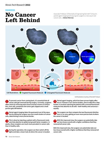
Associate Professor of Biomedical Engineering Kenneth Tichauer’s paired-agent imaging technique gives surgeons a new way to see cancer cells. —Simon Morrow
7
1
2
6
Targeted
Healthy Cell
4
Cancer Cell
Untargeted
Paired-agent imaging
Healthy Cell
Cancer Cell
5
3
Blood Plasma
Cell Illustrations:
Targeted Fluorescent Molecule
Blood Plasma
Untargeted Fluorescent Molecule Cell Illustrations Courtesy of Kenneth Tichauer
To prevent cancer from coming back, it’s essential that all cancer cells get removed during surgery. Currently, surgeons often resort to removing extra tissue around a tumor to achieve this. Even still, sometimes the tumor gets clipped and a small portion gets left behind.
Paired-agent imaging, which has been proven highly effective in Tichauer’s non-clinical studies, aims to take this a step further: It involves injecting the patient with a second fluorescent molecule that attaches to all cells—both healthy and cancerous— equally.
Paired-agent imaging takes the guesswork out of the equation by letting the surgeon see even small clusters of cancer cells, determining a more precise border.
The surgeon can then compare the two fluorescent distributions in an area, getting an even more precise look at where the cancer is located.
This is done by injecting a patient with a fluorescent molecule that attaches to epidermal growth factor receptors. In adults, these receptors are much more prevalent in cancer cells than in healthy cells.
With this improved view, the surgeon can potentially take out less tissue overall with a higher confidence that they removed all cancer cells.
1
2
3
4 12
During the operation, the surgeon can then switch off the lights and take a closer look at where the cancer is located. Illinois Tech Research
5
6
7
With this improved view, the surgeon can potentially take out less tissue overall with a higher confidence that they removed all cancer cells.