1 minute read
International Journal for Research in Applied Science & Engineering Technology (IJRASET)


ISSN: 2321-9653; IC Value: 45.98; SJ Impact Factor: 7.538
Advertisement
Volume 11 Issue II Feb 2023- Available at www.ijraset.com
J. Model Preparation And Training
The structure of deep learning-based network is getting deeper as it is evolving[13] Once all the data are prepared , the modelswere prepared before training the data. The architectures that we are using are VGG16 and VGG19. A fully convolutional network has the capability of handling input images of any dimension and can use the de-convolution layer for interpolating the featured map of the previous layer to retrieve it to the same dimension as that of the given image, which permits calculationsat the pixellevel.[14] All these architectures are selected to figure out the best performing architecture that can classify the tumor efficiently.With all the models, we used the transfer learning technique by initializing the weights from models trainedon the ImageNet dataset. By using the transfer learning, we canavoid overfitting the model and achieve faster convergence on the training data. Afterward the models were trained on the training data.
K. Classification And Evaluation

After training, the model testing phase started in order to identify the accuracy of the learning. After sorting the entiredata, we apply various CNN architecture in the testing phase. We have applied two classifications for the testing phase. After training the accuracy level that we got have analyzed result has been visualized in graphs.
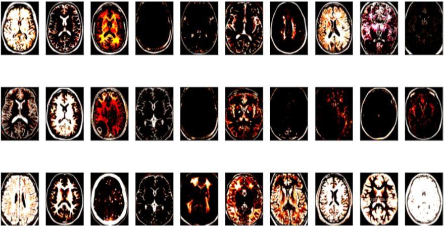
IV. IMPLEMENTATION RESULTS
To test the performance of the proposed algorithm we have tested it on 3200 MRI images, out of which 1835 images have brain syndrome related issues and 1365 do not have tumor. Out of those images which we selected 20 images for testing. Initially, different works on brain tumour classification were reviewed for choosing hyperparameter values [15] The findings of the suggested image segmentation in this methodology, which were achieved using real brain MR data, are presented in this section. Jupyter Notebook of the Anaconda software was used to test the suggested approach First the images were reshaped into 244×244 pixel size and passed through a convolutional layer with three filters. Subsequently, the output passes through maxpool and flattening layers. The model has been trained with 30 epochs in this process. Our model has shown an average accuracy of 92.34 from this model accuracy curve for training and testing has been shown for 30 epochs.