
1 minute read
International Journal for Research in Applied Science & Engineering Technology (IJRASET)


ISSN: 2321-9653; IC Value: 45.98; SJ Impact Factor: 7.538
Advertisement
Volume 11 Issue II Feb 2023- Available at www.ijraset.com
Image pre-processing involves steps such as creating functions to load image datasets into arrays, resizing raw images to an established base size before feeding it to the neural network, applying normalization to rescale the pixel values so they lie within a fixed range, data augmentation to increase the size of the dataset if insufficient number of images are avail- able, among other steps .These preprocessing tasks help improve classification accuracy and alsospeed training processes.
C. Data Segmentation
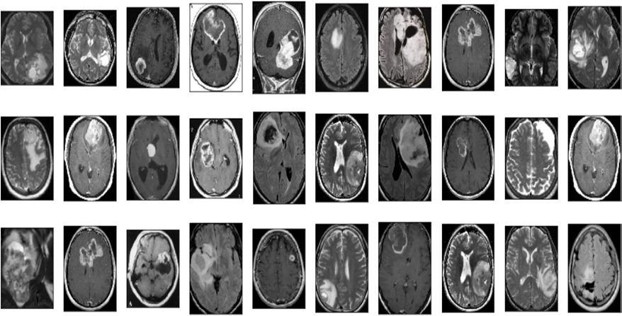
Image segmentation can be partitioned from multiple objects/segments to a single object. It performs labeling of pixel- level for all image pixels which predicts a single label for the whole image.[10] This step aims to differentiate abnormal braintissue from the normal brain tissue. There are manual, semi-automatic and fully automatic segmentation techniques . In manual segmentation, the outline of the affected tissue area is manually traced. This method has the highest accuracy, however, it is time-consuming and cumbersome. Semi-automatic segmentation involves the users inputting some initial data to obtain the final results. In fully automatic methods, the values of the parameters do not have to be set manually and these methods can automatically detect and segment the brain tumor.

D. Data Processing
All the data we got so far from binary classification have been proposed, applying some factors these are data annotation,slice extraction, data normalization, train test split.


