
ISSN: 2321 9653; IC Value: 45.98; SJ Impact Factor: 7.538

Volume 10 Issue X Oct 2022 Available at www.ijraset.com


ISSN: 2321 9653; IC Value: 45.98; SJ Impact Factor: 7.538

Volume 10 Issue X Oct 2022 Available at www.ijraset.com
1
Abstract: Brain tumors being one of the most fatal illnesses for people across all ages, they make up to 90% of tumors that principally affect the central nervous system. To ensure recovery and highest possible life expectancy for the patient doctors must ensure precise diagnosis and properly plan out treatment courses. Manual screening of tumors of the brain is highly complex. Having a tendency to being misdiagnosed owing to Byzantine structure of the brain and varying characteristics of tumors depending on their location and other factors. With the rise of computer sciences, there are automatic detection and classification solutions available that can outperform manual diagnosis and classification in terms of accuracy using machine learning techniques, Medical Professionals around the globe could use such systems. Comprehensive experiments were conducted by us on the selected datasets and it’s shown that the proposed model is in a position to realize competitive results.
Keywords: Magnetic Resonance Image(MRI), Brain tumor, Machine Learning, CNN, Deep Learning, etc.

In order to get a view of the interior of the human body, we use medical imaging technology in which diagnosis and classification of tumors or cancer is the most challenging task. The statistics indicate that brain tumors are one the crucial types of cancer when it comes to lethality. International Agency for Research on Cancer (IARC) reports that greater than 1 million people worldwide are being diagnosed with brain tumors annually and the mortality rate is continuously increasing year on year, with brain tumors being second foremost cause of deaths related to carcinomas for people younger than 34. Hitherto, doctors have been implementing more advanced methods for identifying more painful tumors in patients. Using CT (Computed Tomography) scans and MRIs (Medical Inference Imaging), doctors are now able to anatomize abnormalities in various parts of the body. Using MRI scans for studying brain tumors has recently picked up pace due increase in demand for laborsaving and clinical assessment of extensive amount of medical data. Such type of image processing and analysis involves complex computation of data and visualization of this data to get an understanding of the same. The source of brain tumors can be traced back to aberrant cells forming in the brain, some of which are precancerous, some of which are cancerous or damaging, and others which are innocuous or noncancerous. Malignant tumors are classified into two types: those that begin in the brain and those that are subsequent cancerous growths that can spread to different areas of the human body; in such circumstances, the carcinoma is said to have metastasized in the patient and is most lethal, with a very poor survival rate.
The source of brain tumors can be traced back to aberrant cells forming in the brain, some of which are precancerous, some of which are cancerous or damaging, and others which are innocuous or noncancerous. Malignant tumors are classified into two types: those that begin in the brain and those that are subsequent cancerous growths that can spread to different areas of the human body; in such circumstances, the carcinoma is said to have metastasized in the patient and is most lethal, with a very poor survival rate.
Brain tumors can be classified into two main types, firstlythere are Benign tumors which are not cancerous and secondly, Malignant or harmful tumors that are cancerous in nature.
1) Benign Tumor: Benign tumors in the brain are usually identified as groups of same type of cells that have an abnormal cell division and growth process, and eventually transform into masses of cells that do not have a typical appearance of cancer. The characteristics of benign tumors are as follows:
a) Majority of benign tumors are identified and diagnosed using CT and MRI brain scans
b) Benign tumors usually have a very slow rate of growth and do not spread into neighboring tissues.
c) It grows slowly and does not invade surrounding tissues or spread to other organs.
d) They can be easily seen of CT scan by identifying their patently visible edges and boundaries
ISSN: 2321 9653; IC Value: 45.98; SJ Impact Factor: 7.538 Volume 10 Issue X Oct 2022 Available at www.ijraset.com

2) Malignant Tumor: Malignant brain tumors are cancerous cells and often having boundaries and edges that are not easily visible or identified. With highly rapid growth rate, Malignant tumors are the most life threatening growths due their tendency to aggressively spread and invade the surrounding tissues. The characteristics of a malignant tumors are as follows:
a) Rapid development and inclination to spread to various regions of the brain and spinal cord, making them extremely deadly with a high mortality and poor survival rate
b) Tumors can be graded on various levels, for instance grade 1 and 2 tumors can be either harmless or cancerous, but grade 3 and 4 are considered to be definite malignant growths.

Diagnosing tumors in medical imaging is time consuming due to its manual nature as it mainly relies on human ability and judgment. Specialists in this field, such as radiologists, examine images from CT scans, MRIs, and PET scans and make decisions on which treatment depends. This assiduous process takes a couple of hours to complete. Automation of the detection process helps to cut down a significant amount of time and effort needed.
The primary goal of this work is to create a model that can predict whether or not MRI images include cancer. We created and trained a model that could detect the tumor, presenting an efficient and effective way for assisting in the segmentation and identification of brain tumors that eliminates the need for manual labor. Finally, when we compared the outcomes of all tests, we discovered that certain models performed better in terms of accuracy and loss metrics.
The topic of medical image processing has seen a lot of diverse work done recently. The topic of medical image processing is now home to scientists from a variety of disciplines, such as computer vision and machine learning. Consequently, we examined some of the studies conducted to determine the most efficient and advanced techniques that might be significant for us. Devkota et al. [1] developed the complete segmentation process on the basis of Mathematical Morphological Operations and the spatial FCM technique, which decreases computational effort, although the suggested solution has not been tested to the assessment stage. It identifies malignancy with 92% accuracy and labels it with 86.6% accuracy. In the study by Dr. Chinta Someswararao [2] , where he used a combined Convolution neural network classifier model for determining whether or not the patient has a brain tumor along with machine vision to automatically crop the patient's brain from MRI scans. His overall accuracy is much higher than, say, the criterion of 50%. However, it might be significantly enhanced by using more train data or alternative models and approaches. By combining a clever edge detection technique with adaptive thresholding, Badran et al. [3] were able to extract the ROI. The dataset contained 102 images. A Canny algorithm for edge detection and adaptive thresholding were applied to the initial and next following set of the neural network respectively after the images had been preprocessed. The removal of brain tumors was made simple, fully automated, and effective by Khurram Shahzad and Imran Siddique [4]. The use of morphological gradients and thresholds, as well as morphological operations like erosion and dilation, is made. The morphological gradient is used to calculate the threshold. When the image is converted to black and white using threshold, a tumor and some noise appear on the screen. By compressing the image and employing erosion techniques to reduce noise or unwanted little elements, the image is thinned. Following erosion, dilation is used to rebuild the portion of the removed tumor that erosion has destroyed. In order to excel in a variety of ,machine vision based systems, Muhammed Talo et al. [5] designed AlexNet, a CNN architecture. A Dearth of datasets that are pre tagged is one the main factors holding back the progress of deep learning techniques in the medical sector. In order to enhance general accuracy, a data augmentation strategy was used that addresses this by increasing the quantity of data points from easily accessible annotated picture data sources. The performance of transfer learning models derived from convolutional neural networks was good when weight sharing generated a network large enough to conduct computerized malignancy detection or prediction using Computed tomography data. Ravikumar Guruswamy and Dr. Vijayan Subramaniam [7] reprocessed and retrieved the MRI image characteristics in study. This study made use of both real time and simulated visuals. Next, to eliminate the undesired disturbances, an intensive preprocessing procedure is used.
ISSN: 2321 9653; IC Value: 45.98; SJ Impact Factor: 7.538 Volume 10 Issue X Oct 2022 Available at www.ijraset.com

This work presents a novel approach for noise removal, retrieval, and malignancy identification on MRI images. This stage has a significant success rate, which ensures the system's overall reliability. For segmentation and pattern maintenance, Joseph et al. [8] used Lloyd's algorithm (k means) and Support vector machine algorithms, and they built a relationship between Support Vector Machine and the skull masking strategy. A mix of Lloyd's segmentation and Support Vector Machine technique with skull masking is used to produce a better result. They altered the feature extraction method as well as the previously used Lloyd's k means approach to conceal more of the cranial tissue and generate a more accurate tumor detecting scan. As a result, identifying the type of tumor, its location, and the stage, which has yet to be precisely identified, might allow us to achieve far more. According to Agravat et al. [9], the major purpose of their work is to separate malignant cells from the BRATS 2018 dataset and use variables such as age, contours, and volumetric factors to predict overall patient survival rate. They also tackled the difficulty of identifying brain cancer types and estimating survival rates, for which they employed several ways and determined the accuracy of each method so that they might modify that method. The proposed method utilizes fewer features but achieves more accuracy than state of the art approaches. They divide the mortality prognosis into three categories based on factors such as age and tumor type: short term, medium term, and long term survivors. Several research papers on brain tumor classification and detection have been published, some of the researchers employed traditional classifiers, while others utilized deep learning techniques. Some works that employed traditional methods to achieve a significant result, while others did not. However, we may deduce from these results that deep learning outperforms traditional classifiers owing to its learning process and network memory consumption.

The Database was gathered from Kaggle, named ‘Brain MRI Images for brain tumor Detection’ By Navoneel Chakrabarty.[6] The dataset comprises 253 Brain MRI Images in the folders yes and no. The folder yes contains 155 timorous brain MRI images, whereas the folder no has 98 non timorous brain MRI images.


ISSN: 2321 9653; IC Value: 45.98; SJ Impact Factor: 7.538

Volume 10 Issue X Oct 2022 Available at www.ijraset.com

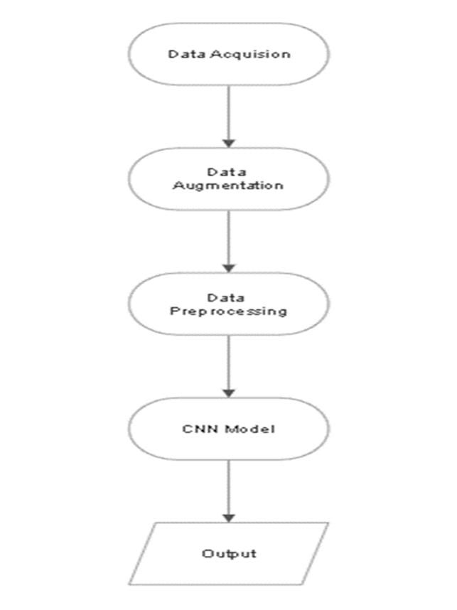
Data augmentation is a technique used in data analysis to expand the volume of information by inserting reproductions of pre existing data that have been significantly altered or newly created synthetic data from preexisting data. Before data augmentation, the dataset contained 155 positive and 98 negative samples, producing 253 sample pictures. After data augmentation, a new dataset is created with 2065 example photographs from 1085 positive and 980 sample photos.
Pre processing is important in order to create a seamless training experience because the MRI scans differ in intensity, contrast, and size. In the first preprocessing step, warping as well as cropping is performed, which will prepare the input picture. During warping, the supplied image is compared to the primary subject in the window. The image's outermost boundary is set in order for the subject to remain intact after cropping. Because the photographs in the dataset are of varying sizes, the image is reshaped to 240 x 240 x 3. Normalization is employed to scale pixel values to the range of 0 1, which will be beneficial to the training process.


Our model consists of the following layers:
1) The zero padding layer, used to control the dimension loss and loss of features present at the boundaries.
2) The convolution layer, this is a core layer that carries out the main convolution computation operations.
3) The max pooling layer, this layer outputs a feature map of the elements with the highest value based depending on the filter size.
4) The batch norm layer, this layer normalizes the values in the feature map and helps in controlling overfitting.
5) The flatten layer, is used to convert the feature map array into a one dimensional array and finally
6) The dense layer performs the classification task based on the input value from the previous layer.
ISSN: 2321 9653; IC Value: 45.98; SJ Impact Factor: 7.538 Volume 10 Issue X Oct 2022 Available at www.ijraset.com

This work also incorporates an activation function in addition to the layers used in the CNN approach. To combat overfitting L2 regularization was employed at all the convolution layers along with strategically placed batch normalization layers.
Model: "BrainDetectionModel" Layer (type) Output Shape Param # ================================================================= input_1 (InputLayer) [(None, 240, 240, 3)] 0 zero_padding2d (ZeroPadding (None, 244, 244, 3) 0 2D) conv0 (Conv2D) (None, 242, 242, 32) 896 activation (Activation) (None, 242, 242, 32) 0 max_pool0 (MaxPooling2D) (None, 121, 121, 32) 0 bn0 (BatchNormalization) (None, 121, 121, 32) 128 conv1 (Conv2D) (None, 119, 119, 32) 9248 activation_1 (Activation) (None, 119, 119, 32) 0 max_pool1 (MaxPooling2D) (None, 59, 59, 32) 0 bn1 (BatchNormalization) (None, 59, 59, 32) 128 conv2 (Conv2D) (None, 57, 57, 32) 9248 activation_2 (Activation) (None, 57, 57, 32) 0 max_pool2 (MaxPooling2D) (None, 28, 28, 32) 0 bn2 (BatchNormalization) (None, 28, 28, 32) 128 conv3 (Conv2D) (None, 26, 26, 32) 9248 activation_3 (Activation) (None, 26, 26, 32) 0 max_pool3 (MaxPooling2D) (None, 13, 13, 32) 0 bn3 (BatchNormalization) (None, 13, 13, 32) 128 flatten (Flatten) (None, 5408) 0 fc0 (Dense) (None, 512) 2769408 bn6 (BatchNormalization) (None, 512) 2048 fc1 (Dense) (None, 1) 513
Total params: 2,801,121 Trainable params: 2,799,841 Non trainable params: 1,280



ISSN: 2321 9653; IC Value: 45.98; SJ Impact Factor: 7.538 Volume 10 Issue X Oct 2022 Available at www.ijraset.com
Figure5:Loss vs Epoch


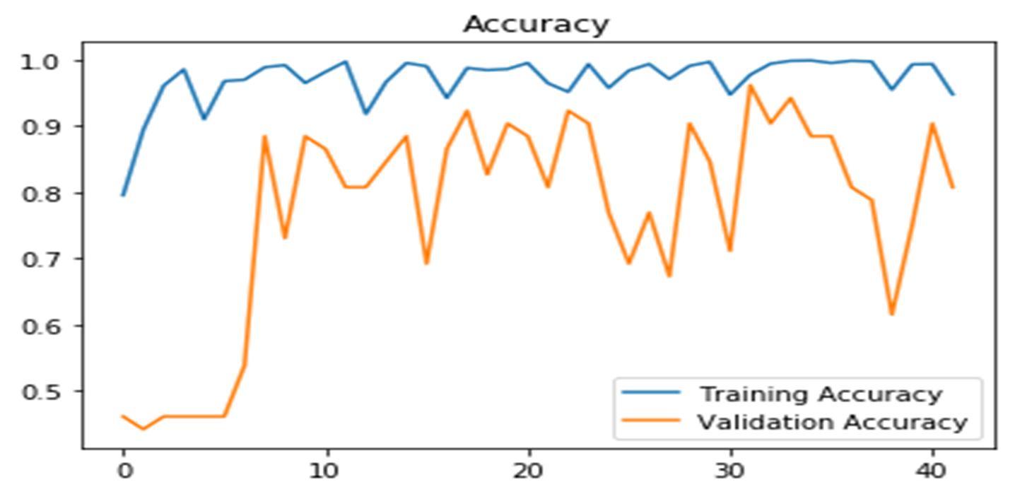
Figure6:Accuracy vs Epoch


Figure7:Best model loss results
ISSN: 2321 9653; IC Value: 45.98; SJ Impact Factor: 7.538


Volume 10 Issue X Oct 2022 Available at www.ijraset.com
Figure8: Best model accuracy results


Figure9: Best model F1 Score results

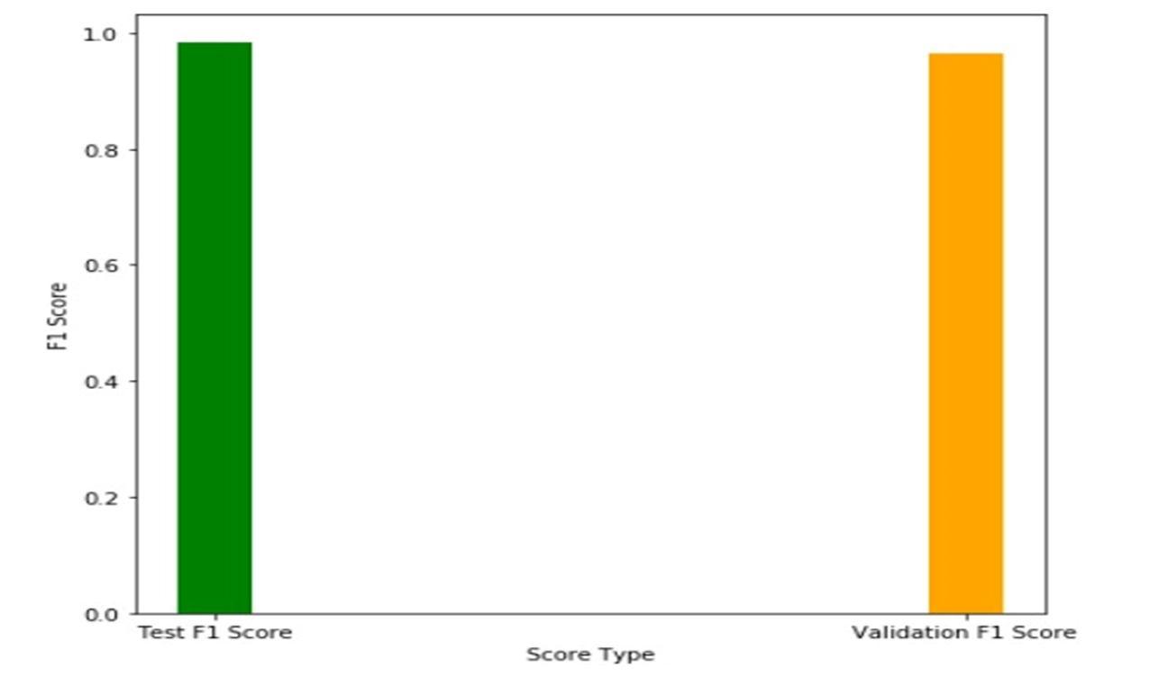
Figure10:Accuracy comparison with other methods.

ISSN: 2321 9653; IC Value: 45.98; SJ Impact Factor: 7.538 Volume 10 Issue X Oct 2022 Available at www.ijraset.com
Experiments were conducted on 2065 photos, 1085 of which had malignancies and 980 of which did not. The dataset is further split as: 70% as training, 10% as validation, and 20% as testing; each experiment was conducted for up to 50 epochs with early stopping to control overfitting. On the 32nd epoch, the model had a test accuracy of 97.74% and a test loss of 0.3033, as well as a validation accuracy of 96.15% and a validation loss of 0.3476.

CNNs are excellent tools for detecting brain cancers in MRI images. In the 32nd epoch, this investigation yielded a validation accuracy of 96% and a loss value of 0.3476. The model performance can be improved in the future we have access to additional amount of data and more powerful hardware to handle computations involving such a large amount of data.
[1] B. Devkota, Abeer Alsadoon, P.W.C. Prasad, A. K. Singh, A. Elchouemi, “Image Segmentation for Early Stage Brain Tumor Detection using Mathematical Morphological Reconstruction,” 6th International Conference on Smart Computing and Communications, ICSCC 2017, 7 8 December 2017, Kurukshetra, India.
[2] Dr. Chinta Someswararao “Brain Tumor Detection Model from MR Images using Convolutional Neural Network” IEE May [June 2020]
[3] Ehab F. Badran, Esraa Galal Mahmoud, Nadder Hamdy, “An Algorithm for Detecting Brain Tumors in MRI Images”, 7th International Conference on Cloud Computing, Data Science & Engineering Confluence, 2017.
[4] Khurram Shahzad and Imran Siddique “Efficient Brain Tumor Detection Using Image Processing Techniques “International Journal of Scientific & Engineering Research [December 2019]
[5] Muhammed Talo, Ozal Yildirim, Ulas Baran Baloglu, Galip Aydin, U Rajendra Acharya, Convolutional neural networks for multi class brain disease detection using MRI images, Computerized Medical Imaging and Graphics,Volume 78,2019,101673, ISSN 0895 6111,
[6] NAVONEELCHAKRABARTY Kaggle dataset [April 2019] (online) Available: https://www.kaggle.com/datasets/navoneel/brain mri images for braintumor detection
[7] Ravikumar Gurusamy and Dr Vijayan Subramaniam,” A Machine Learning Approach for MRI Brain Tumor Classification”, CMC, vol.53, no.2, pp.91 108, 2017.
[8] Rohini Paul Joseph, C. Senthil Singh and M.Manikandan, “Brain MRI Image Segmentation and Detection in Image Processing”, International Journal of Research in Engineering and Technology, 2014.
[9] Rupal R. Agravat; Mehul S. Raval, ”Prediction of Overall Survival of Brain Tumor Patients”, In proceedings of the 2019 IEEE Region 10 Conference (TENCON 2019).

