Education a
Tobacco Lesions C. Moireschi & Al. Canu take you through the effects of tobacco consumption on oral mucosa Most lesions caused by tobacco can be classified with the white lesions of the oral mucosa, with the exception of the smoker's melanosis, which is a brown lesion. Tobacco is also one of the primary factors implicated in the development of oral squamous cell carcinoma. The most compelling statistical and clinical evidence supports tobacco and alcohol as primary factors. These lesions are typical in older people, but the increasing number of smokers in the younger population is causing them to appear earlier.
Nicotine stomatitis
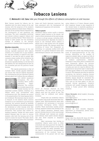
This is a benign thickening of the oral mucosa, typically associated with tobacco smoking. It is usually seen in pipe smokers, but may also develop in cigarette and cigar smokers. It can also be seen in people who consume a large number of hot drinks but do not smoke tobacco of any kind. The alterations typically develop on the hard and soft palates, but can infrequently be seen on the retromolar pad and the posterior buccal mucosa. The typical presentation is that of multiple, white, circular papules exhibiting red centres that may be slightly depressed. The erythematous portion represents dilated salivary gland duct orifices that are inflamed and exhibit squamous metaplasia. The sur足 rounding white surface represents hyper足 keratosis. The nodules are initially separated by normal mucosa, but the individual nodules coalesce, resulting in a diffuse white area interspersed with erythematous dots. The changes can extend to the gingiva where the tissue appears white and thickened. The keratosis may be smooth or fissured. In the western world, nicotine stomatitis is not considered a pre-cancerous condition, but patients should be carefully observed for changes in the involved area and for alterations at other mucosa sites that could represent pre-cancerous or cancerous alterations. This process may resolve on smoking cessation. Similar but more severe palatal changes may be seen in reverse smokers, who hold the burning end of the cigarette inside the mouth. This habit, which is not rare in India and some other Southeast
MEDICAL STUDENT INTERNATIONAL
Asian and South American countries, has been associated with the development of displasia and squamous cell carcinoma.
Tobacco pouch
Smokeless tobacco (either snuff or chewing tobacco), placed directly in the mouth, can result in direct alterations of the oral mucosa. It occurs in all age groups, including child足 hood and adolescence. Typically, the lesion is found in the vestibule where tobacco is placed, and it may extend onto the gingiva and buccal mucosa. The changes range from wrinkling of tissue that disappears on stretching, to a granular surface with mild keratosis, to a greatly thickened tissue with well-developed fissures and kerathosis. The
extent of changes depends on host susceptibility, the form and quantity of tobacco used, and the period of use. The gingiva may be inflamed and exhibit recession. People who use smokeless tobacco are at higher risk of oral carcinoma than nontobacco users. Well-documented cases exist of squamous cell carcinomas or verrucous carcinomas developing within tobacco pouches. These carcinomas usually present as large, exophytic lesions that develop at the site of tobacco placement. However some patients develop cancer at other oral mucosal sites. Although such tumours are usually quite obvious, more subtle clinical changes can also show atypical or dysplastic alterations. Most carcinomas that develop in smokeless tobacco users occur in older individuals who have practiced the habit for many years, typically 30 years or more. Many tobacco pouches are easily reversible once the habit is discontinued. Histopathologic examination of the involved tissue is recommended if the patient will not stop
www.ifmsa.org
using tobacco or if tissue changes persist after cessation. Biopsy is also suggested if the clinical lesion is markedly papillary or demonstrates areas of redness or ulceration.
Smoker's melanosis
Individuals who smoke may develop benign areas of hyperpigmentation of the oral mucosa. Depending on the number of cigarettes smoked daily, as many as 31% of smokers may develop clinically visible areas of melanin pigmentation. Smoker's melanosis is seen more often in females and most frequently affects the anterior mandibular and maxillary gingiva. However, virtually any oral site may be affected. The pigmentation varies from light to dark brown and may appear diffuse or be more localised in nature. The diagnosis can often be made by correlating the patient's smoking history with the clinical presentation of the lesions. However, biopsy should be considered for pigmented lesions, either in unusual loca足 tions such as the hard palate, or with an unusual clinical appearance. Smoker's melanosis will usually disappear gradually during a 3-year period after smoking cessation. Chiara Moireschi, dental student, University of Brescia, Italy isaac23@virgilio.it Alessandro Canu, dental student, University of Brescia, Italy aracnoide@libero.it
Acknowledgements
Checked by Prof. Corrado Paganelli, Department of Orthodontics, Dental Clinic, University of Brescia, Italy.
References
Images taken from Neville BW, Damm DD, White DK, The color atlas of clinical oral pathology.
M EDICAL S TUDENT I NTERNATIONAL MAGAZINE Now on -line at: www.ifmsa.or g/publications 21