23 minute read
Tay-Sachs Disease: Causes and Treatments


Brittany Kemp1*
¹Geisinger Commonwealth School of Medicine, Scranton, PA 18509 *Master of Biomedical Sciences Program Correspondence: britkemp9@gmail.com
Abstract
Tay-Sachs disease (TSD) is a rare autosomal recessive neurodegenerative disease. It is one of several lysosomal storage disorders. Neurodegeneration results from disordered lipid metabolism that causes a buildup of GM2 gangliosides in the central nervous system (CNS). The three forms of TSD, infantile, juvenile, and late-onset, are classified by the amount of residual activity of the enzyme β-hexosaminidase A (HEX A) and can be caused by over 100 different mutations. TSD has a low prevalence in the general population. It is therefore a lesserknown disease in communities that do not frequently encounter affected individuals. There are currently no treatments or cure for any of the three forms of TSD. The aim of this review is to summarize and discuss current studies that investigate potential treatments for TSD to determine where future studies should be focused. Two databases, Medline and EBSCOhost, were used to search the literature for articles that were published between 2000 and 2021. Several animal models, such as mice and sheep, have been used to analyze the effects of bone marrow transplantation and gene therapies using different vectors. Stem cell transplantation, pyrimethamine, modification of protein quality control mechanisms, and a modified amino acid have been tested in vitro in human cells or in a small cohort of patients. The rarity of this disease and the many mutations that cause it present challenges to testing potential treatments on larger groups of patients. Several gene therapy delivery systems and administration of L-acetyl-leucine have shown promising results that could lead to more widely available therapies if successful in more patients.
Introduction
TSD is one of several lysosomal storage disorders, and is closely related to another lysosomal storage disorder, Sandhoff disease. TSD is caused by a mutation in the HEXA gene, which codes for the α-subunit of β-hexosaminidase A (HEX A) and is inherited in an autosomal recessive manner. TSD is commonly studied with Sandhoff disease because Sandhoff disease is caused by a mutation in the β-subunit of HEX A. Both TSD and Sandhoff disease are the main forms of GM2 gangliosidosis. There are currently over 100 mutations identified that cause TSD and impact the function of the α-subunit of HEX A (1). β-hexosaminidase A is a heterodimeric lysosomal protein that breaks down GM2 gangliosides. Without this enzyme, GM2 gangliosides build up in lysosomes of the brain and nerve cells. This buildup results in neurological decline and progression of symptoms as patients age. The age of onset and severity of TSD symptoms is determined by the mutation’s effect on the α-subunit of HEX A. Therefore, the three forms of TSD are characterized based on the amount of residual HEX A activity. Infantile TSD is the classical form of TSD in which patients have very low or no HEX A activity. The physiological effects of the buildup of GM2 gangliosides in the brain and nerve cell lysosomes can be recognized in the patient’s first 3 to 6 months of life. The patient’s symptoms begin with progressive weakness that results in a loss of motor skills as well as a lack of visual attentiveness. Patients begin experiencing seizures within their first or second year of life as the amount of GM2 gangliosides building up increases. Death typically occurs around ages 2 to 3 years, although survival has been recorded up to 5 to 7 years. Juvenile TSD is characterized by symptoms that appear around 2 years of age. Until the age of 2, patients reach normal developmental milestones because they have more residual HEX A activity than infantile TSD patients. Once the levels of built up GM2 ganglioside reach disease levels, the developmental milestones previously reached are slowly lost. Patients walk with an abnormal gait and have difficulty with speech. By age 10, patients have more severe symptoms such as dysphagia and seizures. Death occurs before or around age 20 for these patients. Late-onset TSD occurs in individuals who have a less severe mutation in HEXA resulting in a higher residual HEX A activity than infantile or juvenile TSD patients. This delays their neurological decline and onset of symptoms. Patients with late-onset TSD typically begin showing TSD symptoms in their late teens or young adult years. Many of the same symptoms of decline are present for these patients including muscle weakness, abnormal gait, and speech difficulties.
There is currently no cure for any of the three forms of TSD. Several animal models have been used to identify possible treatments such as using bone marrow stromal cells (2) and several forms of gene therapy (3–5) for treating or slowing the progression of TSD. Other treatment methods have been tested in vitro using human cells or on a small cohort of TSD patients. These include the use of hematopoietic stem cell transplantation (6–7), administration of pyrimethamine (8–9), and manipulation of cellular quality control mechanisms (10) to increase HEX A activity. Application of a modified amino acid (11–12) was proposed as a therapy to protect cells of the cerebellum from damage due to the buildup of GM2 gangliosides. In 2018, TSD treatment methods were presented by Soloveyeva et al., but more potential treatments have since emerged (13). The purpose of this review is to present previous and current attempts at finding a treatment method for TSD as well as to highlight methods that will be explored in future studies. Table 1 summarizes the treatment method and primary outcomes of each study examined in this review.
Methods
Two databases were used to retrieve articles for this review, Medline and EBSCOhost. Medline was used first and resulted in seven articles. Three keywords were used to search on
Therapy Primary outcome
Bone marrow transplant Proper association of Hex A with Hex B in transduced BMSCs. Hydrolysis of GM2 ganglioside by transduced BMSCs.
Reference
Martino et al. (2)
Gene therapy All sheep treated with AAV vector containing HEXA and HEXB cDNA showed delayed onset or prevention of clinical signs of TSD.
Gene therapy AAV vector was able to be detected in small amounts in the brain indicating that it crossed the blood brain barrier. Increases in both MUG and MUGS activity was indicated for the Hex M construct in the liver and brain of treated SD mice.
Gene therapy Hex M was distributed in the brain and liver of treated TSD and SD mice. However, a B and T cell immune response was mounted against the Hex M construct as well as the AAV vector.
Enzyme replacement Supraphysiological MUG and MUGS activity was achieved in NCs of SD mice when transduced with bicistronic LV and persisted upon NPCs differentiation. Transduced fibroblasts from a SD patient were able to clear GM2 ganglioside.
Enzyme replacement Symptoms of a long-standing tremor, gait ataxia, speech stammer, and swallowing problems were resolved in a patient with late-onset TSD. Following treatment, his symptoms had decreased to only an occasional mild tremor. Eight years after treatment was administered, his cells retained full donor chimerism.
Pharmacological chaperone Low doses of PYR administered to late-onset TSD patients was able to increase leukocyte Hex A activity.
Pharmacological chaperone Administration of PYR can increase Hex A activity in patients with late-onset TSD. Optimal doses should be determined on an individual basis to prevent decreasing of biochemical effects.
Endogenous chaperone manipulation Modulating components of cell’s intrinsic ERAD pathway can increase cellular Hex A activity by promoting proper mutant α-subunit folding.
Neuroprotection Acetyl-DL-leucine was able to reduce gait variability during slow walking in patients with cerebellar ataxia. Gray-Edward et al. (3)
Ou et al. (4)
Kot et al. (5)
Ornaghi et al. (6)
Stepien et al. (7)
Clarke et al. (8)
Osher et al. (9)
Derish et al. (10)
Schniepp et al. (12)
Table 1. Primary outcomes of therapeutic approaches for TSD treatment.
this database: Tay-Sachs, Tay-Sachs disease, and treatment. Searches were first narrowed to the date range of 2000–2021 and were further narrowed to 2010–2021 to decrease the number of search results and include more relevant articles. EBSCOhost was searched using four keywords: Tay-Sachs, Tay-Sachs disease, treatment, and GM2-gangliosidosis. The date range was narrowed to include only articles from 2010–2021, as this resulted in the most relevant articles from the first database. Searches on EBSCOhost resulted in six articles that are discussed in this review. No journal exclusion was applied when searching either database.
Animal Studies
Bone Marrow Transplant Several animal models have been used for studying both Sandhoff disease and TSD. Martino et al. (2) studied TSD mice to determine if their bone marrow stromal cells (BMSCs) could be transduced with a Molony leukemia retroviral vector encoding the α-subunit of human HEXA in order to restore enzyme function. This methodology was used because the enzymatic defect must be corrected for cells of the CNS which has presented as a challenge for other treatment approaches. BMSCs were chosen because they have been found to produce circulating progenitors that can repopulate nonhemopoietic tissues. Therefore, they could potentially carry gene therapies to these tissues and, in the case of TSD, restore enzyme function. Several important conclusions can be made based upon findings from this study. First, the strategy used resulted in proper association between the α-subunit and β-subunit of HEX A without the formation of the undesired HEX S (αα) protein product. Upon further analysis, the HEX A protein formed when using transduced HEXA sequence displayed kinetic and thermal stability properties like the HEX A obtained from wild-type fibroblasts. Second, transduced cells from TSD mice were able to hydrolyze GM2 ganglioside that reached their lysosomes while non-transduced cells from TSD mice had accumulation of GM2 ganglioside in vitro. One limitation of using mice as a TSD model for in vivo studies, is that they have an alternative method for breaking down GM2 gangliosides preventing them from showing signs of neurological disease in the same manner as humans (3).
Gene Therapy Gray-Edwards et al. (3) used sheep instead of mice as a model for TSD to evaluate the potential of the adeno-associated virus (AAV) as a vector for administering gene therapy. Sheep were used because they are the only experimental model for TSD that show clinical signs of the disease in a manner like affected humans. Additionally, the sheep’s CNS more closely resembles the size and complexity of the human CNS when compared to other models. Another advantage of this animal model is that it could uncover possible treatment limitations that would arise in humans; however, these limitations would not arise in other
animal models. In this study, the AAV vector was administered intracranially at 2 to 4 months of age. The vector was injected into a single lateral ventricle and bilaterally into the thalamus. These injection sites were chosen to distribute the vector into both the cerebellum and the cerebral cortex. All sheep treated with the AAV vector showed a delayed onset or prevention of clinical signs of TSD. The lifespan was also increased by up to 50% when compared to untreated sheep. Sheep treated with α-subunit and β-subunit cDNA-containing vectors exhibited the most clearance of the sialic acid component of GM2 gangliosides in many CNS areas except for the midbrain. Delivery to the spinal cord and cerebellum revealed a limitation of the delivery based on AAV vector. In both areas, storage of GM2 gangliosides remained equal to or surpassed the levels in untreated sheep. Other locations of injection will need to be studied to determine how deliver the AAV vector more effectively to the spinal cord and cerebellum. AAV was also used as a vector delivery method for the CRISPR system to form edited hepatocytes in Sandhoff mice. The vector delivered cDNA encoding a modified α-subunit incorporating partial β-subunit sequence, yielding, the protein product identified as HEX M. Delivery of this modified sequence allowed Ou et al. (4) to overcome difficulties presented by packaging both HEXA and HEXB in the same vector and also reduced the cost from using separate vectors containing individual subunit sequences. Additionally, usage of HEX M allows for the potential treatment of both TSD and Sandhoff disease using the same vector. Following treatment of Sandhoff mice with the AAV vector containing HEX M and CRISPR machinery, the amount of GM2 gangliosides was significantly reduced in the liver, heart, and spleen. However, the GM2 gangliosides in the brain were not significantly reduced. 4-methylumbelliferyl-6-sulfa2-acetoamido-2-deoxy-β-D-glucopyranoside potassium salt (MUGS) and 4-methylumbelliferyl N-acetyl-β-D-glucosaminide (MUG) activities were measured to determine the amount of HEX activity in each of the four tissues of treated mice in comparison to normal and untreated mice. MUG and MUGS activities in the liver, heart, and spleen increased above the activity of both untreated and normal mice. Additionally, the MUG and MUGS activity in the brain of treated mice was significantly increased when compared to untreated mice. Therefore, a small amount of HEX M was able to cross the blood-brain barrier. Kot et al. (5) identified and investigated a potential limitation of introducing HEX M due to it being a hybrid variant of native subunits of the HEX A protein. Patients who do not express the native HEX A or HEX B protein could have an immune response to the HEX M protein. When the human-derived HEX M enzyme was expressed in both TSD and Sandhoff disease mice a T and B cell immune response was triggered. The immune response reported in this study indicates the need for immunomodulatory gene therapy to be used for long-term treatment of diseases such as TSD and Sandhoff disease. Ornaghi et al. (6) identified another potential treatment for TSD and Sandhoff disease using a different vector delivery system. The bicistronic lentiviral vectors (LV) were found to be able to transduce human neurons and glia as well as CD34+ hematopoietic stem/progenitor cells (HSPCs). The efficacy of these bicistronic LVs to transduce neural stem/progenitor cells (NPCs) of Sandhoff mice was tested first. The NPCs were not functionally impacted by transduction as indicated by the ability to proliferate, self-renew, and maintain multipotency after LV transduction. The expression of both α- and β-subunits of HEX A was detected in transduced cells. Exogenous expression of these subunits did not alter the endogenous expression of HEXA or HEXB genes. Furthermore, supraphysiological MUG- and MUGS-related enzyme activity was produced; this activity persisted through the differentiation of NPCs to neurons and glia. One advantage of administering bicistronic LVs is that a half dose was required when compared to the necessary dosage for monocistronic LVs. Another suggested advantage of using bicistronic LVs is that they drive stoichiometric expression of the α- and β-subunits unlike monocistronic LVs. Stoichiometric expression of the α- and β-subunits can be beneficial for preventing the unwanted formation of HEX S.
Human Clinical Trials
Stem Cell Therapy The usage of bicistronic LVs to effectively and safely reconstitute HEX A activity in vitro for human neuron/glial cells and CD34+ HSPCs was also evaluated by Ornaghi et al. (6). iPSC-derived neural progeny and CD34+ HSPCs were both successfully transduced with the bicistronic LVs and were able to produce functional HEX A. Fibroblasts from a human Sandhoff disease patient were then transduced with the bicistronic LV to test whether enzymatic activity for clearing stored GM2 gangliosides could be rescued in these cells. The composition of α- and β-subunits expressed was comparable to the un-transduced healthy donor fibroblast composition of subunits. GM2 ganglioside storage was cleared in the transduced fibroblasts indicating the efficacy of this treatment in patient-derived cells. The larger cargo capacity of LVs allows genes for both the α- and β-subunits to be delivered in one vector. This could potentially overcome the issue presented when using an AAV vector as well as the HEX M construct (5).
One patient with late-onset TSD was found to have been successfully treated with HSPC transplantation as described by Stepien et al. (7). At 15 years of age the patient was given a bone marrow transplant from a healthy matched-sibling donor. The patient described in this study had a long-standing tremor since age 7 in addition to gait ataxia, a speech stammer, swallowing problems, and neurological deterioration prior to treatment. The patient was diagnosed with a deficiency in HEX A consistent with late-onset TSD. Two attempts for treating his tremor were made using first propranolol and then primidone, but both were eventually stopped due to lack of effectiveness and side effects respectively. Stepien et al. (7) indicated that the patient’s neurological regression was stabilized postHSPC transplantation. Eight years after the transplant was performed, the patient had only mild tremors in his upper limbs and occasionally in his body, with MRI scans showing no further progression when compared to scans done prior to transplantation. Additionally, at age 23 the patient retained full donor chimerism with his white cell HEX A levels comparable to normal control levels and a reduction in GM2 ganglioside in circulation. Further testing on this treatment method is needed, but it marks potential progress in treating one of the forms of TSD.
Cellular Quality Control Manipulation The drug pyrimethamine (PYR) was studied as another potential treatment for late-onset TSD. Pyrimethamine is a pharmacological chaperone (PC) that binds and stabilizes its target enzyme by maintaining the native folded conformation. PYR inhibits its target enzyme better in the neutral pH of the endoplasmic reticulum (ER) rather than in the acidic environment of the lysosome. PYR is currently approved for the treatment of some malaria and toxoplasmosis infections. Clarke et al. (8) examined the clinical benefit of administering PYR to patients with late-onset TSD and Sandhoff disease in doses similar to those used for treating parasitic diseases. When given in low doses (25 mg/day), all 8 subjects in this study tolerated the drug well. When the dosage was raised from 50 to 75 mg per day all but one of the subjects showed significant adverse neurological side-effects. Two noteworthy conclusions can be drawn from this study. Three subjects exhibited definitive increases in HEX A levels when compared with their baseline levels, and no inhibition of HEX A activity was indicated even at the highest plasma PYR level. Further studies need to be performed to fully evaluate the efficacy of PYR as a treatment method for TSD and Sandhoff disease. None of the patients were able to reach the maximum dosage of 100 mg due to adverse side effects developing on the intermediate doses (50–75 mg/day). This study also showed the importance of monitoring plasma PYR levels as the relationship between plasma PYR levels and dosage is highly variable between different subjects. Another study by Osher et al. (9) also assessed whether pyrimethamine could be used to increase HEX A activity in late-onset TSD patients. The dosing for this study began at a lower dose of only 6.25 mg and increased up to 75 mg daily. Participants in this study showed some doserelated increase in HEX A activity, but the dose varied markedly between subjects and doses beyond the optimal resulted in a decline in HEX A activity. The researchers were also unable to rule out if the maximal rise in HEX A activity was due to the dose or if it was time-related. Some participants in this study also experienced negative side effects such as nausea, vertigo, and vomiting; however, serum PYR was not measured, and these symptoms occurred at different dosages for different participants. The study results suggest that PYR administration at low doses or for a short period can increase HEX A activity in late-onset TSD patients, but high doses and/or long-term usage of PYR is associated with negative side effects. The investigators proposed further studies to be performed to evaluate different dosing regimens for treatment of late-onset TSD with PYR. Derish et al. (10) studied two HEX A α-subunit mutants that are both degraded through the endoplasmic reticulumassociated degradation (ERAD) pathway shown in Figure 1. This study evaluated how manipulating endoplasmic reticulum (ER) quality control could impact folding and lysosomal expression of HEX A. It was first confirmed that both E482K and G269S mutations caused misfolding of the α-subunit and its subsequent degradation through the ERAD pathway. Next, several ER quality control mechanisms were manipulated with the objective to properly fold the α-subunit, prevent its
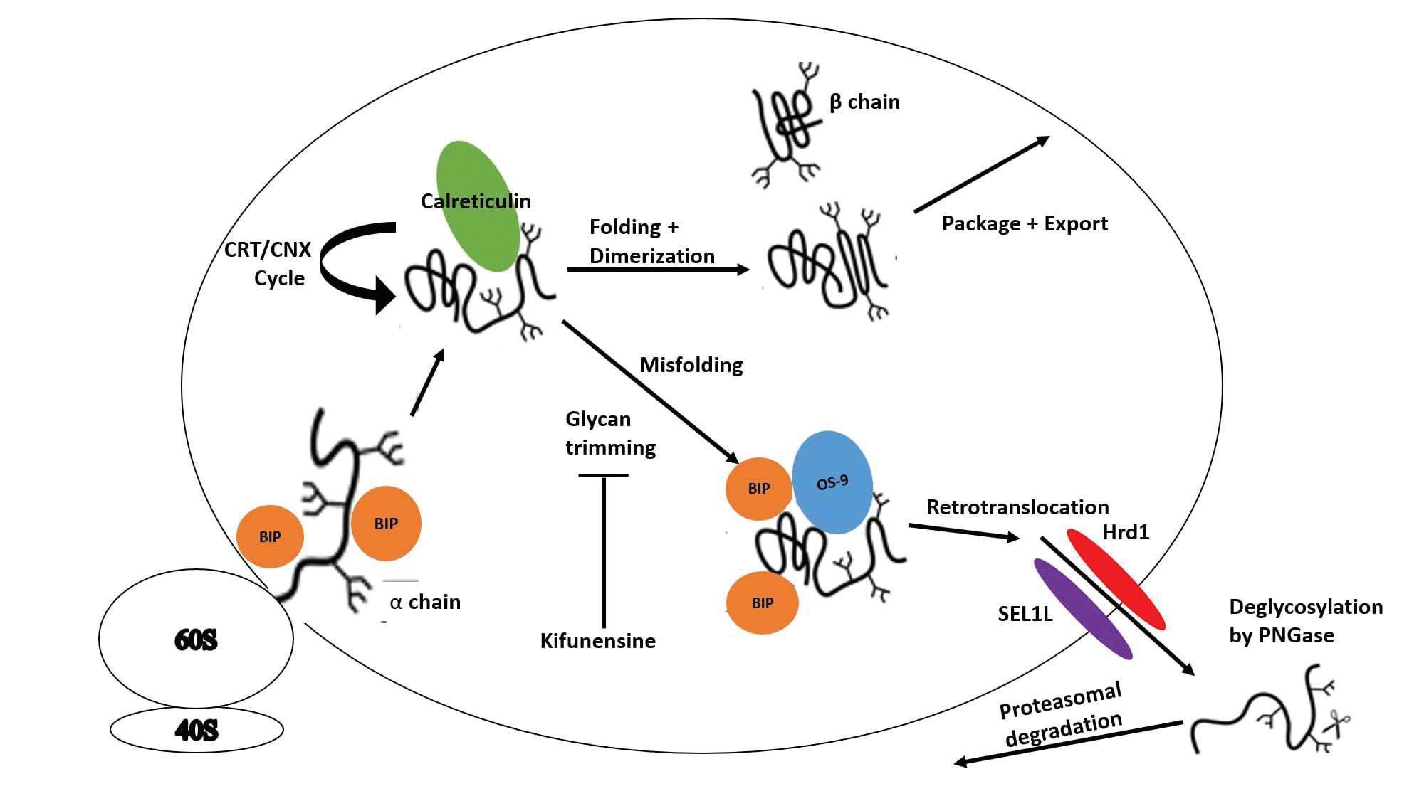
Figure 1. Model for manipulation of the endoplasmic reticulum-associated degradation pathway to treat Tay-Sachs Disease. HEX A α is translocated into the ER where it is cotranslationally glycosylated, then folded in the calreticulin/calnexin cycle. Upon proper folding, α and β subunits dimerize and are packaged for export to the Golgi. Stalling, caused by either the G269S or E482K mutation, glycan trimming begins the subunit’s disposal. Kifunensine blocks glycan trimming, preventing disposal. Chaperones BiP and OS-9 escort the misfolded α-subunit to the retrotranslocon formed by SEL1L and Hrd1 for removal from the ER. Cytosolic PNGase removes glycans prior to degradation by the proteosome (10).
degradation, increase HEX A secretion, and increase lysosomal HEX A activity. First, cells were incubated with kifunensine, a mannosidase inhibitor. It was hypothesized that more correctly folded α-subunit mutants would be formed when given longer time to properly fold. The degradation of both E482K and G269S was decreased; however, the number of secreted molecules was not significantly increased for either mutant. Calreticulin (CRT), a major ER-resident chaperone for glycoproteins, was studied next to determine if it could prevent E482K from ERAD recognition. This was done by overexpressing CRT in some cells. Cells treated to overexpress CRT showed delayed identification of the E482K mutant by ERAD when compared to cells not treated with CRT and cells transduced with mutant, inactive H153A CRT. Finally, chemical chaperones were studied to determine their ability to stabilize G269S to increase its activity. The G269S mutant was chosen because some active forms of this α-subunit can escape ER quality control. Trimethylamine N-oxide (TMAO) and glutamic acid both were able to significantly increase HEX A activity when administered to patient fibroblasts. Since upregulation of G269S alone does not increase HEX A activity, it was proposed that TMAO and glutamic acid were able to promote proper folding of this mutant. Due to the many different mutations that cause TSD, ERAD manipulation needs to be studied in the context of more mutants to determine the full potential of this treatment approach. Neuroprotection Fields et al. (11) identified N-acetyl-L-leucine, a modified amino acid, as a potential therapy for treating three rare neurodegenerative diseases one of which was GM2gangliosidosis. The rationale for utilizing N-acetyl-L-leucine for treatment of these diseases is based on its potential to protect cells of the cerebellum by reducing neuroinflammation, and therefore, preventing neurodegeneration. Previously, the racemic mixture of N-acetyl-leucine was determined to be well tolerated by TSD and Sandhoff disease patients. Racemic N-acetyl-DL-leucine was studied by Schniepp et al. (12) for its ability to reduce cerebellar ataxia in patients. The patients of this study had cerebellar ataxia that was either sporadic or hereditary. The patients in did not have TSD, although cerebellar ataxia is one of the symptoms for juvenile and lateonset TSD. Participants in this study showed improvements in gait variability during slow walking, the stability of which is potentially reliant on the cerebellum for sensory integration. Based on the demonstrated ability of N-acetyl-DL-leucine to improve cerebellum function through measuring improvement in ataxia, Fields et al. (11) created a protocol for using N-acetylL-leucine as a therapy for GM2-gangliosidosis including both TSD and Sandhoff disease. Only the L-enantiomer was proposed for use in this protocol because the L-enantiomer was found to be the only pharmacologically active form. Recruitment for the trial using this protocol was delayed due to the COVID-19 pandemic, but it was planned to resume during 2021.
Conclusion
It is difficult to study potential treatments for TSD as well as many other autosomal recessive diseases because of how rare they are in the population as evidenced by the small number of patients in human trials. Increasing awareness of this disease in populations that have a lower rate of prevalence could be one way of increasing the number of participants in clinical trials. This could also allow for each proposed treatment approach to be studied in the context of many different causative mutations. Due to the many mutations that cause TSD, and the variable phenotypes caused by these mutations, several treatment methods may need to be investigated for a patient to determine the best personalized approach. The increasing prevalence of genetic screenings could also aid in earlier detection of TSD. Screening for mutations prior to neurological decline could allow for earlier intervention once there is an approved preventative therapeutic. Many of the treatment methods detailed in this review need to undergo more rigorous testing to determine proper dosing and avoid adverse side effects. Distributing working enzyme into all necessary areas of the brain is the largest hurdle that must be overcome with all the vector delivery systems. Once a vector is determined to distribute effectively in the brain, combining enzyme replacement therapy with the neuroprotective effects of L-acetyl-leucine should be a focus in future studies. The neuroprotective effects of this modified amino acid could increase the effectiveness of enzyme replacement on slowing neurological decline.
Acknowledgments
I would like to thank Dr. Darina Lazarova for her assistance throughout writing this review.
Disclosures
The author has no conflicts of interest to declare.
References
1. Mistri M, Datar C, Sheth F, Gupta S, Sheth J. Identification of novel mutations in HEXA gene in children affected with
Tay-Sachs disease from India. Mol Cytogenet. 2014;7. 2. Martino S, Cavalieri C, Emiliani C, Dolcetta D, Angelis
MGCD, Chigorno V, et al. Restoration of the GM2 Ganglioside Metabolism in Bone Marrow–Derived Stromal
Cells from Tay-Sachs Disease Animal Model. Neurochem
Res. 2002;27.
3. Gray-Edwards HL, Randle AN, Maitland SA, Benatti HR,
Hubbard SM, Canning PF, et al. Adeno-Associated Virus
Gene Therapy in a Sheep Model of Tay-Sachs Disease. Hum Gene Ther. 2018;29(3):14.
4. Ou L, Przybilla MJ, Tăbăran A-F, Overn P, O’Sullivan MG,
Jiang X, et al. A novel gene editing system to treat both Tay–Sachs and Sandhoff diseases. Gene Ther. 2020;27:10.
5. Kot S, Karumuthil-Melethil S, Woodley E, Zaric V,
Thompson P, Chen Z, et al. Investigating Immune
Responses to the scAAV9-HEXM Gene Therapy Treatment in Tay-Sachs Disease and Sandhoff Disease Mouse Models.
Int J Mol Sci. 2021;22.
6. Ornaghi F, Sala D, Tedeschi F, Maffia MC, Bazzucchi M,
Morena F, et al. Novel bicistronic lentiviral vectors correct β-Hexosaminidase deficiency in neural and hematopoietic stem cells and progeny: implications for in vivo and ex vivo gene therapy of GM2 gangliosidosis. Neurobiol Dis. 2020;134.
7. Stepien KM, Lum SH, Wraith JE, Hendriksz CJ,
Church HJ, Priestman D, et al. Haematopoietic
Stem Cell Transplantation Arrests the Progression of
Neurodegenerative Disease in Late-Onset Tay-Sachs
Disease. JIMD Rep. 2017;41. 8. Clarke JTR, Mahuran DJ, Sathe S, Kolodny EH, Rigat BA,
Raiman JA, et al. An open-label Phase I/II clinical trial of pyrimethamine for the treatment of patients affected with chronic GM2 gangliosidosis (Tay-Sachs or Sandhoff variants). Mol Genet Metab. 2011;102(1):6.
9. Osher E, Fattal-Valevski A, Sagie L, Urshanski N, Amir-
Levi Y, Katzburg S, et al. Pyrimethamine increases β-hexosaminidase A activity in patients with Late Onset Tay
Sachs. Mol Genet Metab. 2011;102(3):8. 10. Derish D, Iwamoto Y, Argon Y. Tay-Sachs disease mutations in HEXA target the α chain of hexosaminidase A to endoplasmic reticulum–associated degradation. Mol Biol
Cell. 2016;27.
11. Fields T, Patterson M, Bremova-Ertl T, Belcher G, Billington
I, Churchill GC, et al. A master protocol to investigate a novel therapy acetyl-L-leucine for three ultra-rare neurodegenerative diseases: Niemann-Pick type C, the
GM2 gangliosidoses, and ataxia telangiectasia. Trials. 2021;22(1):15.
12. Schniepp R, Strupp M, Wuehr M, Jahn K, Dieterich M,
Brandt T, et al. Acetyl-DL-leucine improves gait variability in patients with cerebellar ataxia—a case series. Cerebellum Ataxias. 2016;3(8).
13. Solovyeva VV, Shaimardanova AA, Chulpanova DS, Kitaeva
KV, Chakrabarti L, Rizvanov AA. New Approaches to Tay-
Sachs Disease Therapy. Front Physiol. 2018;9.


