SEPTEMBER / OCTOBER 2009
14
A
Ca2+ Ion channel Cochlea
Stereocilia
Tectoral membrane
Cu hairtor cell
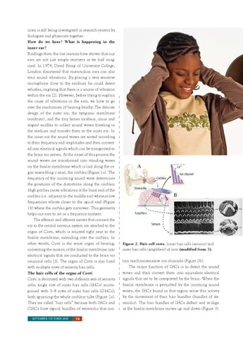
nism is still being investigated in research centers by biologists and physicists together. How do we hear? What is happening in the inner ear? Findings from the last century have shown that our ears are not just simple receivers as we had imagined. In 1979, David Kemp of University College, London discovered that mammalian ears can also emit sound vibrations. By placing a very sensitive microphone close to the eardrum he could detect whistles, implying that there is a source of vibration within the ear [2]. However, before trying to explain the cause of vibrations in the ears, we have to go over the mechanism of hearing briefly: The delicate design of the outer ear, the tympanic membrane (eardrum), and the tiny bones (malleus, incus and stapes) enables to collect sound waves traveling in the medium and transfer them to the inner ear. In the inner ear the sound waves are sorted according to their frequency and amplitudes and then converted into electrical signals which can be transported to the brain via nerves. At the onset of this process the sound waves are transformed into standing waves on the basilar membrane which is laid along the organ resembling a snail, the cochlea (Figure 1a). The frequency of the incoming sound wave determines the positions of the distortions along the cochlea: High pitches create vibrations at the basal end of the cochlea (i.e. adjacent to the middle ear) whereas low frequencies vibrate closer to the apical end (Figure 1b) where the cochlea gets narrower. This geometry helps our ears to act as a frequency analyzer. The efferent and afferent nerves that connect the ear to the central nervous system are attached to the organ of Corti, which is situated right next to the basilar membrane, extending over the cochlea. In other words, Corti is the sense organ of hearing, converting the motion of the basilar membrane into electrical signals that are conducted to the brain via neuronal cells [3]. The organ of Corti is also lined with multiple rows of sensory hair cells. The hair cells of the organ of Corti Corti is decorated with two different sets of sensory cells: single row of inner hair cells (IHCs) accompanied with 3–4 rows of outer hair cells (OHCs), both spanning the whole cochlear tube (Figure 2a). They are called “hair cells� because both IHCs and OHCs have typical bundles of stereocilia that con-
B
Basilar membrane
Amplifiers Sensors
Figure 2. Hair cell rows. Inner hair cells (sensors) and outer hair cells (amplifiers) of corti (modified from 7).
tain mechanosensitive ion channels (Figure 2b). The major function of IHCs is to detect the sound waves and then convert them into equivalent electrical signals that are to be interpreted by the brain. When the basilar membrane is perturbed by the incoming sound waves, the IHCs found in that region sense this activity by the movement of their hair bundles (bundles of stereocilia). The hair bundles of IHCs deflect and re-align as the basilar membrane moves up and down (Figure 3).