23 minute read
LIPOM I GJËNDRËS PAROTIDE: PREZENTIM RASTI DHE PASQYRIM I LITERATURËS LIPOMA OF PAROTI GLAND: A CASE REPORT AND LITERATURE REVIEW


UDK: 616.316,5-006,326 PUNIME PROFESIONALE (PP)
UDK: 616.316,5-006,326 PROFESSIONAL PAPER (PP)
Advertisement
Nedim Kasami, Vladimir Popovski
Klinika Univerzitare e kirurgjisë maksilofaciale Qendra klinike “Nëne Tereza” Rr. Vodnjanska 17, 1000 Shkup, Republika e Maqedonisë Veriore Nedim Kasami, Vladimir Popovski
University Clinic for Maxillofacial Surgery Clinical Center “Mother Teresa“ Vodnjanska str. 17, 1000 Skopje, Republic of N. Macedonia
ABSTRAKTI
Hyrje: Lipomi paraqet një tumor mezenhimal me origjine nga qelizat adipose. Ne regjionin e kokës dhe qafës, ky tumor nuk ështe i shpeshtë. Paraqitja e lipomit ne gjëndrën parotide, është shumë e rallë, midis 0,6 dhe 4%. Pareza e nervit facial zakonisht nuk ështe pasojë e këti tumori, pasi që nuk e lëndon nervin facial meqë zen pozicion sipërfaqësor. Mjetet diagnostike janë të njëjta si per tumorët tjerë të gjëndrës parotide, duke përfshirë ultrazërin, biopsinë aspirative, CT dhe MRI.
Qëllimi i studimit: Në këtë artikull, prezentojmë rast klinik i lipomit ne lobin sipërfaqësor të gjëndrës parotide të majtë, i shoqëruar me një parezë të lehtë të anës së majtë të fytyrës, ptozë të kapakut të syrit dhe rjedhje e lotëve nga syri i majtë, që shkakton një dyshim se degët e sipërme të nervit facial mund të jenë afektuar si pasojë e këti tumori.
Materialet, metodat dhe rezultatet: Analizat përfshinë anamnezën, egzaminimin klinik, dokumentacionin mjekësor të pacientit dhe përshkrimi i protokollit operativ. Studimi i rastit u krahasua me literaturën bashkëkohore. Në anestezion të përgjithshëm, duke përdorur qasje klasike sipas Blair, lezioni i ndërtuar nga indi adipoz u hoq me trajtim kirurgjik një ditor. Nuk pati komplikime postoperative. Analiza pato-histologjike konfirmoi dijagnozen e lipomit. Kontrollat ndjekëse pas 2 muajsh dhe pas 1 viti treguan se nuk kishte recidiv, por simptomat e përmendur mbetën edhe më tutje.
Diskusioni: Diagnostifikimi klinik i lipomit është relativisht i lehtë, pasi që është asimptomatik dhe paraqitet si ënjti e padhimbshme në gjëndrën parotide. Imazhet tomografike janë shumë të sakta dhe të dobishëm në fazën e diagnostifikimit preoperativ. Disa autorë nuk preferojnë biopsi aspirative dhe e tregojnë atë si të panevojshëm në rastin e lipomit. ABSTRACT
Introduction: Lipoma is a benign mesenchymal tumor originating from adipose tissue cells. In the head and neck, this tumor is not common. The occurrence of lipomas in the parotid gland is extremely rare, ranging between 0.6 and 4%. Facial nerve paresis usually is not consequence of presence of this tumor while it does not affect the facial nerve or it’s branches due to the superficial placement of lipoma. Diagnostic tools are the same as for other tumors of the parotid gland including ultrasound, fine needle aspiration citology, CT and MRI.
Aim of study: In this article, we present a clinical case of lipoma of the superficial lobe of the left parotid gland, associated with a slight paresis of the left side of the face, ptosis and tears leaking from the left eye, which aroused suspicion that the upper branches of facial nerve may be involved as the consequence of this tumor.
Material, methods and results: The analysis covered anamnesis, clinical examination, medical documentation of the patient and description of operative protocol. The case study has been complemented with the review of up-to-date literature. Under general anesthesia using a classic Blair’s incision, a lesion composed of fat tissue was removed during one-day surgery. There were no post-surgery complications. The wound healed correctly and the sutures removed at day 7 after surgery. Histological examination confirmed the diagnosis of lipoma. A follow-up recall at 2 months and 1 years later showed that no recurrence was seen but the symptoms like paresis, ptosis and tears leaking further persisted.
Discussion: Clinical diagnosis of parotid gland lipomas is ordinary, while usually, they are asymptomatic and presented as a painless swelling of the parotid gland. Tomographic imaging modalities are
Nedim Kasami, Vladimir Popovski LIPOM I GJËNDRËS PAROTIDE: PREZENTIM RASTI DHE PASQYRIM I LITERATURËS
Nedim Kasami, Vladimir Popovski LIPOMA OF PAROTI GLAND: A CASE REPORT AND LITERATURE REVIEW
Ky entitet patologjik duhet marë parasysh gjatë diagostifikimit diferencial të tumorëve të gjëndrës parotide. Indikacioni më i shpeshtë për ndërhyrje kirurgjikale është arsyeja kozmetike. Operacion minucioz duhet zbatuar për shmangjen e pasojave të padëshiruaua.
Konkluzioni: Lipomi i gjëndrës parotide, paraqet një patologji beninje të rallë, që duhet të konsiderohet në diagnozën diferenciale të masave të gjëndrës parotide. Egzaminimi klinik, anamneza e zgjëruar në kombinim me investigimet përkatësore, rezultojne me diagnoze efikase. Ekscizion i përpiktë kirurgjikal duhet performuar, pa nënçmim, duke zbatuar parimet kirurgjikale, për të shmangur ngjarjet e dëmshme të padëshiruara.
Fjalët kyçe: Lipomi, gjëndra parotide, lobi sipërfaqësor, parotidektomia.
HYRJE
Tumoret e gjëndrës parotide janë tumoret më te shpeshta të gjëndrave pështymore. Analizat histologjike të numrit më të madh të këtyre tumorëve tregojnë sëmundje beninje ( 80%), derisa vetëm 20% janë me natyre malinje (1). Lipomi paraqet tumor beninj mezenhimal me prejardhje nga qelizat adipoze. Regjioni i kokës dhe qafës është më pak i prekur dhe përfshihet me 15-20% (2). Lipomi i gjëndrës parotide është edhe më i rallë me përfshirje prej 0,6 deri 4% (3). Klinikisht është asimptomatik dhe paraqitet si një ënjti pa dhimbje e gjëndrës parotide, shumë e butë në palpacion për dallim nga konzistenca e fortë e adenomit pleomorf, tumorit më të shpeshtë të gjëndrës parotide (4). Tumoret parotide malinje mund të paraqiten shumë të ngjajshëm me një proces beninj, pasi që mund të riten ngadalë dhe të duken të livizshëm në palpacion. Vetëm reth nji e treta e tumorëve malinje paraqiten inicijalisht me parezë te fytyrës, infiltrim të lëkurës ose metastaza të qafës si shenja klinike të malignitetit (5,6). Lobusi sipërfaqësor i gjëndrës parotide lehtë vizualizohet me ultratingull me frekuencë të lartë –high frequency ultrasound- US (7). Specificiteti i ultratingullit në vlerësimin histologjik të tumorit është i ulët (8). Pastaj, ultratingulli mund të përdoret si udhërrëfyes i gjilpërës gjatë aspiracionit citologjik ( Fine Needle Aspiration Citology- FNAC ). Ky kombinim unik i ultratingullit dhe aspiracionit citologjik, ka nivel të lartë të saktësisë diagnostike dhe siguri (9). Aspiracioni citologjik është bërë test standard diagnostik në vlerësimet fillestare të masave parotide në shume vende. Metoda mësohet very accurate in preoperative diagnosis. Some authors do not advice FNAB and advocate it as helpless in the case of lipoma. This pathological entity should be considered in the differential diagnosis of parotid gland’s mass lesions. The most frequent indication for surgical intervention is cosmetic appearance. Meticulous surgical excision should be performed to avoid unintended consequences.
Conclusions: In conclusion, lipoma of the parotid gland is a rare benign pathology, which should be considered in the differential diagnosis of parotid gland’s mass lesions. Clinicalexamination, comprehensive anamnesis with appropriate imaging leads to efficient diagnosis. Meticulous surgical excision should be performed without underestimation, applying all the rules of the surgery, to avoid disturbing adverse events.
Key words: Lipoma, Parotid gland, Superficial lobe, Parotidectomy.
INTRODUCTION
Parotid tumors are the most frequent salivary gland tumors. Histological examination of most parotid tumors proves benign disease (80%), whereas only 20% are malignant [1]. Lipoma is a benign mesenchymal tumor originating from adipose tissue cells. Head and neck area is less frequently affected, and its involvement has ranged between 15 and 20% [2]. Parotid gland lipoma is, even more, rare ranging between 0.6 and 4% [ 3]. Clinical diagnosis is ordinary, they are asymptomatic and presented as a painless swelling of the parotid gland, very soft during palpation in contrast to a hard consistency of the pleomorphic adenomas, the most usually encountered tumors of the parotid gland [4]. Malignant parotid tumors can appear very similar to a benign process as they can grow slowly, displacing instead of infiltrating neighboring structures and seem to be mobile. Only about one third of the malignant tumors present initially with a facial palsy, skin infiltration, or obvious neck metastasis as clinical signs of malignancy [5,6 ]. The superficial lobe is readily visualized with high frequency ultrasound (US) [7]. Like for any imaging method the specificity of ultrasound in assessment of the histology of a tumor is low [8]. Furthermore, US can be used to guide fine-needle aspiration cytology (FNAC). This unique combination of US and FNAC has a high level of diagnostic accuracy and safety [9]. Fine-needle aspiration cytology (FNAC) has become a standard diagnostic test in the initial
Nedim Kasami, Vladimir Popovski LIPOM I GJËNDRËS PAROTIDE: PREZENTIM RASTI DHE PASQYRIM I LITERATURËS
Nedim Kasami, Vladimir Popovski LIPOMA OF PAROTI GLAND: A CASE REPORT AND LITERATURE REVIEW
lehtë, ka nivel të ulët të komplikimeve dhe mund të zbatohet shpejt. Nuk ka evidencë për shpërndarjen e tumorit pas zbatimit të FNAC. Disa meta analiza e kanë konfirmuar saktësinë e lartë diagnostifikuese të FNAC. Në një të fundit, kjo metodë pati saktësi të përgithshme diagnostifikuese në dallimin e tumorëve parotide beninje nga ato malinje reth 96% (10). Sidoqoftë, disa kirurg dhe spitale ende e refuzojne këtë metodë. Sipas Furlong dhe bashkëpunëtorëve (11) dhe Ethunandan dhe bashkp.( 12), edhe pse aspiracioni citologjik është valid për hulumtimin e masave të gjëndrës parotide, në rast të lipomit nuk ndihmon. Arsyet sa duken nuk janë të natyrës shkencore por përsëri të asaj strukturore. Kombinim thelbësor i investigimeve klinike, sonografike dhe citologjike qysh në vizitën e parë, mundëson përpunim të shpejtë të pacientit. Kirurgu ka benefitin e skenimeve reale në korelacion direkt me historiatin dhe rezultatet e egzaminimeve fizike (13). Në rast të nji tumori parotid beninj, të konfirmuar me analize citologjike dhe karakteristika beninje me ultratingull pa ekstenzion në lobin e thellë, vetëm ultratingulli mundëson informacione të mjaftueshme për kirurgun para se të fillon me nji operacion parotid. Shumica e rasteve të raportuara të lipomëve, janë të vendosur në lobin sipërfaqësor të gjëndrës parotide. Në rastin tonë, përshkruajmë një paciente me lipom të madh të lobit sipërfaqësor të gjëndrës parotide, me simptome klinike: parezë e lehtë e anës së majtë të fytyrës, ptozë të kapakut të syrit si dhe rrjedhje e lotëve nga syri i majtë dhe së fundmi me problem kozmetik. Dimenzionet e tumorit edhe pse nuk janë kritike për ekscizion kirurgjikal, sa më i madh të jetë dimenzioni i tumorit më të madhaja janë gjasat e involvimit të nervit facial dhe pareza e pritur e tij.
PREZENTIM RASTI
Një grua 67 vjeçare paraqitet ne klinikë me një ënjti në regjionin parotid nga ana e majtë. Ajo ka qenë e vetëdishme për ënjtinë që ritet ngadalë dhe pa dhimbje gjatë 4 viteve të fundit. Egzaminimet klinike konfirmuan një masë mobile, të butë dhe të lehtë me dimension afër 6 cm. Tumori nuk ishte i ngjitur me lëkurën. Funksioni i nervit facial nuk ishte i qartë, pasi që pacientja kishte mbyllje inkomplete të kapakut të syrit të majtë, rjedhje të lotëve nga syri i majtë dhe refleks të redukuar të mbylljes së syrit të majtë. Duke u bazuar në këto të dhëna, dukej sikur bëhej fjalë për parezë të nervit facial nga ana e majtë të shkallës së tretë sipas House Brackmann-it ( 14). Pacientja nuk evaluation of a parotid mass in many places. The method is easy to learn, has a low complication rate, and can be performed quickly. There is no evidence for tumor seeding after FNAC. Several meta-analyses have confirmed the high overall diagnostic accuracy of FNAC. In the latest one, FNAC had a 96% overall diagnostic accuracy in distinguishing benign from malignant parotid tumors [10]. Although the good accuracy of FNAC has been proven in several trials and meta-analyses, some otolaryngologists and hospitals still refuse FNAC. According to Furlong et al.( 11) and Ethunandan et al.(12), FNAC even though is valuable to investigate parotid gland masses, in the case of lipoma is not helpful. The reasons seem not to be of scientific but again of structural nature. The combination of a thorough clinical, sonographic and cytologic evaluation at the initial visit enables a rapid workup of the patient. The surgeon has the benefit of real-time imaging and direct correlation with history and physical exam findings [13]. In case of a cytologically confirmed benign parotid tumor and benign US features and no obvious deep lobe extension, US alone provides enough imaging information for the surgeon before parotid surgery.
Most of the reported cases of lipomas, are located at superficial lobe of parotid gland. In our case, we describe a patient with a large lipoma of the superficial lobe of the parotid gland, with clinical symptoms: a slight paresis of the left side of the face, ptosis and tears leaking from the left eye and finally a cosmetic problem. The dimensions of the tumor even though are not critical for surgical excision, the bigger the dimension of tumor, the greater the involvement of the facial nerve and its expected weakness.
CASE STUDY
A 67-year-old female presented with a mass of the left parotid region. She had been aware of a slowgrowing, painless swelling for 4 years. Clinical examination confirmed a mobile, soft, non-tender mass that measured about 6 cm . The tumor did not adhere to the skin and was just below the skin. The function of the facial nerve was unclear, while the patient had an incomplete closure of the left eyelid, tears leaking from the left eye and reduced eye blink reflex of the left side. Based on these findings, it looked like paresis of the left facial nerve and the House Brackman (14) grade I. The patient did not confirm any earlier disorder of the facial nerve. By cross-examination, yet we found out that she had a
Nedim Kasami, Vladimir Popovski LIPOM I GJËNDRËS PAROTIDE: PREZENTIM RASTI DHE PASQYRIM I LITERATURËS
Nedim Kasami, Vladimir Popovski LIPOMA OF PAROTI GLAND: A CASE REPORT AND LITERATURE REVIEW
konfirmoi ndonji çregullim të mëparshëm të nervit facial. Me pyetje të kryqëzuara sidoqoftë, zbuluam se ajo kishte pasur insult cerebrospinal para 10 viteve dhe sipas rëfimit të saj nuk ka pasur pasoja.
Fig 1. Fotografi preoperative e pacientes 67 vjeçare me lipom të gjëndrës parotide. Vëreni kapakun e syrit të majtë dhe rjedhjen e lotëve nga syri.
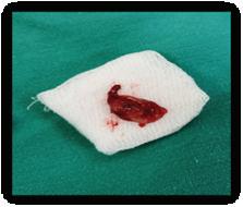
Ne menduam se nuk kishte nevojë për investigime të mëtejme dhe vendosëm ta nxjerim tumorin. Operacioni u kry me anestezion të përgjithshëm, duke shfrytëzuar incizionin klasik për parotidektomi sipas Blair-it. Masa u hoq, derisa gjëndra parotide mbeti intakte, pasi lipomi ishte qartë i kufizuar dhe i lehtë për disektim. Egzampleri ishte një masë e butë, e verdhë, mire e mbështjellur me dimenzion reth 6 cm. Ishte lehtë të dallohet lipomi nga indet përreth si dhe nga vetë gjëndra parotide.
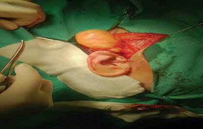
Fig. 2. Fotografi intraoperative gjatë hekjes së tumorit (Blair incision) Fig. 1. Preoperative picture of 67 years-old patient with a lipoma of the parotid gland. Notice the left eyelid and tears leaking from the eye.
We felt that there was no need for further investigations and decided to remove the tumor. Under general anesthesia using a classic Blair’s incision, an operation was performed. The mass was removed while the parotid gland remained intact, because the lipoma was clearly limited and easy to dissect. The specimen was a soft, yellowish, wellcircumscribed mass, measuring 6 cm. It was easy to distinguish the lipoma from the surrounding tissue and parotid gland itself.
Fig.2. Intraoperative picture removing the lipoma
Vëreni qartë kufirin mes tumorit dhe indeve përeth dhe lehtësia e disektimit. cerebrovascular insult 10 years ago, and according to her testimony, no consequences were present.
Nedim Kasami, Vladimir Popovski LIPOM I GJËNDRËS PAROTIDE: PREZENTIM RASTI DHE PASQYRIM I LITERATURËS
Nedim Kasami, Vladimir Popovski LIPOMA OF PAROTI GLAND: A CASE REPORT AND LITERATURE REVIEW
Fig. 3. Pamje intraoperative (gati se në tërësi i nxjerë tumori)
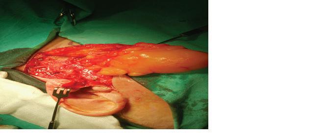
Fig 4. Shtrati operativ pas nxjerjes se lipomit. Gjëndra parotide është intakte poashtu edhe nervi i madh aurikular ( Greater Auricular Nerve - GAN ).
Prerjet histologjike treguan lobuluse te indit adipoz të ndara me një zone të hollë të stromës së indit lidhor. Egzaminimet pato-histologjike konfirmuan dijagnoze të lipomit. Nuk u vërejt recidiv i tumorit 12 muaj pas ndërhyrjes kirurgjikale.
Histological sections showed lobules of adipose tissue separated by a thin core of connective tissue stroma. Histological examination confirmed the diagnosis of lipoma. No recurrence was seen 12 months after surgery.
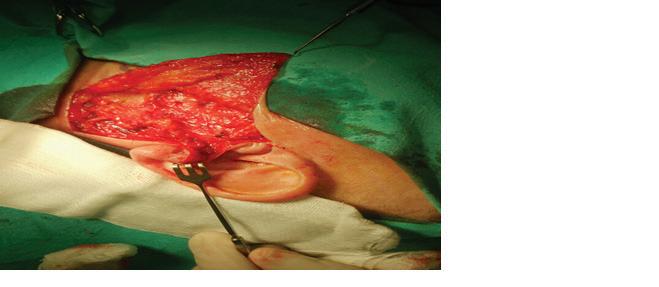
Fig. 5. Prerje histologjike e lipomit të gjëndrës parotide (Hematoxylin–Eosin) Fig.3. Intraoperative picture
Fig 4. Operative bed after the removal of the lipoma. The parotid gland is intact as well as greater auricular nerve.
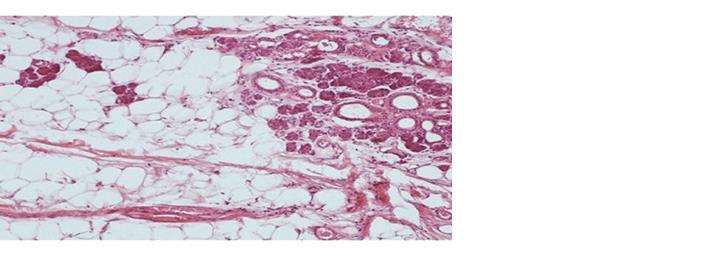
Fig. 5. Histological section of parotid gland lipoma (Hematoxylin–Eosin)
Nedim Kasami, Vladimir Popovski LIPOM I GJËNDRËS PAROTIDE: PREZENTIM RASTI DHE PASQYRIM I LITERATURËS
Nedim Kasami, Vladimir Popovski LIPOMA OF PAROTI GLAND: A CASE REPORT AND LITERATURE REVIEW
DISKUSION
Lipoma e regjionit të kokës dhe qafës paraqet tumor beninj mezenhimal, me paraqitje prej 15-20% të krejt lipomave të trupit ku llogariten prej 0.1-5% nga krejt tumoret beninje (2). Sipas nji studimi të madh të lipomave që e përfshinë regjionin maxilofacial (12), distribuimi i lipomave është gjetur të lokalizojnë zonën parotide dhe bukale me gati se përqindje të ngjajshme 23%, pastaj vinë buzët, area submandibulare, gjuha dhe qiellza.
Në një studim tjetër, autorët zbuluan vetëm tetë raste (1,3 %) të jenë lezione lipomatoze (12). Termini lezion lipomatoz është nji përshkrim i lezioneve që kryesisht përfshinë dy lloje histologjike, lipomi fokal dhe lipomatozë infiltrative difuze. Në rastin e lipomit fokal, investigimet histologjike tregojnë ind adipoz të pjekur të ndarë nga parenhimi i gjëndrës parotide me një kapsulë fibroze, derisa te lipomatoza difuze qelizat e indit adipoz duket të infiltrojnë parenhimin e gjëndrës parotide (12). Në rastin tonë bëhej fjalë për një lezion lipomatoz fokal. Lipomat e gjëndrës parotide kanë pedileksion te meshkujt. Distribuimi sipas moshës është i gjërë, edhe pse lipomët janë gjetur në dekadën e pestë dhe të gjashtë. Disa raste janë përshkruar te fëmijët (15).
Dijagnostikimi klinik i lipomit të parotidës nuk është i vështirë. Ato të vendosur në lobin supreficijal zakonisht paraqiten si një ënjti pa dhimbje, asipmtomatike dhe që ritet ngadalë. Koha mesatare e paraqitjes së lipomit para ekscizionit është 3,2 vjet (11). Në rastin tonë koha e paraqitjes së lipomit para hekjes ishte reth 4 vjet. Sipas literaturës, lipomat mund të shkaktohen nga trashëgimi, obesiteti, diabeti, rezatimi, çregullimet endokrine, injekcionet e insulinës, terapia kortikosteroide dhe trauma (16). Në rastin tonë, obesiteti, diabeti dhe injeksionet e insulinës konsiderohen si faktorë të mudshëm etiologjik.
Hulumtimet preoperative luajnë rol kyç në dijagnostikimin e saktë të natyrës dhe vendit të lezioneve. Rezonanca magnetike dhe Tomografija e kompjuterizuar mund të jenë të dobishëm në dhënien e informatave reth karakteristikave patologjike, ta vlerësojnë lokacionin si dhe marëdhenien me strukturat anatomike të rëndësishme.
Mirëpo, CT nuk ndihmon shumë në diferencimin e lipomit nga indet adipoze përeth. Kjo informatë mund të meret më qartë nga rezonanca magnetike (17). Te rasti jonë investigimet preoperative me US dhe FNAC raportuan ind lipomatoz pa përfshirje të gjëndrës parotide dhe përkundër qëndrimeve të Furlongut (11) DISCUSSION
Lipoma of the head and neck region is benign mesenchymal tumor, accounting for 15–20% of total lipoma of the body, where they account for 0.1–5% of all benign tumors [2]. According to a large study of lipomas affecting the oral and maxillofacial area [12] in a total of 125 cases, the distribution of lipoma has been found to locate in the parotid and buccal area with almost similar percentages 23%, followed by the lips, submandibular area, tongue and palate. In another study of 638 parotidectomies, the authors found only eight cases (1.3%) to be lipomatous lesions [12]. The term lipomatous lesion is an extensive description of these kinds of lesions mainly including two histological types, focal lipoma and diffuse infiltrating lipomatosis. In the case of focal lipoma, histopathologic investigation reveals mature adipose tissue to be separated from parotid gland parenchyma with a fibrous capsule while in diffuse lipomatosis adipose tissue cells found to infiltrate parotid gland parenchyma [12]. In our case it was focal lipomatous lesion. Lipomas of the parotid gland have a predilection to males. The age distribution is wide even though lipomas of the parotid gland are found in the fifth and sixth decade. Those in deep lobe have been reported to be highest in the fourth decade. Few cases have been described in pediatric patients [15].
Clinical diagnosis of the parotid gland lipoma is not difficult. Those situated at superficial parotid lobe usually appear as a painless, asymptomatic and slowgrowing swelling. Mean time of lipoma presence before its excision is 3.2 years [11]. In our case, the presence of lipoma before its surgical excision was 4 years.
According to the literature, lipomas can be caused by heredity, obesity, diabetes, radiation, endocrine disorders, insulin injection, corticosteroid therapy and trauma [16]. In our case, obesity, diabetes and insulin injection are considered as possible etiological factors.
The preoperative imaging has a crucial role to correctly diagnose the nature and the location of lesions. CT scan and MRI can be helpful in giving information about pathological features of the tumors, to evaluate its location and its relation to important anatomic structures.
However, CT scan help does not much in differentiation of lipoma from surrounding adipose tissue. This information can be clearly collected from the MRI scan [17]. In our case preoperative US and FNAC revealed lipomatous tissue without interfering the parotid gland, and despite the attitude of Furlong et al.( 11) and Ethunandan et al.(12) who advocate that
Nedim Kasami, Vladimir Popovski LIPOM I GJËNDRËS PAROTIDE: PREZENTIM RASTI DHE PASQYRIM I LITERATURËS
Nedim Kasami, Vladimir Popovski LIPOMA OF PAROTI GLAND: A CASE REPORT AND LITERATURE REVIEW
dhe Ethunandanit (12) të cilët pohojnë se FNAC te gjëndra parotide ka vlerë të vogël dijagnostike, ne mendojmë se US dhe FNAC janë të mjaftueshëm dhe vendosëm të operojmë pa invesitigime të mëtejme.
Lipomët e gjëndrës parotide superficiale mund të zhvillohen lirshëm dhe të marin përmasa të mëdhaja para se të operohen. Deformimi i fytyrës ishte shkaku kryesor i pacientit për kërkese tretmani. Në studimin tonë ajo çfar na brengoste ishin shenjat e parezës së nervit facial, por sa duket sipas krejt gjasave, këta simptoma janë pasojë e insultit cerebrospinal të mëhershëm dhe përfshirjes së sistemit trigeminal, me pasoja të lehta ndaj nervit facijal.
KONKLUZION
Lipomi i gjëndrës parotide paraqet tumor të rallë beninj, i cili duhet të konsiderohet gjatë dijagnostikimit diferencijal të lezioneve të masave të gjëdrës parotide. Egzaminimi klinik, anamneza e gjërë dhe inçitimet adekuate, çojnë deri te diagnoza e saktë. Operacion minucioz duhet kryer me qëllim të shmangjes së ngjarjeve të padëshiruara shqetësuese.
LITERATURA
1. Eneroth CM, Bradley PJ, Mcgurk M. Salivary gland tumors in the parotid gland, submandibular gland, and the palate region. Cancer (1971) 27(6):1415–8. 2. Weiss SW, Goldblum JR (2001) Chapter 16:
‘‘Benign Lipomatous Tumors’’. In: Weiss SW,
Goldblum JR (eds) Enzinger and Weiss’s soft tissue tumors, 4th edn. Mosby, St Louis, p 571. 3. Dispenza F, De Stefano A, Romano G, Mazzoni A (2008) Posttraumatic lipoma of the parotid gland: case report. Acta Otorhinolaryngol Ital 28:87–88. 4. Srinivasan V, Gamesan S, Premachandra J (1996)
Lipoma of the parotid gland presenting with facial palsy. J Laryngol Otol 110:93–95. 5. Vander Poorten VL, Balm AJ, Hilgers FJ, Tan IB,
Loftus-Coll BM, Keus RB, et al. The development of a prognostic score for patients with parotid carcinoma. Cancer (1999) 6. Guntinas-Lichius O, Wendt TG, Buentzel J, Esser
D, Böger D, Mueller AH,et al.Incidence, treatment, and outcome of parotid carcinoma, 1996-2011: a population-based study in Thuringia, Germany. J
Cancer Res Clin Oncol(2015) 141(9):1679–88.
FNAC in parotid gland is of little diagnostic value, we think that US and FNAC are sufficient and decided to perform surgery without further imaging investigations ( CT and MRI ).
Lipomas of the superficial parotid gland could develop freely and take long dimensions before its excision. Facial disfigurement was the main incentive for the patient to seek treatment. In our study, what worries us are the signs of paresis of facial nerve, but in all likelihood, it seems that the symptoms are consequence of previous cerebrovascular insult and affection of the trigeminal system, with slight sequels to the facial nerve.
CONCLUSION
In conclusion, lipoma of the parotid gland is a rare benign pathology, which should be considered in the differential diagnosis of parotid gland’s mass lesions. Clinical examination, comprehensive anamnesis and appropriate imaging leads to efficient diagnosis. Meticulous surgical excision should be performed to avoid disturbing adverse events.
REFERENCES
1. Eneroth CM, Bradley PJ, Mcgurk M. Salivary gland tumors in the parotid gland, submandibular gland, and the palate region. Cancer (1971) 27(6):1415–8. 2. Weiss SW, Goldblum JR (2001) Chapter 16:
‘‘Benign Lipomatous Tumors’’. In: Weiss SW,
Goldblum JR (eds) Enzinger and Weiss’s soft tissue tumors, 4th edn. Mosby, St Louis, p 571. 3. Dispenza F, De Stefano A, Romano G, Mazzoni A (2008) Posttraumatic lipoma of the parotid gland: case report. Acta Otorhinolaryngol Ital 28:87–88. 4. Srinivasan V, Gamesan S, Premachandra J (1996)
Lipoma of the parotid gland presenting with facial palsy. J Laryngol Otol 110:93–95. 5. Vander Poorten VL, Balm AJ, Hilgers FJ, Tan IB,
Loftus-Coll BM, Keus RB, et al. The development of a prognostic score for patients with parotid carcinoma. Cancer (1999) 6. Guntinas-Lichius O, Wendt TG, Buentzel J, Esser
D, Böger D, Mueller AH,et al.Incidence, treatment, and outcome of parotid carcinoma, 1996-2011: a population-based study in Thuringia, Germany. J
Cancer Res Clin Oncol(2015) 141(9):1679–88.
Nedim Kasami, Vladimir Popovski LIPOM I GJËNDRËS PAROTIDE: PREZENTIM RASTI DHE PASQYRIM I LITERATURËS
Nedim Kasami, Vladimir Popovski LIPOMA OF PAROTI GLAND: A CASE REPORT AND LITERATURE REVIEW
7. Brennan PA, Ammar M, Matharu J. Contemporary management of benign parotid tumours - the increasing evidence for extracapsular dissection.
Oral Dis (2017) 23(1):18–21. 8. Gritzmann N, Rettenbacher T, Hollerweger A,
Macheiner P, Hübner E. Sonography of the salivary glands. Eur Radiol (2003) 13(5):964–75. 9. Brennan PA, Herd MK, Howlett DC, Gibson D,
Oeppen RS. Is ultrasound alone sufficient for imaging superficial lobe benign parotid tumours before surgery? Br J Oral Maxillofac Surg (2012) 50(4):333–7. 10.Liu CC, Jethwa AR, Khariwala SS, Johnson J, Shin
JJ. Sensitivity, specificity, and posttest probability of parotid fine-needle aspiration: a systematic review and meta-analysis. Otolaryngol Head Neck
Surg (2016) 154(1):9–23. 11.Furlong MA, Fanburg-Smith JC, Childers ELB (2004) Lipoma of the oral and maxillofacial region: site and subclassification of 125 cases.
Oral Surg Oral Med Oral Pathol Oral Radiol
Endod 98(4):441–450. 12.Ethunandan M, Vura G, Anand R, Macpherson
DW, Wilson AW (2006) Lipomatous lesions of the parotid gland. J Oral Maxillofac Surg 64:1583–1586. 13.Haidar YM, Moshtaghi O, Mahmoodi A, Helmy
M, Goddard JA, Armstrong WB. The utility of
In-office ultrasound in the diagnosis of parotid lesions. Otolaryngol Head Neck Surg (2017) 156(3):511–7. 14.House JW, Brackmann DE. Facial nerve grading system. Otolaryngol Head Neck Surg. 1985;93:146-7. 15.Esposito C, Califano L, D’Armiento M, Longo F (2000) Lipomatosis of the parotid gland in a child.
Br J Plast Surg 53:699–701. 16.Layfield IJ, Glasgow BJ, Goldstein N et al (1991)
Lipomatous lesions of the parotid gland—potential pitfalls in fine needle aspiration biopsy diagnosis.
Acta Cytol 35:553–556. 17.Som PM, Sacher M, Stollman MD, Biller HF,
Lawson W (1988) Common tumors of the parapharyngeal space: refined imaging diagnosis.
Radiology 169:81–85. 7. Brennan PA, Ammar M, Matharu J. Contemporary management of benign parotid tumours - the increasing evidence for extracapsular dissection.
Oral Dis (2017) 23(1):18–21. 8. Gritzmann N, Rettenbacher T, Hollerweger A,
Macheiner P, Hübner E. Sonography of the salivary glands. Eur Radiol (2003) 13(5):964–75. 9. Brennan PA, Herd MK, Howlett DC, Gibson D,
Oeppen RS. Is ultrasound alone sufficient for imaging superficial lobe benign parotid tumours before surgery? Br J Oral Maxillofac Surg (2012) 50(4):333–7. 10.Liu CC, Jethwa AR, Khariwala SS, Johnson J, Shin
JJ. Sensitivity, specificity, and posttest probability of parotid fine-needle aspiration: a systematic review and meta-analysis. Otolaryngol Head Neck
Surg (2016) 154(1):9–23. 11. Furlong MA, Fanburg-Smith JC, Childers ELB (2004) Lipoma of the oral and maxillofacial region: site and subclassification of 125 cases.
Oral Surg Oral Med Oral Pathol Oral Radiol
Endod 98(4):441–450. 12.Ethunandan M, Vura G, Anand R, Macpherson
DW, Wilson AW (2006) Lipomatous lesions of the parotid gland. J Oral Maxillofac Surg 64:1583–1586. 13.Haidar YM, Moshtaghi O, Mahmoodi A, Helmy
M, Goddard JA, Armstrong WB. The utility of
In-office ultrasound in the diagnosis of parotid lesions. Otolaryngol Head Neck Surg (2017) 156(3):511–7. 14.House JW, Brackmann DE. Facial nerve grading system. Otolaryngol Head Neck Surg. 1985;93:146-7. 15.Esposito C, Califano L, D’Armiento M, Longo F (2000) Lipomatosis of the parotid gland in a child.
Br J Plast Surg 53:699–701. 16.Layfield IJ, Glasgow BJ, Goldstein N et al (1991)
Lipomatous lesions of the parotid gland—potential pitfalls in fine needle aspiration biopsy diagnosis.
Acta Cytol 35:553–556. 17.Som PM, Sacher M, Stollman MD, Biller HF,
Lawson W (1988) Common tumors of the parapharyngeal space: refined imaging diagnosis.
Radiology 169:81–85.