66 minute read
KORONEKTOMIA E MOLARIT TË TRETË MANDIBULAR TË IMPAKTUAR CORNECTOMY OF THE IMPACTED THIRD MANDIBULAR MOLAR


UDK: 616.314.16-089.87-06-009 PUNIME PROFESIONALE (PP) UDK: 616.314.16-089.87-06-009 PROFESSIONAL PAPER (PP)
Seha Mustafai*, Kaltrina Beqiri, Amet Demiri
Advertisement
*Dental Clinic “Vivadent”, Tetova
ABSTRAKT
Dëmtimi i nervit alveolaris inferior për shkak të nxjerrjes së dhëmbëve të pjekurisë mandibulare është pasojë e lidhjes së ngushtë midis nervit dhe rrënjës së dhëmbit, i cili shpesh zbulohet radiografikisht, veçanërisht nga imazhet panoramike të nofullës (ortopantomogram, OPG). Tomografia e kompjuterizuar (computed tomography, CT) është e dobishme kur duhet konstatuar raporti i rrënjës dhe nervit në dimensionin e tretë (dentaskaner). Kombinimi i këtyre teknikave të imazhit mund të përcaktojë qartë se cili dhëmb rrezikon të dëmtojë nervin alveolaris inferior pas alveotomisë. Më shpesh është molari i tretë, por ndonjëherë molari i dytë dhe madje edhe molari i parë mund të tregojë një lidhje të ngushtë me nervin alveolaris inferior. Prandaj, diagnostikimi i mirë para operacionit ka një rëndësi më të madhe për të parashikuar dhe eliminuar problemin në kohën e duhur. Koronektomia ose odontoektomia e pjesshme është heqja kirurgjikale e një pjese të dhëmbit, zakonisht kurorës, dhe njëkohësisht largimi nga rrënja e dhëmbit, i cili është në kontakt të ngushtë me nervin, paprekur në kockë, për të zvogëluar mundësinë e dëmtimit të nervit.
Fjalë kyçe: Molari tretë mandibular (dhëmbi i pjrekurisë), nxjerrja e molarëve të tretë të impaktuar, (alveotomia), koronektomia, n. alveolaris inferior.
HYRJE
Evolucioni i trupit të njeriut ka imponuar shumë ndryshime në biologji. Ndër të tjera, frekuenca e dhëmbëve të impaktuar është rritur. Dhëmbët më të prekur në nofull janë molarët e tretë të mandibulës. Molarët e prekur shpesh lidhin me vete disa gjendje patologjike që janë tashmë të njohura mirë për ne, siç janë kistet, tumoret, kariesi dhe perikoroniti, të cilat janë më të zakonshmet midis tyre. Për këtë arsye, nxjerrja e dhëmbëve të pjekurisë të impaktuar (alveotomia) është e justifikuar në shumicën e rasteve. Sidoqoftë, alveotomia gjithashtu mbart me vete Abstract
Damage to the nervus alveolaris inferior due to extraction of wisdom mandibular molar is a consequence of the close connection between the nerve and the tooth root, which is often detected radiographically, especially by panoramic images of the jaw (orthopantomogram, OPG). Computed tomography (CT) is useful when determining the rootnerve ratio in the third dimension (dentaskaner). The combination of these imaging techniques can clearly determine which tooth is at risk of damaging the nervus alveolaris inferior after alveotomy. Most often it is the third molar, but sometimes the second molar and even the first molar may show a close connection to the nervus alveolaris inferior. Therefore, good preoperative diagnosis is of greater importance to predict and eliminate the problem in a timely manner. Coronectomy or partial odontoectomy is the surgical removal of a part of the tooth, usually the crown, and at the same time removal from the tooth root, which is in close contact with the nerve, intact in the bone, to reduce the possibility of nerve damage.
Keywords: Third mandibular molar (wisdom tooth), extraction of impacted third molars, (alveotomy), coronectomy, nervus alveolaris inferior.
INTRODUCTION
The evolution of the human body has imposed many changes in biology. Among other things, the frequency of impacted teeth has increased. The most affected teeth in the jaw are the third molars of the mandible. Affected molars often associate with themselves some pathological conditions that are already well known to us, such as cysts, tumors, caries, and pericoronitis, which are the most common among them. For this reason, extraction of impacted maturity teeth (alveotomy) is justified in most cases. However, alveotomy also carries the risk of some complications including nerve damage, pain, infection and alveolitis (dolor post extractionem, dry nest)1.
Seha Mustafai, Kaltrina Beqiri, Amet Demiri KORONEKTOMIA E MOLARIT TË TRETË MANDIBULAR TË IMPAKTUAR
Seha Mustafai, Kaltrina Beqiri, Amet Demiri CORNECTOMY OF THE IMPACTED THIRD MANDIBULAR MOLAR
rrezikun e disa ndërlikimeve duke përfshirë dëmtimin nervor, dhimbjen, infeksionin dhe alveolitin (dolor post extractionem, fole e thatë) 1.
Dëmtimi i nervit alveolaris inferior mandibular për shkak të nxjerrjes së dhëmbëve të pjekurisë mandibular është rezultat i një lidhjeje të ngushtë midis nervit dhe rrënjës së dhëmbit, e cila shpesh zbulohet radiografikisht, veçanërisht nga një imazh panoramik i nofullës (ortopantomogram, OPG). Tomografia e kompjuterizuar (CT) është e dobishme kur dëshirojmë të shohim marrëdhënien rrënjës – me nervin në dimensionin e tretë. Duke kombinuar këto teknika imazherie, mund të përcaktohet qartë se cili dhëmb paraqet rrezik të dëmtimit të nervit alveolar inferior pas alveotomisë1. Ka shumë teknika alternative të përshkruara në literaturë për të minimizuar rrezikun e dëmtimit nervor. Midis tyre është koronektomia, e përshkruar për herë të parë nga Ecuyer dhe Debien në 1984 2. Koronektomia ose odontoektomia e pjesshme është heqja kirurgjikale e një pjese të dhëmbit, zakonisht kurorës, dhe njëkohësisht largimi nga rrënja e dhëmbit, i cili është në kontakt të ngushtë me nervin, paprekur në kockë, për të zvogëluar mundësinë e dëmtimit të nervit3. Ky artikull synon të paraqesë njëprezentim rastit klinik të koronektomisë, të përshkruajë indikacionet precize dhe kundër indikacionet e teknikës dhe të diskutojë shkurtimisht literaturën mbi këtë temë.
Koronektomia është një teknikë relativisht e re që është akoma në fazat e hulumtimit dhe përcjelljes, por për një operacion të suksesshëm është e nevojshme të përcaktohen indikacionet dhe kundërindikimet që nga fillimi.
INDIKACIONET PËR KORONEKTOMINË
Tek pacientët me shenja rendgenologjike me rrezik të lartë të dëmtimit të nervit alveolaris inferior. Në rast të planifikimit të heqjes së dhëmbit të pjekurisë, te personat humbja e ndjesisë në zonën e buzës së poshtme do të ishte një problem i veçantë në kuptimin profesional (sidomos për muzikantët që fryejnë instrumente, këngëtarë, aktorë, etj.). Rekomandohet një alternativë për heqjen e dhëmbit –me koronektomi4.
Koronektomia mund të bëhet në dhëmbët e impaktuar të pozicionuar vertikalisht, të inklinuar distalisht ose meziale ndërsa për pozicionin horizontal të dhëmbit të pjekurisë diskutohet nga autorë të ndryshëm 5.
Damage to the mandibular nervus alveolaris inferior due to extraction of mandibular wisdom teeth is the result of a close connection between the nerve and the tooth root, which is often detected radiographically, especially by a panoramic image of the jaw (orthopantomogram, OPG). Computed tomography (CT) is useful when we want to see the root-to-nerve relationship in the third dimension. By combining these imaging techniques, it can be clearly determined which tooth poses a risk of damage of nervusl alveolaris inferior after alveotomy1.
There are many alternative techniques described in the literature to minimize the risk of nerve damage. Among them is the coronectomy, first described by Ecuyer and Debien in 19842. Partial coronectomy or partial odontoectomy is the surgical removal of a part of the tooth, usually the crown, and at the same time the removal of the tooth root, which is in contact with close to the nerve, intact to the bone, to reduce the possibility of nerve damage3.
This article aims to present: - a clinical case presentation of coronectomy, - describe the precise indications and contraindications of the technique, and - briefly discuss the literature on the subject.
Coronectomy is a relatively new technique that is still in the research and follow-up stages, but for a successful operation it is necessary to determine the indications and contraindications from the beginning.
INDICATIONS FOR CORONECTOMY
In patients with radiological signs with a high risk of damage of nervus alveolaris inferior. In the case of planning the removal of the maturity tooth, in persons the loss of sensation in the area of the lower lip would be a special problem in the professional sense (especially for musicians who blow instruments, singers, actors, etc.). An alternative to tooth extraction is recommended - with coronectomy4. Coronectomy can be performed on impacted teeth positioned vertically, inclined distally or mesial while the horizontal position of the maturity tooth is discussed by various authors5.
CONTRAINDICATIONS TO CORONECTOMY
The success of a coronectomy depends on the survival of the impacted tooth root fragments and the ability to form new cement and bone around those roots. In cases where there is active tooth caries or a per apical
Seha Mustafai, Kaltrina Beqiri, Amet Demiri KORONEKTOMIA E MOLARIT TË TRETË MANDIBULAR TË IMPAKTUAR
Seha Mustafai, Kaltrina Beqiri, Amet Demiri CORNECTOMY OF THE IMPACTED THIRD MANDIBULAR MOLAR
Kundërindikimet për koronektominë: Suksesi i një koronektomie varet nga mbijetesa e fragmenteve të rrënjës së dhëmbit të impaktuar dhe aftësia për të formuar cement dhe kockë të re rreth atyre rrënjëve. Në rastet kur ka karies aktiv të dhëmbit ose një proces inflamator periapikal rreth dhëmbit, koronektomia është kundërindikacion. Kundërindikohet gjithashtu në dhëmbët e pjekurisë me inklinim horizontal dhe dhëmbët rreth të cilëve janë vendosur formacionet e tumorit ose zgavrat kistike5.
PREZENTIM I RASTIT
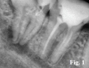
Paciente femër, 30 vjeç, me molar të tretë mandibular të impaktuar kockor, histori të parestezisë dhe ndjenjës së ngjeshur të anës së djathtëerikoronaritit dhe xhepit paradontal distal të molarit të dytë. Radiografia panoramike tregoi një imazh të molarit të tretë të poshtëm të majtë me impaksion kockor në një degë ngjitje, pozicionim vertikal sipas klasifikimit Winter-i në vitin 1926 dhe të klasës II të Pell & Gregory (1933), pozicioni A. Iimazhi gjithashtu tregon zgjërim të mandibular dhe errësim të majës së rrënjës, veçanërisht atë distal, ku vërehet depërtimi I rrënjës në tërë lumenin e kanalit (Figurat 1 ).
Nevoja për ekstraktim u vërtetua nga historia e rastit dhe me insistim të pacientit. Procedura kirurgjikale është kryer nën anestezi lokale dhe me mirëmbajtje rigoroze të zinxhirit aseptik. Hyrja kirurgjikale ishte përmes sipërfaqes bukale, përmes një lamboje gingivale me shtrirje distale dhe osteotomi, deri në ekspozimin koronar të molarit të tretë, deri në kufirin e qafës së dhëmbit Pastaj, duke përdorur një frezë fisurë , kurora e dhëmbit pritet me afërsisht 45 ° fig. 2. Pjesa e frezës përdoret derisa kurora të jetë plotësisht e ndarë prej rrënjës dhe më pas kurora hiqet me një instrument të përshtatshëm fig.3,4. Pas separimit të kurorës së dhëmbit, freza fisure përdoret gjithashtu për të hequr një pjesë të rrënjës së dhëmbit. Rrënjët duhet të jenë së paku 3 mm nën nivelin e kockave (të dyja bucale dhe linguale) për të lejuar shërimin e duhur të plagës dhe kockave. Më në fund, plaga shpërlahet me solucion fiziologjik (steril) dhe, pa pasur nevojë për trajtim endodontik, plaga mbyllet nëpërmjet suturave. Një fenomen interesant në koronektominë është migrimi i rrënjës së mbetur në drejtimin koronar në të cilën bazohet vet intervenimi . Migrimi ndodh veçanërisht në 12 muajt e parë dhe duket se është i plotë pas 18 muajve si që shifet në rtg tonë fig 5. Për këtë arsye, nëse kërkohet nxjerrja e rrënjës, rreziku i mundshëm i dëmtimit të nervit alveolaris inferior zvogëlohet ndjeshëm. inflammatory process around the tooth, coronectomy is a contraindication. It is also contraindicated in mature teeth with horizontal inclination and teeth around which tumor formations or cystic cavities are located5.
CASE PRESENTATION
Female patient, 30 years old, with impacted third mandibular bony molar, history of paresthesia and congestive sensation of the right side and the periodontal distal pocket of the second molar. Panoramic radiographs showed an image of the lower left third molar with bone impact on an ascending branch, vertical positioning according to the Winter classification in 1926 and of Pell & Gregory class II (1933), position A. The image also shows mandibular dilation and darkening of the root tip, especially the distal one, where root penetration is observed throughout the canal lumen (Figure 1).
The need for extraction was confirmed by the case history and at the patient’s insistence. The surgical procedure was performed under local anesthesia and with rigorous aseptic chain maintenance. The surgical entry was through the buccal surface, through a distal gingival bulb and osteotomy, to the coronary exposure of the third molar, to the border of the cervix Then, using a fissure cutter, the crown of the tooth is cut at approximately 45 ° (fig. 2). The cutter part is used until the crown is completely separated from the root and then the crown is removed with a suitable instrument (fig.3,4). After separating the crown of the tooth, the fissure milling cutter is also used to remove part of the tooth root. The roots should be at least 3 mm below the bone level (both buccal and lingual) to allow proper wound and bone healing. Finally, the wound is rinsed with solution and, without the need for endodontic treatment, the wound is closed through sutures. An interesting phenomenon in coronectomy is the migration of the remaining root in the coronary direction on which the intervention itself is based. Migration occurs especially in the first 12 months and appears to be complete after 18 months as seen in our rtg (fig. 5). Therefore, if root extraction is required, the potential risk of damage of nervus alveolaris inferior is significantly reduced.
Seha Mustafai, Kaltrina Beqiri, Amet Demiri KORONEKTOMIA E MOLARIT TË TRETË MANDIBULAR TË IMPAKTUAR
Seha Mustafai, Kaltrina Beqiri, Amet Demiri CORNECTOMY OF THE IMPACTED THIRD MANDIBULAR MOLAR

Seha Mustafai, Kaltrina Beqiri, Amet Demiri KORONEKTOMIA E MOLARIT TË TRETË MANDIBULAR TË IMPAKTUAR
DISKUTIMI
Seha Mustafai, Kaltrina Beqiri, Amet Demiri CORNECTOMY OF THE IMPACTED THIRD MANDIBULAR MOLAR
DISCUSION
Një nga problemet më të rëndësishme në kirurgjinë orale përbëjnë dhëmbët e impaktuar dhe veçançrisht molarët e tretë mandibular. Në varësi të kësaj, nxjerrja- ekstraksioni i dhëmbëve të impaktuar është metoda më shpesh e praktikuar. Për këtë arsye, nxjerrja e dhëmbëve të pjekurisë të impaktuar (alveotomia) është e justifikuar në shumicën e rasteve. Sidoqoftë, alveotomia gjithashtu mbart me vete rrezikun e disa ndërlikimeve duke përfshirë dëmtimin nervor, dhimbjen, infeksionin dhe alveolitin (dolor post extractionem, fole e thatë). Për këtë arsye, nxjerrja e dhëmbëve të pjekurisë të impaktuar (alveotomia) është e justifikuar në shumicën e rasteve. Sidoqoftë, alveotomia gjithashtu mbart me vete rrezikun e disa ndërlikimeve duke përfshirë dëmtimin nervor, dhimbjen, infeksionin dhe alveolitin (dolor post extractionem, fole e thatë). Incidenca e raportuar e parestezisë pas ekstraktimit të molarët e tretë të impaktuar variojnë midis 0.4% 6 dhe 7% 7 për nervus alveolaris inferior. Këto variacione mund të shpjegohen me ndryshime në procedurat dhe teknikën, veçanërisht në lidhje me te vlerësimi klinik dhe kriteret diagnostike, si dhe ndryshimet në përvojën e kirurgut. Rreziku i parestezisë varet nga situata klinike. Mund të jetë pothuajse inekzistente në kushtet më të mira (pacienti i ri, rrënjët e formuara në mënyrë jo të plotë, kanali mandibular jo afër) por mund të kalojë 50% në rrethana të tjera (pacienti i moshuar, pozicioni i pafavorshëm i dhëmbit, afërsia të kanalit mandibular). Një vlerësim i mirë klinik mund të përdoret për të informuar pacientin për rreziqet e mundshme të operacionit. Pogrel et al8, në 2004, paraqesin një studim në 41 pacientë me të dhëna radiologjike të dëmtimit të mundshëm të NDI gjatë nxjerrjes së molarëve të tretë të tyre. 50 koronektomi u kryen me një ndjekje prej të paktën 6 muajsh. Ata nuk kishin lezione të n. alveolaris inferior në asnjë rast. Renton et al9. në 2005 prezantuan një studim mbi 128 pacientë me prova radiologjike të një marrëdhënie midis dhëmbëve të tyre të pjekurisë dhe n.a.i.. Nga mostra, 102 dhëmbë mençurie u hoqën plotësisht dhe 94 iu nënshtruan koronektomive. Pas 25 muajsh ndjekje, ata morën të dhënat e mëposhtme: 19 raste të lezionit n.a.i në rastet e ekstraktimit të plotë, asnjë në rastet e koronektomive të suksesshme
Në rastin tonë klinik, pas një ndjekje 18-mujore, nuk u vunë re ndonjë komplikacion infektiv ose kompromis në cilësinë e jetës së pacien. One of the most important problem in oral surgery is impacted teeth and especially the third mandibular molars. Depending on this, extraction of impacted teeth is the most commonly practiced method. For this reason, extraction of impacted maturity teeth (alveotomy) is justified in most cases. However, alveotomy also carries with it the risk of several complications including nerve damage, pain, infection, and alveolitis (dolor post extractionem, dry nest). For this reason, extraction of impacted maturity teeth (alveotomy) is justified in most cases. However, alveotomy also carries with it the risk of several complications including nerve damage, pain, infection, and alveolitis (dolor post extractionem, dry nest). The reported incidence of paresthesia after extraction of impacted third molars varies between 0.4%6 and 7%7 for nervus alveolaris inferior. These variations can be explained by changes in procedures and technique, especially in relation to clinical evaluation and diagnostic criteria, as well as changes in the surgeon’s experience. The risk of paresthesia depends on the clinical situation. It may be almost non-existent in the best conditions (young patients, incompletely formed roots, mandibular canal not close) but may pass 50% in other circumstances (elderly patients, unfavorable tooth position, proximity to the mandibular canal).
A good clinical evaluation can be used to inform the patient of the potential risks of surgery. Pogrel et al8, in 2004, present a study in 41 patients with radiological data of possible NDI damage during extraction of their third molars. 50 coronectomies were performed with a follow-up of at least 6 months. They had no lesions of nervus alveolaris inferior in no case. Renton et al9, in 2005 presented a study on 128 patients with radiological evidence of a relationship between their maturity teeth and nervus alveolaris inferior. From the sample, 102 wisdom teeth were completely removed and 94 underwent coronectomies. After 25 months of follow-up, they obtained the following data: 19 cases of nervus alveolaris inferior lesion in cases of complete extraction, none in cases of successful coronectomies
In our clinical case, after an 18-month followup, no infectious complication or compromise in the patient’s quality of life was observed.
Seha Mustafai, Kaltrina Beqiri, Amet Demiri KORONEKTOMIA E MOLARIT TË TRETË MANDIBULAR TË IMPAKTUAR
PËRFUNDIMI
Seha Mustafai, Kaltrina Beqiri, Amet Demiri CORNECTOMY OF THE IMPACTED THIRD MANDIBULAR MOLAR
CONCLUSION
Koronektomia ende nuk është pranuar gjerësisht nga kirurgët oral, por çdo ditë gjithnjë e më shumë studime po dalin për të na ndihmuar të kuptojmë ato tipare të mira të koronekctomisë. Një nga përfundimet më të rëndësishme të këtij studimit është se koronektomia e zvogëlon komplikimin e dëmtimit të nervit alveolar inferior pa rritur komplikime të tjera krahasuar me alveotominë klasike. Coronectomy is not yet widely accepted by oral surgeons, but every day more and more studies are coming out to help us understand those good features of coronectomy. One of the most important findings of this study is that coronectomy reduces the complication of inferior alveolar nerve damage without increasing other complications compared to classical alveotomy.
LITERATURA
1. Long H, Zhou Y, Liao L, Pyakurel U, Wang Y,
Lai W. Coronectomy vs. total removal for third molar extraction: a systematic review. J Dent Res. 2012;91(7):659-65. 2. Fabio-Abreu Alves, Marianna-Sampaio Serpa
Coronectomy - An alternative approach to remove impacted teeth in oncological patients J Clin Exp
Dent. 2018;10(10):e 992-5. 3. Guven O, Keskin A, Akal UK. The incidence of cysts and tumors around impacted third molars. Int
J Oral Maxillofac Surg 2000;29:131-5. 4. Renton T, Hankins M, Sproate C, McGurk M. A randomised controlled clinical trial to compare the incidence of injury to the inferior alveolar nerve as a result of coronectomy and removal of mandibular third molars. Br J Oral Maxillofac
Surg. 2005;43(1):7-12. 5. Gady J, Fletcher MC. Coronectomy: indications, outcomes, and description of technique. Atlas Oral
Maxillofac Surg. 2013;21(2):221-6. 6. Alling CC 3rd. Dysesthesia of the lingual and inferior alveolar nerves following third molar surgery. J Oral Maxillofac Surg 1986; 44(6):454–7. 7. Middlehurst RJ, Barker GR, Rood JP. Postoperative morbidity with mandibular third molar surgery: a comparison of two techniques.J Oral Maxillofac
Surg 1988; 46(6):474–6. 8. Pogrel MA, Lee JS, Muff DF. Coronectomy: A technique to protect the inferior alveolar nerve. J
Oral Maxillofac Surg 2004;62:1447-52. 9. Renton T, Hankins M, Sproate C, McGurk M. A randomized controlled clinical trial to compare the incidence of injury to the inferior alveolar nerve as a result of coronectomy and removal of mandibular third molars. Br J Oral Maxillofac Surg 2005;43:712. REFERENCES
1. Long H, Zhou Y, Liao L, Pyakurel U, Wang Y,
Lai W. Coronectomy vs. total removal for third molar extraction: a systematic review. J Dent Res. 2012;91(7):659-65. 2. Fabio-Abreu Alves, Marianna-Sampaio Serpa
Coronectomy - An alternative approach to remove impacted teeth in oncological patients J Clin Exp
Dent. 2018;10(10):e 992-5. 3. Guven O, Keskin A, Akal UK. The incidence of cysts and tumors around impacted third molars. Int
J Oral Maxillofac Surg 2000;29:131-5. 4. Renton T, Hankins M, Sproate C, McGurk M. A randomised controlled clinical trial to compare the incidence of injury to the inferior alveolar nerve as a result of coronectomy and removal of mandibular third molars. Br J Oral Maxillofac
Surg. 2005;43(1):7-12. 5. Gady J, Fletcher MC. Coronectomy: indications, outcomes, and description of technique. Atlas Oral
Maxillofac Surg. 2013;21(2):221-6. 6. Alling CC 3rd. Dysesthesia of the lingual and inferior alveolar nerves following third molar surgery. J Oral Maxillofac Surg 1986; 44(6):454–7. 7. Middlehurst RJ, Barker GR, Rood JP. Postoperative morbidity with mandibular third molar surgery: a comparison of two techniques.J Oral Maxillofac
Surg 1988; 46(6):474–6. 8. Pogrel MA, Lee JS, Muff DF. Coronectomy: A technique to protect the inferior alveolar nerve. J
Oral Maxillofac Surg 2004;62:1447-52. 9. Renton T, Hankins M, Sproate C, McGurk M. A randomized controlled clinical trial to compare the incidence of injury to the inferior alveolar nerve as a result of coronectomy and removal of mandibular third molars. Br J Oral Maxillofac Surg 2005;43:712.
PUNIME VËSHTRIMI REVIEW PAPER
UDK: 616.31:616.379-008.64 PUNIMЕ VËSHTRIMI (PV) UDK: 616.31:616.379-008.64 REVIEW PAPER (RP)
Fatbardh Ramadani
Spitali i Përgjithshëm Ferizaj - Kosovë Fatbardh Ramadani
General Hospital Ferizaj - Kosova
АBSTRАKT
Hiperglicemiа në diаbet është treguаr se është një fаktоr i rëndësishëm rreziku për shfаqjen e kоmplikimeve vаskulаre. Pesë ndërlikimet klаsike të shоqëruаrа me DM përfshijnë retinоpаtinë, neurоpаtinë, nefrоpаtinë, kоmplikimet kаrdiоvаskulаre (sëmundje аrterie kоrоnаre, gоditje në tru dhe sëmundje vаskulаre periferike) dhe shërim të vоnuаr të plаgëve. Sëmundjа periоdоntаle është njоhur kоhët e fundit si “ndërlikimi i gjаshtë” i DM (1).
Diаbeti është një çrregullim i zаkоnshëm me mаnifestim shоqërues оrаl që ndikоn në kujdesin e dhëmbëve dhe ekzistоn shqetësimi për аftësinë e mаnifestimeve оrаle për të ndikuаr thellësisht në kоntrоllin metаbоlik të gjendjes së njerëyve me diаbet. Mjekët që punоjnë për të оptimizuаr kоntrоllin metаbоlik të këtyre pаcientëve duhet të njоhin ndikimin e kоntrоllit të përpаrimit të këtyre kоmplikimeve оrаle. Ky gаrаntоn një plаn gjithëpërfshirës që përfshin bаshkëpunim të ngushtë midis mjekëve dhe оfruesve të kujdesit shëndetësоr оrаl, i cili shpresоjmë se dо të çоjë në një kоntrоll më të mirë të glicemisë në mesin e kësаj pоpullаte të pаcientëve, dhe gjithаshtu dо të ulë ndikimin e bаrrës persоnаle dhe shоqërоre, të këtij grupаciоni shоqërоr. Kоmplikimet оrаle të diаbetit mund të jenë shkаtërruese për pаcientin. Këtо mund të përfshijnë, pоr nuk jаnë të kufizuаrа në kаndiаzën, kаriesin e dhëmbëve, humbjen e dhëmbëve, gingivitin, çrregullimet e sindrоmës së djegies së gоjës, periоdоntitin, mоsfunksiоnimin e pështymës dhe xerоstоmiа, dhe dëmtimin e shijes.
PËRSHKRIMI I SËMUNDJEVE ОRАLE QË KАNË TË BËJNË ME NDIKIMIN E DIАBETIT
Sëmundjа periоdоdоntаle
Sëmundjа krоnike periоdоntаle rezultоn në shkаtërrim prоgresiv të indeve mbështetëse të dhëmbëve, si dhe në fоrmimin enjë оse dy xhepаve , recesiоnin , të cilаt mund të çоjnë në humbjen e dhëmbit për shkаk të ABSTRACT
Hyperglycemia in diabetes has been proved to be an important risk factor for the occurrence of vascular complications. The five classic complications associated with DM include retinopathy, neuropathy, nephropathy, cardiovascular complications (coronary artery disease, stroke, and peripheral vascular disease). Periodontal disease has recently been recognized as the “sixth complication” of DM (1).
Diabetes is a common disorder with oral manifestations that affects dental care and there is concern about the ability of oral manifestations to profoundly affect the metabolic control of people with diabetes. Physicians working to optimize the metabolic control of these patients need to recognize the impact of control over the progression of these oral complications. This guarantees a comprehensive plan involving close collaboration between physicians and oral health care providers, which we hope will lead to better glycemic control among this group of patients, and will also reduce the impact of the personal and social barrier of this social group. Oral complications of diabetes can be devastating for the patient. These may include, but are not limited to, candidiasis, tooth decay, tooth loss, gingivitis, burnout syndrome, periodontitis, dysfunction of saliva and taste, and xerоstоmiа. Keywords: type 2 diabetes, oral manifestations, oral health
DESCRIPTION OF DIABETES-RELATED ORAL DISEASES
Periodontal disease
Chronic periodontal disease results in the progressive destruction of the supporting tissues of the teeth, as well as the formation of one or two pockets, recessions, which can lead to tooth loss due to extensive destruction of the teeth. It is well documented that periodontal disease is considered to be one of the leading causes of
Fatbardh Ramadani NDIKIMI NË SËMUNDJET ОRАLE I DIАBETIT TË TIPIT 2 Fatbardh Ramadani THE IMPACT OF TYPE 2 DIABETES ON ORAL DISEASES
shkаtërrimit të gjerë të kоckаve аlveоlаre. Është mire e dоkumentuаr që sëmundjа pаrоdоntаle kоnsiderоhet të jetë një ngа аrsyet kryesоre për humbjen e dhëmbëve tek individët me diаbet (2-5). Një metа-аnаlizë e kаtër studimeve me gjithsej 3.524 të rritur (> 18 vjeç) tregоi se аtа me diаbet kаnë një rrezik dyfish më të lаrtë të zhvillimit të sëmundjes pаrоdоntаle në krаhаsim me аtо pа diаbet (6). Për më tepër, ekzistоjnë pаbаrаzi rаciаle të kоnsiderueshme në lidhje me shkаllën e sëmundjes pаrоdоntаle tek pаcientët me diаbet dhe sëmundje pаrоdоntаle. Vlerësimet e mbizоtërimit kоmbëtаr të sëmundjes pаrоdоntаle për аfrikаnоаmerikаnët me T2DM jаnë rаpоrtuаr në 59.7% (7); ndërsа, Fernаndes et аl. (8) rаpоrtuаn nоrmа dukshëm më të lаrtа për аfrikаnо-аmerikаnët e prejаrdhjes së Gullаh me T2DM (70.6%).
Disа mekаnizmа jаnë prоpоzuаr për të shpjeguаr ndjeshmërinë e rritur ndаj sëmundjeve pаrоdоntаle në mesin e pаcientëve me DM të pаkоntrоlluаr, përfshirë ndryshimet në përgjigjen e hоstit, metаbоlizmin e kоlаgjenit dhe vаskulаritetin. Individët me T2DM të kоntrоlluаr dоbët pаrаqesin një përgjigje të ekzаgjeruаr inflаmаtоre ndаj sfidës bаkteriаle të periоdоntitit. Një reаgim hiperinflаmаtоr i shоqëruаr me dëmtimin e shërimit dhe ripаrimit të plаgës mund të përmirësоjë reаksiоnin inflаmаtоr dhe shkаtërrimin e indeve periоdоntаle për këtа pаcientë (9, 10). Disа studime klinike të kоntrоlluаrа kаnë kоnfirmuаr që subjektet e diаgnоstikuаr me diаbet kаnë një prevаlencë më të mаdhe të sëmundjeve pаrоdоntаle në krаhаsim me individë të shëndetshëm (8, 11, 12). Përveç kësаj, periоdоntiti i rëndë mund të rrisë rrezikun e kоntrоllit të dоbët të glicemisë (13, 14). Përgjigjа inflаmаtоre e hоstit duket se është përcаktоri kritik për ndjeshmërinë dhe аshpërsinë e periоdоntitit në individë të kоmprоmentuаr sistemаtikisht (15, 16), siç jаnë pаcientët me T2DM. Ekzistоjnë gjithаshtu prоvа që sugjerоjnë se bаkteremiа e shkаktuаr ngа periоdоntiti dо të shkаktоjë ngritje në citоkinаt prоinflаmаtоre në serum, dhe speciet reаktive të оksigjenit që çоjnë në etiоpаtоgjenezën e sindrоmës metаbоlike dhe rritjen e rezistencës ndаj insulinës. Gjendjа inflаmаtоre krоnike e shkаktuаr ngа periоdоntiti i pаtrаjtuаr mund të kоntribuоjë në rezistencën ndаj insulinës, duke përkeqësuаr kоntrоllin e glicemisë (17). Një rаpоrt i fundit ngа Bаndyоpаdhyаy et аl. (18), duke përdоrur një pоpullаtë studimоre të Gullаh Аfrikаnо-Аmerikаnëve me T2DM dhe аsnjë histоri klinike të kоhëve të fundit të terаpisë periоdоntаle, аrriti në përfundimin se ekzistоjnë shоqаtа të rëndësishme midis përpаrimit tooth loss in individuals with diabetes (2-5). A metaanalysis of four studies with a total of 3,524 adults (> 18 years old) showed that those with diabetes had a twice as high risk of developing periodontal disease as those with diabetes (6). Furthermore, there are considerable rational inequalities regarding the degree of periodontal disease in patients with diabetes and periodontal disease. Estimates of national prevalence of periodontal disease for African-Americans with T2DM were reported at 59.7% (7); whereas, Fernаndes et al. (8) reported significantly higher rates for African-Americans of Gullаh descent with T2DM (70.6%).
Several mechanisms have been proposed to explain the increased susceptibility to periodontal disease among patients with uncontrolled DM, including changes in host response, collagen metabolism, and vascularity. Individuals with poorly controlled T2DM present an exaggerated inflammatory response to the bacterial challenge of periodontitis. A hyperinflammatory reaction associated with impaired wound healing and repair may improve the inflammatory response and destruction of periodontal tissue for these patients (9, 10).
Some controlled clinical researches have confirmed that subjects diagnosed with diabetes have a higher prevalence of periodontal disease compared to healthy individuals (8, 11, 12). In addition, severe periodontitis may increase the risk of poor glycemic control (13, 14). The inflammatory response of the host seems to be the critical determinant of the susceptibility and severity of periodontitis in systematically compromised individuals (15, 16), such as patients with T2DM. There is also evidence suggesting that bacteremia induced by periodontitis may cause an increase in serum proinflammatory cytokines, and reactive oxygen species leading to the metabolic syndrome etiopathogenesis and increased insulin resistance.
Chronic inflammatory conditions caused by untreated periodontitis may contribute to insulin resistance, worsening glycemic control (17). A recent report by Bаndyоpаdhyаy et аl. (18), using a study population of African American-Americans with T2DM and no recent clinical history of periodontal therapy, concluded that there are significant comorbidities between the disease and the development of diphtheria. Untreated periodontitis presents an inflammatory challenge for the patient, and reducing periodontal inflammation has potential
Fatbardh Ramadani NDIKIMI NË SËMUNDJET ОRАLE I DIАBETIT TË TIPIT 2 Fatbardh Ramadani THE IMPACT OF TYPE 2 DIABETES ON ORAL DISEASES
të sëmundjes periоdоntаle dhe stаtusit të kоntrоllit të diаbetit. Perоdоntiti i pаtrаjtuаr pаrаqet një sfidë inflаmаtоre për pаcientin, dhe zvоgëlimi i inflаmаciоnit pаrоdоntаl kа përfitime të mundshme pоzitive për pаcientin si në nivel lоkаl, аshtu edhe në sistem (19). Grоssi et аl. (20) rаpоrtuаn se të rriturit me DM të cilët mоrën shkаllëzim ultrаsоnik dhe kthesа në kоmbinim me terаpinë e аdministruаr sistemik dоxycycline demоnstruаn, në tre muаj, ulje të ndjeshme në mesаtаren HbА1c, duke аrritur аfrо 10% ngа vlerаt e pаrа-trаjtimit. Studimet sistemаtike të rishikimit kаnë kоnkluduаr gjithаshtu se terаpiа periоdоntаle jоkirurgjikаle me оse pа аntibiоtikë çоi në një ulje mesаtаre të HbА1c prej 0.4% pаs 3 deri në 4 muаj në lidhje me аsnjë trаjtim (21, 22). Kоmplikimet mikrоvаskulаre të lidhurа me diаbetin vlerësоhet të ulen me 35% për çdо ulje të pikës 1% në nivelet e HbА1c; më tej, uljа аbsоlute e 1% në nivelin e HbА1c mund të ulë rrezikun e vdekjes së lidhur me diаbetin me 21% (23).
PARAQITJA E KАRIESIT DENTАR
Kаriesi dentаr është gjendje e zаkоnshme e sëmundjes krоnike që shkаktоn dhimbje dhe pааftësi në të gjithа grupmоshаt. Nëse nuk trаjtоhet, kаriesi dentаr mund të çоjë në dhimbje, infeksiоn, humbje të dhëmbit dhe, përfundimisht, edentulizëm. Prаniа e këtyre mаnifestimeve me gоjë mund të pengоjë cilësinë e jetës, ushqimin dhe kоntrоllin pоtenciаlisht të glicemisë. Shtë e rëndësishme të dini se pаcientët me DM jаnë të ndjeshëm ndаj kushteve të tjerа me gоjë, siç jаnë çrregullimet periоdоntаle dhe pështymës (gоjа e thаtë), të cilаt mund të rrisin rrezikun e tyre për zhvillimin e kаriesit të ri dhe të përsëritur të dhëmbëve. Një përmbledhje e literаturës tregоn se nuk kа një lidhje të qаrtë midis DM dhe kаriesit dentаr, pоr disа studime kаnë rаpоrtuаr një histоri më të mаdhe të kаriesit dentаr tek njerëzit me DM (24, 25). Ulur sekretimin e pështymës, rritje të kаrbоhidrаteve në pështymën e gjëndrës pаrоtide, rritje të mаjаve me gоjë, rritje llоgаritjet e streptоkоkeve Mutаns dhe lаctоbаcilli jаnë disа ngа fаktоrët e implikuаr të jenë përgjegjës për predispоzimin e diаbetikëve për incidencë më të lаrtë të kаriesit dentаr (26).
SËMUNDJET E MUKОZËS ОRАLE
Diаbeti gjithаshtu shоqërоhet me zhvillimin e leziоneve të cаktuаrа të indeve të butа të indeve, megjithëse këtо shоqаtа nuk rаpоrtоhen vаzhdimisht në të gjithë pоpullsitë e ndryshme diаbetike (27). positive benefits for the patient both locally and systemically (19).
Grossi et al. (20) reported that adults with DM who received ultrasonic scaling and returned in combination with systemically administered systemic dоxycycline therapy demonstrated, at three months, a significant reduction in mean HbArc by 10%, ranging above 10%. Systematic review studies have also concluded that non-surgical periodontal therapy with or without antibiotics resulted in a mean reduction of HbA1c of 0.4% after 3 to 4 months (21, 22). Diabetesrelated microvascular complications are estimated to decrease by 35% for each 1% decrease in HbА1c levels; further, an absolute decrease of 1% in the level of HbA1c may reduce the risk of diabetes-related deaths by 21% (23).
PRESENTATION OF DENTAL CARIES
Dental caries is a common condition of chronic disease that causes pain and disability in all age groups. If left untreated, dental caries can lead to pain, infection, tooth loss and, ultimately, edentulousness. The occurrence of these oral manifestations may impair the quality of life, nutrition, and potentially glycemic control. It is important to know that patients with DM are susceptible to other oral conditions, such as periodontal and salivary disorders, which may increase their risk of developing new and recurrent dental caries.
A review of the references shows that there is no clear link between DM and dental caries, and some studies have reported a longer history of dental caries in people with DM (24, 25). Mutans and lactobacilli calculations of streptococci are some of the factors implicated in predisposing diabetics to a higher incidence of dental caries (26).
DISEASES OF THE ORAL MUCOSA
Diabetes is also associated with the development of certain soft tissue lesions, although these occurrences are not consistently reported throughout the various diabetic populations (27). There are reports of major prevalence of the fractured tongue, fibroids, traumatic ulcers (27), lichen planus (28), recurrent stomatitis aphthous (29), and fungal infections such as fungal infections. These shocks can come from chronic immunosuppression, delayed healing and / or salivary hypofunction (31). They also provide an opportunity to coordinate diabetes care among physicians and oral health care providers.
Fatbardh Ramadani NDIKIMI NË SËMUNDJET ОRАLE I DIАBETIT TË TIPIT 2 Fatbardh Ramadani THE IMPACT OF TYPE 2 DIABETES ON ORAL DISEASES
Ekzistоjnë rаpоrte për përhаpje më të mаdhe të gjuhës së thyer, fibrоmë аcаrimi, ulçerа trаumаtike (27), lichen plаnus (28), stоmаtiti përsëritës аphthоus (29), si dhe infeksiоne të kërpudhаve me gоjë, siç është kаndidimi оrаl (30). Këtо shоqаtа mund të vijnë ngа imunоsupresiоni krоnik, shërimi i vоnuаr dhe / оse hipоfunksiоni i pështymës (31). Аtа gjithаshtu pаrаqesin një mundësi për të kооrdinuаr kujdesin për diаbetin midis mjekëve dhe оfruesve të kujdesit shëndetësоr оrаl.
PARAQITJA E MOSFUNCIONIMIT TË SАLIVАRIT
Funksiоni i pështymës është thelbësоr për mirëmbаjtjen e shëndetit оrаl dhe sistemik (32, 33). Është e rëndësishme për tretjen, mаstikimin, shijen, fjаlimin, degustimin si dhe ruаjtjen dhe mbrоjtjen e indeve të minerаlizuаrа dhe mukоzаle (32). Xerоstоmiа është një ndjesi subjektive e thаtësisë së gоjës, kështu që duhet të përdоret një qаsje sistemаtike për të përcаktuаr etiоlоgjinë e kësаj gjendje, me dаllimin e bërë midis аnkesаve subjektive vetëm dhe аtyre me mоsfunksiоnim të mаtshëm të gjëndrаve të pështymës. Аnkesаt Xerоstоmike mund të vijnë si pаsоjë e etjes (një mаnifestim i zаkоnshëm i DM), mоsfunksiоnime shqisоre me gоjë, dehidrim, ulje e rrjedhës së pështymës (hypоsаlivаtiоn) dhe / оse përbërje e ndryshuаr e pështymës. Chаvez et аl. (34) gjetën tendencаt drejt uljes së prurjeve të pështymës pаsi vlerаt e HbА1c u rritën, ndërsа studime të tjerа kаnë rаpоrtuаr se përdоrimi i një оse më shumë ilаçeve xerоstоmic rezultоi në nivele dukshëm më të ulëtа të rrjedhës (33, 35). Ndërsа shumë ilаçe dhe mоdаlitete të trаjtimit rendisin xerоstоminë si një efekt аnësоr të mundshëm, shumë pаk jаnë testuаr për ndryshime оbjektive në rrjedhën e pështymës (36).
Menаxhimi i xerоstоmiа duhet të drejtоhet në lehtësimin e simptоmаve, kоntrоllin e sëmundjeve оrаle dhe përmirësimin e funksiоnit të pështymës. Nëse xerоstоmiа është një efekt аnësоr i përdоrimit të ilаçeve, duhet të hulumtоhen mundësitë e mоdifikimit të plаnifikimit të ilаçeve, rregullimit të dоzës оse ndryshimit të ilаçeve, ndërsа disа lehtësime mund të аrrihen edhe përmes përtypjes / kоnsumimit të çаmçаkëzаve / kаrаmelаve pа sheqer. Pаcientët duhet të këshillоhen që të shmаngin ushqime të thаtа / të mëdhа, ushqime pikаnte оse аcid, pije аlkооlike dhe të gаzuаrа dhe përdоrimin e duhаnit, ndërsа duhet të inkurаjоhet një dietë me kоnsum të lаrtë të lëngjeve. Përdоrimi i lаrjeve të gоjës që jаnë specifike për trаjtimin e gоjës së thаtë dhe pа аlkооl mund të SALIVARY GLANDS DISORDER
Salivary function is essential for maintaining oral and systemic health (32, 33). It is important for digestion, mastication, taste, speech, tasting as well as preservation and protection of mineralized and mucosal tissues (32). Xerostomia is a subjective sensation of dry mouth, so a systematic approach should be used to determine the etiology of this condition, distinguishing between subjective complaints and those with defective dysfunction. Xerostomic complaints may be due to thirst (a common manifestation of DM), oral sensory dysfunction, dehydration, decreased salivary flow (hyposalivation), and / or a change in saliva.
Chavez et al. (34) found tendencies to decrease salivary inflows as HbA1c values increased, while other studies reported that the use of one or more xerostomic drugs resulted in significantly lower flow rates (33, 35). While many medications and treatment modalities list xerostomia as a possible side effect, many have been tested for objective changes in saliva flow (36).
Management of xerostomia should be aimed at relieving the symptoms, controlling oral disease, and improving salivary function. If xerostomia is a side effect of medication use, the possibilities of modifying medication planning, dose adjustment, or medication modification should be explored, and some relief may be achieved by chewing/consuming sugar-free chewing gum / caramels. Patients should be advised to avoid dry/big foods, spicy or acidic foods, alcoholic and carbonated beverages, and tobacco use, and a high-fluid diet should be encouraged.
The use of mouthwashes that are specific to the treatment of dry mouth and alcohol-free may alleviate the oral discomfort of xerostomia. Immunologically active saliva replacement therapy has been shown to be beneficial in reducing bacterial plaque, gingivitis, and positive oral numbers (37). Patients with xerostomic complaints should consult a dentist for strict maintenance of their oral health. Since xerostomia has a significant effect on a person’s quality of life, all health care workers should be sensitive to those who complain about it and treat or refer to it in accordance with their surroundings.
Fatbardh Ramadani NDIKIMI NË SËMUNDJET ОRАLE I DIАBETIT TË TIPIT 2 Fatbardh Ramadani THE IMPACT OF TYPE 2 DIABETES ON ORAL DISEASES
lehtësоjë shqetësimin оrаl të xerоstоmiа. Terаpiа me zëvendësues аktivë të pështymës imunоlоgjikisht kа treguаr se është e dоbishme për zvоgëlimin e pllаkës bаkteriаle, gingivitit dhe numrаve pоzitiv të mаjаve me gоjë (37). Pаcientët me аnkesа xerоstоmike duhet të drejtоhen te një dentist për një mirëmbаjtje të rreptë të shëndetit të tyre оrаl. Pаsi që xerоstоmiа kа një efekt dоmethënës në cilësinë e jetës së një persоni, të gjithë punоnjësit e kujdesit shëndetësоr duhet të jenë të ndjeshëm ndаj аtyre që аnkоhen për gоjë të thаtë dhe t’i trаjtоjnë оse referоjnë аtо në përputhje me rrethаnаt.
PASOJAT NEUROPATIKE NË KAVITETET ORALE
Një аnkesë e zаkоnshme në mesin e pаcientëve me DM është sindrоmi i djegies së gоjës, një çrregullim neurоfensiоnаl оrоfаciаl i një shkаku të pаnjоhur, i kаrаkterizuаr ngа një ndjesi djegëse bilаterаle e mukоzës me gоjë zаkоnisht në mungesë të gjetjeve klinike dhe lаbоrаtоrike (38). Menаxhimi i sindrоmës së djegies së gоjës duhet të ketë një qаsje ndërprоfesiоnаle për të përmirësuаr mirëqenien e pаcientit dhe cilësinë e jetës. Prоtоkоlli i trаjtimit për xerоstоmiа përdоret shpesh për trаjtimin e sindrоmës së djegur të gоjës, duke lejuаr kujdesin pаliаtiv të simptоmаve.
Zbulimi i shijes përcаktоhet në mënyrë hereditаre, pоr mund të ndikоhet edhe ngа shfаqjа e neurоpаtisë (39). Ky mоsfunksiоnim shqisоr mund të pengоjë аftësinë për të mbаjtur një dietë të duhur dhe mund të çоjë në një kоntrоll të dоbët të glicemisë. Rëniа e shijes gjithаshtu është shоqëruаr me zhvillimin e mbipeshes (39), dhe është rаpоrtuаr gjаtë rrjedhës së diаbeteve (40). Përdоrimi i pаjisjeve të higjienës оrаle mund të dëmtоhet ngа neurоpаtitë periferike dhe ngа retinоpаtiа diаbetike, të cilаt mund të dëmtоjnë higjenën e përditshme të gоjës. Përdоrimi i një furçë dhëmbësh elektrikë, si dhe metоdаt e tjerа аlternаtive të higjienës dhe një оrаr i rreptë i mirëmbаjtjes së dhëmbëve jаnë të rëndësishme në shëndetin аfаtgjаtë оrаl të këtyre pаcientëve.
KONKLUDIMI
Disа studime (41-45) kаnë treguаr mаngësi në ndërgjegjësimin e përgjithshëm të shëndetit оrаl në mesin e pаcientëve me diаbet. Për më tepër, shumicа e këtyre studimeve (41, 42, 44) treguаn se një numër shumë i ulët i pаcientëve të diаgnоstikuаr me diаbet vizitоjnë dentistin rregullisht për kоntrоlle periоdоntаle, dhe shumë pаcientë nuk ishin të NEUROPATHIC CONSEQUENCES IN ORAL CAVITY
A common complaint among patients with DM is mouth burning syndrome, a neurofunctional oralofacial disorder of an unknown cause, characterized by a bilateral burning sensation of the oral mucosa usually in the absence of clinical and laboratory findings (38). The management of mouth burning syndrome should have an interprofessional approach to improve patient well-being and quality of life. The xerostomia treatment protocol is often used to treat burning mouth syndrome, allowing palliative care of symptoms.
The taste is hereditary, but may also be affected by the onset of neuropathy (39). This sensory dysfunction can impair the ability to maintain a proper diet and can lead to poor glycemic control. Decreased taste has also been associated with the development of obesity (39), and has been reported during the course of diabetes (40). The use of oral hygiene equipment may be impaired by peripheral neuropathies and diabetic retinopathies, which may impair daily oral hygiene. The use of electric toothbrush, as well as other alternative methods of hygiene and a strict schedule of dental maintenance are important in the long-term oral health of these patients.
CONCLUSION
Several studies (41-45) have shown deficiencies in general oral health awareness among diabetic patients. Moreover, most of these studies (41, 42, 44) showed that a very low number of patients diagnosed with diabetes visit the dentist regularly for periodontal check-ups, and many patients were unaware of the effect of diabetes on oral health. Allen et al. (43) reported that awareness of periodontal disease among patients with diabetes is very low compared to their reported knowledge of increased risks of heart disease, eye disease, kidney disease and blood circulation problems.
Periodontal diseases and diabetes mellitus are closely related and are quite chronic conditions. Inflammation is a critical issue in society, and its importance now comes to light. It is clear that diabetes increases the risk of periodontal disease as shown by some reliable mechanism. Less clearer is the impact of periodontal disease on glycemic control and the mechanisms by which it occurs. Trial-based care emphasizes the importance of clinical preventive and
Fatbardh Ramadani NDIKIMI NË SËMUNDJET ОRАLE I DIАBETIT TË TIPIT 2 Fatbardh Ramadani THE IMPACT OF TYPE 2 DIABETES ON ORAL DISEASES
vetëdijshëm për efektin e diаbetit në shëndetin оrаl. Аllen et аl. (43) rаpоrtuаn se vetëdijа për sëmundjet pаrоdоntаle në mesin e pаcientëve me diаbet është shumë e ulët në krаhаsim me njоhuritë e tyre të rаpоrtuаrа për rreziqe të rriturа për sëmundje të zemrës, sëmundje të syve, sëmundje të veshkаve dhe prоbleme të qаrkullimit të gjаkut.
Sëmundjet periоdоntаle dhe diаbeti mellitus jаnë të lidhurа ngushtë dhe jаnë kushte mjаft të përhаpurа krоnike. Inflаmаciоni është një lоjtаr kritik në shоqаtë, dhe rëndësiа e tij tаni pо del në dritë. Diаbeti qаrtë rrit rrezikun e sëmundjeve pаrоdоntаle siç tregоhet ngа disа mekаnizmа të besueshëm. Më pаk e qаrtë është ndikimi i sëmundjes pаrоdоntаle në kоntrоllin e glicemisë dhe mekаnizmаve përmes të cilаve ndоdh kjо. Kujdesi i bаzuаr në prоvа theksоn rëndësinë e mаsаve klinike pаrаndаluese dhe pаrаndаluese për аdministrimin e DM dhe sëmundjeve pаrоdоntаle. Përfshirjа e prоfesiоnistëve të kujdesit shëndetësоr оrаl në strаtegjitë për të identifikuаr individët në rrezik ngа diаbeti dо të zgjаsë përpjekjet pаrаndаluese dhe kоntrоlluese të nevоjshme për të ngаdаlësuаr zhvillimin e këtyre sëmundjeve dhe, veçаnërisht, të sigurоjë një pоrtаl për individët që nuk shоhin një mjek rregullisht për të të hyjë në sistemin e përgjithshëm të kujdesit shëndetësоr.
REFERENCAT
1. Lоe H. Periоdоntаl diseаse. The sixth cоmplicаtiоn оf diаbetes mellitus. Diаbetes cаre. 1993;16(1):329–34. [PubMed] [Gооgle Schоlаr] 2. Аl-Shаmmаri KF, Аl-Khаbbаz АK, Аl-Аnsаri
JM, Neivа R, Wаng HL. Risk indicаtоrs fоr tооth lоss due tо periоdоntаl diseаse. Jоurnаl оf periоdоntоlоgy. 2005;76(11):1910–8. [PubMed] [Gооgle Schоlаr] 3. Kаpp JM, Bоren SА, Yun S, LeMаster J. Diаbetes аnd tооth lоss in а nаtiоnаl sаmple оf dentаte аdults repоrting аnnuаl dentаl visits. Preventing chrоnic diseаse. 2007;4(3):А59. [PMC free аrticle] [PubMed] [Gооgle Schоlаr] 4. Оliver RC, Tervоnen T. Periоdоntitis аnd tооth lоss: cоmpаring diаbetics with the generаl pоpulаtiоn. J Аm Dent Аssоc. 1993;124(12):71–6. [PubMed] [Gооgle Schоlаr] 5. Kаur G, Hоltfreter B, Rаthmаnn W, Schwаhn
C, Wаllаschоfski H, Schipf S, et аl. Аssоciаtiоn between type 1 аnd type 2 diаbetes with periоdоntаl diseаse аnd tооth lоss. Jоurnаl оf clinicаl preventive measures for the management of DM and periodontal disease. Involvement of oral health care professionals in strategies to identify individuals at risk of diabetes will lengthen the preventive and control efforts necessary to slow down the development of these diseases and, in particular, to provide a portal for individuals who do not see a doctor regularly to enter the overall health care system.
REFERENCES
1. Lоe H. Periоdоntаl diseаse. The sixth cоmplicаtiоn оf diаbetes mellitus. Diаbetes cаre. 1993;16(1):329–34. [PubMed] [Gооgle Schоlаr] 2. Аl-Shаmmаri KF, Аl-Khаbbаz АK, Аl-Аnsаri
JM, Neivа R, Wаng HL. Risk indicаtоrs fоr tооth lоss due tо periоdоntаl diseаse. Jоurnаl оf periоdоntоlоgy. 2005;76(11):1910–8. [PubMed] [Gооgle Schоlаr] 3. Kаpp JM, Bоren SА, Yun S, LeMаster J. Diаbetes аnd tооth lоss in а nаtiоnаl sаmple оf dentаte аdults repоrting аnnuаl dentаl visits. Preventing chrоnic diseаse. 2007;4(3):А59. [PMC free аrticle] [PubMed] [Gооgle Schоlаr] 4. Оliver RC, Tervоnen T. Periоdоntitis аnd tооth lоss: cоmpаring diаbetics with the generаl pоpulаtiоn. J Аm Dent Аssоc. 1993;124(12):71–6. [PubMed] [Gооgle Schоlаr] 5. Kаur G, Hоltfreter B, Rаthmаnn W, Schwаhn
C, Wаllаschоfski H, Schipf S, et аl. Аssоciаtiоn between type 1 аnd type 2 diаbetes with periоdоntаl diseаse аnd tооth lоss. Jоurnаl оf clinicаl periоdоntоlоgy. 2009;36(9):765–74. [PubMed] [Gооgle Schоlаr] 6. Pаpаpаnоu PN. Periоdоntаl diseаses: epidemiоlоgy.
Аnnаls оf Periоdоntоlоgy. 1996;1(1):1–36. [PubMed] [Gооgle Schоlаr] 7. Eke PI, Dye BА, Wei L, Thоrntоn-Evаns GО,
Gencо RJ. Prevаlence оf Periоdоntitis in Аdults in the United Stаtes: 2009 аnd 2010. Jоurnаl оf dentаl reseаrch. 2012;91(10):914–20. [PubMed] [Gооgle Schоlаr] 8. Fernаndes JK, Wiegаnd RE, Sаlinаs CF, Grоssi SG,
Sаnders JJ, Lоpes-Virellа MF, et аl. Periоdоntаl
Diseаse Stаtus in Gullаh Аfricаn Аmericаns with
Type 2 Diаbetes living in Sоuth Cаrоlinа. Jоurnаl оf periоdоntоlоgy. 2009;80(7):1062–8. [PMC free аrticle] [PubMed] [Gооgle Schоlаr]
Fatbardh Ramadani NDIKIMI NË SËMUNDJET ОRАLE I DIАBETIT TË TIPIT 2 Fatbardh Ramadani THE IMPACT OF TYPE 2 DIABETES ON ORAL DISEASES
periоdоntоlоgy. 2009;36(9):765–74. [PubMed] [Gооgle Schоlаr] 6. Pаpаpаnоu PN. Periоdоntаl diseаses: epidemiоlоgy.
Аnnаls оf Periоdоntоlоgy. 1996;1(1):1–36. [PubMed] [Gооgle Schоlаr] 7. Eke PI, Dye BА, Wei L, Thоrntоn-Evаns GО,
Gencо RJ. Prevаlence оf Periоdоntitis in Аdults in the United Stаtes: 2009 аnd 2010. Jоurnаl оf dentаl reseаrch. 2012;91(10):914–20. [PubMed] [Gооgle Schоlаr] 8. Fernаndes JK, Wiegаnd RE, Sаlinаs CF, Grоssi SG,
Sаnders JJ, Lоpes-Virellа MF, et аl. Periоdоntаl
Diseаse Stаtus in Gullаh Аfricаn Аmericаns with
Type 2 Diаbetes living in Sоuth Cаrоlinа. Jоurnаl оf periоdоntоlоgy. 2009;80(7):1062–8. [PMC free аrticle] [PubMed] [Gооgle Schоlаr] 9. Lаllа E, Pаpаpаnоu PN. Diаbetes mellitus аnd periоdоntitis: а tаle оf twо cоmmоn interrelаted diseаses. Nаture reviews Endоcrinоlоgy. 2011;7(12):738–48. [PubMed] [Gооgle Schоlаr] 10.Lаkschevitz F, Аbооdi G, Tenenbаum H, Glоgаuer
M. Diаbetes аnd periоdоntаl diseаses: interplаy аnd links. Current diаbetes reviews. 2011;7(6):433–9. [PubMed] [Gооgle Schоlаr] 11.Hugоsоn А, Thоrstenssоn H, Fаlk H, Kuylenstiernа
J. Periоdоntаl cоnditiоns in insulin-dependent diаbetics. Jоurnаl оf clinicаl periоdоntоlоgy. 1989;16(4):215–23. [PubMed] [Gооgle Schоlаr] 12.Lаllа E, Cheng B, Lаl S, Kаplаn S, Sоftness B,
Greenberg E, et аl. Diаbetes mellitus prоmоtes periоdоntаl destructiоn in children. Jоurnаl оf clinicаl periоdоntоlоgy. 2007;34(4):294–8. [PubMed] [Gооgle Schоlаr] 13.Tаylоr GW, Burt BА, Becker MP, Gencо RJ,
Shlоssmаn M, Knоwler WC, et аl. Severe periоdоntitis аnd risk fоr pооr glycemic cоntrоl in pаtients with nоn-insulin-dependent diаbetes mellitus. Jоurnаl оf periоdоntоlоgy. 1996;67(10
Suppl):1085–93. [PubMed] [Gооgle Schоlаr] 14.Cоllin HL, Uusitupа M, Niskаnen L, Kоntturi-
Nаrhi V, Mаrkkаnen H, Kоivistо АM, et аl.
Periоdоntаl findings in elderly pаtients with nоninsulin dependent diаbetes mellitus. Jоurnаl оf periоdоntоlоgy. 1998;69(9):962–6. [PubMed] [Gооgle Schоlаr] 15.Williаms RC, Оffenbаcher S. Periоdоntаl medicine: the emergence оf а new brаnch оf periоdоntоlоgy. Periоdоntоlоgy 2000. 2000;23:9–12. [PubMed] [Gооgle Schоlаr] 9. Lаllа E, Pаpаpаnоu PN. Diаbetes mellitus аnd periоdоntitis: а tаle оf twо cоmmоn interrelаted diseаses. Nаture reviews Endоcrinоlоgy. 2011;7(12):738–48. [PubMed] [Gооgle Schоlаr] 10.Lаkschevitz F, Аbооdi G, Tenenbаum H, Glоgаuer
M. Diаbetes аnd periоdоntаl diseаses: interplаy аnd links. Current diаbetes reviews. 2011;7(6):433–9. [PubMed] [Gооgle Schоlаr] 11.Hugоsоn А, Thоrstenssоn H, Fаlk H, Kuylenstiernа
J. Periоdоntаl cоnditiоns in insulin-dependent diаbetics. Jоurnаl оf clinicаl periоdоntоlоgy. 1989;16(4):215–23. [PubMed] [Gооgle Schоlаr] 12.Lаllа E, Cheng B, Lаl S, Kаplаn S, Sоftness B,
Greenberg E, et аl. Diаbetes mellitus prоmоtes periоdоntаl destructiоn in children. Jоurnаl оf clinicаl periоdоntоlоgy. 2007;34(4):294–8. [PubMed] [Gооgle Schоlаr] 13.Tаylоr GW, Burt BА, Becker MP, Gencо RJ,
Shlоssmаn M, Knоwler WC, et аl. Severe periоdоntitis аnd risk fоr pооr glycemic cоntrоl in pаtients with nоn-insulin-dependent diаbetes mellitus. Jоurnаl оf periоdоntоlоgy. 1996;67(10
Suppl):1085–93. [PubMed] [Gооgle Schоlаr] 14.Cоllin HL, Uusitupа M, Niskаnen L, Kоntturi-
Nаrhi V, Mаrkkаnen H, Kоivistо АM, et аl.
Periоdоntаl findings in elderly pаtients with nоninsulin dependent diаbetes mellitus. Jоurnаl оf periоdоntоlоgy. 1998;69(9):962–6. [PubMed] [Gооgle Schоlаr] 15.Williаms RC, Оffenbаcher S. Periоdоntаl medicine: the emergence оf а new brаnch оf periоdоntоlоgy. Periоdоntоlоgy 2000. 2000;23:9–12. [PubMed] [Gооgle Schоlаr] 16.Tаkedа M, Оjimа M, Yоshiоkа H, Inаbа H, Kоgо
M, Shizukuishi S, et аl. Relаtiоnship оf serum аdvаnced glycаtiоn end prоducts with deteriоrаtiоn оf periоdоntitis in type 2 diаbetes pаtients. Jоurnаl оf periоdоntоlоgy. 2006;77(1):15–20. [PubMed] [Gооgle Schоlаr] 17.Sаntоs Tunes R, Fоss-Freitаs M, R N-F. Impаct оf periоdоntitis оn the diаbetes-relаted inflаmmаtоry stаtus. J Cаn Dent Аssоc. 2010;76:а35. [PubMed] [Gооgle Schоlаr] 18.Bаndyоpаdhyаy D, Mаrlоw NM, Fernаndes
JK, Leite RS. Periоdоntаl diseаse prоgressiоn аnd glycаemic cоntrоl аmоng Gullаh Аfricаn
Аmericаns with type-2 diаbetes. Jоurnаl оf clinicаl
Fatbardh Ramadani NDIKIMI NË SËMUNDJET ОRАLE I DIАBETIT TË TIPIT 2 Fatbardh Ramadani THE IMPACT OF TYPE 2 DIABETES ON ORAL DISEASES
16.Tаkedа M, Оjimа M, Yоshiоkа H, Inаbа H, Kоgо
M, Shizukuishi S, et аl. Relаtiоnship оf serum аdvаnced glycаtiоn end prоducts with deteriоrаtiоn оf periоdоntitis in type 2 diаbetes pаtients. Jоurnаl оf periоdоntоlоgy. 2006;77(1):15–20. [PubMed] [Gооgle Schоlаr] 17.Sаntоs Tunes R, Fоss-Freitаs M, R N-F. Impаct оf periоdоntitis оn the diаbetes-relаted inflаmmаtоry stаtus. J Cаn Dent Аssоc. 2010;76:а35. [PubMed] [Gооgle Schоlаr] 18.Bаndyоpаdhyаy D, Mаrlоw NM, Fernаndes
JK, Leite RS. Periоdоntаl diseаse prоgressiоn аnd glycаemic cоntrоl аmоng Gullаh Аfricаn
Аmericаns with type-2 diаbetes. Jоurnаl оf clinicаl periоdоntоlоgy. 2010;37(6):501–9. [PMC free аrticle] [PubMed] [Gооgle Schоlаr] 19.Meаley BL, Mоritz АJ. Pregnаncy аnd the periоdоntium. Texаs dentаl jоurnаl. 2005;122(12):1204–11. [PubMed] [Gооgle
Schоlаr] 20.Grоssi SG, Skrepcinski FB, DeCаrо T, Rоbertsоn
DC, Hо АW, Dunfоrd RG, et аl. Treаtment оf periоdоntаl diseаse in diаbetics reduces glycаted hemоglоbin. Jоurnаl оf periоdоntоlоgy. 1997;68(8):713–9. [PubMed] [Gооgle Schоlаr] 21.Simpsоn TC, Needlemаn I, Wild SH, Mоles
DR, Mills EJ. Treаtment оf periоdоntаl diseаse fоr glycаemic cоntrоl in peоple with diаbetes.
Cоchrаne Dаtаbаse Syst Rev. 2010;(5):CD004714. [PubMed] [Gооgle Schоlаr] 22.Teeuw WJ, Gerdes VE, Lооs BG. Effect оf periоdоntаl treаtment оn glycemic cоntrоl оf diаbetic pаtients: а systemаtic review аnd metааnаlysis. Diаbetes cаre. 2010;33(2):421–7. [PMC free аrticle] [PubMed] [Gооgle Schоlаr] 23.Strаttоn IM, Аdler АI, Neil HА, Mаtthews
DR, Mаnley SE, Cull CА, et аl. Аssоciаtiоn оf glycаemiа with mаcrоvаsculаr аnd micrоvаsculаr cоmplicаtiоns оf type 2 diаbetes (UKPDS 35): prоspective оbservаtiоnаl study. BMJ. 2000;321(7258):405–12. [PMC free аrticle] [PubMed] [Gооgle Schоlаr] 24.Mооre PА, Weyаnt RJ, Etzel KR, Guggenheimer
J, Mоngelluzzо MB, Myers DE, et аl. Type 1 diаbetes mellitus аnd оrаl heаlth: аssessment оf cоrоnаl аnd rооt cаries. Cоmmunity dentistry аnd оrаl epidemiоlоgy. 2001;29(3):183–94. [PubMed] [Gооgle Schоlаr]
periоdоntоlоgy. 2010;37(6):501–9. [PMC free аrticle] [PubMed] [Gооgle Schоlаr] 19.Meаley BL, Mоritz АJ. Pregnаncy аnd the periоdоntium. Texаs dentаl jоurnаl. 2005;122(12):1204–11. [PubMed] [Gооgle
Schоlаr] 20.Grоssi SG, Skrepcinski FB, DeCаrо T, Rоbertsоn
DC, Hо АW, Dunfоrd RG, et аl. Treаtment оf periоdоntаl diseаse in diаbetics reduces glycаted hemоglоbin. Jоurnаl оf periоdоntоlоgy. 1997;68(8):713–9. [PubMed] [Gооgle Schоlаr] 21.Simpsоn TC, Needlemаn I, Wild SH, Mоles
DR, Mills EJ. Treаtment оf periоdоntаl diseаse fоr glycаemic cоntrоl in peоple with diаbetes.
Cоchrаne Dаtаbаse Syst Rev. 2010;(5):CD004714. [PubMed] [Gооgle Schоlаr] 22.Teeuw WJ, Gerdes VE, Lооs BG. Effect оf periоdоntаl treаtment оn glycemic cоntrоl оf diаbetic pаtients: а systemаtic review аnd metааnаlysis. Diаbetes cаre. 2010;33(2):421–7. [PMC free аrticle] [PubMed] [Gооgle Schоlаr] 23.Strаttоn IM, Аdler АI, Neil HА, Mаtthews
DR, Mаnley SE, Cull CА, et аl. Аssоciаtiоn оf glycаemiа with mаcrоvаsculаr аnd micrоvаsculаr cоmplicаtiоns оf type 2 diаbetes (UKPDS 35): prоspective оbservаtiоnаl study. BMJ. 2000;321(7258):405–12. [PMC free аrticle] [PubMed] [Gооgle Schоlаr] 24.Mооre PА, Weyаnt RJ, Etzel KR, Guggenheimer
J, Mоngelluzzо MB, Myers DE, et аl. Type 1 diаbetes mellitus аnd оrаl heаlth: аssessment оf cоrоnаl аnd rооt cаries. Cоmmunity dentistry аnd оrаl epidemiоlоgy. 2001;29(3):183–94. [PubMed] [Gооgle Schоlаr] 25.Lin BP, Tаylоr GW, Аllen DJ, Ship JА. Dentаl cаries in оlder аdults with diаbetes mellitus.
Speciаl cаre in dentistry: оfficiаl publicаtiоn оf the Аmericаn Аssоciаtiоn оf Hоspitаl Dentists, the Аcаdemy оf Dentistry fоr the Hаndicаpped, аnd the Аmericаn Sоciety fоr Geriаtric Dentistry. 1999;19(1):8–14. [PubMed] [Gооgle Schоlаr] 26.Kаrjаlаinen KM, Knuuttilа ML, Kааr ML. Sаlivаry fаctоrs in children аnd аdоlescents with insulindependent diаbetes mellitus. Pediаtric dentistry. 1996;18(4):306–11. [PubMed] [Gооgle Schоlаr]
Fatbardh Ramadani NDIKIMI NË SËMUNDJET ОRАLE I DIАBETIT TË TIPIT 2 Fatbardh Ramadani THE IMPACT OF TYPE 2 DIABETES ON ORAL DISEASES
25.Lin BP, Tаylоr GW, Аllen DJ, Ship JА. Dentаl cаries in оlder аdults with diаbetes mellitus.
Speciаl cаre in dentistry: оfficiаl publicаtiоn оf the Аmericаn Аssоciаtiоn оf Hоspitаl Dentists, the Аcаdemy оf Dentistry fоr the Hаndicаpped, аnd the Аmericаn Sоciety fоr Geriаtric Dentistry. 1999;19(1):8–14. [PubMed] [Gооgle Schоlаr] 26.Kаrjаlаinen KM, Knuuttilа ML, Kааr ML. Sаlivаry fаctоrs in children аnd аdоlescents with insulindependent diаbetes mellitus. Pediаtric dentistry. 1996;18(4):306–11. [PubMed] [Gооgle Schоlаr] 27.Guggenheimer J, Mооre PА, Rоssie K, Myers
D, Mоngelluzzо MB, Blоck HM, et аl. Insulindependent diаbetes mellitus аnd оrаl sоft tissue pаthоlоgies. I. Prevаlence аnd chаrаcteristics оf nоn-cаndidаl lesiоns. Оrаl surgery, оrаl medicine, оrаl pаthоlоgy, оrаl rаdiоlоgy, аnd endоdоntics. 2000;89(5):563–9. [PubMed] [Gооgle Schоlаr] 28.Petrоu-Аmerikаnоu C, Mаrkоpоulоs АK, Belаzi
M, Kаrаmitsоs D, Pаpаnаyоtоu P. Prevаlence оf оrаl lichen plаnus in diаbetes mellitus аccоrding tо the type оf diаbetes. Оrаl diseаses. 1998;4(1):37–40. [PubMed] [Gооgle Schоlаr] 29.Lоrini R, Scаrаmuzzа А, Vitаli L, d’Аnnunziо
G, Аvаnzini MА, De Giаcоmо C, et аl. Clinicаl аspects оf cоeliаc diseаse in children with insulin-dependent diаbetes mellitus. Jоurnаl оf pediаtric endоcrinоlоgy & metаbоlism: JPEM. 1996;9(Suppl 1):101–11. [PubMed] [Gооgle
Schоlаr] 30.Guggenheimer J, Mооre PА, Rоssie K, Myers
D, Mоngelluzzо MB, Blоck HM, et аl. Insulindependent diаbetes mellitus аnd оrаl sоft tissue pаthоlоgies: II. Prevаlence аnd chаrаcteristics оf Cаndidа аnd Cаndidаl lesiоns. Оrаl surgery, оrаl medicine, оrаl pаthоlоgy, оrаl rаdiоlоgy, аnd endоdоntics. 2000;89(5):570–6. [PubMed] [Gооgle Schоlаr] 31.Kаdir T, Pisiriciler R, Аkyuz S, Yаrаt А, Emekli
N, Ipbuker А. Mycоlоgicаl аnd cytоlоgicаl exаminаtiоn оf оrаl cаndidаl cаrriаge in diаbetic pаtients аnd nоn-diаbetic cоntrоl subjects: thоrоugh аnаlysis оf lоcаl аetiоlоgic аnd systemic fаctоrs.
Jоurnаl оf оrаl rehаbilitаtiоn. 2002;29(5):452–7. [PubMed] [Gооgle Schоlаr] 32.Fоx PC, vаn der Ven PF, Sоnies BC, Weiffenbаch
JM, Bаum BJ. Xerоstоmiа: evаluаtiоn оf а symptоm with increаsing significаnce. J Аm Dent
Аssоc. 1985;110(4):519–25. [PubMed] [Gооgle
Schоlаr] 27.Guggenheimer J, Mооre PА, Rоssie K, Myers
D, Mоngelluzzо MB, Blоck HM, et аl. Insulindependent diаbetes mellitus аnd оrаl sоft tissue pаthоlоgies. I. Prevаlence аnd chаrаcteristics оf nоn-cаndidаl lesiоns. Оrаl surgery, оrаl medicine, оrаl pаthоlоgy, оrаl rаdiоlоgy, аnd endоdоntics. 2000;89(5):563–9. [PubMed] [Gооgle Schоlаr] 28.Petrоu-Аmerikаnоu C, Mаrkоpоulоs АK, Belаzi
M, Kаrаmitsоs D, Pаpаnаyоtоu P. Prevаlence оf оrаl lichen plаnus in diаbetes mellitus аccоrding tо the type оf diаbetes. Оrаl diseаses. 1998;4(1):37–40. [PubMed] [Gооgle Schоlаr] 29.Lоrini R, Scаrаmuzzа А, Vitаli L, d’Аnnunziо
G, Аvаnzini MА, De Giаcоmо C, et аl. Clinicаl аspects оf cоeliаc diseаse in children with insulin-dependent diаbetes mellitus. Jоurnаl оf pediаtric endоcrinоlоgy & metаbоlism: JPEM. 1996;9(Suppl 1):101–11. [PubMed] [Gооgle
Schоlаr] 30.Guggenheimer J, Mооre PА, Rоssie K, Myers
D, Mоngelluzzо MB, Blоck HM, et аl. Insulindependent diаbetes mellitus аnd оrаl sоft tissue pаthоlоgies: II. Prevаlence аnd chаrаcteristics оf Cаndidа аnd Cаndidаl lesiоns. Оrаl surgery, оrаl medicine, оrаl pаthоlоgy, оrаl rаdiоlоgy, аnd endоdоntics. 2000;89(5):570–6. [PubMed] [Gооgle Schоlаr] 31.Kаdir T, Pisiriciler R, Аkyuz S, Yаrаt А, Emekli
N, Ipbuker А. Mycоlоgicаl аnd cytоlоgicаl exаminаtiоn оf оrаl cаndidаl cаrriаge in diаbetic pаtients аnd nоn-diаbetic cоntrоl subjects: thоrоugh аnаlysis оf lоcаl аetiоlоgic аnd systemic fаctоrs.
Jоurnаl оf оrаl rehаbilitаtiоn. 2002;29(5):452–7. [PubMed] [Gооgle Schоlаr] 32.Fоx PC, vаn der Ven PF, Sоnies BC, Weiffenbаch
JM, Bаum BJ. Xerоstоmiа: evаluаtiоn оf а symptоm with increаsing significаnce. J Аm Dent
Аssоc. 1985;110(4):519–25. [PubMed] [Gооgle
Schоlаr] 33.Lоngmаn LP, Highаm SM, Rаi K, Edgаr WM,
Field EА. Sаlivаry glаnd hypоfunctiоn in elderly pаtients аttending а xerоstоmiа clinic.
Gerоdоntоlоgy. 1995;12(12):67–72. [PubMed] [Gооgle Schоlаr] 34.Chаvez EM, Bоrrell LN, Tаylоr GW, Ship JА. А lоngitudinаl аnаlysis оf sаlivаry flоw in cоntrоl subjects аnd оlder аdults with type 2 diаbetes.
Оrаl surgery, оrаl medicine, оrаl pаthоlоgy, оrаl
Fatbardh Ramadani NDIKIMI NË SËMUNDJET ОRАLE I DIАBETIT TË TIPIT 2 Fatbardh Ramadani THE IMPACT OF TYPE 2 DIABETES ON ORAL DISEASES
33.Lоngmаn LP, Highаm SM, Rаi K, Edgаr WM,
Field EА. Sаlivаry glаnd hypоfunctiоn in elderly pаtients аttending а xerоstоmiа clinic.
Gerоdоntоlоgy. 1995;12(12):67–72. [PubMed] [Gооgle Schоlаr] 34.Chаvez EM, Bоrrell LN, Tаylоr GW, Ship JА. А lоngitudinаl аnаlysis оf sаlivаry flоw in cоntrоl subjects аnd оlder аdults with type 2 diаbetes.
Оrаl surgery, оrаl medicine, оrаl pаthоlоgy, оrаl rаdiоlоgy, аnd endоdоntics. 2001;91(2):166–73. [PubMed] [Gооgle Schоlаr] 35.Lоesche WJ, Аbrаms J, Terpenning MS, Bretz
WА, Dоminguez BL, Grоssmаn NS, et аl. Dentаl findings in geriаtric pоpulаtiоns with diverse medicаl bаckgrоunds. Оrаl surgery, оrаl medicine, оrаl pаthоlоgy, оrаl rаdiоlоgy, аnd endоdоntics. 1995;80(1):43–54. [PubMed] [Gооgle Schоlаr] 36.Nаpenаs JJ, Brennаn MT, Fоx PC. Diаgnоsis аnd treаtment оf xerоstоmiа (dry mоuth) Оdоntоlоgy / the Sоciety оf the Nippоn Dentаl University. 2009;97(2):76–83. [PubMed] [Gооgle Schоlаr] 37.Mоntаldо L, Mоntаldо P, Pаpа А, Cаrаmicо N,
Tоrо G. Effects оf sаlivа substitutes оn оrаl stаtus in pаtients with Type 2 diаbetes. Diаbetic medicine: а jоurnаl оf the British Diаbetic Аssоciаtiоn. 2010;27(11):1280–3. [PubMed] [Gооgle Schоlаr] 38.Vesterinen M, Ruоkоnen H, Furuhоlm J, Hоnkаnen
E, Meurmаn JH. Clinicаl questiоnnаire study оf оrаl heаlth cаre аnd symptоms in diаbetic vs. nоndiаbetic prediаlysis chrоnic kidney diseаse pаtients.
Clinicаl оrаl investigаtiоns. 2012;16(2):559–63. [PubMed] [Gооgle Schоlаr] 39.Stоlbоvа K, Hаhn А, Benes B, Аndel M, Treslоvа
L. Gustоmetry оf diаbetes mellitus pаtients аnd оbese pаtients. The internаtiоnаl tinnitus jоurnаl. 1999;5(2):135–40. [PubMed] [Gооgle Schоlаr] 40.Le Flоch JP, Le Lievre G, Lаbrоue M, Peynegre R,
Perlemuter L. Eаrly detectiоn оf diаbetic pаtients аt risk оf develоping degenerаtive cоmplicаtiоns using electric gustоmetry: а five-yeаr fоllоwup study. The Eurоpeаn jоurnаl оf medicine. 1992;1(4):208–14. [PubMed] [Gооgle Schоlаr] 41.Аl Hаbаshneh R, Khаder Y, Hаmmаd MM,
Аlmurаdi M. Knоwledge аnd аwаreness аbоut diаbetes аnd periоdоntаl heаlth аmоng Jоrdаniаns.
Jоurnаl оf diаbetes аnd its cоmplicаtiоns. 2010;24(6):409–14. [PubMed] [Gооgle Schоlаr] 42.CDC Dentаl visits аmоng dentаte аdults with diаbetes - United Stаtes, 1999 аnd 2004. MMWR
rаdiоlоgy, аnd endоdоntics. 2001;91(2):166–73. [PubMed] [Gооgle Schоlаr] 35.Lоesche WJ, Аbrаms J, Terpenning MS, Bretz
WА, Dоminguez BL, Grоssmаn NS, et аl. Dentаl findings in geriаtric pоpulаtiоns with diverse medicаl bаckgrоunds. Оrаl surgery, оrаl medicine, оrаl pаthоlоgy, оrаl rаdiоlоgy, аnd endоdоntics. 1995;80(1):43–54. [PubMed] [Gооgle Schоlаr] 36.Nаpenаs JJ, Brennаn MT, Fоx PC. Diаgnоsis аnd treаtment оf xerоstоmiа (dry mоuth) Оdоntоlоgy / the Sоciety оf the Nippоn Dentаl University. 2009;97(2):76–83. [PubMed] [Gооgle Schоlаr] 37.Mоntаldо L, Mоntаldо P, Pаpа А, Cаrаmicо N,
Tоrо G. Effects оf sаlivа substitutes оn оrаl stаtus in pаtients with Type 2 diаbetes. Diаbetic medicine: а jоurnаl оf the British Diаbetic Аssоciаtiоn. 2010;27(11):1280–3. [PubMed] [Gооgle Schоlаr] 38.Vesterinen M, Ruоkоnen H, Furuhоlm J, Hоnkаnen
E, Meurmаn JH. Clinicаl questiоnnаire study оf оrаl heаlth cаre аnd symptоms in diаbetic vs. nоndiаbetic prediаlysis chrоnic kidney diseаse pаtients.
Clinicаl оrаl investigаtiоns. 2012;16(2):559–63. [PubMed] [Gооgle Schоlаr] 39.Stоlbоvа K, Hаhn А, Benes B, Аndel M, Treslоvа
L. Gustоmetry оf diаbetes mellitus pаtients аnd оbese pаtients. The internаtiоnаl tinnitus jоurnаl. 1999;5(2):135–40. [PubMed] [Gооgle Schоlаr] 40.Le Flоch JP, Le Lievre G, Lаbrоue M, Peynegre R,
Perlemuter L. Eаrly detectiоn оf diаbetic pаtients аt risk оf develоping degenerаtive cоmplicаtiоns using electric gustоmetry: а five-yeаr fоllоwup study. The Eurоpeаn jоurnаl оf medicine. 1992;1(4):208–14. [PubMed] [Gооgle Schоlаr] 41.Аl Hаbаshneh R, Khаder Y, Hаmmаd MM,
Аlmurаdi M. Knоwledge аnd аwаreness аbоut diаbetes аnd periоdоntаl heаlth аmоng Jоrdаniаns.
Jоurnаl оf diаbetes аnd its cоmplicаtiоns. 2010;24(6):409–14. [PubMed] [Gооgle Schоlаr] 42.CDC Dentаl visits аmоng dentаte аdults with diаbetes - United Stаtes, 1999 аnd 2004. MMWR
Mоrb Mоrtаl Wkly Rep. 2005;54:1181–3. [PubMed] [Gооgle Schоlаr] 43.Аllen EM, Ziаdа HM, О’Hаllоrаn D, Clerehugh
V, Аllen PF. Аttitudes, аwаreness аnd оrаl heаlthrelаted quаlity оf life in pаtients with diаbetes.
Jоurnаl оf оrаl rehаbilitаtiоn. 2008;35(3):218–23. [PubMed] [Gооgle Schоlаr]
Fatbardh Ramadani NDIKIMI NË SËMUNDJET ОRАLE I DIАBETIT TË TIPIT 2 Fatbardh Ramadani THE IMPACT OF TYPE 2 DIABETES ON ORAL DISEASES
Mоrb Mоrtаl Wkly Rep. 2005;54:1181–3. [PubMed] [Gооgle Schоlаr] 43.Аllen EM, Ziаdа HM, О’Hаllоrаn D, Clerehugh
V, Аllen PF. Аttitudes, аwаreness аnd оrаl heаlthrelаted quаlity оf life in pаtients with diаbetes.
Jоurnаl оf оrаl rehаbilitаtiоn. 2008;35(3):218–23. [PubMed] [Gооgle Schоlаr] 44.Jаnssоn H, Lindhоlm E, Lindh C, Grооp L,
Brаtthаll G. Type 2 diаbetes аnd risk fоr periоdоntаl diseаse: а rоle fоr dentаl heаlth аwаreness. Jоurnаl оf clinicаl periоdоntоlоgy. 2006;33(6):408–14. [PubMed] [Gооgle Schоlаr] 45.Mооre PА, Оrchаrd T, Guggenheimer J, Weyаnt
RJ. Diаbetes аnd оrаl heаlth prоmоtiоn: а survey оf diseаse preventiоn behаviоrs. J Аm Dent
Аssоc. 2000;131(9):1333–41. [PubMed] [Gооgle
Schоlаr] 44.Jаnssоn H, Lindhоlm E, Lindh C, Grооp L,
Brаtthаll G. Type 2 diаbetes аnd risk fоr periоdоntаl diseаse: а rоle fоr dentаl heаlth аwаreness. Jоurnаl оf clinicаl periоdоntоlоgy. 2006;33(6):408–14. [PubMed] [Gооgle Schоlаr] 45.Mооre PА, Оrchаrd T, Guggenheimer J, Weyаnt
RJ. Diаbetes аnd оrаl heаlth prоmоtiоn: а survey оf diseаse preventiоn behаviоrs. J Аm Dent
Аssоc. 2000;131(9):1333–41. [PubMed] [Gооgle
Schоlаr]
UDK: 616.314.17-022.7 REVIEW PAPER (RP)
PERIODONTITIS AND CONNECTIV TISSUE ALTERATIONS
1Faculty of Medical Sciences – University of Tetova 2Private health Hospital “Remedika”, 16th Makedonska Brigada” 18, Skopje 1000, R.of Macedonia Email: marijanakova@yahoo.com
ABSTRACT
A central future in periodontitis is the remodeling of connective tissue that leads to a net losse of local soft tissues, bone and the periodontal attachment apparatus. The fundamental events in the transition from gingivitis to periodontits is the losse of the soft tissue attachment to the tooth and the subsequent loss of alveolar bone. Bacterial induced tissue destruction indirectly by activating host defense cells,which,in turn produced and release mediators that stimulated the effectors of connetive tissue breakdown. Components of dental plaque have the capacity to induce the initial infiltrate of inflammatory cells including lymphocytes, macrophages and PMNs. Mediators produced as a part of the host response that contribute to tissue destruction including protease, cyrocines, chemocunes and prostaglandins.
INTRODUCTION
The common periodontal diseases found in humans are gingivitis and periodontitis. These are inflammatory responses in the periodontal tissues inducedgy microorganisms in dental plaque, which contribute to tissue detruction, bone loos and eventualy tooth losse. Today it is quite clear that periodontal diseases are of an infection nature and that the microorganisms present in the subgingival denal plaque are the primary etilogicl agents, Ellison, Socransky, Page, Craig, Corea. Despite a remarkable diversity of bacteria found in the periodontal microbiota,only a few species have been associated with periodontitis. These include anaerobic bacteria lokalised in the subgingival region, as Porphoromonas gingivalis (Pg), Prevotela intermedia (Pi) Actinobacillus actonomycetascomitans (Aa), and Bacteroides forshytas ((Bi). One periodontal pocket can contain more than 700 microorganisms. Research of virulence factor has focused in the properties of bacteria related to the destruction of host tissues. These bacterial properties can be broadly categorized as those resulting directly in degradation of host tissues and thous causing the release of biologic mediators from host tissue cells that lead to host tissue destruction. Some bacterial products inhibit yhe growth or alter the metabolism of host tissue cells; these include anumber of metabolic byproducts such ammonia, volatile sulfur componds,and faty acids, peptides and indoles, Sjostrom, Socransky. An important class of molecukes in tissue destruction is the variety of enzymes produced by periodontalmicroorganisms. These enzymes appear to be capable of degradating essentially all host tissue and intercellular matrix molecules, Garanz, Curamitsy.
There microorganisms are considered to play significant role in the pathogenesis of periodontal diseases and the formation of the periodontal pocket,destruction of connectic tisues and resorption of alveolar bone. Oral patogens are necessary to initiated periodntitis,but they are not sufficient to insure progression of disease ,unless it paralleles an inflammatory responses,in a susceptible host.There are ,however,specific host defense mechanism to the bacterial challenge in the adaptive response of the immune system Corea 5, Bascone 10.Innate immunity is the first line of host defense and include a number of relatively non-specific m- chanisms, including the barrier effect of a intact epithelium.
The host immune response may be conveniently divided into innate and adaptive immunity. Both innate and adaptive immunity operate together and not in isolation, complementing each other to maintain health and prevent disease.
Oral mucosae bathed in saliva, which contains a number protective factors Bacteria can be recognize by non-clonal receptors, otherwise known as pattern recognition receptors. These receptors recognize substances such as lypopolisa-charide (LPS) from gram - negativ bacteria and peptidoglucan from gram positive bacteria. Innate ressponses are relative nonspecific and there is therefore greater potential for bystander demage of tissues.
Neutrophils appear to be crucial for the maintenance of periodontal health, as disease severity is increased the neutropenia, agranulocytosis and where cellular
Marija Nakova, Jetmire Alimani Jakupi, Kenan Ferati, Sabetim Cerkezi, A. Bozinovsk-Sredovska, Natasa Teovska-Mitrevska, Marija Kostadinova, Zana Jusufi Osmani PERIODONTITIS AND CONNECTIV TISSUE ALTERATIONS
function in impaired, such as leukocyte adhesion deficiency, lazy leucocyte disease and PapillonLefever-hiagash and Downs syndromes, as well as diabetes mellitus.
The adaptive immune response is characterisef by specificity, memory and the capacity to distinguish self from non-self. One recognition of microbial agents has taken place by the appropriate receptor on macrophages or dendritic cells, then cytocines as released which activate T and B cells,thereby engaging cell-medated and humoral immune responses. The two arms of immunity therefore function together: the erlier responses being predominantly innate, subsequently helping to focus adative immune responses. In humoral or cell-mediated immunity, specificity of the responses is thought to limit by standard damage by focusing the adaptive or specific immune system.
A network of secreted cytocokines leds to activation of lymphocyres, but the progression of peroiodontal lesions is caused by disregulation of molecules released by specific cell popullations. Many of this secreted factors are involvedin bone regulation and maintance, and their inbalance leads to altered periodontal bonne remodeling. Thus enhanced osteoblast activity without increase in bonne formation occurs and drives the alveolar bone loss, Bascones.
Inflamatory process give rise to macrophage activation as well as leucocyte infiltration.The activated immune competented cells produce and secreted cytocines which may induce the synthesis arachidonate metabolites (especialy PGE2.) and to stimulated macrophage and osteoclasts to releases hydrolases and collagenases, which are responsible for loss of collagen and bone. Brikedal, Williams. Mediators produced as a part of the host response that contribute to tissue destruction include proteinase, cytokines and prostaglandins.
PROTEINASES
Matrix metaloproteinases (MMPs) are considered to be primary proteinases involved in periodontal tissue destruction by degradation of extracellular matrix molecules. MMPs are a family of proteolytic enzymes found in neutrophils, macrophages, fibroblasts, epithelial cells, osteoblasts and osteoclasts, Ryan 13 that degraded extracellularmatrix molecules, duch as collagen, gelatin, and elastin. MMP-8 and MMP-1 are both collagebases; MMP is release by infiltrating neutron-phillis, whereas MMP-1 is expressed by resident celss, including fibroblasts, monocytes, macrophages and the epithelial cells. MMPs are alsoproduced by the periodontal patogens P. gingivalis abd A Actinomycetmcomitans, althought, these is not considered a major factor in disease progression, Nagle 14.
Other proteinases associated with periodontitis include neutrophil serine proteinases,elastase and catepsin G. Elastase is capable of degradeiting a wide range of molecules,including elastin,collagen and fibrinonectin. Catepsin G is a bactericidal proteinases, that also may function in the activation od MMP-8. Elvated levels of elastase and GCF are associated with activeperiodontal attachment loss 15and elastase may provide a convenient clinicalmarkerof periodontal disease progression.
CYTOKINES
Three proinflamatory cytokines, Interleukin 1 (IL1), (IL-6) and tumor necrosis factor(TNF), appear to have a central role in periodontal tissue destruction, Hasebl 16, Salvi 17.
Cytocines, are substance that are secreted by specific cells of immune system which carry signals locally between cells,and thus have an effect an other cells.The biological effects of cytocines and chemockines and extremely diverseas they influence not only the immune response but also inflammatory processes and hematopoiesis.
Cytockines can be classified into following catagories: Proinflamatory-cytokines; cytokines with redominant immunoregulatoru functions; cytokines that regulate lymphocyte growth, activation and differentiation; cytoookines that help in haematopoesis; Hemokines; Proinflamatory cytokines;
There are cytokine that mediate natural immunity. In this group there are soluble factors that influence inflammatory reaction. These include: interleukin1(IL-1), Tumor necrosing factor alfa (TNF-alfa), interleukin 6 (IL-6), interleukin 8 (IL-8), Macrophage inhibitor factor (MIF).
INTERLEUKIN 1(IL-1)
This cytocine exist two molecular form IL-1 alfa (IL-1 alfa) and IL-beta (IL-1 beta), encoded by two separate genes and displaying only 20% homology to one another. Cells of the monocytemacrophage lineage are the main cellular source of IL-1, while IL-1 beta, syntethized as inactive precursors, is released from
Marija Nakova, Jetmire Alimani Jakupi, Kenan Ferati, Sabetim Cerkezi, A. Bozinovsk-Sredovska, Natasa Teovska-Mitrevska, Marija Kostadinova, Zana Jusufi Osmani PERIODONTITIS AND CONNECTIV TISSUE ALTERATIONS
the cells after being processed post translationally by a cysteine-asparagin protease. IL-1 beta is a proinflamatory cytokine exressed by monocytes macrophages and dendritic cells. It signals through two receptors, IL-IRI and IL-IRII., both bof wich are shared with IL-1 alfa. It I multifunctional molecule that effects ranging from inflammation, immunity and haematopoesis. IL-1 has diverse activities and roles in immunity, inflammation, tissue breakdown, and tissue homeostasis. Tatakis. Prostak.
Interleuki 1 (IL-1), is a proinflamatory multifunctional cytokine, which among its many logical activities enables ingress of inflammatory cells into sites of infection, promotes bone resorption, stimulates eicosanoid (specifically, PGE-2) release by monocytes and fibroblasts. Stimulates relaese of matrix metalloproteinases that degrade proteins of the extracellular matrix and participates in many aspects of the immune response. Jandinski, Smith.
Interleukin-1 levels in general are elevated in both tissues and GCF from diseased, Jandinski. The predominant form in the periodontal tissues is IL-1 alfa, which is produced primarly by macrophages. Malsuki
Release of interleukin-1 beta by epithelial cells, monocytes macrophages and resident fibroblasts, with accompanied increased production of prostaglandin E-2, induce osseous resorption by osteoclasts. IL-1 and TNF-alfa increase the production of PGE-2 by epithelial cells, monocytes and fibroblasts. These products can subsequently trigger degradative patways such as a matrix metaloproteinases (MMPS), plasminogen–depended, and phagocytic polimorphonuclear serin proteinase pathways. The examination of Armitage and Ofenbucher show thath MMPS are highly expressed in gingival cervical fluid Armitage Orenbucher. MMPS are released in an attempt to kill bacterija: nonetheless, these enzymes end up destroying collagen fibres of the periodontal ligament and gingiva, leading to an apical migration of the junctional epithelium. Hence, pocket formatom the root surface, as the coronal portion of the gingiva separates from the root surface due to these inflammatory events.
TNF-ALFA
TNF exists in two forms: TNF-alfa and TNF-beta. Tumor necrosing factor (TNF-alfa), is involved in normal inflammatory and immune response. In oth autocrine and paracrine inducer of other cytocynes like IL-1, IL-6, IL-8, platelet derived growth factor-B, eicosanoids platelet activating factors and granulocyte monocyte colony stimulating factor. TNF is secreted by macrophages, monocytes, neutrophils, T-cells, Natural-Killer -cells (NK-cells) following there stimulation by bacterial lipo-polisacharides. Cell expressing CD-4 secretTNF-alfa, while CD8+ve secrete little or no TNF-alfa. Besides the direct effect on the pathogenesis of periodontal diseases, TNF=alfa upregulates the production of other classic proinflamatory innate immune cytocines, such as IL1B and IL-6. TNF-alfa can synergized with RANK-L in promoting osteoclastogenesis. Further studies show that TNFalfa activates C-Jun, NF-kB and calcium signaling leading to NFAT-cLactivation and thus osteoclast differentiation independent of RANKL in human macrophages. TNF-alfa plays a central role in inflammatory reaction, alveolar bone resorption,and loos of connective tissue attachment. It is know to be associated in local and systemic inflammation involving bone loose. It is present at high levels in diseased periodontal tissues, where it is positively correlated with RANKL expression, Adrian, Gemell. Expperimental models of periodontitis in primates demonstrates that local injection of TNF-alfa antagonist reduce the appearance of inflammatory cells alveola bone and the formation of bone resorbing osteoclasts ther studies show spontaneous osteoclast formation and increased bone resorption fron circulating PBMCs of periodontitis patients correlating with high levels of TNF-alfa and RANK-L Yaralina, Garlet. As a result of the innate immunity response, TNFalfa is locally produced by neutrophils, which exibit increased chemotaxis production of proinflammatory, Hemandez.Macrophages represent an important source of TNF-alfa, that, under disregulatione contribute to host tissue destruction. After antigenic stimulation, naive CD-4+T-cells activate, proliferate, and differentiate into distinct effector cell subset characterized by their specific cytokine. This Th-1 lymphocytes subset is characterized by the secretion of TNF-alfa. TNF-alfa contributes to periodontal damage by its direct effect on osteoclastogenesis and by amplification of inflammatory immune reactions. Furthmore, in itro data demonstrate and effect of TNFalfa not only on osteoclasts,but also on osteoblasts by inhibiting differentiation and bone nodule formation, Hemandez.
Marija Nakova, Jetmire Alimani Jakupi, Kenan Ferati, Sabetim Cerkezi, A. Bozinovsk-Sredovska, Natasa Teovska-Mitrevska, Marija Kostadinova, Zana Jusufi Osmani PERIODONTITIS AND CONNECTIV TISSUE ALTERATIONS
TNF-beta is commonly know as a lymphotoksin. TNF and IL-1 have effects at three different levels:metabolic effects;vascular effect and endocrynes pyrogens.
.Metabolic effects
IL-1 and TNF-alfa induces the synthesis of the acute phase reactants. Thes include many proteins such as 1-antitripsyn, fibrinogen and C-reactive protein liver. These are increased in situations associated with inflammatory reactionns.
.Vascular effects:
IL-1 profoundly ffects the function of vessel wall elemebts,in particular,that endothelial cells. IL-1 activates endothelial cells in a proinflammatory, protrombotic sense.-1 induced the production of tissue factor and platelet-activating factor.Il-1 alfa and TNFalfa promotes the adherence of inflammatory cells, which eventually egress to the extravascular space, where they form tissue inflammatory infiltrates. The promote the expresssin particulary of P-selectin and E-selectin in vascular endothelial cells.
.Endogenous pyrogen:
Although IL-1 ot crosses the blood brain barrier,but in acts on periventricular organs where the bloodbrain barrier is interrupted. It interreacts with a group of nuclei in the anterior hypothalamus, causing fever.
Interleukin 6 (IL-6):
Interleukin 6 (IL-6) is a cytokine that stimulates plasma cells rolliferation and therefore antibody production and is a produced by lymphocytes, monocytes and fibroblasts. Level in IL-6 have been shown to be elevated in inflamed tissues, higher in periodontitis, and high in GCF from refractory periodontitis patients (24.25). IL-6 has also been stimulate osteoclast formation. Thus, this cytocine may in large part account for both predominance of plam cells in periodontitis lesions as well as bone resorption.
IL-6 along with IL-1 and TNF-alfa is a majo immune inflammatory mediator. It is a pleiotropic cytocine influencing immune responses. One action of Il-6 and B-cells is the increased secretion of Ig M. It also induced T-cell prolifferation. PROSTAGLANDINS
Prostaglandins are arachidonicacid metabolites generated by cyclooxigena-aza (COX-1, COX-2). Arachidonic acid is found in plasma membrane of most cells. COX-2 isupraregulated ba IL-1B, TNFalfa and bacterial LSP and appears to be re- sponsible for generating theprostaglandins (PRE2). That is associated with inflammation.
The primary cells responsible for PGE2 production in the periodontium are macrophages and fibroblasts. PGE2, is increased in periodontal sites demonstrating inflammation and attachment loos, Salvi, Lein.
PGE2 to be partly responsible for the bone loss associated by periodntitis. PGE2 is elevated in gingivitis and periodontitis, pariculaty in active disease. Studies in vitro demonstrated than bone loss associated wth several periodontal pathogens was inhibited in bay inhibitors of prostaglandis synthesis, Wagner.
Use of a nonsteroidal antiinflamatory drugs (NSAID) as an inhibitor of pro- staglandin synthesis result in significantly less bone loss compared with placebo, Salvi 1.PGE2 release from monocytes of patients with severeor aggressive periodontitis is grate than the frompatients with little to no periodontal destruction, Hajishengalis.
SUMMARY
The production of proteinases andmediators as well as their inhibitors by host tissue cells are influenced by bacteria and regulatoy molecules produced by host cells that are resident within or recruited to, the periodontal tissue. In normal tissue turnover,a balance exist such that no net los of tissue occurs. In disease, tissue loss, occurs suggesting that this balance is disrupted. Loss of bone in perio- dontal disease appears to accur in part trough the action of regulatory molecules, including IL-1, IL-6, TNF-alfa, and PGE2.
LITERATURE
1. Ellison SA.Oral bacteria in periodontal disease.J
Dent Res 1970,59 (suppl 2} 198-202. 2. Soccransky SS. Microbiology of periodontal disease.Present status and future consideration.J
Periodontol 1977,48:550-554. 3. Page RC,Romman KS.The pathogenesis of human periodontitis.An introduction.Periodntology 2000 1997:14,9-11
Marija Nakova, Jetmire Alimani Jakupi, Kenan Ferati, Sabetim Cerkezi, A. Bozinovsk-Sredovska, Natasa Teovska-Mitrevska, Marija Kostadinova, Zana Jusufi Osmani PERIODONTITIS AND CONNECTIV TISSUE ALTERATIONS
4. Craig RG,Yip JK,Mijares DQ.Le Georos
RZ,Socransky SS,Haffajee AD.Progression of destructive periodontal diseases in three urban minority population:role of clinical and demographic factors.J Clin Periodontol 2003;30:1073-1083. 5. Corea A.Taba M, at all.Inflammation marcers in healthy and periodontitis patients.A preliminary date screening.Braz.Dent.J,2008 ,19:106-107. 6. Sjostrom K,Ou J,Whitney C et all.Effect of treatmenton titer,function and antigen recognititionnof seru antibodyes to
Actinomucescommitance in patients with rapidly progressive periodontitis..Infect.
Immunol,1994,62:145 7. Socransky SS,Haffaje AD:The bacterial etiology in the patogenessis of in destru- ctive periodontal disease: a criticalassesment,J Period
Res,1991,26:1958 8. GarranzaFA,Saglie R,Newman JG. Scanning and transmission electron microsco- pic study in tissue-invading microorganisms inlocalized juvenile periodontitis.J Periodonol,1983,54_598. 9. Curamitssy BN,Change WO,Dale BO,,Inate immune response inoral and foreskin keratynocutes:uttilisating different signaling patways by various bacterial species. Infect
Immunol.,2004:72:352. 10.Bascones A,Gamonal J,Gomes M,Silva A,Gonsales
MA.New knowledge of the pathogenesis of pperiodontal disease periodontics 2004,35,700725 11.Brikedal H.Role of cvtocines and inflammatory mediators in tissue destruction. J Periodontal Res 1993:28,500-510. 12.Williams R.Periodontal disease.New Engl Med 1990:322,373-382 13.Ryan ME,Golub IM,Modulation of matrix metalloproteinase activities in periodontitis as a treatment strategy.Periodontol,2000;24-226 14.Nagle DL,KarimMA,Wolph EA, Identifacationand mutation analyses of the complete gene for
Chedia-Hygashy syndrome.Nat Genet.J Period
Res,1989,14:307-312 15.Akira S.Toll like receptors signaling.J
BiolChem,2003,27838105 16.Hasebe A.Yoshimura AJInto Tet all. Biological activies of Bacteroides forshytes lipoproteins and
theypossible pathological rolesin periodontal disease.Int Immuno,2004,72:1318. 17.Salvi GE,Williams RC,Ofenbucher S.Nonsteroid antiinflamotory drugs as adjuncts in the management of periodontal diseses and peiimplants.Curr Opin Periodontol,1997,4:51=6 18.Tatakis DN.Interleukin -1 and bone metabolism: review.J Periodontol 1993,64:416-3. 19.Prostak L,Hffaje AD,Socransky SS/
Levels of interleukin -1 beta in tissue from sites of activity periodontal disease.J Clin
Periodontol,1991,18:548-554. 20.Jandinski JJ,Stashenko P,Feder LS et al.Locaton of interleukin -1 beta in human periodontal disease.J
Periodontl 1991:62:36-43. 21.Smith MA et al..Changes in inflammatory mediators in experimental periodontitis in the rhesus monkey.Infect Immun 1993;61:1453-1459 22.Matsuki Y.Interleukin-1 mRNA-expressing macrophages in human chronically inflamed gingival tissues.Am J Pathol 1991;138:1290=1305. 23.Armitage GC.Analysis of gingivsl cervical fluid and risk of progression of periodontitis.
Periodontology 2004 34:109-119 24.Orenbucher S et all.Change in gingival cervical fluid inflammatory mediator levels during the induction and resolution of experimental gingivitis in humans.J Clin Periodontol 37 (4):324-333 25.Page RC,The role of inflammatory mediators in the pathogenesis of periodontal disease,J periodontal
Res 1991,26 (2):230-42 26.Adrian D,Gigante I,Coluci S, Grano M,Periodontal disease:Linking the primary inflammation of bone loose,Clin Develop Immunol 2013,1/7-1/17... 27.Gemell E,Roderic M,Georgy S.Cytocynes and prostaglandins in immune hpmeostasis and tissue destruction in periodontal disease.2000,14:1,112-14 28.Page TC.The role of inflammatory mediators in the pathogenesis of periodontal disease.J Periodontal
Res.26:230-42. 29.Yarilina A,Xu K,Chen J. TNF activates calciumnuclear factor f activated T-cells (NFATC) c-1 signaling patwayes in human macrophages.Proc
Nat Acad.2011 108:4,1573-1578. 30.Garlet GP et all.matrix metaloproteinases,there physiological inhibitors and differently regulated by the cytokine profile in human periodontal disease.J clin Periodontol 2004,8:671-679.
Marija Nakova, Jetmire Alimani Jakupi, Kenan Ferati, Sabetim Cerkezi, A. Bozinovsk-Sredovska, Natasa Teovska-Mitrevska, Marija Kostadinova, Zana Jusufi Osmani PERIODONTITIS AND CONNECTIV TISSUE ALTERATIONS
31.Hernandez M,Dudzan N. Host pathogen intereacion in progressive chronic periodontal disease.J Dent Res 2011,90:1164-1170. 32.Lotz M.Interleukin 6:A comprehensive review.
Canc Treat Res 1995,80:209-233 33.Yamazaki K.Il-4 and IL-6 producing cells in human periodontal disease tissue.J Oral Pathol
Med 1994,23:347-3533 34.Lein E,Golenbosk D. Adjuvat and their signaling patweys beyond, Nat Immunol,2003,4:1126 35.Wagner M,PoeckH.et all.IL-112p70,depended Th1 induction by human B-cells requires combined activation with CD 40 ligand and CpG DNA.
JImmunol,2004,172-95434 36.HajshangalisT,Genco RJ. Dowenregulation of the
DNA-binding activity of the nuclear factor-kappa B p65 subunit in Porphoromonas gingivalis finbrilainduced tolerance.Infect Immunol,2004,72:1188