Spanning the Globe for Specialty Surgery by Heather Chronis
T
he first encounter with Gadi Ronen was with a 3D translucent model of his skull and tumor. He followed soon after to meet his surgeons, Carl Snyderman and Paul Gardner, Co-Directors of the UPMC Center for Skull Base Surgery. The model was created by Stratasys, a company in Israel where Mr. Ronen is employed as an industrial engineer. Once the model was completed, Stratasys sent an employee to Pittsburgh with the 3-D skull to ensure that Dr. Snyderman received the final product intact. Gadi Ronen tackled his battle with a sinus cancer in the same fashion he handles everything in his life – with intense research and the support of family and friends. When Mr. Ronen was advised that his cancer had reoccurred and that surgery was his remaining option, his physician recommended the best surgeon to perform this intricate surgery, Dr. Carl Snyderman and the Center for Skull Base Surgery in the Department of Otolaryngology at the University of Pittsburgh. Mr. Ronen then consulted with a friend, an ordained rabbi, who operates an ambulance company in his home country of Israel and ‘matches sick people with the best hospitals and doctors in the world’ [https://en.wikipedia.org/wiki/ Avraham_Elimelech_Firer]. “He told me that the only person that I should see is Carl Snyderman, MD. I scheduled an appointment with Dr. Snyderman and left immediately for Pittsburgh,” states Mr. Ronen. The UPMC Center for Skull Base Surgery was the first such center in North America and pioneered many of the techniques used today, including minimally invasive surgical procedures for the removal of sinus cancers and brain tumors. Depending on the location of the tumor, Dr. Snyderman and his colleagues can surgically remove it through the nose, avoiding extensive facial incisions and opening of the skull. In Mr. Ronen’s case, the tumor was behind his nose, but close to the eye and major vessels to the brain. During the ten-hour surgery at UPMC in October, Dr. Snyderman and Dr. Gardner
6
were able to completely remove the tumor through the nose. The 3D model provided by Mr. Ronen was helpful in planning the surgery and visualizing its relationships to major vessels and nerves. 3D visualization and printing technology is revolutionizing surgery, especially in complex areas such as the base of skull. Surgeons can now create 3D images of tumors and important anatomical structures prior to surgery to study their relationships and plan the best surgical approach. 3D models can be printed on site prior to surgery for use as visual aids or for surgical training. (see below for a discussion of 3D printing technology). The 3D model provided by Mr. Ronen now sits on Dr. Snyderman’s desk in the Center for Skull Base Surgery as a reminder, not only of how far Mr. Ronen traveled to receive surgery, but also how far new technologies have advanced the care of patients with tumors in “impossible” locations.
For more on Pittsburgh CREATES, please see the article on the back cover.
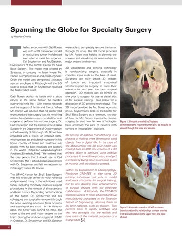
Figure 1: 3D model provided by Stratasys demonstrates the recurrent tumor (pink) as it would be viewed through the nose and sinuses.
3D printing, or additive manufacturing, is a process of making three dimensional solid objects from a digital file. In the case of the above article, the 3D skull model was created from an MRI. The creation of a 3D printed object is achieved using additive processes. In an additive process, an object is created by laying down successive layers of material until the object is created. Within the Department of Otolaryngology, Pittsburgh CREATES is also using 3D printing technology, not only to model anatomical structures for surgical training, but to also develop new enhancements to surgical devices with our corporate collaborators. Additionally, the CREATES team has access to other advanced additive manufacturing processes at the Swanson School of Engineering, allowing them to 3D print materials, such as titanium. This permits them to rapidly prototype and test new concepts that are realistic and have many of the material properties of a final product.
Figure 2: 3D model created at UPMC of a tumor (translucent) and its relationships to major arteries (red) and veins (blue) in the upper neck and base of skull.