3 minute read
Arkaprava Chakraborty, Deep Das, Souvik Dubey Alak Pandit, Biman Kanti Ray


JOURNAL OF THE INDIAN MEDICAL ASSOCIATION, VOL 118, NO 07, JULY 2020
Pictorial CME Thunderclap headache, beyond subarachnoid haemorrhage
Advertisement
Subhadeep Gupta 1 , Arkaprava Chakraborty 1 , Deep Das 2 , Souvik Dubey 3 , Alak Pandit 4 , Biman Kanti Ray 4
19-year-old female presented with acute onset holocranial headache, reached its peak within 1 minute, associated with recurrent vomiting. On examination patient was conscious and alert. Her supine blood pressure was 130/70 mm Hg. Neurological examination was normal except presence of left abducens nerve palsy. 1. What is your diagnosis? 2. Can we suspect the etiology from the parenchymal image only? 3. How do you confirm your diagnosis? 4. What is the best possible management of the patient?
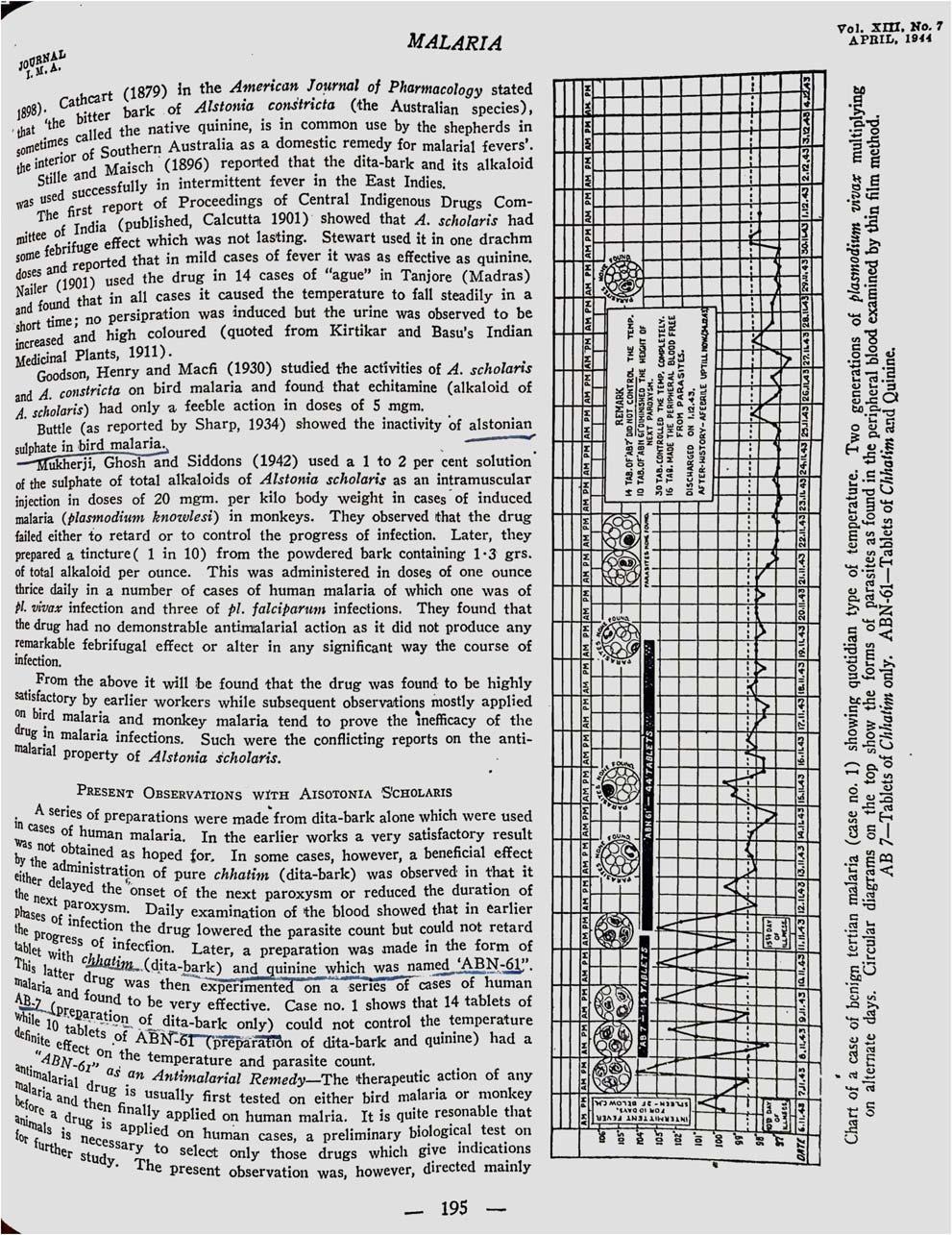
Thunderclap headache is defined as intensely painful headache reaching its peak within seconds to few minutes, often described as the worst headache of life as in our patient. Our patient had thunderclap headache and features of raised intracranial tension as evidenced by left abducens nerve palsy as false localising sign. Possibilities considered were aneurysmal subarachnoid haemorrhage, cerebral sinus thrombosis, cryptococcal meningitis and rarely intracerebral haemorrhage (ICH). Computed tomography scan of brain without contrast (NCCT) shows right temporal bleed (Fig A).
ICH in young can be due to several etiologies including aneurysm, coagulopathies, vascular malformations and rarely intracranial dissections. History of addiction, bleeding diathesis and renovascular & endocrine hypertension were ruled out. Magnetic resonance angiography (MRA) [Fig B] failed to identify underlying pathology as it was largely obscured by blood and proximal right middle cerebral artery (MCA) anatomy was not clear. We decided to perform digital subtraction angiography (DSA) next. DSA [Fig C & D] revealed a dissecting aneurysm involving the entire right M1 segment and distal right internal carotid artery was narrowed. There was good distal flow with the transmural dissection extending till about right M2 segment; there was no extravasation of contrast material.
1 DM Neurology Post-doctoral trainee, Bangur Institute of Neurosciences, IPGMER Annex 1, Kolkata 2 Senior consultant, Dept, of Neurology, Bangur Institute of Neurosciences, IPGMER Annex 1, Kolkataand Consultant Neurologist, Woodlands multi-speciality hospital and C K Birla Hospitals 3 Assistant Professor, Dept, of Neurology, Bangur Institute of Neurosciences, IPGMER Annex 1, Kolkata 4 Professor, Department of Neurology, Bangur Institute of Neurosciences, IPGMER Annex 1, Kolkata
Definitive treatment is necessary because it can have 2 possible outcomes. If the dissecting aneurysm gets thrombosed it can cause MCA territory infarctor it may rerupture leading to significant morbidity and mortality. Ideal treatment in this patient is superficial temporal artery to middle cerebral artery bypass (STA-MCA) where STA is dissected, pushed through a burrhole and anastomosed to a branch of MCA along with sacrifice of the proximal vessel.
Extra-cranial dissections are common in clinical settings but isolated MCA dissection [MCAD] is infrequently reported as a cause of stroke. It is more common in Asian males. Almost 2/3 patients of MCAD have ischemic presentation. DSA is the most effective imaging modality for diagnosis. In 3/4th cases of MCAD, pathology is noted in the M1 segment. Outcome is not favourable in nearly 1/3rd patients of MCAD. These data are taken from a systematic review by Asaithambi G et al 1 .
REFERENCE
1 Asaithambi, G., Saravanapavan, P., Rastogi, V., Khan, S.,
Bidari, S., Khanna, A. Y., … Hedna, V. S. (2014). Isolated middle cerebral artery dissection: a systematic review.
International Journal of Emergency Medicine, 7(1).
51
JOURNAL OF THE INDIAN MEDICAL ASSOCIATION, VOL 118, NO 07, JULY 2020

52
JOURNAL OF THE INDIAN MEDICAL ASSOCIATION, VOL 118, NO 07, JULY 2020

53
JOURNAL OF THE INDIAN MEDICAL ASSOCIATION, VOL 118, NO 07, JULY 2020
54
JOURNAL OF THE INDIAN MEDICAL ASSOCIATION, VOL 118, NO 07, JULY 2020
55