10 minute read
Rohit Bhardwaj, Ankur Gupta, Kirti Khandelwal, Sabarirajan Ponnusamy Chirayata Basu, Karthika Nathan


JOURNAL OF THE INDIAN MEDICAL ASSOCIATION, VOL 118, NO 07, JULY 2020
Case Report Adenocystic carcinoma of palate masquerading benign cystic palatal lesion : A rare case report
Advertisement
Rohit Bhardwaj 1 , Ankur Gupta 2 , Kirti Khandelwal 3 , Sabarirajan Ponnusamy 4 , Chirayata Basu 4 , Karthika Nathan 4
Adenocystic carcinoma (ACC) is a rare epithelial malignancy of salivary gland origin, accounting for <1% of all head & neck malignancies. Palate is a preferred site. It shows female predominance, preference in 5th and 6th decade of life, slow growth rate, perineural invasion, distant metastasis and potential for local recurrence. Surgery with radiotherapy is the treatment modality of choice. We present a case of 34 years old female, who was diagnosed to have an infected cystic lesion on FNAC. HPE of resected specimen confirmed it as ACC. Patient received combined treatment (Surgery + Radiotherapy), and
now free of disease even after 2 years of follow up. [J Indian Med Assoc 2020; 118(7): 48-50]
Key words : Adenocystic carcinoma (ACC), epithelial malignancy, minor salivary glands, perinural invasion, local recurrence.
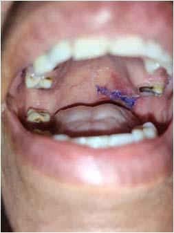
Adenocystic carcinoma (ACC) is a rare epithelial Editor's Comment : malignancy of salivary gland origin. It accounts for less ACCs are very much capable of masquerading a than 1 % of all head and neck malignancies & almost 10 % benign lesion because of its slow growth rate, of salivary gland tumours belong to this variety 1 . It mainly indolent course, asymptomatic presentation (most affects minor salivary glands however sites of respiratory of the times) and cellular pleomorphism leading to tract like larynx & lungs have also been reported to be involved inaccurate histo-pathological diagnosis on FNAC or by ACC owing to the presence of submucosal or small punch biopsy. seromucinous glands 2 . The preferred location of this tumour is palate, especially area of junction between soft and hard palate 3 . Demographically females are affected more by ACC, A low threshold for combining radiotherapy along with surgery for treatment should be practiced since the tumour has recurrence potential. I hope this delivers the desired information. I hope for the in 5th or 6th decade of life 4 . Tumour usually follows a slow positive response from your esteemed journal with growth pattern with indolent disease course which makes respect to this article. it look more like a benign rather a malignant lesion & can be held responsible for its delayed presentation. ACC has gathered various names based on its histological studies which includes basiloma, cylindroma, adenoepithelioma and adenoid basilod carcinoma 5 . Histopathological classification holds prognostic values for ACC 6 . Based on histology this tumour has three varients i.e. cribriform, tubular and solid. Cribriform or “swiss cheese” variant is most common and also has the best prognosis. A single tumour can show more than one histological pattern. A peculiar feature of this tumour is the neurotropic tendency for metastasis 7 . Gasserian ganglion has been reported to be the intracranial site involved by ACC 8 . Lymphatic and haematogenous spread occurs rarely, however cases of distant metastasis to bone, lungs and soft tissues by haematogenous route exists 9 . Tumour can be dealt with either by single modality (Surgery/ Radiotherapy/ chemotherapy) or as combined therapy 10 . Wide local excision with adequate margins and post op radiotherapy is the preferred modality of treatment for this tumour 11 . We present a case of 34 years old lady who was misdiagnosed based on disease course and FNAC findings and had to undergo repeat surgical excision of the lesion. CASE REPORT 34 years old lady presented to our outpatient department with complain of a slowly progressing swelling on left side of palate for past 8 months. She experienced no pain, difficulty in chewing / swallowing or loosening of teeth and any other swelling in head and neck region. She never had smoking, alcoholism and tobacco chewing habits. On detailed clinical evaluation she was found to have a soft to firm swelling involving left side of soft palate (Fig 1). Swelling measured approximately 3cm X 2cm & had smooth surface. This non mobile swelling did not have ulcerations on surface. Department of Otorhinolaryngology, VMMC and Safdarjung Patient had carious teeth. No other swelling or neck lymph
Hospital, New Delhi 110029 nodes were palpable. Palatal movements were also 1 MS (ENT), DNB (ENT), Senior Resident bilaterally symmetrical. Nasal endoscopy also failed to report 2 MS (ENT), Senior Resident and Corresponding Author anything relevant. 3 MS (ENT), Senior Resident Fine needle aspiration cytology was done from the 4 MS (ENT), Junior Resident lesion. FNAC reported the lesion as benign appearing Received on : 15/04/2020 Accepted on : 11/06/2020 squamous cells and polymorphs in a mucoid background
48
JOURNAL OF THE INDIAN MEDICAL ASSOCIATION, VOL 118, NO 07, JULY 2020
suggestive of a benign infected cystic lesion. A contrast enhanced computed tomography scan also reported this as a ‘well-defined hypodense lesion’ approx. 31 cm x 23cm, showing minimal post contrast enhancement of 10-15 HU, suggestive of benign nature of lesion (Fig 2).
Based on clinical and radiological evaluation, patient was planned for complete surgical excision of this benign palatal lesion. The lesion was excised meticulously without damaging macroscopically uninvolved palatal musculature and surgical site repaired primarily to avoid any fistula formation (Fig 3). Fig 2 — Showing CT appearance of the Histopathological examination of this specimen reported it as an adenocystic carcinoma with positive tumour margins. After discussing the nature of disease and possibility of palatal defect following revision surgery with patient, she agreed for complete surgical excision of the tumour. We excised the palatal tissue taking adequate margins all around the previous surgical site (Fig 4). Considering the large size of the palatal defect intraoperatively, no attempts of primary repair were made. HPE revealed uninvolved margins all around the lesion; even the nearest positive margin had a disease free distance of >10mm. Later patient also received radiotherapy to further sterilize the surgical site in an attempt to minimize the chances of recurrence. Excision of carious teeth was advised by dental surgeons prior to radiotherapy but patient denied for this. She developed trismus following tumours have a slow indolent course, they are seldom diagnosed early, more so when the palatal is involved. As the palatal lesions are mostly asymptomatic and appear as submucosal, smooth surfaced swellings without having any overlying ulceration, delayed diagnosis is not so uncommon. Besides this, tumour histology also contributes in this diagnostic confusion. The microscopic architectural patterns of this tumour can show wide variations; individually these variations might fail to suggest the malignant nature of the lesion. FNACs and small incisional biopsies obtained away from the true representing area, report inaccuracies in diagnosis owing to this pleomorphism (a confusing feature of these tumours). We hold these factors attributable to the delayed presentation and the misdiagnosis in our case. MRI has a role in describing the soft tissue extension and perineural invasion Fig 1 — Showing preoperative palatal swelling, marked by star lesion marked by solid arrow surgery and radiotherapy which was dealt by active mouth opening exercises. Prosthesis was made to overcome the difficulties caused by palatal defect (Fig 5). She has been under our follow up for past 3 years and is free of recurrence.
D ISCUSSION
ACCs usually arise in intercalated ducts of the mucous secreting glands from a cell type which can differentiate in either epithelial or in myoepithelial cells. Owing to the cellular origin, these tumours are mainly confined to minor and major salivary glands and mucous secreting glands of respiratory tract 12 . Since these Fig 3 — Post-operative image showing repaired surgical site Fig 4 — Intraoperative photograph showing completely resected tumour margins


49
JOURNAL OF THE INDIAN MEDICAL ASSOCIATION, VOL 118, NO 07, JULY 2020 diagnosis on FNAC or small punch biopsy. This calls for a high index of suspicion for diagnosing these lesions. We also recommend a low threshold for combined or duel modality treatment (surgery along with radiotherapy), since the tumour has recurrence potential.
REFERENCES
Cytodiagnosis of adenoid cystic carcinoma of the parotid metastatic to kidney and lung. J Cytol 2007; 24: 201-2. 2 Florentine BD, Fink T, Avidan S, Braslavasky D, Raza A, Cobb carcinoma: A reportof three cases. DiagnCytopathol 2006; 34: 4s91-4. Journal 2008; 8(4): 172-80. salivary glands: a demographic and histologic study of 426 cases. Oral Surg Oral Med Oral Pathol 1988; 66(3): 323-33. 2165-7920.
Fig 5 — Photograph showing palatal fistula while CT helps in showing the bony involvement, besides being important in surgical planning and follow up. ACCs also appear as benign on CT unless the lesion ulcerate or cause bony destruction, this also happened with our case where radiology also suggested a possibility of benign nature of the lesion. Detailed histopathological evaluation of the excised specimen reported the lesion as ACC. While searching for the optimal treatment modality for the tumour, we found diverse opinions in literature. Possible treatment options are surgery, radiotherapy and chemotherapy as a single modality or combination of these. Surgery (wide local excision along with adequate safety margins) was favored by few authors 13,14 . Others proposed combining the two, as surgery or radiotherapy alone is not sufficient enough to prevent disease recurrence and distant metastasis 15 . Owing to its slow growth rate, the response of ACCs towards chemotherapy was not very convincing 16 . After analyzing the various prognostic factors like histopathological grade, cervical lymphatic metastasis, surgical margins & we discussed it with radiotherapy team in multidisciplinary team meeting and decided to re-excise the tumour margins and subject the patient for radiotherapy to sterilize the tumour bed. After receiving this combined duel modality treatment, patient is free of any recurrence even after 3 years.
CONCLUSION
ACCs are very much capable of masquerading a benign lesion because of its slow growth rate, indolent course, asymptomatic presentation (most of the times) and cellular pleomorphism leading to inaccurate histopathological
1 Srivastava S, Jaiswal R, Agarwal A, Singh PK, Singh SN — CJ — Extra-salivary gland presentations of adenoid cystic 3 Moore, Burkey, Netterville, et al — Surgical management of minor salivary gland neoplasms of the palate. The Ochsner 4 Waldron, El-Mofty, Gnepp — Tumors of the intraoral minör 5 Deepak C — Adenoid Cystic Carcinoma of the Maxilla - A Case Reportand 5 Year Follow-up. J Clin Case Rep 2012; 2: microscopic perineural invasion with respect to our case,
6 El-Naggar AK, Huvos AG — Tumors of the salivary glands:
Adenoidcysticcarcinoma. In: Barnes EL, Eveson JW, Reichart
P, Sidransky D, editors. World Health Organization classification of tumours: Pathology and Genetics. Head and Neck Tumours.
Lyon: IARC Press; 2005. 221-2. 7 Rinaldo A, Shaha AR, Pellitteri PK, Bradley PJ, Ferlito A—
Man¬agement of malignant sublingual salivary gland tumors.
Oral Oncol 2004; 40: 2-5. 8 Wakisaka S, Nonaka A, Morita Y, Fukui M, Kinoshita K —
Adenoid cystic carcinoma with intracranial extension: report of three cases. Neurosurgery 1990; 26: 1060-5. 9 Ellis GL, Auclair PL— Atlas of tumor pathology: Tumors of the salivary glands. Third Series fascicle 17. Washington, DC:
Armed Forces Institute of Pathology; 1996, 203-16. 10 Chundru, Amudala, Thankappan, et al — Adenoid Cystic
Carcinoma of Palate: A Case Report And Review Of Literature
Dent Res J (Isfahan) 2013; 10(2): 274-8. 11 Tripathi, Nahar, Padmavathi, et al — Adenoid Cysic Carcinoma of the Palate: A Case Report with Review of Literature.
Journal of Cancer Science & Therapy 2010; 2(6): 160-2. 12 Ellis GL, Auclair PL, Gnepp DR — Adenoid cystic carcinoma,
Surgical Pathology of Salivary glands, Philadelphia. WB
Saunders. 1991; 333-346. 13 Kokemueller H, Eckardt A, Brachvogel P, Hausamen JE —
Adenoid cystic carcinoma of the head and neck – a 20 years experience. Int J Oral Maxillofac Surg 2004; 33: 25-31. 14 Jayalakshmi S, Agarwal S, Nachiappan PL, Prasad RR,
Bhuthra S, SharmMC, et al — Intracranial adenoid cystic carcinoma, a case report. J Neuro-oncol 2000; 47: 47-50. 15 Maciel Santos MES, Ibrahim D, Neto JC, Da Silva JC, Da Silva
UH, Sobral APV — Carcinoma adenóidecístico: relato de caso.
Rev Cir Traumatol Buco-Maxilo-Fac 2005; 5: 49-54. 16 Vincentelli F, Grisoli F, Leclercq TA, Ardaud B, Diaz-Vasquez
P, Hassoun J — Cylindromas of the base of the skull. J
Neurosurg 1986; 65: 856-9.

50