


JOURNAL

JOURNAL
JOURNAL OF THE INDIAN MEDICAL ASSOCIATION, VOL 118, NO 08, AUGUST 2020
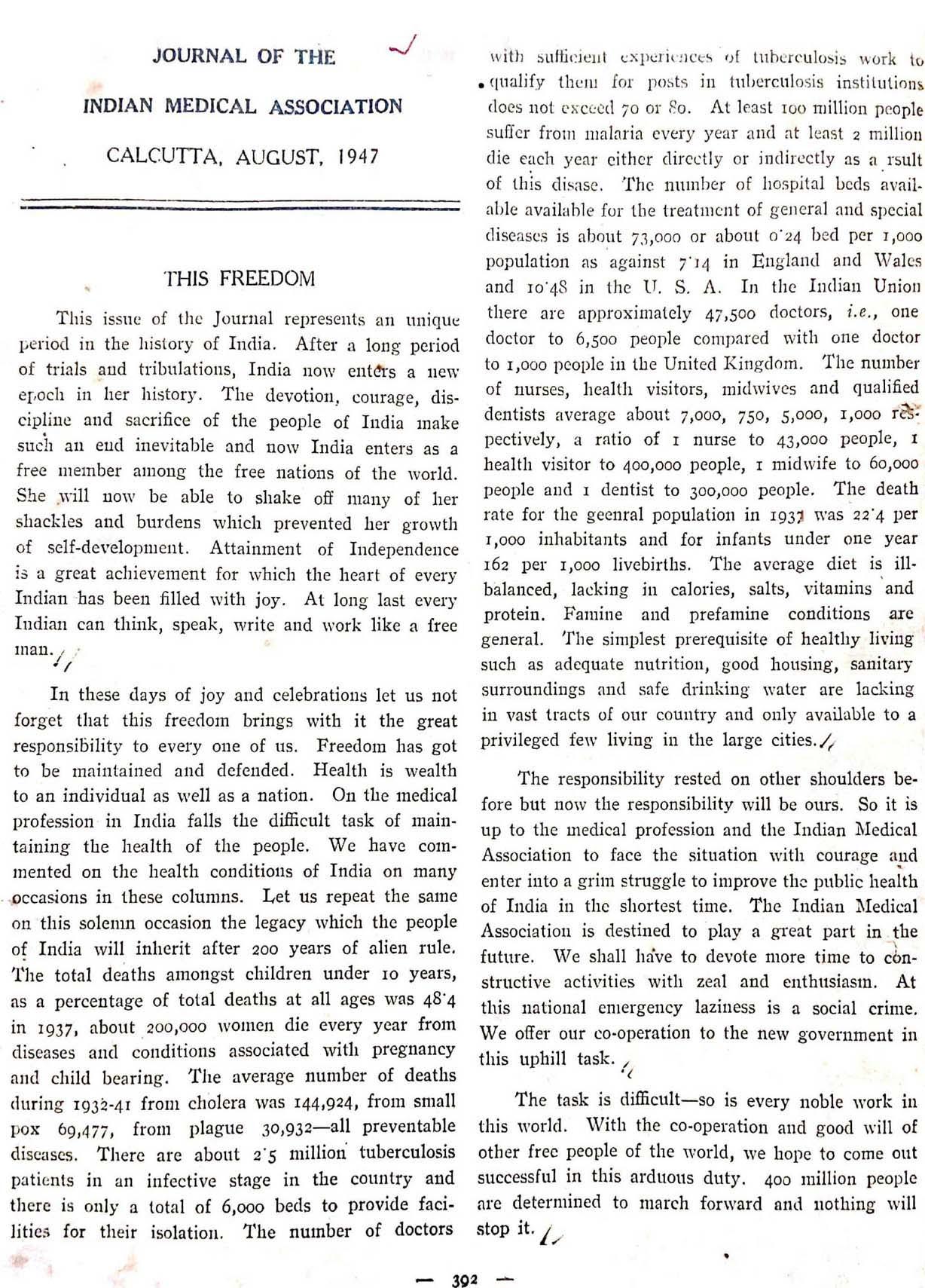
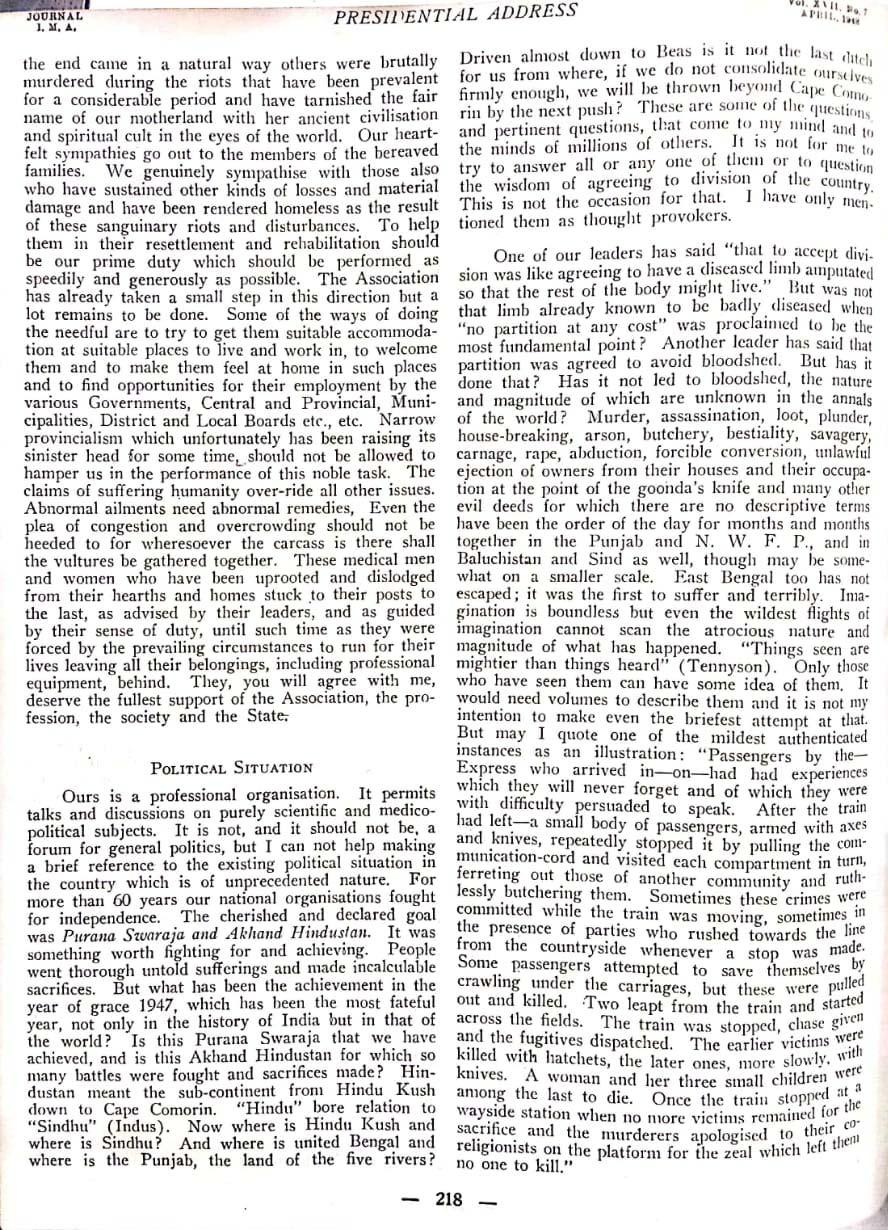
JOURNAL OF THE INDIAN MEDICAL ASSOCIATION, 1947, VOL 17, P-218
JOURNAL OF THE INDIAN MEDICAL ASSOCIATION, VOL 118, NO 08, AUGUST 2020
JOURNAL OF THE INDIAN MEDICAL ASSOCIATION, 1947, VOL 17, P-219
AugustMemories:
“At the stroke of the midnight hour, whole the world sleeps, India will awake to life and freedom.”
— Pandit Nehru at Constituent Assembly of India , 1947

India gained freedom from British colonial rule on Friday, 15 August, 1947. This day of transfer of power was the culmination of years of struggle by people of the subcontinent. Although there was unconscionable loss of lives due to the partition of the area along religious lines, the day of independence is still a proud day in the history of our country. This success was gained due to involvement of all sections of the society, including doctors. This editorial, on this 74th commemoration of our Independence Day, will look back at the contribution of doctors in the Indian freedom movement.
True Freedom has two component – Spiritual attainment and Political attainment. So the Indian freedom struggle had to be fought on two fronts. On one hand, there was the political struggle and on the other, there was the urgent need to improve the condition of the society as a whole. The political struggle was being driven along two different ideologies: armed uprising and non-violent civil movement. The societal movement was fought along multiple lines. The Indian society at that time was stringently divided according to caste (in Hindu community), religion, and gender and of course, socio-economic status. Superstition, lack of education prevailed in society .It was not an easy task to bring the maharajas and nawabs in same platform with the commoners (whom the royalty despised) or to bring the Brahmins together with Dalits (whom the Brahmins considered impure). Raja Rammohan Roy, Vidyasagar, Swami Vivekananda worked to bring changes in society in terms social and spiritual attainment . Freedom movement was a myriad of internal conflicts, ego clashes and antagonistic decisions. Everyone had to do their bit to make this movement a success. Doctors had a very important role in all the phases and sections of this movement. For example, the feat of Pandit Madhusudan Gupta in performing the first human dissection in India and thereby breaking the spell of cultural taboo or challenge taken by Dr Radha Govinda Kar following refusal of Indians to be Medical teachers in Calcutta Medical College to establish a Medical College (R G Kar medical college) entirely by money of Indians can’t be underestimated. Ultimately Dr Kar died in Influenza Pandemic while serving patients during fearful crisis. Mahatma Gandhi was the leader of the non-violent movement while luminaries like Netaji, Surya Sen, Bhagat Singh and Binay-Badal-Dinesh were fighting the British government with weapons. But the struggle for social movement was much more difficult. Indians wanted to get back the power to rule their country but what was their vision for the future? There was a lot of conflict. While people like Nehru and Dr. Bidhan Chandra Roy wanted a modern country with a scientific outlook, there was a significant portion of the leaders who wanted to go back to the old days of glory.
Right from the beginning of Indian freedom struggle, lawyers, journalists and members of aristocratic Indian families established themselves to be the leaders. Other professions like doctors or scientists had very little scope of reaching the upper echelons of the political movement. But still, there are many doctors who had substantial contributions in this national movement and helped their countrymen in many ways. Sadly, subsequent Indian history has been unkind to these doctors who spent their lives struggling for the freedom of India. When Indian freedom movement is discussed by academics, the names of these doctors are never given prominence. The standard texts written by Indian historians are almost silent on the role of doctors in this movement. But we, as doctors, have the duty to commemorate these heroes of our profession and remind the future
generations of the selfless contribution of these legendary medical persons.
Sushila Nayyar :
She was a graduate (MD) of Lady Hardinge Medical College, Delhi. Thus, with her educational qualification, she could easily have become a very successful physician. But she chose to follow the Gandhian path and in 1939, came to join the Gandhian movement at Sevagram.
She became the personal physician of Mahatma Gandhi (thus, although Gandhi decried modern medicine, he had an MD specialist as his personal physician!). She took part in the 1942 Quit India movement and was imprisoned. She testified in the Kapur commission about assassination attempts on Mahatma Gandhi. After the eventual death of Mahatma Gandhi, she went to John Hopkins University, USA where she further did MPH and DrPH, being the first doctoral student of the Maternal and Child Health program of that prestigious institute. She came back to India and had important contributions in developing public health programs of India, including leprosy, tuberculosis and child health. One memorable contribution of Ms Nayyar was setting up of the famous leprosy institute at Agra near Taj Mahal. There was a lot of furore in the Indian society about setting up an “unclean” leprosy hospital near the so-called monument of love. But Ms Nayyar, uncompromising and no-nonsense as she was, just brushed aside these sentimental protests and set up this premier institute for leprosy at Agra.
Laxmi Sehgal :
Born in Malabar, Captain Laxmi Sehgal passed her MBBS from Madras Medical College in 1938 and received further diploma in Gynecology soon. But she left for Singapore, met Netaji Subhas Chandra Bose and was given the responsibility of forming the female battalion of Azad Hind Fauj. She was given the rank of “Captain” as they marched on to Burma. She fought valiantly in the war against the British. After Independence, she continued her Gynecology practice at Kanpur. She did spectacular social work, like helping Bangladeshi refugees during 1971 war, organizing medical camp after Bhopal disaster of 1984 and helping poor woman at Kanpur.
TSS Rajan :
TSS Rajan was a Tamil doctor from Trichinopoly. He was a brilliant student who worked at Middlesex hospital, London. He obtained MRCS in 1911 and started a private practice. But call of his country made
him leave the lucrative profession and join the Indian National Congress. He led Congress agitations against British rule and was imprisoned multiple times.
In 1931, he led the movement to disobey infamous salt laws at Tanjore and was imprisoned. He was a well-respected member of the Congress party and held multiple political portfolios.
Bidhan Chandra Roy was a legendary doctor in Bengal. He had the rare feat of earning both the MRCP and MRCS diplomas. He was a legendary physician in Bengal and India and was personal physician to many of the leaders of Indian freedom struggle, including Mahatma Gandhi and Nehru. He was very active in Indian freedom movement, held many prominent political positions in Bengal and was the first chief minister of West Bengal (after independence, till 1950, the post was known as Prime minister of Bengal. Prafulla Ghosh was the first Prime minister, followed by Bidhan Chandra Roy. Then after election, Roy became the first chief minister). After independence, he wanted to go back to his profession, but at the advice of Mahatma Gandhi, he relented and remained in active politics till his death.
His other prominent political positions included Mayor of Calcutta (1931-33), Vice-Chancellor of Calcutta University (1942-1944), President of the Medical Council of India (1939) and the Governor of the United Provinces (now Uttar Pradesh).He was imprisoned for his role in Indian freedom struggle in 1930. But along with his political activities, he continued his professional work and treated numerous patients for free at his home and clinic. He was one of the rare Indian physicians whose death led to an obituary in the British Medical Journal (14 July, 1962). There, it was remarked that “at his professional zenith he may have had the largest consulting practice in the world, news of his visit to a city or even railway station bringing forth hordes of would-be patients.”
Kadambini Ganguly was the first female graduate of the Calcutta Medical College and the first successful practicing female physician in India. But besides her duties as a doctor, she also had important social work. After the Indian National congress was formed, she became the first female speaker at any convention of the party (1890). After the Bengal Partition Act was passed in 1905, she arranged a women’s conference the very next year in protest. In 1914, when Mahatma Gandhi visited Calcutta, she presided over a
Brahmosamaj meeting held in honour of Gandhi. She also voiced strong opinion against the pathetic treatment of tea workers in Assam and coal workers in Bihar. These movements depict her strong sense of social activism.
Diwan Singh Kalepani :
Diwan Singh was a poet and doctor who worked in the army. He was posted at the Andamans as a punishment for his anti-British views. He wrote his poetry and discussed with the local people the evils of colonial rule. He also tried to educate the local people by forming a school. However, when the Japanese army occupied the Andamans during Second World War, Diwan Singh did not like this new foreign rule either. At first the Indian independence league was formed in the Andamans with Diwan as the president. But the relation between them and the brutal Japanese soldiers soured very quickly. He disobeyed the Japanese commander and was killed in Cellular jail by the Japanese.
Dr Bhupal Bose and Dr Narayan Roy :
The names of these two doctors are mentioned in the list of prisoners at Cellular jail. Dr Narayan Roywas a member of the Yugantar party and had skill in making bombs. He was arrested in the Dalhousie square bombing case. He spent around 9 years in British jail, including cellular jail.
Pattavi Sitaramayya :
Sitaramayya was a Physician graduating from Madras Christian College and developed a thriving practice in current Andhra Pradesh. But he found the call for his country to be greater than his vocation and joined the Indian freedom struggle. He was very active in Andhra congress committee and also served in National congress committee. Bengalis remember Sitaramayya as the candidate whom Netaji Subhas Bose defeated to become President of INC at Tripuri Congress in 1939. Sitaramayya also participated in 1942 quit India movement and was imprisoned for three years. He was active in many social and financial reforms in Andhra Pradesh.
Muthulaxmi Reddy :
Muthulaxmi Reddy is a name which should be known to every Indian. But sadly, following the deplorable tradition of our country, we tend to remember names of film artists and cricketers (with no contribution to the society) while people like Muthulaxmi Reddy are forgotten.
She was a doctor from Madras Medical College, where she passed with numerous honours and medals.
She had a promising career in Medicine but she gave it up for her country. She was greatly influenced by Sarojini Naidu and Mahatma Gandhi. While on one hand, she had important political activities, she also worked tirelessly for women and children emancipation. She also formed one of the biggest cancer hospitals of India, Adyar Cancer Institute, after independence. She also struggled successfully to abolish the notorious devadasi system of India.
Dr Binay Kumar Nandy :
Dr Binay Kumar Nandy passed from Calcutta Medical College and joined the Indian Medical Service in 1941. He was thus at first working for the British government and was posted in Singapore. After Singapore was captured by the Japanese army, he was taken prisoner along with the other soldiers. Then, when the Indian National Army was formed, he joined it under Subhash Bose and fought against the British Army in Burma. He was imprisoned after the INA lost the battle. He was detained in Bhopal and released in 1946. He later ran his own charitable clinic in West Bengal till his death.
Lt Colonel AC Chatterjee :
He was another legendary INA soldier. AC Chatterjee was a doctor of Kolkata who had it all. He joined the IMS during the First World War and was later transferred to various prestigious posts in Bengal. He was the director of Public Health in Bengal Province and worked tirelessly for malaria control. Then, suddenly, at the age of 50, he was recalled back to the army by the British government during the Second World War. He boarded a ship from Bombay and went to the Far East, where the British soldiers were fighting the Japanese army. He set up a medical unit there and tended to the fallen soldiers. But, as the British beat a retreat, he was captured by the Japanese and eventually joined the INA. He quickly became a close confidante of Netaji.
He was made the first finance minister of the provisional government of free India and later, foreign minister of this provisional government by Netaji. Chatterjee evaded capture by the British army in Saigon but was eventually captured and imprisoned a few months later.
After the government files pertaining to Netaji were made public, it was revealed that the British Government was wary of the return of AC Chatterjee to Bengal. The head of the Eastern Command of the British Army requested the Spy chief in Delhi to detain him as long as possible to prevent possible revival of the INA in Bengal. Thus, he was a freedom fighter whom
the British feared.
KB Hedgewar :
This Marathi Doctor studied at the Calcutta National Medical College. He actively participated in the activities of the Indian national Congress in the 1920s. He also was associated for some time with the Anushilan Samiti of Bengal. He was active in many social movements in India.
Medical College, Kolkata, the first institution of modern medicine in India, had significant contributions in the Indian freedom Struggle. Anti-British movement was implemented through Bengal Provincial Students’ Federation (BPSF) the Bengal branch of All India Students’ Federation (AISF). Many students of this college were imprisoned for participating in the Quit India movement. In 1947, a student, Sere Dhiraranjan Sen, was killed on Vietnam Day (24 January) British police firing. A plaque bearing his name was set up in the Students’ common room of this college.
There were many doctors in India, who wanted to build their own institutions to teach medical science, separate from British government institutions. Traditional British institutions like Medical College Kolkata or the PG hospital were always headed by European doctors. Indian doctors, even if highly qualified, could never rise to the top of these places. Thus, these institutions set up by Indian doctors were symbols of Indian identity and Indian entrepreneurship.
Dr Sundari Mohan Das :
Dr Das, a son of Sylhet in erstwhile Bengal province, passed his MD from Medical College Kolkata. He went back to Sylhet but his practice there was marred by his social activity. He converted to Bramho religion and this angered the local upper caste Hindus, who drove him out of his place. But before being driven out by the fanatics, he had already managed to start a Girls’ school in that area. He came to Calcutta and started his activity. He was a staunch nationalist. He was active in the Swadeshi movement of Bengal (starting 1905) and in his personal life, refused European consumer goods till his last days. He wrote a number of songs to inspire the public against British rule. He was instrumental in forming the Bengali technical institute, which later became the Jadavpur University.
He wrote books like “Municipal Darpan” and “Briddha Dhatri Rojnamcha” which were Bengali books on public health. It was probably the first attempt to impart medical knowledge in Bengali to the public. He was also secretly helping the terrorist organizations of Bengal, which were fighting the British underground
and in fact, his home was one of the sites of bomb making! His house was a meeting place for many of the famous revolutionaries of that period like Bipin Chandra Pal and the “Swaraj Samity” was formed there only. He helped set up three famous hospitals in Kolkata: Chittaranjan Seva Sadan, National Medical College and RG Kar Medical College. He had a very flourishing private practice. But he still found time to contribute a lot for his country. When Chittaranajan Das became Mayor of Calcutta, Dr Sundari Mohan Das became the director of Public Health for the city. It is indeed sad that the contributions of this selfless citizen have been totally neglected by later historians of the country and his native city. When the early twentieth century history of Bengal is discussed, writers, musicians, politicians and members of the royal families are shown as sages. But the myriad contributions of doctors like Sundari Mohan Das are relegated to mere footnotes.
One of the most eminent surgeons of India in his time. BaghaJatin, the famous Bengal Revolutionary, was wounded when a tiger attacked him near his native village. It was Dr Sarbadhikary who treated Jatin and cured him of the wounds. With Dr Radha Gobinda Kar, he was instrumental in setting up the Belgachia Medical School, which is modern day RG Kar Medical College. This institution was built up as an indigenous medical school, out of British influence.
“In these days of joy and celebrations let us not forget that this freedom brings with it the great responsibility to every one of us. Freedom has got to be maintained and defended. Health is wealth to an individual as well as to a nation. On the medical profession in India falls the difficult task of maintaining the health of the People …………….. The responsibility rested on other shoulders before but now the responsibility will be ours.”
Thus, the onus was on the medical profession to educate the countrymen on the benefits of modern science. When India gained freedom, the average life expectancy of the population was around 40 years, malnutrition was rife and every known infectious disease from Tuberculosis to Cholera were ravaging through the society. Had India adopted the “return to Satyajug” theme, we would have seen a huge rise in
mortality in the country after independence. But it was through the tireless and often thankless work of multiple modern doctors that the health parameters of the country improved substantially after independence. There are many other unsung heroes of our profession. Doctors had a very important role to play during those days. For example, after the partition, when millions of homeless refugees came into the country, many doctors organized medical camps for them for years to come. But such activities are mostly forgotten. So, we think a revision of Indian history should be done to acknowledge the important contribution of doctors in the history of freedom struggle and post independence era.
Now, during the Covid pandemic also, doctors all over the country are doing selfless and tireless service for the countrymen. At the time of writing of this editorial, more than 200 doctors have died all across the country while battling the pandemic and many more are infected and struggling for their lives. But we are sad to see that this sacrifice is not being properly represented in the media and armchair intellectuals of the country are getting all the limelight. Dr Pradip Bhattacharya physician from a small town near Kolkata seen patients at same fees during Lockdown period even performed home visits of very sick and old patients. He suffered from COVID infection and succumbed to death inspite of all efforts of treating intensivist. To meet hospital bill even rickshaw pullers of locality contributed. Last journey of COVID sufferers are usually friendless, tearless, absolutely alone but here ignoring all fear and protocol thousands of people accompanied his funeral journey with tears and slogans. People gave him respect of martyr. But he deserved more from other corners. His sacrifice no less than sacrifice of a freedom fighter. But I know people may forget him, may forget sacrifice
of his wife who had not pulled back his husband during lockdown period from performing noble duty. Editorial board tribute sacrifices all doctor martyrs. As editor I have specially mentioned contribution of martyr –doctors with hope that even after 100 yrs if somebody open archive of JIMA will read great sacrifice our colleagues and their role will remain immortal in pages of JIMA.

(If you write your name in paper will be torn If you write your name in stone will be eroded If you write in heart of mankind will remain forever)
— Manna Dey
We thus have a duty to preserve the historic feats of our profession. At the headquarters of IMA and in the office of JIMA, a permanent display of the pictures and quotations of these great patriotic medical men should be set up. Also, such exhibitions should be arranged during medical conferences of all disciplines, that will be our real tribute to them. Let us take an oath on the eve of Independence Day; we will give our blood to fulfill dream of Greats of our fraternity.
“The task is difficult – so is every noble work in the world. With the co-operation and good will of other free people of the world, we hope to come out successful in arduous duty. 400 million people are determined to march towards and nothing will stop it.”
EDITORIAL JIMA, AUGUST 1947

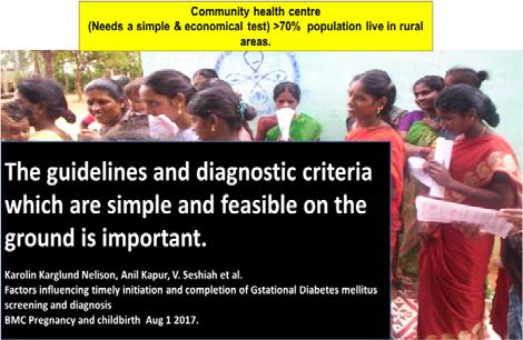
Veeraswamy Seshiah1, Vijayam Balaji2, Hema Divakar3, Anjalakshi Chandrasekar4, Samar Banerjee5, Ashok Kumar Das6
All efforts should be made in planning appropriate and possible methods of delivering health care for pregnant woman in the pandemic ocean of COVID-19,with limited medical facilities.Gestational Diabetes Mellitus (GDM) may play a crucial role in the increasing prevalence of diabetes and obesity and also may be the Origin of Cardio Metabolic Diseases.The Ministry of Health and Family Welfare, Government of India expects health care providers to screen all pregnant woman for glucose intolerance by a feasible, do able, economical and evidence-based test. “A Single Test Procedure” which is also followed by Diabetes in Pregnancy Study Group India. This test is ideal in the pandemic times.For a better perinatal outcome, the fasting plasma glucose (FPG) has to be maintained between 80 mg/dl (4.4 mmol/dl) and 90 mg/dl (5.0 mmol/dl) and 2hr Post Prandial Plasma Glucose (PPPG)110 mg/dl (6.1 mmol/dl) and 120 mg/ dl (6.7 mmol/dl) and mean plasma glucose 105 mg/dl (5.9 mmol/dl).Medical Nutrition Therapy (MNT) and life style modifications are recommended as an initial step to maintain normal maternal glucose, failing which insulin or Oral Hypoglycemic Agent (OHA) may be advised. Both small for gestational age and large for gestational age babies are prone to develop diabetes in the future. Hence, the aim in the treatment is to obtain newborn babies birth weight appropriate for gestational age of 2.5 to 3.5 kg.
[J Indian Med Assoc 2020; 118(8): 18-21]
Key words : Hyperglycemia in Pregnancy (HIP),COVID-19 Pandemic, Single Test Procedure, Post Prandial Plasma Glucose (PPPG).
Editor's Comment :
Covid -19 infection affects pregnant woman less frequently probably due to the development of immunity during pregnancy. Nevertheless, all pregnant woman should be screened with the simple and evidence based “Single Test Procedure” approved by the Ministry of Health and Family Welfare Government of India. Pregnant women should take 60 to 70 grams of protein daily and immune boosters like Zinc, Vitamin C and Vitamin D. The target glycemic control advisedis FPG ~ 90mg/dl and 2hr PG ~ 120 mg/dl so as to obtain birthweight of new borns appropriate for gestational age, between 2.5 and 3.5 kg.
1MD, FRCP, DSc, DSc, DSc (Hony), Consultant, Dr Balaji Diabetes Care Center and Dr Seshiah Diabetes Research Institute, Chennai- 600029 and Corresponding Author
2MD, FRCP (Glasgow), FRCP (Edinburgh), FRCP (London), Consultant, Dr Balaji Diabetes Care Center and Dr Seshiah Diabetes Research Institute, Chennai- 600029
3MD, FRCOG, Director, Divakar’s Specialty Hospital, Bengaluru
The whole world grapples with COVID -19 pandemic and its consequences. This situation adversely affects the medical profession, particularly in the diagnosis and care of people with diabetes. This is going to result in epidemic of diabetes. The prevalence of diabetes is increasing globally from 463 million in 2019 to 700 million in 2045a 51% increase1. While several reasons are ascribed for this rising trend including aging population, urbanization, genetic predisposition, nutrition and lifestyle transition, etc, one factor that has not received adequate attention is Glucose intolerance that occurs during pregnancy. Gestational Diabetes Mellitus (GDM) may play a crucial role in the increasing prevalence of diabetes and obesity2. In 2019 the global prevalence of Hyperglycemia in Pregnancy (HIP) in the age group 20-49 years was estimated to be 20.4 million or 15.8% of live births 1. They had some form of hyperglycemia in pregnancy, of which 83.6% were due to GDM1.Hence, it has become necessary that all pregnant women should be screened for GDM, even if they have no symptoms3
4MD DGO, PhD, Professor, Department of Obstetrics and Gynecology, Madha Medical College, Chennai, Tamil Nadu
5MD, FRCP, Professor, Department of Medicine on Diabetology, Vivekananda Institute of Medical Sciences, Kolkata.
6MD, PhD, FRCP (London), Professor of Medicine & Endocrinology, Pondicherry Institute of Medical Sciences
Received on : 10/07/2020
Accepted on : 05/08/2020
Wide spread anecdotal evidence suggests that both clinicians and pregnant women are increasingly unwilling to recommend or undergo OGTT4. The problem is, the blood glycose test results are available around three hours after the OGTT or next day and then GDM women have to undergo additional health service visits, for
diabetes education, glucose monitoring review, and fetal ultrasonography, all of which carry exposure risk during pandemic. Hence, there is a need for guideline which is universally acceptable4
Problem of Screening:
Unfortunately, there is no uniformity in the guidelines for diagnosing GDM. All the diagnostic criteria require women to be infasting, including that of International Association of Diabetes in Pregnancy Study Group guideline (IADPSG). The concern of this guideline is that, it over diagnoses GDM without clear clinical benefit5. Another inadequacy of IADPSG criteria is, its recommendation for diagnosing GDM with FPG >5.1 mmol/dl (92mg/dl). It was observed in relation to FPG of 5.1 mmol/dl, there is a considerable variability between countries noted in the Hyperglycemia and Adverse Pregnancy Outcome study (HAPO). FPG diagnosing only 22% of GDM in women in Bangkok and Hong Kong compared with up to 71% in some US centers. A low diagnostic rate of 24% of GDM has also been reported in Asian Indians with a fasting plasma glucose of 5.1 mmol/l6.This is due to increased insulin resistance in non-Caucasian population7 . Therefore, IADPSG procedure cannot be recommended as a universal guideline.
Most of the time pregnant women do not come in the fasting state because they may have to travel a long distance6. OGTT is resource intensive and many health services, especially in low resource settings, are not able to routinely perform an OGTT in pregnant women. In these circumstances, many health services do not test for hyperglycemia in pregnancy6.Therefore, options which do not involve an OGTT are required. For a pregnant woman, the request to attend in fasting, for a blood test may not be realistic because of the long travel distance to the clinic in many parts of the world, and increased tendency to nausea in the fasting state. Attending the first prenatal visit in the fasting state is impractical in many settings6, even in developed countries (eg: UK) a fasting blood test at the antenatal booking is often inconvenient8. The dropout rate is very high when a pregnant woman is asked to come again for the glucose tolerance test6. Consequently, nonfasting testing may be the only practical option6.
In this context, a study established that the twohour Plasma Glucose >7.8 mmol/dl with 75g oral glucose administered to a pregnant woman in the fasting or non-fasting state, without regard to the time of the last meal was able to identify woman with GDM9-11. This “Single Test procedure” which is feasible to perform in all resource settings has been adopted by Diabetes in Pregnancy Study Group India(DIPSI) for diagnosing GDM (Fig 1). National Institute of Clinical Excellence (NICE) guidelines also recommend 2hr PG
>7.8 mmol/dlas one of the diagnostic criteria for GDM based on the study performed in multi ethnic population of UK12. The DIPSI procedure is approved by the Ministry of Health & Family Welfare Government of India13, WHO6, FIGO14 & IDF15. This procedure is being followed in Sri Lanka16, Pakistan17, Bangladesh18 and may be in many other countries.
Repeat Testing:
If the first testing is negative the test has to be repeated in the second trimester(between 22 to 28 weeks) and if negative to repeat in the third trimester(between 32 to 34 weeks) plasma glucose calibrated glucometer can be used.
MANAGEMENT :
Treating GDM appreciably reduces the probability of serious neonatal morbidity compared with routine prenatal care19. Maternal–fetal Medicine Units Network conducted a randomized clinical trial for the treatment of gestational diabetes20, the results of which provided further compelling evidence that treatment, as necessary, reduces rates of adverse pregnancy outcomes including perinatal mortality, neonatal hypoglycaemia, neonatal hyperbilirubinemia, elevated cord blood C-peptide level, and birth trauma. This network also, observed lifestyle modification and dietary intervention will be effective in 80–90% of women with GDM.
TARGET GLYCEMIC CONTROL :
The recommended glycemic control is FPG ~ 90 mg/dl (5.0 mmol/dl) and 2-hour postprandial plasma glucose ~ 120 mg/ dl (6.7 mmol/dl) in GDM patients so as to avoid perinatal complications21,22. The goal is to obtain newborn babies birth weight appropriate for gestational age between 2.5 to 3.5 kg. This is to avoid both small for gestational age and large for gestational age new borns, as this is the first step to prevent off spring developing diabetes.
Management Guiding Principles:
•All Pregnant women who test positive for GDM for the first time should be started on MNT and physical
exercise for 2 weeks. Dietary intake is foundational to optimal pregnancy outcomes because nutritional quality and quantity have an important impact on the overall growth and development of the fetus.
•Woman should walk/exercise (which she is used to) for 30 minutes or perform household work.
•If 2hr Post Prandial Plasma Glucose (PPPG) remains > 6.7 mmol/dlwith MNT and lifestyle changes, Metformin or Insulin therapy is recommended.
Medical Nutrition Therapy (MNT): In facilities where nutritionists are not available for diet counselling, a readymade list of diet sheet containing the food items which can be taken in plenty and which should be avoided-is made available. It is difficult to have a personal interaction with pregnant women due to COVID Pandemic.
Drug Management (Metformin or Insulin Therapy):
•Metformin or Insulin therapy is the accepted medical management of pregnant women with GDM not controlled on MNT. Insulin is the first drug of choice
•Insulin can be started any time during pregnancy for GDM if MNT fails.
•If pregnant woman is not willing for insulin, metformin can be recommended provided gestation is more than 12 weeks23. The starting dose of metformin is 500 mg twice daily orally up to a maximum of 2 gm/ day. If the woman's blood sugar is not controlled with the maximum dose of metformin (2 gm/ day) and MNT, there is no other option but to advise Insulin.
•Hypoglycaemia and weight gain with metformin are less in comparison to Insulin.
Insulin Therapy:
•The recommended starting dose of insulin in GDM is 0.1 unit/kg of body weight per day. Dose can be increased on follow up till 2hr PPPG is around 6.7 mmol/dl.
•Rarely a GDM woman may require more than 20 units of insulin per day (two third of the dose before breakfast and one third before dinner. eg: If 18 units required, 12 unit in the morning and 6 unit in the evening preferable to use pre-mixed insulin). If she requires multiple doses of insulin, she may be referred to a higher center where physician is available.
Monitoring Glycemic Control :
•Fasting and 2 hr PPPG can be monitored to adjust the drug dosage. But most importantly monitoring 2hr PPPG is ideal as when 2hr PPPG is around 6.7 mmol/dl, FPG will never exceed 5.0 mmol/dl.
•Laboratory glucose measurement is often not available and testing with a portable plasma glucose standardized metre is the only option6.
•There are very little data on the use of HbA1c to
diagnose diabetes in pregnancy. Consequently, WHO guideline (2013) does not include HbA1c as a means of diagnosing diabetes in a pregnant woman and for monitoring6
•After satisfactory glycemic control is achieved monitoring alternate days may be necessary in women who is taking insulin. Places with limited resources monitoring can be done every 2 weeks between 24th and 28th weeks and from 28th week every week till delivery.
•Self-monitoring of blood glucose: All GDM mothers,partners and family members should be taught about self-monitoring blood glucose.
Post-partum care — All GDM woman after confinement should be tested for glucose intolerance, 6 weeks after delivery. In the post-partum period, the “single test procedure” which was followed in the antepartum period can be followed. This test which was good in the ante-partum period should also be good for the post-partum period.
If GDM woman is on insulin she may not require insulin immediately after the delivery and in the postpartum period. GDM woman who was on metformin may be advised to continue if her post-partum blood glucose is >7.8 mmol/dl. Metformin can be continued during breastfeeding.
Summary and Conclusion :
All available evidence suggests that pregnant women are at no greater risk of becoming seriously unwell than other healthy adults if they develop coronavirus24. Nevertheless, pregnant woman may be advised to undergo“Single Test Procedure” for diagnosing glucose tolerance,which is a do able and evidence-based test. To avoid waiting in the lab area, she may take at home 75g glucose mixed with 300 ml of water in the fasting or non-fasting state irrespective of the last meal timing. The intake of the solution to be completed within 5 minutes and then she can go to the lab around 2hrs after drinking glucose solution to have her venous blood glucose tested. To avoid crowded place like medical facility a plasma standardized glucometer can be used to evaluate capillary blood glucose and this procedure is recommended by the Ministry of Health & Family Welfare Government of India and WHO for diagnosing GDM.
Glycemic control requires health education on life style modifications.This can be done on individual basis or in group sessions. In the present scenario of COVID-19 Pandemic it is advisable to use digital media for sharing the knowledge. If this not possible printed pamphlet with all information can be given to pregnant women in the language they understand. They may be advised to maintain the target glycemic level off asting~ 5.0 mmol/dl or 2hr Post Prandial
Plasma glucose ~6.7 mmol/dl to minimize the risk of fetal macrosomia and to avoid perinatal morbidity. Women who fails to respond to lifestyle changes may be advised Oral Hypoglycemic Agent (metformin) or insulin. Only drawback for recommending insulin is, the person has to be given training in self-injection and needs to be followed frequently.
Optimal glycemic management during pregnancy leads to not only immediate well-being to the mother and the fetus but also to several transgenerational benefits. Hence even in the period of this pandemic of COVID-19, appropriate advice can be provided to GDM women by personal contact or using telephonic communication or other technological methods.
“MostComplicatedProbleminthisUniversehasa SimpleSolution”
— Albert Einstein
ONE Testwith 75gm of oral glucose irrespective of last meal timing.
ONE ValueTo diagnose GDM 2hr PG = 140 mg/dl.
ONE Target2hr PPPG ~ 120mg/dl.
Funding : None
Conflict of Interest : None
1International Diabetes Federation (IDF), Atlas Ninth edition2019. Online version of IDF Diabetes Atlas: www.diabetesatlas.org.
2Asslamira Ferrara — Increasing prevalence of GDM Diabetes Care 30 (2): 2007.S141- 146.
3Kristina Fiore. United states Preventive Service Task force (USPSTF) backs Universal diabetes Screening. Jan 13, 2014.
4David McIntyreand Robert G. Moses — The Diagnosis and Management of Gestational Diabetes Mellitus in the Context of the COVID-19 Pandemic. Diabetes Care - https://doi.org/ 10.2337/dci20-0026.
5Thangaratinam S, Cooray S, Sukumar, Nithya H Mohammed; Devlieger R, Benhalima K; McAuliffe, Fionnuala, Saravanan, Ponnusamy, Teede, Helena — Endocrinology in the time of COVID-19: Diagnosis and Management of Gestational Diabetes Mellitus. Accepted Manuscript published as EJE20-0401.R1. Accepted for publication: 26-May-2020
6Stephen Colagiuri, Maicon Falavigna, Mukesh M. Agarwal, Michel Boulvain, Edward Coetzee, Moshe Hod, Sara Meltzer, Boyd Metzger, Yasue Omori, Ingvars Rasa, Maria Inês, Veerasamy Seshiah, David Simmons, Eugene Sobngwi, Maria Regina Torloni, Hui-xia Yang. Strategies for Implementing the WHO Diagnostic Criteria and Classification of Hyperglycaemia First Detected in Pregnancy. DRCP. 103 (2014) 364-372.
7V W Wong, et al — South-East Asians had the lowest BMI, lowest fasting yet highest 2-hr glucose level on 75-g glucose tolerance test. Diabet. Med. 29, 366–371 (2012).
8Simmons D, Thompson CF, Engelgau MM — Controlling the diabetes epidemic: how should we screen for undiagnosed diabetes and dysglycaemia? Diabet Med 2005; 22(2):207212.
9C. Anajlakshi, V. Balaji, Madhuri S. Balaji, S. Ashalatha, Sheela Suganthu, T. Arthi, V. Thamizharasi, V. Seshiah — A Single Test Procedure to Diagnose Gestational Diabetes Mellitus. Acta Diabetologica (2009) 46: 51-54. DOI 10.1007/s00592008-0060-9.
10Paul W. Franks, Helen C. Looker, Sayuko Kobes, Leslie Touger, P. Antonio Tataranni, Robert L. Hanson, and William C. Knowler
— Gestational Glucose Tolerance and Risk of Type 2 Diabetes in Young Pima Indian Offspring. Diabetes 2006 55: 460 -465.
11Petit, et.al — used the non-fasting 2hour 75 g OGTT Long term effects on offspring, Diabetes 1991; 40(suppl 2):126-30.
12National Institute for Health and Care Excellence. Diabetes in pregnancy: management from preconception to the postnatal period NICE guideline Published: 25 February 2015 nice.org.uk/ guidance/ng3.
13Maternal Health Division Ministry of Health & Family Welfare Government of India, www.mohfw.gov.in &www.nhm.gov.in. February 2018.
14Moshe HOD, Anil Kapur, David A. Sacks, Eran Hadar, Mukesh Agarwal, Gian Carlo Di Renzo, Luis Cabero Ruaro, Harold David Mclyntyre, Jessica L. Morris, Hema Divakar: The International Federation of Gynecology and Obstetrics (FIGO) Initiative on Gestational Diabetes Mellitus; A Pragmatic Guide for Diagnosis, Management and Care. Int J Gynaecol Obstet 2015 Oct;131 Supply 3:S173-211.doi: 10.1016/S00207292(15)30033-3.
15Chittaranjan N Purandare(FIGO), Shaukat Sadikot(IDF), Nam Cho Han(IDF), Moshe Hod (FIGO). FIGO-IDF Joint Statement and Declaration on Hyperglycemia in Pregnancy. IDF Congress.Abu Dhabi,6th December 2017. www.diabetesatlas.org / atlas@idf.org .
16Screening, Diagnosis and Management of Diabetes in Pregnant Women: National Guideline, Sri Lanka. Journal of South Asian Federation of Obstetrics and Gynaecology (SAFOG).
17Musarrat Riaza, Asmat Nawazb, ShabeenNaz Masoodc, Asher Fawwadde, Abdul Basita, A.S. Shera. Frequency of gestational diabetes mellitus using DIPSI criteria, a study from Pakistan. Clinical Epidemiology and Global HealthVolume 7, Issue 2, June 2019, Pages 218-221.
18Sandesh-Panthi, M A Hasanat, Mashfiqul-Hasan, YasminAktar, Nusrat-Sultana, Sharmin-Jahan, M Atiqur-Rahman, M Fariduddin Department of Endocrinology, Bangabandhu Sheikh Mujib Medical University (BSMMU), Shahbag, Dhaka, Bangladesh Frequency of Gestational Diabetes Mellitus in Bangladesh Impact of WHO 2013 Screening Criteria: Efficiency of DIPSI and WHO 1999 Criteria. JCD VOL 2 NO. 2 JUL - SEPT 2015 https://www.researchgate.net/publication/311873204.
19Crowther CA, Hiller FE, Moss JR, McPhee AJ, Jeffries WS, Robinson FS — Australian Carbohydrate Intolerance Study in Pregnant Women (ACHOIS) Trial Group. Effect of treatment of gestational diabetes mellitus on pregnancy outcomes. N Engl J Med 2005;352:2477-86.
20Landon MB, Spong CY, Thom E, Carpenter MW, Ramin SM, Casey B, et al — A multi-center, randomized trial of treatment for mild gestational diabetes. N Engl J Med 2009;361:1339-48.
21Veeraswamy Seshiah, Anil Kapur, Vijayam Balaji, Sidharth N Shah, Ashok Kumar Das, Hema Diwakar, Samar Banerjee, C Anjalakshi — Targeting Glycemic Level in Gestational Diabetes Mellitus to that of Normal Pregnancy would result in a better Maternal-Fetal Outcome. Journal of The Association of Physicians of India Vol. 67 May 2019.
22Committee on Practice Bulletins–Obstetrics. Practice Bulletin No. 137: gestational diabetes mellitus. Obstet Gynecol 2013;122:406–416
23Neeta Singh, Malti Madhu, Perumal Vanamail, Nisha Malik, Sunesh Kumar — Efficacy of metformin in improving glycaemic control & perinatal outcome in gestational diabetes mellitus: A non-randomized study. Department of Obstetrics & Gynaecology, All India Institute of Medical Sciences, New Delhi, India.Indian J Med Res 145, May 2017, pp 623-628 DOI: 10.4103/ijmr.IJMR_1358_15.
24Coronavirus (COVID-19) infection and pregnancy – guidance for healthcare professionals. Royal College of Obstetricians and Gynaecologists. Version 10.1 – 19 June 2020.
Kaushik Bhattacharya1, Neela Bhattacharya2
Ever since the World Health Organisation (WHO) declared on March 11th 2020, Coronavirus Disease 2019 (COVID-19) a ‘pandemic’due to alarming level of spread of the Corona virus infection, doctors and the health care workers are facing discrimination and are socially ostracised. Stigma associated with COVID-19 poses a serious threat to the physical and mental wellbeing of health care workers. This article while highlighting the problems also suggests measures the doctors and health care workers should take so that they can address this stigma efficiently.
[J Indian Med Assoc 2020; 118(8): 22-4]
Key words : COVID-19, pandemic, assault, stigma.
‘I am not afraid of death, but I am afraid of dying. Pain can be alleviated by morphine, but the pain of social ostracism cannot be taken away’
— Derek Jarman
Ever since the COVID-19 was declared as pandemic by WHO, the doctors and other health care workers are being shunned and harassed by the society. Apart from the landlords asking the doctors treating the COVID -19 patients in the hospital to vacate the rented house immediately, there has been reports from across the globe of shocking incidences of physical assault on health care workers. Social ostracism has become malignant during the pandemic causing a lot of anxiety to all the health care workers. Social Ostracism in India :
Though Government of India has warned that strict action under the Epidemic Act would be taken against those indulging in social ostracism of health care workers, the incidences are not showing any decreasing trend. The moment the doctor or a nurse is taken to the hospital when they have fever or feel sick from home in a hospital ambulance, the paranoid residents of the entire apartment complex whip out their mobile cameras to shoot a video of the sick person and it appears in the social media immediately as if a criminal is being taken to the jail. On one side a health care professional fears the risk of contracting the virus even after wearing the tiresome and irritating Personal Protective Equipment (PPE) without drinking
1MS, DNB, MNAMS, FAIS, FACS, FRCS (Glasgow), Specialist Surgery, CAPFs Composite Hospital BSF Kadamtala, Siliguri 734011 and Corresponding Author
2MS, DNB, MNAMS, MCh (Plastic Surgery), Consultant Plastic and Reconstructive Surgeon, Anandaloke Multispeciality Hospital, Siliguri 734001
Received on : 05/07/2020
Accepted on : 09/07/2020
Editor's Comment :
Corona Virus Disease with all its Pandemic behaviour is here to stay.
Doctors and health care workers while dealing with the disease must invariably face the backlash that comes from fear and misinformation regarding the disease from the public. It is by having a clear understanding of the disease,taking safety precautions, and guiding the public with facts and precise knowledge will the medical personnel be able to ride over these testing times.
water or taking a washroom break for 8 to 12 hours on duty, there is tremendous anxiety of they and their entire family being socially boycotted by all if they fall sick due to the virus any time1. There is not only an anxiety of separation from the family members but also being isolated from the society if declared as COVID positive. A nurse of a hospital had to stop sending his daughter to tuition class after some students asked her odd questions and harassed the child. More than 150 house surgeons at MGM Hospital who have passed MBBS from Kakatiya Medical College in Warangal had been asked to vacate the accommodation. A house surgeon from MGM Hospital, Warangal put up a post on Facebook which went viral “ One owner said that we are dirty. Did I study 11 hours a day for this ?”. In Telangana, duty doctors and nurses faced harassment from the police and their vehicles were vandalized too.It is unfortunate to see that these healthcare professionals who are hailed as ‘Coronavirus Warriors’ once have been so blacklisted by the community. All the medical staff put their lives at risk, take the due precautions and yet face such discrimination. Residents of a locality in Indore pelted stones at the health care workers who had gone there to screen the people for COVID-19, injuring two female doctors. Doctors also face a lot of flak and abuses from the
patient attenders due to shortage of essential equipment, ventilators or PPE and the hospital policy of restricting visiting by patient family. The anxiety of the doctors is either due to worry of self and the family, shortage of equipment and social stigma. In Pune’s Wagholi, 22 members of staff of a multi-specialty hospital – including doctors, nurses, medical staff, residential medical officer, and male nurses, were forced to vacate their accommodations as they had come in contact with a COVID-19 positive patient. A country where a lot of Indians stepped onto their balconies to beat plates, ring bells, and clap their hands to thank the doctors, nurses and other healthcare professionals for their tireless efforts, at the call of the Prime Minister of India once, has ultimately degenerated into making many of their health care workers ‘homeless’ as a mark of gratitude! A neurologist from Chennai who died due to COVID 19 was denied dignified burial as a mob vandalised the ambulance where the body was kept, injured staff, and objected to burying the body in the crematorium just a couple of months ago.
Social Ostracism in the rest of the world :
A senior nurse from Mexico City went on national television to make a plea on behalf of her fellow health care workers- “Please stop assaulting us” during this pandemic. In the Philippines, attackers doused a nurse with bleach causing blindness. Nurses in the State of Jalisco were blocked from public transportation because of her occupation. A nurse in Culiacan in Mexico was drenched with Chlorine while walking along the street. In Merida, a city of the Yucatan Peninsula,a nurse was hit by an egg thrown by someone passing on a motorcycle. Since the coronavirus pandemic, many doctors and nurses in Columbia have been facing stigmatization as they are regarded as COVID-19 spreaders. One Italian nurse tragically took her own life – an act that colleagues attributed to the stresses of her work caring for COVID-19 patients. Healthcare workers in China, Thailand, Turkey and Pakistan have faced intimidation or arrest for casting doubt on Government policies or for suggesting patient data has been manipulated. A nurse at Japan was approached by a few mothers and asked to leave a Tokyo park she was visiting with her children. What is the way out for the health care workers ?
Doctors and other health workers have not only taken a hit on their physical health, but even their mental health has been affected very badly. A study has quoted that chronic stress of this type could shorten the life span of a person by 2.8 years2.
Like the spread of the virus, COVID-19-related violence has proliferated around the globe unchecked. It is time for the medical profession to deal with the stigma with a firm hand. Its better to take a break from the news as hearing about the pandemic daily with reports of deaths of doctors and health care workers can be quite depressing. It is important to have a constructive routine daily that one enjoys like floor workouts or Yoga. It is especially important to communicate with the fellow doctors or health professionals daily to lessen the mental tension. It is important for all to keep at the back of the mind that one day this phase will end. If someone is feeling depressed or anxious due to stigma from the society, its always better to seek help from all quarters. Education is one of the most popular tools to deconstruct stigma. In this regard, social media posts from celebrities who have had the disease is also likely to help lift the taboo.
A constructive way for doctors to engage currently is to take the suggestions given in the UNICEF guide to prevent and address social stigma3:
(1) Words matter : It is especially important how doctors and health care workers speak and behave. Our words should be reassuring and positive, our behaviour should be calm and composed, should suggest empathy despite the tensions we bear.
(2)Doyourpart: Doctors and health care staff are in a great position to reach out through the social, print or television media and spread facts and dispel rumours. They can advise regarding healthy measures and safe practices regularly, build a clear image that a doctor is your best friend in these times, engage social influences like prominent citizens to spread knowledge. They should publish success stories of people recovering from COVID and the treatment given by selfless health care workers, implement a “Hero” campaign so that the public would think twice before stigmatizing doctors.
(3)Teamup with health authorities to devise ways in which the pandemic and the issues arising out of it can be efficiently resolved in your locality to garner the trust of the local people. Systematic training and counselling of the health care workers is also especially important4.
Conclusion :
“Theworldneedssomeonetheycanadmirefroma distance, from a very far distance ”
—MichaelBasseyJohnson
Stigma due to COVID-19 is an important factor for burnout and compassion fatigue among health care workers. They are not only facing psychological
distress but also affecting the job performance. Its time for the society to come forward and help the medical community with confidence building measures and not discriminate them. The doctors and the health care workers should have a control over their tongue and should be compassionate to the patients, irrespective of knowing the risk of getting ostracised in the society. The story of Corona survivors should be highlighted in the media to create a positivity.
Limitation of Study :
Since this is an article on social implications of COVID on health care personnel, there is as such no limitation involved.
Conflict of Interest – Nil
Source of Funding - Nil REFERENCES
1https://www.outlookindia.com/website/story/india-newscoronavirus-pandemic-aiims-delhi-doctors-sound-alarmbells-against-govt-apathy/351933
2TommiHärkänen, Kari Kuulasmaa, Laura Sares-Jäske, PekkaJousilahti, Markku Peltonen, Katja Borodulin, Paul Knekt, Seppo Koskinen — Estimating expected life-years and risk factor associations with mortality in Finland: cohort study. BMJ Open, 2020; 10 (3): e033741 DOI: 10.1136/bmjopen2019-033741
3https://www .unicef.org/document s/social-stigmaassociated-coronavirus-disease-covid-19
4Ramaci T, Barattucci M, Ledda C, Rapisarda V — Social Stigma during COVID-19 and its Impact on HCWs Outcomes. Sustainability 2020, 12: 3834.
Ifyouwanttosendyourqueriesandreceivethe responseonanysubjectfromJIMA,pleaseuse the E-mail or Mobile facility.
Website:https://onlinejima.com
For Reception:Mobile : +919477493033
For Editorial:jima1930@rediffmail.com
Mobile : +919477493027
For Circulation:jimacir@gmail.com
Mobile : +919477493037
For Marketing: jimamkt@gmail.com
Mobile : +919477493036
For Accounts: journalaccts@gmail.com
Mobile : +919432211112
For Guideline:https://onlinejima.com
Shounak Ghosh1, Alakendu Ghosh2
Current scientific information in Autoimmune rheumatic diseases ( AIRD ) revolutionised the outlook in dealing with the different diseases, Moving from the concept of NETosis through interferon based pathogenic modification gives a new horizon to have a deep insight into the rheumatological disorders. We have newer classification criterias of different diseases being identified with an objective to pick up the disease very early so that we can start treatment and better outcome may be predicted, In therapeutic armamentarium the availability of oral small molecule along with different biosimilars has made the outcome of different disease a dramatic positive turn. The improvement of quality of life adding to the upliftment of functional classes of different AIRD . In addition the artificial intelligence with the use of machine learning is coming in an exciting way which would really change the dimension of assessing and managing the different AIRDs.
[J Indian Med Assoc 2020; 118(8): 25-8]
Key words : NETosis, Biomarkers, Small molecules, Biosimilars, Artificial Intelligence.
“Medical science has made such tremendous progress that there is hardly a healthy human left”
—AldousHuxley
Thefield of Rheumatology and Clinical Immunology is an amusing paradox. The origin of musculoskeletal conditions can be traced as far back as the origin of the modern man, with evidence of gout having existed in Egyptians around 2640 BC, and skeletal evidence of Ankylosing Spondylitis unearthed by archaeologists, dating back to 1500 BC. Yet the real advent of modern Rheumatology, as we know it now, is a relatively recent phenomenon, mainly accelerated in the last few decades, since the introduction of Glucocorticoids in the 1950s. Even leading medical organisations in Rheumatology were established only recently as far as the history of modern medicine goes, with the American College of Rheumatology being established as late as 1988.
As modern medicine and its changing trends highlight a global shift in interest in auto-immunity and the various pathways leading to rheumatic disease, newer targets for diagnosis and therapy are being thoroughly examined, with newer molecules for targeted treatment, and the incorporation of machine learning and Artificial Intelligence in healthcare. Basic and Translational Sciences : At the core of any scientific research lies the glaring
1MD (Med), DNB, Rheumatology trainee, Medanta - The Medicity, Sector 38, Gurgaon 122001
2DNB , FRCP ( Lond ) Professor & Head, Department of Clinical Immunology & Rheumatology, IPGME&R / SSKM Hospital, Kolkata 700020 and Corresponding Author
Received on : 29/06/2020
Accepted on : 15/07/2020
Editor's Comment :
The concept of treat to target (T2T) in autoimmune rheumatic disease (AIRD) is gaining importance in current scientific literature.
This is extrapolated from the recent advances in translational science by identification of enhanced NET osis with their impaired clearance triggering immune dysregulation Possible targets to inhibit NET osis with identification newer molecules with exciting results are being published in literature in RA, SLE, AAV, APS.
Different potential biomarkers are also evolving as newer diagnostic and prognostic supportive tools in different AIRD.
In the therapeutic domain the oral small molecules and biosimilars have revolutionised therapeutic outcomes of different AIRD.
We are moving into the world of artificial intelligence (AI ) through which we would utilise the focussed machine learning evidence in our future practise balancing the science and art of medicine.
question: “Why?” Molecular research addresses this directly, constantly trying to unveil novel pathways leading to various diseases that may further an understanding of them and shed light on ways to prevent or halt pathogenetic processes in the same. NETs cast wider than expected :
The role of neutrophils in autoimmune/ autoinflammatory conditions has been examined with a fine-toothed comb in the past few years, with the release of neutrophilic granules and NETosis (Neutrophilic Extracellular Trap formation) being highlighted as a key pathway involved in disease pathogenesis.
Enhanced NET formation and their impaired
JOURNAL OF THE INDIAN MEDICAL ASSOCIATION, VOL 118, NO 08, AUGUST 2020
clearance trigger immune dysregulation and tissue damage, a phenomenon already established in autoimmune disease 1 . Three pathways of NET formation are known2 – Suicidal or lytic NETs (infective/ antigenic stimuli leading to activation of neutrophil receptors and ROS mediated cell and nuclear membrane lysis); Vital NETosis (complement mediated, without cell lysis); and mitochondrial (mitochondrial DNA released, involving C5a complement component and Lipopolysaccharides as triggers). Patients with some autoimmune diseases have a distinct population of neutrophils called low-density granulocytes (LDGs), which are more prone to release NETs.
Such NETosis has been profiled in ANCAassociated vasculitides, especially leading to increased thrombosis, a mechanism similar to that noted in Antiphospholipid Antibody Syndrome. SLE patients have enhanced NETosis as well 3 , and dysregulated NET formation leading to increased PAD4 mediated generation of citrullinated proteins and thus a potential pathogenic pathway leading to Rheumatoid Arthritis was extensively studied in 20194 Possible targets to inhibit NETosis are being studied at present, like Calcineurin inhibitors (Calcium mobilisation is essential for NETosis), metformin (reduces the NET-IFNa pathway), and TAK-2425 (a TLR4 inhibitor) – but substantial trials are needed before further comments can be made. Overall, our knowledge of the genetic undercurrent determining NETosis and its implications have farther effects that initially imagined, and this is a fertile land for further research at present.
Systemic Sclerosis – the race for biomarkers : Systemic Sclerosis and its well-established features of autoimmunity, inflammation, vasculopathy and the final frontier of fibrosis have paved their way to a new interest in molecular studies. Skin biopsy specimens have been evaluated for gene signatures using microarrays that have led to sub-classifying Scleroderma into 4 distinct genotypic subsets: proliferative, inflammatory, normal-like and limited6. This has also revealed gene clustering, with certain genes being upregulated more in one subset as opposed to another.
This research then led to the burning question researched in all diseases of a chronic nature: Can this help in identifying newer biomarkers of disease? Analysis of skin samples from diffuse cutaneous SSc patients, revealed a four-gene biomarker panel consisting of THBS1, COMP, SIGLEC1 and IFI44, the expression of which are regulated by TGFß and IFN?,
and they correlated moderately well with the mRSS7. Amidst the search for newer biomarkers, the utility of quantification of Endothelial cell-derived extracellular vesicles8 (EVs) from the body fluids of SSc patients has been a recent topic of interest wherein both positive and negative correlations with cutaneous and internal organ involvement have been found. The content of these EVs are now targets of research to derive any connection between their levels and the amount of disease activity.
Driving damage in RA: synovial fibroblasts :
RA, being the prototypical disease for rheumatologists worldwide, is the gift that keeps giving. 2019 has driven further research in the field of synovial tissue architecture driving joint damage. Synovial fibroblasts cultured from RA joints have shown increased expression of FAPa (a dipeptidyl peptidase) and THY1 (Thymus cell antigen 1). Mass cytometry showed that FAPa+THY1- effector fibroblasts in the synovial lining have been associated with increased bone and cartilage destruction, with very little inflammation; while FAPa+THY1+ fibroblasts in subsynovial layer caused more severe inflammation with very little damage to the joint or cartilage. Now it remains to be seen if such distinct fibroblast signatures can lead to more targeted therapeutic strategies.
Cytokine Cross-talk: IL 16 :
Comprehensive quantitative proteomics analysis of synovial tissue in Rheumatoid Arthritis patients revealed that serum IL-16 levels correlated positively with MMP3 levels, and this was also the biomarker that decreased in serum following therapy with conventional or biologic DMARDs9. IL-16 is a serum biomarker that has also shown correlation with disease severity in primary knee Osteoarthritis, and further claims to fame for this cytokine may well be on their way.
Clinical Criteria Updates :
As a decade ends and another begins, this relatively nascent specialisation of Rheumatology has grown in leaps and bounds, and as expected, academic circles mandate the revision of existing set classification or diagnostic criteria in light of new evidence.
Systemic Lupus Erythematosus :
The American College of Rheumatology and European League Against Rheumatism (ACR/EULAR) have recommended the use of a revised and updated 2019 classification criteria for SLE, which includes ANA positivity at a tire of at least 1:80 as an entry criterion required to classify a patient as having Lupus. Further criteria have been divided into clinical and
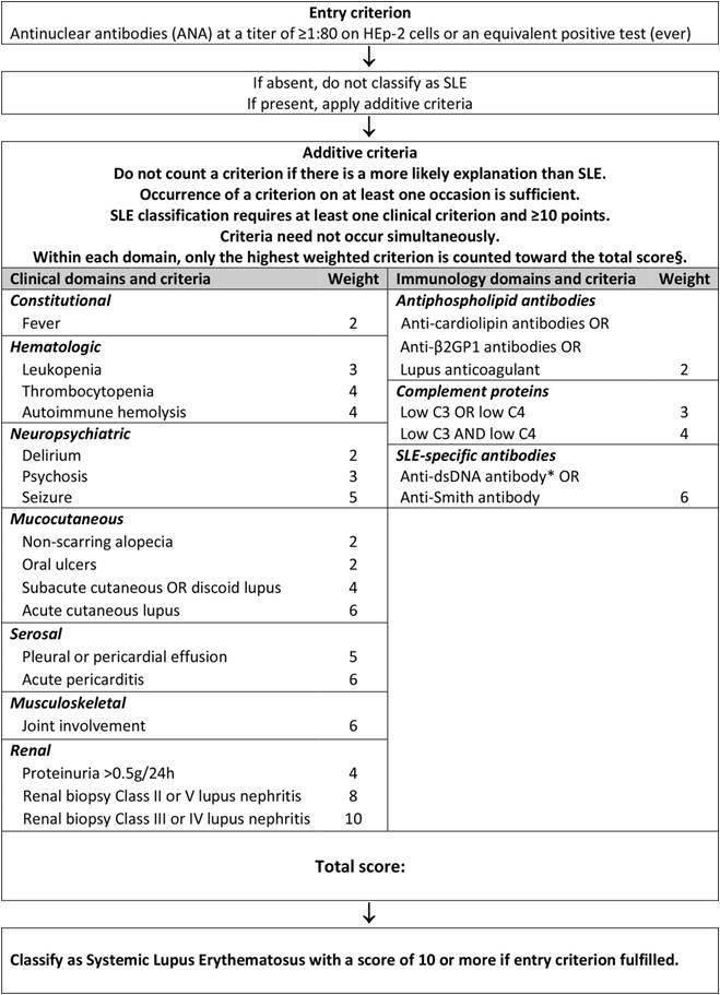
immunological domains and criteria, with weightage being distributed among the parameters. Proliferative lupus nephritis proven on biopsy holds the maximum weightage, and a total score of 10 or more weightage points are needed to classify a patient as a case of Lupus10 . This has demonstrated a sensitivity and specificity of 96.1% and 93.4% (Fig 1).
IgG4-Related Disease :
IgG4-related disease and its propensity to form fibroinflammatory, tumefactive lesions in virtually any organ in the body, also had its classification criteria updated and somewhat complicated. A 3-step process was validated by an international multispeciality groups of physicians: “First, it must be demonstrated that a potential IgG4-RD case has involvement of at least one of 11 possible organs in a manner consistent with IgG4-RD. Second, exclusion criteria consisting of a total of 32 clinical, serological, radiological and
pathological items must be applied; the presence of any of these criteria eliminates the patient from IgG4-RD classification. Third, eight weighted inclusion criteria domains, addressing clinical findings, serological results, radiological assessments and pathological interpretations, are applied11.”
Therapeutics :
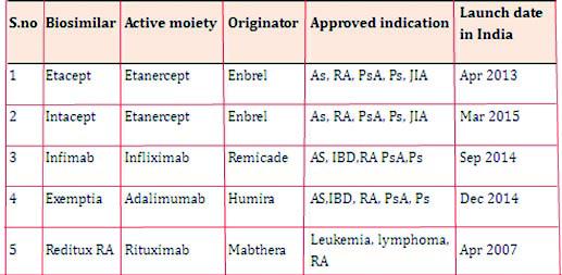
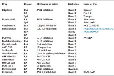
The advent of biologics and small oral molecules has recently changed the existing scenario of pharmacologic treatment of rheumatic diseases. These drugs have innovative mechanisms of action, that target specific parts of the pathogenetic pathways in various diseases. Conventional biologic DMARDs have paved their way to newer biological like Sarilumab (IL-6 antagonist) for Rheumatoid Arthritis, biosimilars (Fig 2), and more recently, the oral small molecules that have brought a “pill in the pocket” alternative to regular injections for disease control. There are many more undergoing trials and in the pipeline for release in the near future (Fig 3), although their safety and efficacy studies need further investigation. JAK-inhibitors like Tofacitinib, Upadacitinib and Baricitinib are upcoming areas of prime interest for rheumatologists worldwide.
Antifibrotics in Scleroderma :
In light of recent advances in the understanding of the pathogenesis of fibrosis in Scleroderma, many agents are being tried in trials to discover their efficacy as anti-fibrotic agents, as currently there are no known effective measures to reverse or halt fibrosis on this
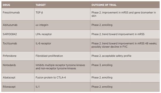
disease. The list of agents and their targets have been illustrated in Fig 4, although it remains to be seen what efficacy and safety data is finally churned out at the end of these trials.
Machine Learning/AI :
Machine learning (ML) is a subset of artificial intelligence finding increasing applications in Rheumatology, with growing datasets providing a basis for application of machine learning via deep learning, supervised/unsupervised learning and reinforcement learning12. Automated image recognition is already in use, and newer programmes in ML, especially using Supervised Learning, are being developed to individualise disease prediction models. Algorithms can now aid in e-diagnosis, disease course and patterns of disease, treatment related modifiable factors and the risk or benefit of an intervention as previously studied in national cohorts.
Hence, in the future, shared decision-making will include the patient’s opinion, the rheumatologist’s evidence-based experience, as well as algorithms drawn up by machine-learned evidence.
In conclusion, one may cite a hundred or so ongoing trials or review analyses in the field of rheumatology, and yet the core components of individualised decisionmaking and a few already well-established treatment protocols are the pillars continuing to support clinical judgement. A new era beckons, where further research in targeted therapy and artificial intelligence may help us take better decisions, thus bringing down the time to diagnosis and better patient outcomes.
Limitation :
Even with all these developments we could not translate significantly the clinical achievement in our daily clinical practise . We would need more evidence based scientific information in future for translating bench to bedside practise.
1Yang H, Biermann MH, Brauner JM, Liu Y, Zhao Y, Herrmann M, et al — New insights into neutrophil extracellular traps: Mechanisms of formation and role in inflammation. Front Immunol. 2016
2Bonaventura A, Liberale L, Carbone F, Vecchié A, DiazCañestro C, Camici GG, et al — The pathophysiological role of neutrophil extracellular traps in inflammatory diseases. Thromb Haemost. 2018
3Barrera-Vargas A, Gómez-Martín D, Carmona-Rivera C, Merayo-Chalico J, Torres-Ruiz J, Manna Z, et al — Differential ubiquitination in NETs regulates macrophage responses in systemic lupus erythematosus. Ann Rheum Dis. 2018
4Odqvist L, et al — Genetic variations in A20 DUB domain provide a genetic link to citrullination and neutrophil extracellular traps in systemic lupus erythematosus. Ann Rheum Dis 2019.
5Gupta S, Kaplan MJ — The role of neutrophils and NETosis in autoimmune and renal diseases. Nat Rev Nephrol 2016
6Milano A, et al — Molecular subsets in the gene expression signatures of scleroderma skin. PLoS ONE 3, e2696. 2008
7Farina G, Lafyatis D, Lemaire R, Lafyatis R — A four-gene biomarker predicts skin disease in patients with diffuse cutaneous systemic sclerosis. Arthritis Rheum 2010; 62: 580-8.
8Michalska-Jakubus M, Kowal-Bielecka O, Smith V, Cutolo M, Krasowska D — Plasma endothelial microparticles reflect the extent of capillaroscopic alterations and correlate with the severity of involvement in systemic sclerosis. Microvasc Res 2017; 110: 24-31.
9Murota A, Suzuki K, Kassai Y, Miyazaki T, Morita R, Kondo Y Takeuchi T — Serum proteomic analysis identifies interleukin 16 as a biomarker for clinical response during early treatment of rheumatoid arthritis. Cytokine 2016; 78: 87-93.
10Aringer M, Costenbader K, Daikh D, et al — 2019 European League against Rheumatism/American College of rheumatology classification criteria for systemic lupus erythematosus. Ann Rheum Dis 2019; 78: 1151-9.
11Wallace ZS, Naden RP, Chari S, et al — The 2019 American College of Rheumatology/European League against rheumatism classification criteria for IgG4-related disease. Ann Rheum Dis 2020.
12Pandit A, Radstake TRD J — Machine learning in rheumatology approaches the clinic. Nat Rev Rheumatol 2020; 16: 69-70.
Anita Babasaheb Tandale1, Shruti Sudhakar Khade2, Karishma Krishnakumar3
In December 2019, a novel coronavirus (2019-nCOV) emerged in Wuhan, China. It has affected the entire globe causing the ongoing pandemic. The SARS-CoV-2 infection could be asymptomatic or mildly symptomatic in most cases of COVID-19. Therefore, there is a difficulty in diagnosis of SARS-Cov-2 infection based only on the clinical findings and hence requires the confirmatory laboratory testing. In dental clinical practice, there is a very high risk of transmission of SARS-Cov-2 infection. Therefore, there is an urgent need to assess and minimize the risk in dental care settings. Each patient in dental clinical practice needs to be considered as the potentially infectious and managed accordingly with the use of appropriate infection prevention and control measures. The approaches and measures for risk alleviation need to be emphasized and practiced appropriately to prevent the infection. This review presents the important changes needed in routine and emergency dental practice in the current pandemic.
[J Indian Med Assoc 2020; 118(8): 29-35]
Key words : COVID-19, Dental practice, Infection prevention control, emergency dental treatment, aerosol transmission.
There were only four coronaviruses known to cause human disease before the current coronavirus emerged in China in 2019. However their virulence was not very much significant1. The severe acute respiratory syndrome (SARS) outbreak reported from the East Asia during the years 2002-03 was the first major pandemic associated with coronavirus2. Since the year 2012, the Middle East respiratory syndrome (MERS) coronavirus outbreaks were reported in Saudi Arabia3 These are the only two most prominent outbreaks of coronaviruses in history.
In December 2019, emergence of 2019 novel coronavirus (2019-nCOV) happened in Wuhan, China4 It was named subsequently as the SARS-CoV-2 virus5. It is the seventh member of the large family of coronaviruses affecting humans, thereby affecting the entire globe causing COVID-19 pandemic declaration by the WHO6. Coronaviruses are commonly associated with respiratory illnesses like common cold, flu-like illness or pneumonia. However very few patients may also present with the gastrointestinal symptoms. The laboratory diagnostic tests are performed on
1MDS, Professor and Postgraduate Teacher, Department of Conservative Dentistry and Endodontics, Padmashree Dr D Y Patil Dental College and Hospital, Pune 411018 and Corresponding author
2MDS, Postgraduate Second year Student
3MDS, Postgraduate First year Student
Received on : 01/06/2020
Accepted on : 30/07/2020
Editor's Comment :
The oral medicine and healthcare professionals are at the heightened risks of the COVID-19. Each patient needs to be considered infectious with the need for infection prevention measures. Strict adherence of infection prevention practices would be very critical in pandemic situation.
nasopharyngeal, oropharyngeal and blood samples. However, the SARS-CoV-2 infection could be asymptomatic or mildly symptomatic in most cases. Therefore, there is difficulty in diagnosis of infection based on clinical findings unless the laboratory testing is done6. There is a need to consider each dental patient in clinical practice as the potentially infectious one 7 . The dental practice needs to be managed accordingly with the use of appropriate infection prevention and control measures8
In dental clinical practice, there is a very high risk of transmission of SARS-Cov-2 infection. Therefore, there is an urgent need to assess and minimize the risk in dental care. The approaches and measures for risk alleviation need to be emphasized and practiced properly. There are various guidelines and recommendations for the risk alleviation. This review presents the important changes needed in routine and emergency dental practice in the current pandemic.
Literature search :
We searched the PubMed using the search term ‘COVID-19’ and ‘dental’ in the text word. We could identify 61 references using the search terms combination. The major article types identified were original articles (14), review articles (23) and other articles (24) - including letters (6), editorials (5), perspectives (3), and 2 each of communication, opinion and guidance and 1 each of correspondence, highlights, interview and recommendation. All publications were critically reviewed for title, abstract and full text. The publications were then appraised for the major thematic areas or aspects and utilized for deciding the structure of the literature review and is presented in the following sections.
Transmission Risks :
Healthcare workers on the frontlines are the most vulnerable to the SARS-CoV-2 infection9. The dentists working with the patients are at the greatest risk8. They can also become potential carriers and thereby act as the source of infection for their patients9. Chances of cross infection between the dental clinician and patient increases because of typical dental clinic setting10 The asymptomatic phase or the incubation period has been reported to be between 2 and 14 days, however a few are reported with symptoms even after 24 days of exposure11. The asymptomatic spread of virus is also confirmed12. Therefore, there is an urgent need for infection control protocols to be implemented strictly and effectively for avoiding spread in dental clinical practices10
The most common transmission route is the direct transmission11. It is mostly by coughing, sneezing, and droplet inhalation. Also, the contact with oral, nasal, mucous membranes and eye is also an important route13. Coronavirus has the affinity for ACE2 receptors, which are abundant throughout respiratory tract and salivary gland duct epithelium in humans. Saliva has been reported to have high viral loads with significant role in human-to-human transmission15. Also, the faceto-face communication is the most common route during the patient management in dental clinics. The aerosols generated during various dental procedures are predominant way of spread7. The dental procedures use handpieces under irrigation. These procedures may lead to handling of instruments and diffusion of aerosols of saliva, blood and body fluids 13 . Therefore, contamination of environment is high with such instruments, apparatuses and surfaces in the dental clinics. Hence, dentists are likely to face a very high risk of acquiring infection if proper infection prevention
control precautions are not implemented in clinical practice12. Therefore, the chains of transmission need to be broken for prevention of SARS-CoV-2 infections by applying prevention measures targeted at the source of infection, mode of transmission and the practices of dental professionals (Table 1).
There are 9 cases of SARS-CoV-2 infection reported among 169 dental practitioners10. This signifies the high risk of transmission in dental practice. Therefore, dentists have an important role to play in preventing SARS-CoV-2 transmission in dental clinics. The toothbrushing has been indicated to be emphasized for prevention of infection16.
Oral Manifestations :
There are no obvious oral manifestations of SARSCoV-2 infection. These may be very nonspecific, like loss or altered taste or smell sensations, thereby unlikely to be reported or enquired during the dental evaluations. The inflammatory process in oral cavity would be mostly unremarkable. Therefore, there are great chances of missing the detections of potentially infectious person based on history and oral examination. This risk is even accentuated with the reports of most infections being asymptomatic. Additionally, the transmission risk is potentially more in asymptomatic, pre-symptomatic and mildlysymptomatic phases of infection17. Thus, the dental practice faces the heightened risk without having tools to identify the infection without high index of suspicion with laboratory testing.
Also, saliva and other oral secretions are highly viraemic with higher viral loads reported than even recommended and usually collected respiratory specimens like nasal, pharyngeal and lower respiratory
Table 1 — Transmission Chain in Dental Clinical Practice with Prevention Opportunities
Transmission Transmission risk - events, chain elements practices and procedures
Source of• Infected patient – asymptomatic, infection pre-symptomatic, mild symptomatic
•Surfaces, objects and instruments
•Infected staff working in the clinic
Mode of•Droplets and aerosolstransmission from the infected source patient or procedures
•Contact (direct/indirect) - surfaces, objects, equipments, instruments
•Airborne transmissionduring dental procedures
Susceptible• Clinic attendees - patients, visitors humans• Clinic staff - dentists, clinic staff and assistants, receptionists
JOURNAL OF THE INDIAN MEDICAL ASSOCIATION, VOL 118, NO 08, AUGUST 2020
specimens 15 . Therefore, a very high risk of transmission is likely in the absence of obvious oral manifestations. Additionally, there could be underreporting and even hiding of important exposures and symptoms due to fear and panic of isolation and quarantine.
Occupational Health :
There are very important issues in routine dental clinical practice that are relevant to infection risk prevention and control associated with transmission of hepatitis B, hepatitis C and HIV. The universal precautions for control of cross-infections are very well known in dental practice. The risk is the highest during the current times of COVID-19 pandemic situation. The risk assessment needs to be considered along with all possible measures to reduce the risk. However, the dentists need to address the risk assessment critically in the current situation. This aspect needs to be considered importantly after lifting of lockdown restrictions as the transmission could be widespread with most asymptomatic infections. The decisionmaking on the prevention measures has been recommended to be based on moral than evidence8
Infection Control :
The American Dental Association (ADA) recommends that all palliative dental care and nonemergency treatments may be postponed during the COVID-19 pandemic. However, dental professionals are recommended to remain available for potentially lifethreatening dental emergencies 18 They are required to remain vigilant during and even after the pandemic. The dental care needs to be provided only after a thorough case history and screening for COVID-19. All dental patients must be screened first via telephone with consultation provided through messages and video communications, whenever possible19 The checklist of information to be enquired is provided in the boxes below.
Prevention Measures :
The patients suspected or confirmed for COVID-19, who are requiring emergency treatments for complaints like severe tooth pain and/ or swelling should be managed primarily with analgesics and antibiotics 20 . This will help them
temporarily in having symptomatic relief so as to refer them to a dental specialist with all appropriate measures to manage COVID-19 positive cases. Patients not responding to pharmacological management must be screened and treatment provided with the PPE and proper infection control protocol21
Triages can be set up at the entrance for measuring temperature of the patients using an infrared thermometer along with the case history of the patients. The signs and posters with instructions about respiratory hygiene including cough etiquette and also hand hygiene may be helpful. These could be pasted at the entrance, in waiting areas, elevators, cafeterias, etc. The patients need to be provided instructions to use hand sanitizers before entering the clinics. Patients with cough and sneezing need to be taken to a separate waiting room and face masks offered to them if they aren’t already wearing so as to minimize the spread of infection22
The indicative flowchart for screening, triaging and management of practice is provided in Fig 1.
Hand Hygiene :
Hand hygiene is critical and must not be overlooked. Hand washing needs to be followed with its proper technique for 20-30 seconds. Even though hand hygiene is considered a part of the normal routine practice, it
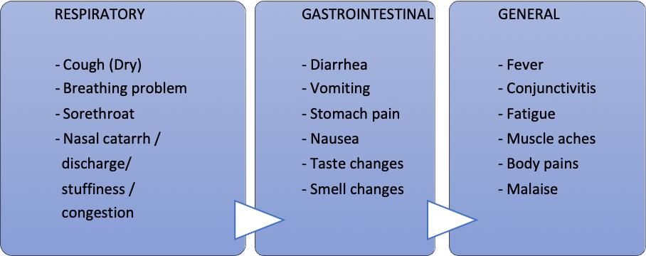
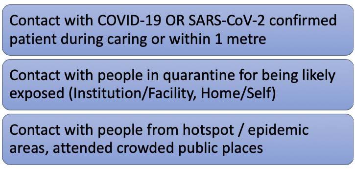
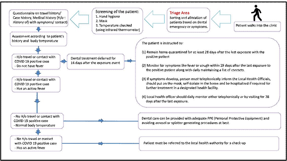
Fig 1 — Screening, Triage and Patient Management in Dental Clinics
must be strictly reinforced among the operators and the dental clinic staff. Washing of hands by the dentists needs to be followed before patient examination, before
and also after dental treatment, after touching surrounding or undisinfected equipments and when hands are visibly soiled. One must be especially watchful to not touch eyes, mouth and nose23
Setting Timing Activities neededTools/ resources needed
OutsideBefore the EnquiryTelephone, Contact log the clinicclinic visitSymptoms and exposuresQuestionnaire checklist (At Home)AppointmentRegistration (Electronic)
In the clinic/At the entryTemperature
Infrared thermometer Waiting area/in the clinicHand hygieneSoap / Sanitizer roomWaiting area
Arrangements for distancing Disinfection of areaDisinfectant chemicals Protection of patientsSurgical mask
Clinic Before dentalPatient preparationsShoe covers, mouth rise procedures/ treatmentStaff hand wash Soap / sanitizer work areaStaff use masksN-95 Mask, goggle, face shield, gloves, gown, head cover, shoe cover
During dental Instruments
Treatment Surfaces
Disinfection, sterilization
Disinfection, barrier, UV Procedures
Non-aerosol, PPE, face shield, Rubber dam, Anti-retraction handpiece, HEPA filter
After dentalVentilationAvoid ACs, cross ventilation
treatment Instruments
Disinfection, Sterilization Protection of staff PPE for waste segregation
Hand hygieneFrequent and thorough wash
PPE (Personal Protective Equipments) :
The ADA guidelines recommend PPE for dental facilities - in asymptomatic patients, patients with history of exposure, negative patients or recovered positive cases. The operator and staff should use N95 respirators with fit testing on the face, along with full-face shields, and eye protection. They should also follow disinfection procedures immediately after every procedure in cases with chances of aerosol generation. In non-aerosol generating procedures and without use of three way syringes, the operator and staff may use 3 ply face masks, with the PPE including eye protection, and approved disinfection procedures. New pair of gloves is must while donning
and doffing the N95 respirators and PPE24. The qualitative fit test (QLFT) must be performed after wearing the N95 respirators to ensure the proper fit to prevent any air leaks. User seal check is performed by positive and negative pressure. In positive pressure seal check, user exhales gently blocking exit paths for air from the facepiece. The successful check should provide slight pressure before outward leakage on increased pressure. In negative pressure seal check, user inhales forcefully after blocking air entry in the facepiece. The successful seal happens when the facepiece slightly collapses under negative pressure25
Emergency Guidelines :
All dental emergencies must be handled through telephone or video communications controlled with pharmacological approach. ADA recommends that management of emergency must focus on relieving severe pain along with efforts for decreasing infection risks. The conditions to be considered for emergency care include acute pain from pulpal inflammation, pericoronitis or third-molar pericoronitis, post-operative osteitis, dry socket, abscess, or localized bacterial infection, traumatic injuries, avulsion/luxation, procedures prior to critical medical procedures, fractured restoration related trauma26
During treatment of emergency cases, the patient must be instructed to perform hand hygiene and rinse mouth with 0.2% povidone iodine solution. It is recommended to use extra-oral radiographs such as panoramic radiograph, CBCT, etc rather than intra-oral radiographs to avoid saliva splatter. On application of the N95 respirator, the operator and the staff must check the seal of the mask by holding and exhaling to check for any leaks. The PPE must be examined for any tear or visible soiling before the use. Application of rubber dam isolation with high ejection saliva should be reinforced in every case. Aerosol generating procedures must be avoided as much as possible. It is advised against treating the patient if the clinic/ hospital lacks enough PPE equipment and consider to refer the patient to the appropriate clinic/ hospital after pharmacologically controlling the emergency26.
Oral and maxillofacial compound injuries, which are life threatening cases, should be hospitalized and chest CT prescribed immediately. In life-threatening maxillofacial compound injury, there is a need to rule out the suspected infection instead of PCR test which is time consuming, patient is advised immediate chest CT / X-ray27
Rationalization and Reuse of Masks :
Reuse strategy for N-95 is not recommended for
aerosol producing procedures and if visibly soiled. CDC recommends following measures for optimizing the supply of N95 respirators28
Cleaning of hands before and after touching or adjusting the N95 or Filtering Facepiece Respirator (FFR) by using soap and water or alcohol-based hand sanitizer.
Avoiding touch to the inside of the N95 or FFR.
Using of a pair of new pair of gloves for donning and doffing.
Making seal check by user to check if respirators are being properly worn.
Inspecting the N95 or FFR to determine integrity.
Rubber dam application
Other infection control practices include preprocedural mouth rinse with 0.2% povidone- iodine along with the use of rubber dam application with high volume saliva ejector which can help minimize the aerosol generation and splatter. Use of CariSolv and hand scalers can be implemented for caries removal and periodontal scaling28
Anti-retraction Handpiece :
To prevent cross-infection, anti-retraction dental handpieces are strongly recommended during the COVID-19 pandemic. The anti-retraction valves present in these handpieces will prevent aspiration of fluids and debris during dental procedures. Dental unit may get contaminated with bacteria and viruses. The use of three way syringe needs to be avoided to prevent unnecessary splatter. Implementation of four handed dentistry is recommended.
All unnecessary personnel must be at least 6 feet away from the patient28.
Sterilization and Disinfection Practices :
Once the curve starts to move down, aerosolgenerating procedures can be initiated, with strict sterilization and disinfection procedures. All critical, heat-resistant semi-critical instruments and handpieces should be cleaned and sterilized after each use or discarded. Heat sensitive semi-critical items need to be disinfected with 2% Glutaraldehyde18
The frequently touched clinical contact surfaces get contaminated directly by aerosols or by contaminated gloved hands of dental health care professionals. The high-touch clinical surfaces those are difficult to clean must be covered using a physical barrier for every patient or disinfected using 1% Sodium hypochlorite or 70% alcohol between patients. The barrier protection can prevent contamination of clinical contact surfaces and equipment that are difficult to
clean. The barriers may be done by using materials impervious to moisture including plastic wrap, bags, sheets, tubing, and plastic-backed paper. These barrier coverings should be removed and discarded between patients by the professionals using gloves. The welltrained dental professional is essential for proper disinfection protocol18
In any operatory, from the dental chair, aerosols can travel up to 10 feet. Ideally, the use of negative pressure room is recommended. Another alternative is use of air filters. The aerosols generated in dental clinics can be reduced by devices like High-Efficiency Particulate Arrestor Air Filters (HEPA) filters as they decrease the concentration of airborne infectious pathogens, trap bioaerosols as small as 0.3 micron (which is the tiniest particle size to get into your lungs) with 99.97% efficiency, thereby reducing the spread of diseases. As HEPA 13 and HEPA 14 have efficiency of 99.97%, they are recommended for medical purpose29
Germicidal lamps (UVGI / UV-C) destroy and inactivate airborne microorganisms on frequently exposed surfaces. UV-C gives off light at the short end of the wavelength band of 253.7 nm. UV-C lamps are used in air, water purification and also decontamination of rooms and surfaces. However exposure of the light on skin and eyes can cause skin and eye lesions29
The floor must be disinfected with 1% Sodium hypochlorite, 3% hydrogen peroxide or EPA approved agents. Mop heads and cleaning cloth must be laundered (heat disinfection) with detergent and drying at 80oC and changed frequently.
Waterborne infections with dental water systems are reported in literature. The potential for transmission of waterborne infections and related diseases is verified in hospital settings and in the community. The transmission in dental practice may occur by inhalation of infectious aerosols from the respiratory equipment. Hence, the dental waterlines must be cleaned by use of 1 ml of 5% NaOCl mixed in 5 liters of dental waterline or 1 ml of 3% NaOCl mixed in 3 liters of dental waterline regularly30
Waste Management :
Infectious waste in dental practice could be solid waste gauze saturated with blood or saliva, hard and soft tissues, extracted teeth and contaminated sharp items like needles, scalpel blades, and wires. The careful containment for treatment or disposal is required for regulated medical waste18. The non-sharp regulated medical waste needs to be managed by using a sturdy leak-resistant biohazard bag. A second biohazard bag
may be used for preventing exterior contamination or puncturing of the bag. The sharps need to be managed by using puncture-resistant containers using biohazard label18
Hydrochloroquine is a derivative of chloroquine used in treatment of malaria since many years. It has been recommended for empiric use in treatment and prophylaxis of COVID-19. The antiviral activity of hydroxychloroquine sulphate is based on inhibition of SARS-CoV-2 viral replication in vitro along with early human clinical application utility. Some in vitro studies suggest that approved doses of hydroxychloroquine sulphate could prevent SARS-CoV-2 infection if taken prophylactically among healthcare workers and close contacts31
The drug is considered safe; however there are some serious side effects such as retinopathy and immunosuppression. Therefore, it is recommended to be used only on physician advice and requirement of closed observation of side effects as recommended for emergency use31.
Use of Non-steroidal Anti-inflammatory Drugs (NSAIDS) :
Many patients will get relief on using analgesics and NSAIDs. The use of ibuprofen is warned in COVID patients, as it is believed that virus binds to ACE2 receptor and it increases accelerated expression of this protein23. Hence, it may potentiate and enhance infection even though there is no strong epidemiological evidence to suggest a harmful effect of ibuprofen on COVID-19 patients. Paracetamol can be used as the first line of treatment in such cases17
Monetary Implications :
In the pandemic situation, there is a need to use PPE and all other additional infection prevention measures in dental practice, although the cost of dental care is likely to go high32. This would ultimately be shifted to the patients as the dentists would not be able to bear the same on their part. This will increase the expenses on patients thereby increasing the costs of the treatment.
Summary :
The health care workers should make the society aware on the possible risks of the COVID-19 disease in dentistry practice and also take the proper actions. The consideration should be given to treat each patient as likely infectious and take utmost measures to prevent infection. The protection of healthcare workers should be the key aspect during the COVID-19
pandemic. Therefore, strict adherence of infection prevention and control measures is an important aspect that needs to be inculcated in dental clinical practice during the pandemic.
Acknowledgements :
We acknowledge Dr. Babasaheb V. Tandale (MD), for his guidance, critical inputs and revision of draft manuscript. Also, we are thankful to Miss. Revati Avadhut Bangar for the help in searching literature, compilation and listing of references.
Funding : None
Conflict of Interest : None
1Chang L, Yan Y, Wang L — Coronavirus Disease 2019: Coronaviruses and blood Safety. Transfusion Medicine Reviews, https://doi.org/10.1016/j.tmrv.2020.02.003 (Accessed on 17th May 2020).
2Lam WK, Zhong NS, Tan WC. Overview on SARS in Asia and the world. Respirology. 2003;8 Suppl(Suppl 1):S2-S5. doi:10.1046/j.1440-1843.2003.00516.x.
3de Groot RJ, Baker SC, Baric RS, et al — Middle East respiratory syndrome coronavirus (MERS-CoV): announcement of the Coronavirus Study Group. J Virol. 2013;87(14):7790-7792. doi:10.1128/JVI.01244-13.
4World Health Organization. https://www.who.int/csr/don/12january-2020-novel-coronavirus-china/en/. (Accessed on 17th May 2020).
5Gorbalenya AE, Baker SC, Baric RS, et al — The species Severe acute respiratory syndrome-related coronavirus: classifying 2019-nCoV and naming it SARS-CoV-2. Nat Microbiol 5, 536–544 (2020). https://doi.org/10.1038/s41564020-0695-z.
6World Health Organization. http s://www.who .int/ emergencies/diseases/novel-coronavirus-2019/technicalguidance/naming-the-coronavirus-disease-(covid-2019)and-the-virus-that-causes-i. (Accessed on 17th May 2020).t. (Accessed on 17th May 2020).
7Ather A, Patel B, Ruparel NB — Coronavirus Disease 19 (COVID 19): Implications for clinical dental care. J Endod 2020; 46(5): 584-95.
8Coulthard P — Dentistry and coronavirus (COVID-19) - moral decision-making. Br Dent J 2020; 228(7): 503-5.
9Fallahi HR, Keyhan SO, Zandian D — Being a front-line dentist during the Covid-19 pandemic: a literature review. Maxillofac Plast Reconstr Surg 2020; 42(1): 12.
10Meng L, Hua F, Bian Z— Coronavirus Disease 2019 (COVID19): Emerging and future challenges for dental and oral medicine. J Dent Res 2020; 99(5): 481-7.
11Peng X, Xu X, Li Y— Transmission routes of 2019-nCoV and controls in dental practice. Int J Oral Sci 2020; 3; 12(1):9.
12Lo Giudice R — The Severe acute respiratory syndrome Coronavirus-2 (SARS CoV-2) in Dentistry. Management of biological risk in dental practice. Int J Environ Res Public Health 2020; 17(9): E3067.
13Wadia R. Transmission routes of COVID-19 in the dental practice. Br Dent J 2020; 228(8): 595.
14Xu H, Zhong L, Deng J, Peng J, Dan H, Zeng X, et al — High expression of ACE2 receptor of 2019-nCoV on the epithelial cells of oral mucosa. Int J Oral Sci 2020 Feb 24;12(1): 8. doi:
10.1038/s41368-020-0074-x.
15Xu R, Cui B, Duan X, Zhang P, Zhou X, Yuan Q.Xu R, et al — Saliva: potential diagnostic value and transmission of 2019nCoV. Int J Oral Sci 2020; Apr 17;12(1):11. doi: 10.1038/ s41368-020-0080-z.
16Addy M — Toothbrushing against coronavirus. British Dental Journal 2020; 228(7): 487
17Odeh ND, Babkair H, Abu-Hammad S, Borzangy S, et al — COVID-19: Present and future challenges for dental practice. Int J Environ Res Public Health. 2020 Apr 30;17(9):E3151
18American Dental Association (ADA) — Interim guidance for management of emergency and urgent dental care. Updated 2020. Accessed April 23, 2020.
19Villa A, Sankar V, Shiboski CVilla A — Tele(oral)medicine: a new approach during the COVID-19 crisis. Oral Dis. 2020; Available at https://onlinelibrary.wiley.com/doi/full/10.1111/ odi.13364 (Accessed 17 May 2020).
20Yu J, Zhang T, Zhao D— Characteristics of endodontic emergencies during Coronavirus disease 2019 Outbreak in Wuhan. J Endod 2020; S0099-2399(20)30238-7.
21Izzetti R, Nisi M, Gabriele M — COVID-19 transmission in dental practice: Brief review of preventive measures in Italy. J Dent Res. 2020; 22034520920580.
22Alharbi A, Alharbi S, Alqaidi S — Guidelines for dental care provision during COVID-19 pandemic. Saudi Dental Journal 2020; 32(4):181-6.
23Abramovitz I, Palmon A, Levy D, et al — Dental care during the coronavirus disease 2019 (COVID-19) outbreak: operatory considerations and clinical aspects. Quintessence Int. 2020; 51(5): 418-29.
24Umer F, Haji Z, Zafar K — Role of respirators in controlling the spread of Novel Coronavirus (Covid-19) among dental health care providers: a review. Int Endod J 2020; Available at https://pesquisa.bvsalud.org/controlecancer/resource/pt/ mdl-32357257 (Accessed 17 May 2020).
25Centres for Disease Control and Prevention (CDC) — Proper N95 respirator use for respiratory protection preparedness. Available at https://blogs.cdc.gov/niosh-science-blog/2020/ 03/16/n95-preparedness/ (Accessed 17 May 2020)
26Guo H, Zhou Y, Liu X — The impact of the COVI 19 epidemic on the utilization of emergency dental services. J Dent Sci 2020; Available at https://www.sciencedirect.com/science/ article/pii/S1991790220300209 (Accessed 17 May 2020).
27Dave M, Seoudi N, Coulthard P — Urgent dental care for patients during the COVID-19 pandemic. Lancet 2020; 395(10232):1257.
28Centers for Disease Control and Prevention. Guidelines for infection control in dental health-care settings — 2003. MMWR 2003;52(No. RR-17):[22-31].
29Yadav N, Agarwal B, Maheshwari C — Role of high-efficiency particulate arrestor filters in control of airborne infections in dental clinics. SRM Journal Res Dent Sci 2015; 6: 240-2.
30Fiorillo L, Cervino G, Matarese M, D'Amico C, Surace G, et al COVID-19 surface persistence: A recent data summary and its importance for medical and dental settings. IntJEnviron Res Public Health 2020 Apr 30; 17(9): E3132.
31Sinha N, Balayla G — Hydroxychloroquine and covid-19. Postgrad Med J. 2020. Available at https://pmj.bmj.com/content/ early/2020/04/15/postgradmedj-2020-137785.long (Accessed 17 May 2020)
32Farooq I, Ali S, et al — COVID-19 outbreak and its monetary implications for dental practices, hospitals and healthcare workers. Postgrad Med J 2020 Apr 3:postgradmedj-2020137781.
Colin Robertson BA(Hons), MBChB, MRCP(UK), FRCSEd, FRCPGlas, FICP(Hon) FSAScot, Professor in Accident and Emergency Medicine and Surgery, University of Edinburgh Editor, Macleod's Clinical Examination 12th Edition
Medical education in the future :
1.What should be future direction of Medical Education – focus on Micro-health or Macro-health, communicable vs non-communicable, community approach vs specialized approach ?
For too long we have neglected the importance of community aspects in Medical education, we have concentrated upon the high-tech. end of medicine with emphasis upon diseases such as cancer, cardio- and cerebro-vascular disease. These diseases are clearly important, but the greatest benefits to a population come from much simpler and cheaper approaches such as the provision of clean water, sanitation, adequate nutrition and childhood immunisation. These subjects are frequently dismissed, or even worse trivialised, in Medical education.
2.Do you think medical education of the future should incorporate non-medical disciplines like economics,anthropologyorsociology?Ifso,won’tthat beanextraburdenonthealreadyoverflowingmedical subjects ?
One problem is that many of our undergraduates come straight from school. They have little world or work experience. Of course the above subjects are important, they are a part of life experience.
3.Asignificantcauseoftheconfrontationbetween medical science and the society involves the rising healthcarecosts,whetheritisout-of-pocketorthrough insurance premium. Do you think doctors should take asignificantroleindeterminingthecostofhealthcare? Ifso,shouldtheybetaughttheconceptofcost-effective healthcare from student days ?
Yes
4. Do you think the teaching of clinical methods hasanyroleincurrenttimes?Especiallyinthemodern times of medical litigation, can a doctor survive with onlyclinicalmethodology?
Clinical methodology is even more important today. Far too often young, and older, doctors rely solely on tests, without appreciating the limitations, and even dangers, of the test they are using. A test should be used to confirm a clinical diagnosis, not the other way round.
1.In view of the ubiquity of social distancing, do you think clinical examination will be less popular in thepost-COVIDera?Whatcanbefalloutandsolution?
The opposite is the case. One of the things that we have learned is that telephone and even video consultations have major limitations, and many of our patients have suffered as a consequence.
2.Since an epidemic causes major financial and socialdisruption,doyouthinktheroleofphysiciansin overall policy making will be enhanced in the coming days? For example, will major multinational corporations include doctors while making their long term strategy ?
I doubt it. Across the world, medical advice has frequently been dismissed, or even ignored.
3.WhatcanbefutureofDoctorPatientrelationship when doctors maintain distance with patients during their visits ?
I sincerely hope that we will return to our previous clinical contact structure. Medicine has to have humanity and compassion at its core. Without that approach, Medicine will become an automated, cold, impersonal area. I don’t believe that our patients want that and neither do I.
1.Do you think the direction of medical research will change in the near future? If so, will preventive aspects of medicine be given more importance in research ?
It would be encouraging if that was the case, however ‘Big Pharma’ are unlikely to buy into that concept – they will not see much in the way of potential reward. This means that governments will have to take the initiative.
2.Pandemics like Covid-19 will become commoner as global warming progresses. Do you think every country should have a pandemic response team ?
Of course; it is absolutely essential. A country that has not prepared is not serving its population as it should.
3.What do you think about the general trajectory of medical research in the last two decades? Is it more concentrated on curative medicine ?
We have concentrated more research and effort into combating diseases that principally affect the last few years of life. Often this leads to increased misery and with mixed benefits for our patients.
1.In many parts of the world like USA, there were a lot of resentment at the government directives for social distancing. People wanted to lead their lives normally,despitethepandemic.Whydoyouthinkthere is so much mistrust on the healthcare system ?
I think it is principally a distrust of the political systems, rather than healthcare system and its providers. Having said that, however, there has been a marked lack of appreciation of the roles played by some members of the health care team – in particular nursing staff, cleaners, therapists and so on. Doctors are only a part – and perhaps not even the most important part – of health care !
2.Why do you think the countries of Europe and USA had such high mortality, despite having a welldeveloped primary healthcare infrastructure ?
The populations in Europe and the US are in general older, and there is clear evidence that age is a major determinant of morbidity and mortality. Older individuals are likely to have more co-morbidities such as hypertension, diabetes, vascular disease, and these factors too affect mortality. In the more affluent countries such as the US and UK obesity is now a major concern and, as we have seen, this significantly impacts upon morbidity and mortality too.
3.A pandemic like the present one often leads to neglectofotheraspectsofpublichealth.Forexample, stress on COVID-19 control has led to a marked backlog in routine childhood immunization in many places. How can we plan in advance to avoid this ?
Immunisation programmes need to be continued irrespective of other aspects. Our children and grandchildren are our future, do not ever let us neglect their care.
4.How are you, in UK, catering to non-COVID patients for the last five months? Are you continuing routine clinical visits? If not, then are those patients kept at home with no treatment ?
We are still evaluating the effect on the reduced routine and clinical visits. Anecdotely, there have been delays in getting pateints with conditions such as acute coronary syndromes, stroke and non-Covid infections timely care. The effects on mental health may be even more significant and very worrying.
1.Many drugs were touted as probable cures for
COVID-19 and given approval for compassionate use. But I think these approvals generate a sense of false hope in the society. What is your take on that ?
The media, and especially the Internet, have played a major role in many of these stories. Claims of ‘Miracle cures’ and ‘Magic bullets’ do nothing except raise false hopes and generate unrealistic expectations. Some political leaders have been particularly at fault for this. As doctors we should be careful, measured and proportionate in what we say to the media.
2.Sometimes,duringapandemic,adoctormaybe under pressure (from the hospital management team or the patient’s next of kin) to use a drug of doubtful efficacy. How do you manage those situations ?
A clinician can only act as they think what is in their patient’s best interest. To be influenced by management or relatives erodes that patient-doctor relationship. However, measured discussion with respected, experienced clinical colleagues is always appropriate.
3.Sometimes, when ventilators are in short supply, a physician may be asked to choose between moribund patients to ration the available resources. How do you approach those cases? For example, is it right to choose a young patient over an elderly patient just because of age ?
The essential aspect of this question has to be that individual patients must always be considered and treated as individuals. Simply to make a decision on the basis of age, sex, religion, wealth or race is, in my view, intrinsically wrong. Additionally, at times of restriction of resource, these crucial decisions should never rest solely on the shoulders of one individual. There is the clear requirement for an ethics committee within a hospital to assist and make transparent these decisions. It is equally essential that the individuals comprising this group are not merely doctors, but include non-medical staff, ethics experts, religious leaders etc.
4.In situations like the present one, doctors can oftenfindPPEinshortsupply.Shouldadoctorriskhis/ her life for patient care in such cases? How do you balance your personal risk vs your duty to humanity?
This must also be a personal decision. Out with this pandemic, clinical staff regularly are at risk – from nosocomial infections, personal injury, mental health issues etc. The role of colleagues should be to support, whatever decision an individual doctor has made. Further, even if a doctor indicates that they, for example, do not wish direct patient contact they may still be able to play crucial roles.
5.Do you think the excessive media coverage of this pandemic is creating hindrance for healthcare workers ?
Unequivocally, YES
Amrisha Sharan1, Dipendra Kumar Sinha2, Nawal Kishore Jha3
Introduction : Candida isolation from intra operative peritoneal fluid in cases of hollow organ perforation is a significant risk factor for post-operative morbidity and mortality. The present study analyses the frequency of fungal isolation and its implications on post-operative course in patients with hollow viscus perforation.
Material and Methods : This prospective observational clinical study was conducted from April 2014 to November 2015 at our institute after approval from the institute’s ethics committee. The study population was selected from the patients presenting to the surgical emergency department with nontraumatic perforation peritonitis.The peritoneal fluid was collected during exploratory laprotomy and sent for microbiological analysis.
Results : The total number of cases included in the study was 127. A total of 44 samples positive for fungus. All of the fungi isolated belonged to Candida species. 77.3% (n=34) of the Candida isolated were Albicans and 22.7% (n=10) were Non-Albicans.
Conclusion : Prompt identification and empirical use of antifungal along with wide spectrum antibiotics may help the patients with non traumatic hollow viscus perforation to recover early.
[J Indian Med Assoc 2020; 118(8): 38-41]
Key words : Peritonitis, Fungus, Candida.
Peritonitis is a spectrum with varied aetiology and clinical course. Primary peritonitis occurs in young females and cirrhotic and is typically mono-microbial in nature. It consists of bacteria from genital tract in former cases and translocated gut bacteria in latter. Secondary peritonitis is more common and a result of anatomic breach of the gastrointestinal tract. The microbial flora is from the adjacent gut and is polymicrobial in nature. It also results from infection due to indwelling catheters, ventriculo-peritoneal shunt and direct extension from retroperitoneal infection. A clinical syndrome evolves with recurrent peritonitis after apparent control of primary or secondary peritonitis due to organisms of low intrinsic pathology. This has been termed tertiary peritonitis.
The gastrointestinal tract is a major reservoir of Candida species and an important portal for intraabdominal and disseminated candidiasis. As a commensal of the digestive tract, Candida may leak into the peritoneal cavity after perforation of a hollow viscus or surgical section of the intestinal wall. However, under most circumstances, Candida is cleared quickly from the peritoneum. Nevertheless, in some patients,
Department of General Surgery, Rajendra Institute of Medical Sciences, Ranchi, Jharkhand 834009
1MS (Gen Surg), Senior Resident and Corresponding Author 2MS, Professor 3MS, Professor & Head
Received on : 27/02/2020
Accepted on : 11/07/2020
Editor's Comment :
Fungal cultures of peritoneal fluid samples obtained intra operatively should be routinely done in the treatment of patients of perforation peritonitis along with bacterial culture.
Positive peritoneal fluid fungal cultures is a significant risk factor for poor outcome.
Antifungal should be used along with wide spectrum antibiotics in patients with non-traumatic hollow viscus perforation to ensure early recovery.
peritoneal seeding results in the development of an intra-abdominal Candida infection, with a risk of dissemination to the bloodstream and to extraabdominal tissues and organs.
The most frequently implicated risk factors include the use of broad-spectrum antibacterial agents, use of central venous catheters, receipt of parenteral nutrition, receipt of renal replacement therapy by patients in ICUs, neutropenia, and receipt of immunosuppressive agents (including glucocorticosteroids, chemotherapeutic agents, and immune-modulators)1. Patients with health care-associated intra-abdominal infection are at higher risk of Candida peritonitis, particularly patients with recurrent gastrointestinal perforations and surgically treated pancreatic infection.
Candida isolation from intra operative peritoneal fluid in cases of hollow organ perforation is a significant risk factor for post-operative morbidity and mortality2.
The present study analyses the frequency of fungal isolation and its implications on post-operative course in patients with hollow viscus perforation.
MATERIAL AND METHODS
This prospective observational clinical study was conducted from April 2014 to November 2015 at our institute after approval from the institute’s ethics committee. The study population was selected from the patients presenting to the surgical emergency department with non traumatic perforation peritonitis. Patients developing peritonitis due to anastomotic leak during hospital stay were also included. The patients who have received any form of antifungal treatment in past one month have been excluded.
The peritoneal fluid was collected during exploratory laprotomy and sent to the microbiology lab for examination. For bacterial culture the sample was inoculated in Blood Agar and MacConkey Agar at 37 degree Celsius for 48 hours. The colony characteristics and bacterial motility were examined. Gram staining was done. Bacterial identification was done by Indole, Methyl Red, Voges Proskaeur and Catalase (IMViC) tests. Antimicrobial susceptibility test (AMST) was done.For fungal culture, the sample was inoculated on Sabouraud’s Dextrose Agar at 37 degree Celsius for 7 days (Fig 1). A colony smear was done to identify fungi. Germ tube test was done by inoculating a yeast colony into 1 ml of human serum and incubating it at 37 degree Celsius for 2 hours. A KOH mount was prepared at the end of 2 hours and filamentous outgrowths could be seen extending from yeast cells if the fungus was of Candida species. As germ tube test is positive for Candida albicans, the candida with germ tube negative results were reported as Candida non albicans. Anti-fungal susceptibility could not be done due to non-availability.
The total number of cases included in the study was 127. The age range was from 5 years to 73 years with mean age of 39.5 years. There were 26 patients who were more than 50 years age. The population under study included 23.6% (n=30) females and 76.4%(n= 97) males. The average age of females in the sample was 40.1, while the average age of males was 39.Of the 127 patients 76 (59.8%) patients came to the hospital after 24 hours of onset of symptoms.The remaining 51 (40.2%) patients presented to the hospital within 24 hours of onset of symptoms.29.9% (n=38) patients had received pre-operative antibiotic therapy for a duration of more than 48 hours.The rest 70.1% (n=89) had either received antibiotics for less than 48 hours before undergoing surgery for perforation peritonitis or one pre-operative dose of antibiotic.
The Duodenum was the commonest site of perforation (n=65) (Fig 2), followed by ileal (n=30) (Fig 3), gastric (n=12), appendicular (n=15) and colonic perforation (n=5). The total gastroduodenal perforations were 77.
Of the 127 peritoneal fluid samples cultured, 90 (70.9%) were culture positive and 37(29.1%) were sterile. The total samples positive for fungal culture were 44(34.6%). Only bacteria were present in 46 cases (36.2% of all samples). All 44 (34.6%) fungal culture positive samples were also positive for bacteria.Of the 90 culture positive samples, 34 (37.8%) were mono-microbial and 56 (62.2%) had more than one microbe isolated. Thus, 26.7% of the total sample (127) was mono-microbial and 44.1% was polymicrobial.
A total of 44 samples positive for fungus. All of the fungi isolated belonged to Candida species. 77.3% (n=34) of the Candida isolated were Albicans and 22.7% (n=10) were Non-Albicans.
Of the total samples, E coli was the most frequently isolated, being present in 61/127 (48.03%) of all samples. Gram positive cocci were present in 49/127 (38.58%) of all samples. Klebsiella was found in 7/ 127 (5.51%) of all samples. Pseudomonas was present in 6/127 (4.72 %) of the total sample.
DISCUSSION
A total of 127 patients were included in this study after taking proper consent, out of which 24 % were females and 76% were males. Worldwide there is predominance of males presenting with this life threatening problem, our study also shows a similar trend. The mean age of the sample was 39.5 years. Of the total patients, 59.84% presented to the hospital 24 hours after onset of symptoms. Of the total patients 29.82% of the patients had been administered antibiotics for more than 48 hours before surgery.
In this study, the commonest site of gastrointestinal perforation was the duodenum (51.18%), followed by ileum (23.62%), appendix (11.81%),stomach (9.44%)
and colon (3.93%). In total, gastroduodenal perforations accounted for 65.09% of all cases. This is comparable to the sample in the study done by Prakash et al1 in 2008 where gastroduodenal perforations accounted for 70% of all perforations and Jindal et al2 in 2015 where gastroduodenal perforation was the commonest site(48.7%).
The results of the present study showed that 70.8% of the samples were culture positive and the rest (21.9 %) were sterile. This is comparable to Jindal et al where the culture positivity was 73.5% but lower than in the study by Prakash et al and Lee et al3 in 2002 where around 50 % of the samples were culture positive. The study by Prakash et al had a smaller sample size and Lee et al3 studied only peptic ulcer perforation. This could be the reason for the difference in frequency of culture positive samples.
In the present study, the prevalence of Aerobic gram negative bacteria was 56.69 %. de Ruiter et al4 in 2009 found 52.9% Aerobic gram negative bacteria which is similar to our results. The commonest aerobic gram negative bacteria isolated in their study were E.coli (45%). Of the 221 samples they cultured, 42.5 % had gram positive cocci. In our study we found 48% E.coli and 38.5% gram positive cocci which is similar to their results.
In our study, 34.6% (44 of 127) of the samples had positive fungal culture. All of the fungi isolated were of Candida species. Various studies have reported the incidence of fungi from 1 to 49%.Prakash et al have reported prevalence of 30.9% and Jindal et al have reported a prevalence of 48.6% of fungal isolation in peritoneal fluid of patients with peritonitis due to hollow viscus perforation. De Ruiter et al reported a frequency of 19.9% for fungal isolation in hollow organ perforation.
In the present study, the frequency of fungal isolation was 44.6% in duodenal perforation, 50% in gastric perforation, 26% in ileal perforation and 20% in colonic perforation. Lee et al studied the incidence
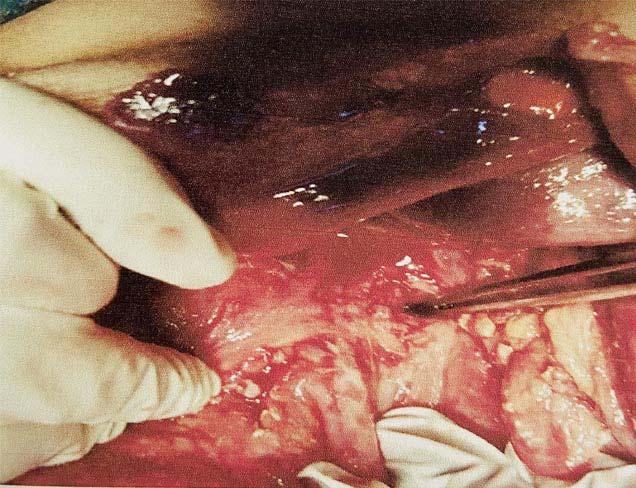
of fungal peritonitis in peptic ulcer perforation. They found a fungal isolation with a frequency of 37.09 % in peptic ulcer perforation cases. Shan etal5 in 2002 found 43.4% fungal isolation in cases of peptic ulcer perforation. De Ruiter et al found it to be 41% in perforated peptic ulcer. Jindal et al reported a prevalence of 70.5% candida isolation in gastroduodenal perforation whereas Prakash et al reported it to be 23%. The findings are variable in all studies.
In the present study, the rate of fungal isolation in ileal perforation was 26%. Jindal et al found it to be 32.8% and Prakash et al reported it to be 77%. There was no fungal positivity in appendicular perforation similar to the results found by de Ruiter et al and Jindal et al. In colonic perforation, fungal positivity was found to be 25% by Jindal et al and 11.8% by de Ruiter et al. Our study found the prevalence of positive fungal culture in colonic perforation to be 20%. The differences in results could be because of different geographical location and patient profile.
Shan et al reported age, preoperative organ failure, delay in operation, high Mannheim Peritonitis Index (MPI) and Acute Physiology and Chronic Health Evaluation (APACHE) II scores, and preoperative antibiotic therapy as risk factors for a positive fungal culture. Dupont et al6 in 2003 found four independent risk factors of yeast isolation in peritoneal fluid (similar odds ratio) in a retrospective cohort: female gender, upper gastrointestinal tract origin of peritonitis, intraoperative cardiovascular failure, and previous antimicrobial therapy at least 48 hours before the onset of peritonitis.
In the present study, we found a significant difference between proportion of age more than or equal to 50 and less than 50 with fungal isolation. In our study, there was significant difference between proportions of males with fungal isolation to females. However in our study, the difference between patients who had a delayed presentation with fungal isolation and those without a delay and fungal isolation was not
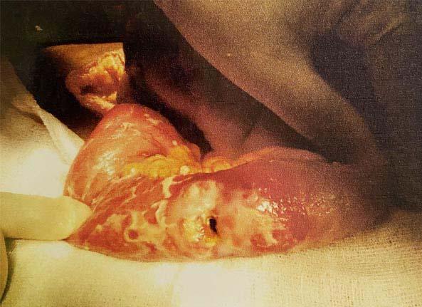
significant. In our study we also found a significant difference between proportions of Pre-operative antibiotic use of more than 48 hours with fungal isolation to Pre-operative antibiotic use of less than 48 hours. There was a significant relationship between site of perforation and isolation of fungus, the commonest site of isolation being gastroduodenal. These results are comparable to the studies mentioned above. The relationship between delay and presentation more than 24 hours and fungal isolation was not significant which is different from the previous studies.
As the usual standard of care all patients were given appropriate antibiotics based on culture sensitivity results and I.V. fluconazole if fungal isolates were present. In the post-operative period surgical site infection was present in 68.16% of fungal positive cases, 43.47% in only bacteria positive and 24.32% in patients with sterile cultures. These results are comparable to Jindal et al who found SSI in 76.5% of patients with fungal positive cultures.The proportion of patients with SSI was significantly larger for patients with positive fungal culture when compared to the patients with no fungus isolated in our study sample. In our study, hospital stay of more than 15 days was seen in 72.72% of fungal positive, 45.65% of bacterial positive and 10.8 % of sterile cultures.This result is comparable to Jindal et al who found 72% patients with fungal culture positive results having prolonged hospital stay.The proportion of patients with duration of hospital stay of more than 15 days was significantly larger for patients with positive fungal culture when compared to the patients with no fungus isolated in the sample in our study. We found an overall mortality of 10.23 %, with fungal positive cases having a mortality of 22.7% and only bacteria positive cases with 6.5% mortality. The fungal positive cases were responsible for 76.92% and bacterial only were responsible for 23.09% of overall mortality. Overall mortality found by Jindal et al was 8.5%, Prakash et al was 35.7% and Lee et al was 9.6%. In the fungal positive cases the mortality was 21.7% in the study by Lee et al, 76% by Prakash et al, 14.7 % by Jindal et al and 13% by Khan et al7 . Our findings are comparable to these results. In our study, the proportion of patient mortality was significantly larger for patients with positive fungal culture when compared to the patients with no fungus isolated in the sample. Shan et al found a significant relationship between fungal isolation and incidence of surgical site infections, hospitalization and mortality. Lee et al concluded that fungal isolation was not a significant risk factor for mortality. Prakash et al found that Candida co infection was a bad prognostic factor.Jindal
et al found that patients with positive fungal culture had higher incidence of surgical site infection, residual abscess formation, longer intensive care unit stay, longer hospital stay and higher mortality rates in comparison to fungal culture negative patients and results were statistically significant. We found that there was a statistically significant relationship between fungal isolation and surgical site infection, prolonged hospital stay and mortality.
In a stepwise multivariate logistic regression,age of patients and pre-operative antibiotic use for more than 48 hours were the most significant independent variables associated with the isolation of fungus in the sample. The third important variable associated with fungal isolation was sex of the patients in our study sample.
The incidence of fungal infection in non-traumatic hollow viscus perforation is quite high. It can thus be concluded that bacterial as well as fungal cultures and antimicrobial sensitivities of peritoneal fluid samples obtained intraoperatively should be routinely done in the treatment of patients of perforation peritonitis. There is high prevalence of positive peritoneal fluid fungal cultures in Institutional set up and fungus is a significant risk factor for poor outcome in these patients. Hence, surgeons should be made aware of the usefulness of fungal culture along with bacterial culture of intra operative peritoneal fluid samples and utility of antifungal therapy in the treatment of gastrointestinal perforation.It may also be the leading cause for increased morbidity and mortality in patients in extreme of ages. Prompt identification and empirical use of antifungal along with wide spectrum antibiotics may help the patients with non-traumatic hollow viscus perforation to recover early.
Funding : None
Conflict of Interest : None
1Prakash A,Sharma D, Saxena A, Somashekar U, et al — Effect of Candida infection on outcome in patients with perforation peritonitis. Indian J Gastroenterol 2008; 27: 107-9.
2Jindal N, Arora S, Pathania S — Fungal Culture Positivity in Patients with Perforation Peritonitis. J Clin Diagn Res 2015; 9(6) : DC01-3
3Lee SC, Fung CP, Chen HY, et al — Candida peritonitis due to peptic ulcer perforation: incidence rate, risk factors, prognosis and susceptibility to fluconazole and amphotericin B. Diagn Microbiol Infect Dis 2002; 44: 23-7.
4de Ruiter J, Weel J, Manusama E, et al — The epidemiology of intra-abdominal flora in critically ill patients with secondary and tertiary abdominal sepsis. Infection 2009; 37: 522-7
5Shan YS, Hsu HP, Hsieh YH, et al — Significance of intraoperative peritoneal culture of fungus in perforated peptic ulcer. Br J Surg 2002; 90: 1215-9.
6Dupont H, Bourichon A, Paugam-Burtz C etal.Can yeast isolation in peritoneal fluid be predicted in intensive care unit patients with peritonitis? Critical Care Med 2003; 31: 752-7
Md Aolad Hossain1, Aminur Rahman2, S M Monowar Hossain3, Nadira Majid4, Shahjada Mohammad Dastegir Khan5, Zahed Ali6, Md Nurul Amin Miah7, Firoz Ahmed Quraishi8, Uttam Kumar Saha9
Background : The aim of the study to assess the association of C-reactive protein (CRP) with severity of acute ischemic stroke (AIS). Materials and Methods: This study was a hospital based crosssectional descriptive study and conducted in the department of Neurology& Medicine in Sir Salimullah Medical College & Mitford Hospital, Dhaka during January 2018 to December 2018. Clinically and radiologically diagnosed 100 admitted AIS patients were assessed and interviewed according to prefix selection criteria. Serum CRP assay was done within 24 hour of hospitalization. The severity of stroke was evaluated by using National Institutes of Health Stroke Scale (NIHSS). Interpretation of NIHSS were correlates with CRP. Results: Out of 100 patients, Mean age of group A and group B were respectively 61.18±10.48 years and 60.40 ± 10.58 years with no significant difference (p>0.05). Distribution of gender, occupation, economic status and risk factors were similar across the group (p>0.05). Mean CRP level of group A and group B were 14.07 ± 4.69 and 3.67 ± 1.29 mg/dl, respectively. Severity of stroke which was measured by NIHSS score were significantly higher in CRP raised group than others (p<0.05). Similarly, lower GCS score were observed in group A patients than group B ((p<0.05). Moreover, CRP positive stroke patients had significantly higher number of deaths at day 7 follow up after stroke than CRP negative patients (Group A-14% versus group B-2%, p <0.05). Conclusion : CRP is elevated in the acute phase of AIS and elevated CRP level is significantly associated with severity of AIS patients. [J Indian Med Assoc 2020; 118(8): 42-7]
Keywords : C-reactive protein, Stroke, Atherosclerosis.
Strokeis a worldwide health problem. It is a major cause of morbidity, mortality and disability in developed as well as developing countries. Ischemic strokes, which account for 80% of strokes, are caused
1 MBBS, MD (Neurology), Consultant Neurology, Ibn Sina Diagnostic and Consultation Center, Sylhet, Bangladesh
2MBBS, FCPS(Medicine), MD(Neurology), MACP(USA), MAAN(USA), FINR(Switzerland) Assistant Professor, Department of Neurology, Sir Salimullah Medical College, Dhaka 1100 and Corresponding Author
3MBBS, MD(Neurology), Assistant Professor, Department of Neurology , US Bangla Medical College and Hospital, Narayangonj, Bangladesh
4MBBS, MD(Pathology), Associate Professor, Department of Pathology, Delta Medical College and Hospital, 26/2 Principal Abul Kashem Road, Mirpur-1, Dhaka 1216
5MBBS, MD(Neurology), Assistant Professor, Department of Neurology, Sir Salimullah Medical College, Dhaka 1100
6MBBS, FCPS (Medicine), MD(Neurology), Professor, Department of Neurology, Sir Salimullah Medical College, Dhaka 1100
7MBBS, FCPS (Medicine), MACP(USA),Professor, Department of Medicine, Sheikh Hasina Medical College, Tangail 1900
8MBBS, FCPS(Medicine), MD(Neurology), Professor ,Department of Neurology, Anwar Khan Modern Medical College, Dhaka 1205
9MBBS, MCPS(Medicine), MD(Neurology), Professor (Rtd), Department of Neurology, National Institute of Neurosciences & Hospital, Dhaka 1207
Received on : 04/07/2020
Accepted on : 25/07/2020
Editor's Comment :
High CRP is associated with a greater risk for ischemic stroke or Transient Ischaemic Attack (TIA).
C-reactive protein is elevated in the acute phase of acute ischaemic stroke
Elevated CRP level is significantly associated with severity of AIS patients.
CRP positive stroke patients had significantly higher number of deaths at day 7 follow up after stroke than CRP negative patients.
by the obstruction or clogging of the major arteries in the cerebral circulation. Cerebral atherosclerosis, a major cause of ischemic stroke, can be divided into extracranial atherosclerosis (ECAS) and intracranial atherosclerosis (ICAS), and anterior and posterior circulation atherosclerosis 1. Inflammation plays a major role in all phases of pathophysiology in atherosclerosis2,3. Therefore, it might be hypothesized that a more severe stroke is associated with greater inflammatory response. High-sensitivity C-reactive protein (hsCRP) is a sensitive marker of inflammation and tissue injury. Stable plaques are characterized by a chronic inflammatory infiltrate, whereas vulnerable and ruptured plaques are characterized by an ‘‘active’’
inflammation involved in the thinning of the fibrous cap, predisposing the plaque to rupture4. Although a single vulnerable atherosclerotic plaque rupture may cause the event, there are many other types of plaques, several of which are vulnerable. The existence of multiple types of vulnerable plaques suggests that atherosclerosis is a diffuse inflammatory process2. CRP is a phylogenetically highly conserved plasma protein, with homolog in vertebrates and many invertebrates, and it participates in the systemic response to inflammation5. The role of CRP as a marker during and after stroke is less extensively studied in comparison to coronary artery disease. The Rotterdam study shows that although high CRP is associated with the risk for future stroke, it is not useful for individual stroke prediction6. On the other hand, the Framingham study shows that high CRP is associated with a greater risk for ischemic stroke or TIA5. Acute ischemic stroke may trigger an inflammatory response that leads to increased levels of C-reactive protein (CRP). High levels of CRP may be associated with poor outcome because they reflect either an inflammatory reaction or tissue damage7. Several studies have found an association between increased CRP levels and clinical outcome in the ischemic stroke. The results of previous studies that have aimed to assess the prognostic value of CRP in the very early phase of stroke are ambiguous. Two prospective studies did not observe a relation between CRP levels obtained within 6 or 12 hour after symptom onset and death or dependency at follow-up 8,9 Increased CRP levels following ischemic stroke may also reflect concurrent infections. Secondary infections are common in the first week of stroke and are associated with poor outcome but they usually occur more than 12 h after stroke onset10,11. In another study, only two clinically overt infections were reported within 24 hour after stroke onset. In addition, only few studies have analyzed the relationship between elevated admission CRP levels and stroke severity12. The aim of our study was therefore to determine the prognostic value of CRP measured in the very early phase of ischemic stroke for poor functional outcome in patients with acute ischemic stroke.
MATERIALS AND METHODS
This study was a hospital based cross-sectional descriptive study and conducted in the department of Neurology & Medicine in Sir Salimullah Medical College & Mitford Hospital, Dhaka during January 2018 to December 2018 for duration of one year. Clinically and radiologically diagnosed total (admitted) Acute Ischemic Stroke (AIS) patients within 24 hours were assessed and interviewed according to selection
criteria. Patients who were admitted more than 24 hours after the onset of symptoms were excluded. Patients who were found with stroke or awoke with stroke were included if it was known that the patient had been normal. A questionnaire was made and pretested for data collection. A detailed clinical history including age, sex, socioeconomic status, occupation, duration of symptoms, risk factors, neurological problems were elicited and recorded in all cases.
At admission, plain CT scan of the head was done to rule out haemorrhage. Serum CRP assay was done within 24 h of hospitalization and analyzed by Tinaquant latex method using Modular P (Roche Diagnostics). The NIH stroke score (NIHSS) was assessed by a neurologist at the time of admission. The NIHSS was categorized as 0, 1–4, 5– 15, 16-20 & 21-42 and the NIHSS score were correlates with CRP. For analysis of these subjects were stratified into two groups: group A- AIS with raised CRP and Group B consist of AIS without raised CRP. Both groups of the patients were observed in similar manner and outcome assessment was also done in according to the protocol. Total follow up was done up to 7 days from admission. Severity was evaluated by using National Institutes of Health Stroke Scale (NIHSS).
Their informed written consent was taken in a consent form before collecting data. Proper permission was taken from the concerned departments and local ethical committee.
Exploratory data analysis were carried out to describe the study population where categorical variables were summarized using frequency tables while continuous variables were summarized using measures of central tendency and dispersion such as mean, median, percentiles , standard deviation and Chi-square test. All statistical analysis were performed using SPSS 23.0 for Windows (SPSS Inc, Chicago, Illinois, USA) level of significance was set at .05 and p-value <0.05 was considered significant
Operational Definitions :
Stroke13 : Stroke is defined by the World Health Organization as a clinical syndrome consisting of 'rapidly developing clinical signs of focal (aphasia/ dysphasia/ dysarthria/ dysphasia/any cranial nerve palsy/weakness) (at times global) disturbance of cerebral function, lasting more than 24 h or leading to death with no apparent cause other than that of vascular origin. In the spring of 2013, the AHA/ASA published an expert consensus document with a new definition of stroke. The major fundamental change compared with older definitions is that the new broader definition of stroke includes any objective evidence of
permanent brain, spinal cord or retinal cell death due to a vascular cause.
Acute Stroke 14,15: The acute phase of stroke includes 24 hours to weeks of post acute period. This time is to the key interventions involved in the assessment, treatment or management, and early recovery in the first days after stroke onset. This may represent initial diagnostic procedures undertaken to identify the nature and mechanism of stroke, interprofessional care on designated care units to prevent further complications and promote early recovery. At the “acute” stage, the following neurologic and medical complications have been recorded: stroke progression; seizures; increased intracranial pressure; fever; urinary and chest infections; severe hypertension; congestive heart failure; falls; and deep vein thrombosis and pulmonary embolism. The presence of these complications is associated with deterioration and higher mortality. Previous studies focused on the first week after stroke. Broadly speaking it is refers to the first days to weeks of inpatient treatment with stroke. National Institutes of Health Stroke Scale (NIHSS)16:
1ALevel of consciousness0-3
1BLevel of consciousness questions0-2
1CLevel of consciousness commands0-2
2Best Gaze0-2
3Visual fields0-3
4Facial palsy(paresis)0-3
5AMotor-left arm
0-4, UN
5BMotor-right arm0-4, UN
6AMotor-left leg0-4, UN
6BMotor-right leg0-4, UN
7Limb ataxia
0-2, UN
8Sensory0-2
9Best language0-3
10Dysarthria(articulation of words)0-2, UN
11Extinction0-2
Severity of Stroke17 : The National Institutes of Health Stroke Scale (NIHSS) is considered as the Gold Standard systematic assessment tool that provides a quantitative measure of stroke-related neurologic deficit. The NIHSS was originally designed as a research tool to measure baseline data on patients in acute stroke clinical trials. The NIHSS is composed of 11 items, each of which scores a specific ability between a 0 and 4. For each item, a score of 0 typically indicates normal function in that specific ability, while a higher score is indicative of some level of impairment. The individual scores from each item are summed in order to calculate a patient's total NIHSS score. The
maximum possible score is 42, with the minimum score being a 0.
ScoreStroke severity
0No stroke symptoms
1-4Minor stroke
5-15
16-20
21-42
Moderate stroke
Moderate to severe stroke
Severe stroke
RESULTS AND OBSERVATIONS
100 patients with AIS were included in the study 64(64.00%) males and 36(36%) females were included in the study, female-to-male ratio of 1:1.8.
In Table 1 the distribution was same in both group A and group B. The difference in distribution was not statistically significant (p>0.05).
This Table 2 shows that the mean age of presentation was 60.79 ± 10.49 years. Mean age of group A and group B were respectively 61.18±10.48 years and 60.40±10.58 years. There was no statistically significant difference between those two groups (p>0.05). Majority of the patients were aged between 61-70 years in both groups (44%).
This Table 3 shows among all patients 58% had hypertension, 24% had DM, 10% had dyslipidemia, 11% had obesity and 24% had smoking habit. The difference in distribution of risk factors between two groups was not statistically significant (p>0.05).
Table 1 — Distribution of the study subjects according to sex (n=100)
SexGroup AGroup BTotalP value (CRP positive)(CRP negative) (n=100) (n=50)(n=50)
n%n%n%
Male3264326464641.00NS
Female183618363636
NS = Not significant ; P-value reached from χ2 test
Table 2 — Distribution of the study subjects according to age (n=100)
Age inGroup AGroup BTotalP value years (CRP positive)(CRP negative) (n=100) (n=50)(n=50)
n%n%n%
41 – 50102012242222 0.445NS
51 – 60122414282626
61 – 70224422444444
71 – 806122488
Mean ± SD 61.18±10.4860.40±10.5860.79±10.49 0.712NS Range (42-80)(41-75)(41-80)
NS = Not significant ; P-value reached from χ2 test
Table 3 — Distribution of the study subjects according to Risk factors (N=100)
RiskGroup A Group BTotalP value factors (CRP positive)(CRP negative) (n=100) (n=50)(n=50)
n%n%n%
Hypertension306028565858 0.685NS
DM163281624240.061NS
Dyslipidaemia7143610100.182NS
Obesity6125101111 0.749NS
Smoking112213262424 0.640NS
NS = Not significant ; P-value reached from χ2 test
Table 4 — CRP level of the study subjects (N=100)
CRP levelGroup AGroup BTotal (CRP positive)(CRP negative) (n=100) (n=50)(n=50)
In Table 4 shows mean CRP level of group A patients was 14.07±4.69 and of group B was 3.67 ± 1.29 and of all was 8.87 ± 6.25. Less than 6 mg/dl CRP was considered negative CRP.
In Table 5 shows among patients who were CRP positive (group A) 26% had NIHSS between 21 – 42, 30% had 16 – 20, 34% had 5 -15 and 10% had 1- 4. While among those who had CRP in negligible amount (group B) 10% had NIHSS 21 – 42, 18% had 16 – 20, 42% had 5 – 15 and 30% had 1 – 4. The difference in distribution of NIHSS score between two groups was statistically significant (p<0.05).
In Table 6 shows CRP positive stroke patients had significantly higher number of deaths at day 7 after stroke than CRP negative patients (14% vs 2% deaths among group A and group B respectively, p <0.05).
In Table 7 shows Descriptive analysis of CRP values in relation to admission NIHSS score and outcome of patients shows that mean CRP values increased with increasing NIHSS score and CRP values was significantly higher among patients who were dead than
Table 5 — NIHSS score of the study subjects in relation to CRP (N=100)
Age inGroup AGroup BTotalP value years (CRP positive)(CRP negative) (n=100) (n=50)(n=50)
n%n%n%
1 – 4510153020200.015S
5 – 15173421423838
16 – 2015309182424
21 – 4213265101818
NS = Not significant ; P-value reached from χ2 test
those who were alive after stroke.
In Table 8 shows univariate regression analysis of different risk factors for dying at follow-up after stroke was done. Patients with age >60 years, being male, CRP positive, HTN, DM, dyslipidaemia, smoking and obesity had higher odds of dying at 7 days after stroke. But none of the factors were significant at 0.05 level. At <0.1 level age > 60 years, CRP positivity and DM was found to be significant predictor of death at 7 days in stroke patients.
In Table 9 shows multivariate regression analysis of different risk factors for dying at follow-up after stroke was done. Only those risk factors found significant (at <0.1 level) in univariate analysis was included. When adjusted for other factors, patients with CRP positivity was found to have higher odds of dying at 7 days after stroke (OR 6.99; 95%CI 0.81- 60.18, p=0.076). It was
Table 6 — Outcome of study subjects at day 7 after stroke in relation to CRP (N=100)
Age inGroup AGroup BTotalP value years (CRP positive)(CRP negative) (n=100) (n=50)(n=50) n%n%n% Alive4386499892920.027S Dead7141288
NS = Not significant ; P-value reached from χ2 test
Table 7 — CRP level in relation to admission NIHSS score and outcome of patients (N=100)
Outcome NIHSS scoreCRP value (mean ± SD) CategorynIn relation toIn relation to NIHSS scoreoutcome
Alive1 – 4205.02±2.667.87±5.16
P value determined by Student’s t test
Table 8 — Univariate analysis risk factors for dying at 7 days after stroke (n=100)
Table 9 — Multivariate analysis of risk factors for dying at 7 days after stroke (n=100)
Variables
* p value significant at <0.1 level
significant at <0.1 level.
DISCUSSION
Total 100 patients of acute ischaemic stroke were studied. Among them 50 patients were CRP positive and another 50 patients had negligible CRP. The mean age of all patients was 60.79±10.49 years. This is nearly similar to the finding of a stroke registry conducted by Bhowmik and colleagues (2016)18. They included 679 patients of ischaemic stroke and found a mean age of 60.6 years. In the present study majority patients were aged between 61 to 70 years (44%). In comparison Siddiqui et al (2013) studied all types of stroke and found majority patients aged between 51 to 60 years (29%) followed by 22% aged between 61 – 70 years19.The difference could be attributed to the difference in study population. But, overall 69% were aged =50 years in their study which is similar to the findings (78% aged =50 years) of this study. The majority patients were male (66%) and rest (34%) were female with similar sex distribution across groups in this study. This is also similar to the findings of Bhowmik et al (2016) who reported 67.7% patients being male in their study18. Another study conducted by Islam et al (2013) reported a male-female ratio of 3.44:2.41 among stroke patients20. Mohammad et al (2014) noted in review that one of the risk factor of stroke is male sex21. Therefore, findings of the present study are consistent with previous findings. The most common risk factor found in this study was HTN (58%) followed in decreasing order by DM (24%), smoking habit (24%), obesity (11%) and dyslipidaemia (10%) 20. Islam et al (2013) found majority 86.3% cases of hypertension, 55% cases of smoking, 11.3% cases of diabetes among ischemic stroke cases 20 Mohammad et al (2014) found that hypertension was the most common modifiable risk factor found in stroke patients (57.6%) followed by smoking (44.6%), tobacco use (24.3%), Oral Contraceptive Pill (OCP) use in female (40% of female stroke), diabetes (23%), ischemic heart disease (17.1%), obesity (10.6%) and dyslipidaemia (5.3%) 21. Sharmin et al found 46% diabetic patients among their study population22 Siddiqui et al (2013) found that about 77% of patient had history of hypertension, 22% diabetes mellitus,
20% dyslipidaemia, 13% previous history of stroke and 27% ischaemic heart disease19. Badiuzzaman et al (2009) fond 58.62% patients had hypertension with other risk factors in their study and this was followed by risk factors associated with smoker (53.9%), lipid disorder (48.01%), heart diseases (25.75%), diabetes mellitus (20.01%) and previous history of stroke (10.61%)23. All the above comparison implies that HTN was the most common risk factor with ischemic stroke. Among others, diabetes and dyslipidaemia are also common.
In this study, CRP positivity was found to be significantly associated with poor outcome in followup at 7 days (p<0.05). CRP positive stroke patients had significantly higher number of deaths at day 7 after stroke than CRP negative patients (14% vs 2% deaths among group A and group B respectively, p <0.05). Similar result was encountered by Dewan and Rana (2011) in their study 3. In their study total 13 out of 100 stroke patients died. Among them 12 (92.3%) were CRP positive and 1 (7.7%) were CRP negative and the difference was statistically significant.
Univariate analysis showed that CRP positivity is associated with higher odds of dying at 7.97 (95% CI 0.05-7.97; p value =0.057). When adjusted for other significant (at <0.1 level) risk factors of stroke including age >60 years, and DM the OR of dying for CRP positive ischemic stroke remains similar OR 6.99 (95%CI 0.8160.18, p=0.076), in which CRP positivity at 24 hours after admission in ischemic stroke patients is an important prognostic factor of death at 7 days after discharge from hospital. Dewan and Rana (2011) followed up patients for 18 months and found that an increasing CRP values at discharge predicted adverse prognosis and had strongest association with outcome at one year in a multivariate model 3. Di Napoli et al (2001) followed up 193 ischemic stroke patients for 1year. They found that CRP at hospital discharge was the strongest independent marker of adverse outcome (HR 7.42, 95% CI 2.75 to 20.03; P=0.0001) 24.In spite of small number of cases studied within 7 days, findings of this study supported the previous observations that an elevated CRP reflects the severity and the extent of brain infarct and is related to early mortalities24-26.
Conclusion :
In our present study, mean CRP levels were significantly higher in patients with ischemic stroke when compared to controls. This study confirms that C-reactive protein is elevated in the acute phase of ischaemic stroke and could present a prognostic marker.
Limitations :
This study has small sample size and study populations were confined to only one tertiary care hospital and the long term follow up were not assessed.
Funding : No funding sources
Conflict of interest : None declared
Recommendation: Further population based study is necessary to infer the findings over the general population.
1Kim JS, Nah HW, Park SM, Kim SK, Cho KH, Lee J, Lee YS, Kim J, Ha SW, Kim EG, Kim DE — Risk factors and stroke mechanisms in atherosclerotic stroke: intracranial compared with extracranial and anterior compared with posterior circulation disease. Stroke. 2012; 43(12):3313-8.
2Spagnoli LG, Bonanno E, Sangiorgi G, Mauriello A — Role of inflammation in atherosclerosis. Journal of Nuclear Medicine 2007 Nov 1; 48(11): 1800-15.
3Dewan KR, Rana PV — C–reactive protein and early mortality in acute ischemic stroke. Kathmandu University Medical Journal 2011; 9(4):252-5.
4O'Brien EC, Rose KM, Shahar E, Rosamond WD — Stroke mortality, clinical presentation and day of arrival: the atherosclerosis risk in communities (ARIC) study. Stroke research and treatment. 2011; 2011.
5Rost NS, Wolf PA, Kase CS, Kelly-Hayes M, Silbershatz H, Massaro JM, D’Agostino RB, Franzblau C, Wilson PW — Plasma concentration of C-reactive protein and risk of ischemic stroke and transient ischemic attack: the Framingham study. Stroke 2001; 32(11): 2575-9.
6Bos MJ, Schipper CM, Koudstaal PJ, Witteman JC, Hofman A, Breteler MM — High serum C-reactive protein level is not an independent predictor for stroke: the Rotterdam Study. Circulation 2006; 114(15): 1591-8.
7Shoaeb MA, Shehata MA, Taema KM, Hammouda MA — CRP in cerebrovascular stroke: Prognostic implications. The Egyptian Journal of Critical Care Medicine 2014 Apr 1; 2(1):43-52.
8Topakian R, Strasak AM, Nussbaumer K, Haring HP, Aichner FT — Prognostic value of admission C-reactive protein in stroke patients undergoing iv thrombolysis. Journal of neurology 2008; 255(8): 1190-6.
9Winbeck K, Poppert H, Etgen T, Conrad B, Sander D — Prognostic relevance of early serial C-reactive protein measurements after first ischemic stroke. Stroke 2002 Oct 1; 33(10): 2459-64.
10Aslanyan S, Weir CJ, Diener HC, Kaste M, Lees KR — GAIN International Steering Committee and Investigators. Pneumonia and urinary tract infection after acute ischaemic stroke: a tertiary analysis of the GAIN International trial. European journal of neurology 2004; 11(1): 49-53.
11Emsley HC, Hopkins SJ — Acute ischaemic stroke and infection: recent and emerging concepts. The Lancet Neurology 2008; 7(4): 341-53.
12den Hertog HM, Van Rossum JA, Van Der Worp HB, Van Gemert HM, de Jonge R, Koudstaal PJ, Dippel DW, PAIS
investigators — C-reactive protein in the very early phase of acute ischemic stroke: association with poor outcome and death. Journal of neurology 2009; 256(12):2003-8.
13Sacco RL, Kasner SE, Broderick JP, Caplan LR, Connors JJ, Culebras A, Elkind MS, George MG, Hamdan AD, Higashida RT, Hoh BL, Janis LS, Kase CS, Kleindorfer DO, Lee JM, Moseley ME, Peterson ED, Turan TN, Valderrama AL, Vinters HV — An updated definition of stroke for the 21st century: a statement for healthcare professionals from the American Heart Association/American Stroke Association. Stroke 2013; 44: 2064-89.
14Bahouth M, LaMonte MP — Acute ischemic stroke: evaluation and management strategies. Topics in Advanced Practice Nursing 2005; 5(4).
15Rocco A, Pasquini M, Cecconi E, Sirimarco G, Ricciardi MC, Vicenzini E, Altieri M, Di Piero V, Lenzi GL — Monitoring after the acute stage of stroke: a prospective study. Stroke 2007; 38(4):1225-8.
16NIH Stroke Scale. National Institutes of Health (2019,January 9,).Retrieved from website.http s://www.stroke.nih.gov/ documents/NIH_Stroke_Scale.pdf.
17Hage V. The NIH stroke scale: a window into neurological status. Nurse Com Nursing Spectrum (Greater Chicago) 2011; 24(15): 44-9.
18Bhowmik NB, Abbas A, Saifuddin M, Islam M, Habib R, Rahman A, Haque M, Hassan Z, Wasay M — Ischemic strokes: Observations from a hospital based stroke registry in Bangladesh. Stroke research and treatment 2016; (5):13.
19Siddiqui MR, Islam QT, Iqbal MJ, Binte-Mosharraf SS — Sociodemographic Status & Associated Risk Factors of the Stroke Patient's in a Tertiary Care Hospital of Bangladesh. Anwer Khan Modern Medical College Journal 2013 Nov 9; 4(2):1822.
20Islam MN, Moniruzzaman M, Khalil MI, Basri R, Alam MK, Loo KW, Gan SH. Burden of stroke in Bangladesh. International journal of stroke 2013 Apr; 8(3): 211-3.
21Mohammad QD, Habib M, Mondal BA, Chowdhury RN, Hasan MH, Hoque MA, Rahman KM, Khan SU, Chowdhury AH, Haque B — Stroke in Bangladeshi patients and risk factor. Mymensingh medical journal: MMJ 2014; 23(3): 520-9.
22Sharmin N, Sultana N, Rahman H, Rahman T, Chowdhury S — Association of Diabetes Mellitus with Acute Ischemic Stroke. Bangladesh Journal of Medical Biochemistry 2018; 9(2): 45-8.
23Badiuzzaman M, Mohammed F, Chowdhury F, Bari M, Alam M, Ahasan H 1 — Prevalence of Modifiable Risk Factors among Stroke Patients in a Tertiary Care Hospital in Dhaka. Journal of Medicine 2009; 10, 3 (1), 18-21.
24Di Napoli M, Papa F, Bocola V — C-reactive protein in ischemic stroke: an independent prognostic factor. Stroke 2001; 32(4): 917-24.
25Di Napoli M, Di Gianfilippo G, Sollecito A, Bocola V — Creactive protein and outcome after first-ever ischemic stroke. Stroke 2000; 31(1): 231-9.
26Idicula TT, Brogger J, Naess H, Waje-Andreassen U, Thomassen L — Admission C-reactive protein after acute ischemic stroke is associated with stroke severity and mortality: the 'Bergen stroke study'. BMCNeurol 2009; 9(1):18.
Nabaruna Paul1, Dwijen Das2
Background : Poisoning, both accidental and suicidal, contributes to significant mortality and morbidity. Poisoning refers to the development of dose related adverse effects following exposure to chemicals, drugs and other xenobiotics, although individual responses to a given dose may vary because of genetic polymorphism, enzymatic induction or inhibition in the presence of other xenobiotics, or acquired tolerance.
Aims and Objectives : To study the epidemiological and clinical profile of acute poisoning cases admitted in Department of Medicine.
Materials and Methods : A total of 300 cases admitted in the Department of Medicine, Silchar Medical College and Hospital, from 1st July, 2017 to 30th June, 2018, with diagnosis of acute poisoning and satisfying the inclusion and exclusion criteria were taken up for evaluation after obtaining informed written consent.
Results : Males and females represented 52% and 48% of the total cases and 58.33% of them were in the age group 20-29 years. Married persons and rural inhabitants contributed to 73.67% and 66% respectively. Clinical presentations were varied based on the type, nature, amount and toxicological status of the compound ingested. Poisoning with pesticides was most common occurrence and 74% attempts were suicidal. The overall mortality rates were 13%.
Conclusion : It is of utmost importance to generate awareness among the general population about the harmful effects of various chemicals. Education, self-employment, small family size and psychological counselling should be encouraged.
[J Indian Med Assoc 2020; 118(8): 48-51]
Key words : Accidental, Morbidity, Xenobiotics, Pesticides, Psychological Counselling.
Poisoning refers to the development of dose related adverse effects following exposure to chemicals, drugs and other xenobiotics1. The word poison is derived from the latin word potionem that means deadly draught2 Poisoning occurs by the absorption of any physical, chemical or organic substances via gastrointestinal tract, skin, mucosa, respiratory tract or parenteral route that causes damage to cells, tissue organ and organ system. Individual responses to a given poison or dose may vary and depends on route of exposure, chemical and physical properties of poison, mechanism of action and enzyme induction and inhibition in presence of other xenobiotics. The effect of a particular poison may also depend on individual parameters like status of hepatic and renal function, genetic polymorphism, or acquired tolerance1,3
1MBBS, Postgraduate Trainee, Department of Medicine, Silchar Medical College and Hospital, Silchar, Assam 788014
2MD, FACP (USA), FRCP (Glasg), FIACM, Associate Professor, Department of Medicine, Silchar Medical College and Hospital, Silchar, Assam 788014 and Corresponding author.
Received on : 04/02/2020
Accepted on : 14/03/2020
Editor's Comment :
It is important to create awareness among the general population on safe agricultural practices and use of protective wear at workplaces and emphasize the importance of education, gender equality and social harmony to help the flatten the curve of rising cases of poisoning. At the same time, upgradation of emergency care services is required to reduce the burden of morbidity and mortality associated with the same.
Poisoning has emerged as a major medico- social problem all over the world that may relate to determinants like social and emotional disturbances, unemployment, work dissatisfaction, chronic disease states etc. According to WHO, approximately three million acute poisoning cases with 2, 20,000 deaths occur every year. Out of this, 90% of fatal poisoning occur in developing countries4. Although most of the poisoning are unintentional in children, suicidal mode of poisoning accounts for majority among adults. The epidemiological pattern of poisoning varies from region to region because of varying social structure, religious influence, economic status, educational level, awareness among general population and availability
of drugs5.
This study was conducted to bring in light the clinico-epidemiological profile of acute poisoning cases prevailing in North-eastern part of the country and thereby to uplift the emergency care management, level of education and awareness and psycho-social support to reduce the burden of the same.
AIMS
To evaluate the epidemiological and clinical profile of acute poisoning cases admitted in Department of Medicine, of a tertiary care teaching hospital of Northeastern India.
METHODS AND METHODOLOGY
This is a single centered hospital based observational study conducted over a period of 1 year,from 1st July, 2017 to 30th June, 2018, in a tertiary care teaching hospital in North-eastern India.
All the cases satisfying the inclusion and exclusion criteria for the study were takenafter obtaining informed and written consent from the patient/ guardian.
Inclusion Criteria :
Patients with acute poisoning aged >12 years.
Exclusion Criteria :
1.Patients with chronic exposure to a particular poison, alcohol intoxication, bee sting, snake bite.
2.Patients who were brought dead to hospital.
A through history and physical examination were done for a total of 300 cases included in the study. All routine blood investigations, urine examinations, chest X- ray and ECG were done and patients were evaluated during their hospital stay. The data obtained wererecorded in a preformed proforma. Psychiatric consultation was also taken for the patients where possible.The results for each parameter were represented in numbers, percentages and average (mean, standard deviation).The t test and Chi-square (χ2) test were used to compare mean and proportion respectively. Data were analyzed using Microsoft Excel 2016 and SPSS version 21.0 and p<0.05 was taken as statistically significant.
RESULTS
Total number of study subjects were300, with 156(52%) male and 144(48%) female. Most common age group affected with acute poisoning was 20-29years accountingfor 58.33%. Only 1.33% of the study subjects aged more than 60 years. Rural and urban population were 66% and 34% respectively. Poisoning was commonest among married individuals accounting for 73.67% (79.86% female and 67.94% male). Out of 300 patients, 38% of the population were housewives, 35.67% were students, 20% were farmers and 6.33%
were businessman (Table 1). Muslim religion predominated comprising 57.69% and 58.33% among male and female respectively followed by Hindu religion (42.30% male and 40.27% female) and Christian religion (1.38% male) (Fig 1).
Table 1 — Characteristics of Study Subjects
Gender :
Male 156 (52%)
Female 144 (48%)
Age Group (In Years) : 10-1959 (19.67%)
20-29 175 (58.33%)
30- 3939 (13%)
40- 4918 (6%)
50- 595 (1.67%)
60 And Above4 (1.33%)
Occupation : Housewife114 (38%)
Student 107 (35.67%)
Farmer60 (20%)
Buisnessman19 (6.33%)
Inhabitation : Urban102 (34%)
Rural198 (66%)
Marital Status : MarriedTotal 221(73.67%), Male 106 (67.94%), Female 115 (70.86%)
UmmarriedTotal79 (26.33%), Male 50 (32.05%),
Female 29 (20.13%)
Mode of poisoning was suicidal, accidental and homicidal in 74 %, 20% and 6% of respectively. The various poisons in the study were pesticides 39.67%, phenol 20.67%, kerosene 15%, sleeping pills 12.33%, rat kill 7.67%, iron tablets 2.67% and yellow oleander 2% (Table 2).
In the present study, 55% of the cases presented with gastrointestinal signs and symptoms (nausea, vomiting, pain abdomen, diarrhea), 27% presented with neurological manifestations (dizziness, headache, drowsiness, altered sensorium), 8% presented with respiratory signs and symptoms (breathlessness, cough, coarse crepitations), 1% with cardiovascular manifestations (tachycardia, arrhythmia) and 9% with others (frothing, burning eyes, increased lacrimation) (Fig 2). GCS on presentation was <8 in 47 cases and >8 in 252 cases.
After appropriate evaluation and treatment 67% of the study subjects were discharged, 20% absconded and 13% expired. A psychiatric evaluation was done in possible 231 cases on 2nd and 3rd day of the hospital stay and reactive depression, financial stress
and manic depression were found in 49.78%, 30.30% and 19.91% of the cases respectively (Table 3).
Table 2 — Mode of Poisoning and Poisonous Agents
After thorough analysis it has been observed that male gender, poisoning with pesticides, GCS <8 on presentation and suicidal mode of poisoning contributed significantly to mortality (Fig 3). DISCUSSION
Poisoning has emerged as a significant cause of emergency care hospitalization. Although in developed countries mortality due to poisoning is 1-2%, low level of education and awareness, delayed presentation and resource poor emergency care settings contributes to a large proportion of cases in developing and underdeveloped countries. In India mortality attributable to poisoning varies from 15-30%6,7
In the present series poisoning was more prevalent in males (52%) as compared to females (48%) and most common age group was 20-29 years that comprised of 58.33% of the cases. These findings were similar to study conducted by Surendra Khosya et al5 where males (61.7%) were more commonly affected than
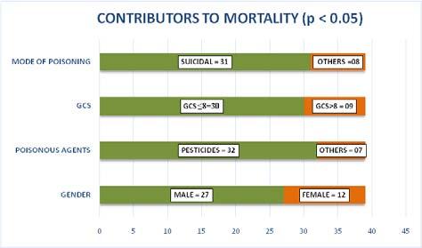
Table 3 — Psychiatric Evaluation and Outcome of Patients
Psychiatric EvaluationNumber of Patients
Reactive Depression115 (49.78%)
Financial Stress70 (30.30%)
Manic Depression46 (19.91%)
Outcome Number of Patients
Patients Discharged201 (67%)
Patients Expired39 (13%)
Patients Absconded60 (20%)
females (38.27%) and 42.92% of the study subjects were in 21-30 years age group. In the present series, 66% of the cases were inhabitants of rural area and 34% of the cases were from urban area. In the study conducted by Subash Vijaya Kumar et al8 65% and 35% were rural and urban inhabitants respectively. In the present study, 73.67% of the cases were married and this finding corroborated to the study conducted by Acharya et al9 where 71% of the cases were married. Among the married population, poisoning predominated in females and this may relate to low level of education, social disharmony and early marriage.
In the index study maximum number of cases were housewives (38%) followed by students (35.67%), farmer (20%) and businessman (6.33%). Whereas, in the study of Sandesh Datir et al10, maximum cases of acute poisoning were in farm owners, clerical and shop owners (25.68%) followed by students and housewives each contributing 21.18% and 08.98% were among unemployed population. Of all the cases Muslim religion predominated with 58%, Hindu religion 41.33% and Christian 1.38% where as in the study of Mukul Joshi et al11, Hindu religion contributed to 94.2%, Muslim and Sikh were 3.3 % and 2.5 % respectively. The discrepancy observed may be due to a predominance of Muslim religion in the region, large family size, ignorance, lack of protective clothing and unsafe practices at work places.
In this study suicidal mode of poisoning contributed to 74%, accidental 20% and homicidal 6% of the total cases and most of them were due to pesticides (39.67%) followed by phenol (20.67%).Whereas, Vaddadi Srinivas et al12 in their study found 70% had suicidal intention, 19% had accidental ingestion and 11% were unknown. Prashant Gupta et al13 in their study found that 57.1% of the cases were due to organophosphorus. In another study conducted by J Jesslin et al14 39.5 % were due to pesticides, 26.1% due to medicines, 22.1% were due to household products,12.1 % were environmental and 0.2 % were due to heavy metals.
In the present series, 55% of the cases presented with nausea, vomiting, abdominal pain and diarrhea, 27% presented with neurological manifestations
(dizziness, headache, drowsiness, altered sensorium), 8% presented with respiratory signs and symptoms (breathlessness, cough, coarse crepitations), 1% with cardiovascular manifestations (tachycardia, arrhythmia) and 9% with others (frothing, burning eyes, increased lacrimation). Amit Patil et al15 had observed that 64.8% of the cases presented with neurological manifestations whereas gastrointestinal manifestations were present in 37% of the study subjects. The prevalence of gastrointestinal manifestations was more in the index study when compared to observation made by Amit Patil et al15. A mortality of 13% was observed in this study whereas J Lavanya et al16 in their study observed 5% mortality. Majority of those who expired presented to the hospital late. The various parameters that contributed significantly to mortality in the present series were male gender, poisoning with pesticides, suicidal mode of poisoning and GCS <8 on presentation. The other factors on which light could be shed for a better outcome may be the time of presentation to hospital, amount of a particular substance taken and various comorbid conditions.
Limitations of the Study :
The present study is a single centered one, with a small sample size in a fixed geographical area and undertaken for a short duration of time. A multicentric study covering a wider geographical area and among different ethnic groups for an extended period of time would have been more beneficial.
CONCLUSION
As poisoning is growing in magnitude in this developing world of increased emotional and social stress, it is of immense importance to uplift the management protocols not only in tertiary care centres but also in primary care centres. Hand in hand education, self-employment, psychosocial counseling should be provided to the society to reduce the overall burden.
Funding : None
Conflict of Interest : None
1Mycyk MB — Poisoning and Drug Overdose. Chap- 449. In: Jameson JL, Fauci AS, Kasper DL, et al, eds.Harrrison’s Principles of Internal Medicine, 20thedn.New York: McGrawHill Education 2018: 3300 - 3313.
2Maheswari E, Abraham L, Chacko CS, et al — Assessment of Pattern, Severity and Outcome of Poisoning in Emergency Care Unit. Journal of Applied Pharmaceutical Science. 2016 December; 6(12):178-83.
3Preston RJ, Hoffmann GR — Genetic Toxicology. Chap- 9. In; Klaassen CD eds.Casarett and Doull’s Toxicology. The Basic Science of Poisons. 8thedn. New York: McGraw-Hill Education 2013; 445-80.
4Murat S, Guiven M — Intensive care management of organophosphate insecticide poisoning. Crit Care 2001; 5(4): 211-5.
5Khosya S, Meena SR — Current Trends of Poisoning: An Experience at a Tertiary Care Hospital Hadoti Region, Rajasthan, India. J Clinic Toxicol 2015; 6(2): 298.
6Pillay VV — Introduction. Chap- 1. Modern Medical Toxicology. 4thedn. New Delhi. JAYPEE 2013:3-6.
7Taruni NG, Bijoy TH, MomonchandA. A profile of poisoning cases admitted to RIMS Hospital Imphal. JournForsenic Med Toxicol 2001; 18: 31-3.
8Kumar SV, Venkateswarlu B, Sasikala M, et al — A study on poisoning cases in a tertiary care hospital. J Nat Sci Biol Med 2010 Jul-Dec; 1(1): 35-9.
9Acharya S, Lakshminarayana K, Sharanappa — Assessment of poisoning cases in a tertiary care hospital. IJBR 2014; 5(9): 578-81.
10Datir S,Petkar M, Farooqui J, etal— Profile of Acute Poisoning Cases at Pravara Rural Hospital, Loni. J Indian Acad Forensic Med 2015 October- December; 37(4): 400-4.
11Joshi M, Patel DV — A Study on Clinical Profile of Patients with Acute Poisoning; GCSMC J Med Sci 2015 July- December; IV(II): 97-100.
12Srinivas V, Srinivas VR — A clinical profile of acute poisoning; JEMDS 2015 April; 4(29): 4923-5.
13Gupta P,Kumar A,Singh SP, et al — Pattern of Cases ofAcute Poisoning in a Rural Tertiary Care Center in Northern India. Ntl J Community Med 2016; 7(4): 307-10.
14Jesslin J, Adepu R, Churi S — Assessment of Prevalence and Mortality Incidences Due To Poisoning In a South Indian Tertiary Care Teaching Hospital. Indian J Pharm Sci 2010; 72(5): 587-91.
15Patil A, Peddawad R, Verma VCS, et al — Profile of Acute Poisoning Cases Treated in a Tertiary Care Hospital: A Study in Navi Mumbai. APJMT 2014 March; 3(1): 36-40.
16Lavanya J, Sivaranjani V, Arshiya BS, et al —A Retrospective Analysis of Patterns, Severity and Clinical Outcome of Different Poisoning Cases in A Tertiary Care Teaching Hospital. IOSR Journal of Pharmacy and Biological sciences 2018 Jan – Feb; 13(1): 09-15.
A study on association of ankle brachial index in patients with ischemic stroke in a tertiary care
Swarup Kanta Saha1, Debasish Dey2, Goutam Biswas3, Vivek Choudhary4, Aritra Kumar Roy5, Atanu Chandra6, Jyotirmoy Pal7, Partha Sarathi Karmakar8
Peripheral arterial disease (PAD) is common in elderly population especially those with underlying atherosclerotic risk factors. The objective of our study was to know the percentage of underlying PAD among ischemic stroke patients and to determine the association between abnormal ankle brachial index (ABI) and different risk factors of stroke. We conducted a cross sectional observational study over 18 months in 144 ischemic stroke patients. ABI was measured using USG Doppler device. Among the 144 patients with ischemic stroke 38.89% had ABI <0.9, suggestive of PAD. ABI also showed significant correlation with difference of SBP in upper & lower limb at each side. Low ABI was found to be associated with recurrence of vascular events. Patients with comorbidities such as hypertension, diabetes, ischemic heartdisease, previous stroke, altered lipid profile were more in low ABI group.
We conclude that screening for PAD by measurement of ABI should be done in acute stroke patients because it has important implication not only for evaluation but also for prognosis of such patients.
[J Indian Med Assoc 2020; 118(8): 52-5]
Key words : Peripheral Arterial Disease, Ankle Brachial Index, Ischemic Stroke.
Stroke or cerebro-vascular accident (CVA) is defined as a neurologic deficit, which is sudden onset and ascribable to focal vascular cause1 Ischemic stroke contributes roughly 85% of total stroke incidence2. One of the important factor in pathophysiology of ischemic stroke is atherosclerosis3. These patients often have intrinsic peripheral arterial disease (PAD). As PAD is mostly asymptomatic, hence it remains undiagnosed in majority of patients4. Ankle Brachial Index (ABI) is a measurement that can be utilised not only for diagnosis but also for quantification of PAD5. In this study we wanted to get an idea regarding the
1MD (Medicine), Senior Resident, Department of General Medicine, Sagardighi Super Speciality Hospital, Murshidabad
2MD (Radiodiagnosis), Assistant Professor, Department of Radiology, R.G. Kar Medical College and Hospital, Kolkata 700004
3MD (Medicine), Department of General Medicine, Raiganj Government Medical College and Hospital, Uttar Dinajpur 733134
4MD (Medicine), Senior Resident, Department of General Medicine, Khatra Sub division Hospital, Bankura
5MBBS, Resident, Department of General Medicine, R G Kar Medical College and Hospital, Kolkata
6MD (Medicine), DNB (Med), MRCP (UK), Assistant Professor, Department of General Medicine, RG Kar Medical College and Hospital, Kolkata and Corresponding Author
7MD (Medicine), FRCP, FRCP, FICP, FACP, Professor, Department of General Medicine, RG Kar Medical College, Kolkata.
8MD (Medicine), DNB (Med), FICP, FACP; Professor, Department of General Medicine, RG Kar Medical College, Kolkata
Received on : 04/07/2020
Accepted on : 25/07/2020
Editor's Comment :
Considerable number of patients with ischemic stroke present with low ABI indicating peripheral artery disease, which is significantly associated with recurrence of vascular events. Hence evaluation of ABI for PAD can help us detect patients with increased risk of recurrent stroke so that they can be addressed for different treatment modalities or lifestyle changes.
USG Doppler is an easy, cost effective equipment available in various health setup hence USG Doppler can be incorporated as routine investigations for patients presenting with ischemic stroke to cut the social burden of recurrent stroke & subsequent health consequences.
prevalence of PAD in our study population and whether any association is present between the different risk factors of ischemic stroke and abnormal ABI.
MATERIALS AND METHODS
This cross sectional observational study was conducted in R G Kar Medical College, Kolkata. New onset ischemic stroke patients aged more than 45 years of either sex admitted in Medicine indoor ward was included in our study.
Patients less than 45 years, those with haemorrhagic stroke, patients with deep vein thrombosis or limb ischemia, those with lymphedema of lower limbs were excluded from our study. After scrutinising total 161 patients, 144 patients met inclusion criteria and were considered for evaluation Our study was conducted for 18 months (March 2017-
August 2018). ABI was measured with the help of USG Doppler soon after admission in the ward. The patient used to lie down for 5 to 10 minutes in supine position and the temperature of the room adjusted at comfortable level. An appropriately sized blood pressure cuff was used. The cuff used to be wrapped encircling the elbow and ankle joint. At ankle, the lower margin of the cuff was placed 2 cm superior to the medial malleolus. At elbow, BP cuff used to be wrapped parallel 2 cm above joint line.Measurement was done using 6-12 MHz Doppler ultrasound probe with gel applied over the sensor. The ultrasound probe was angled at 45° to 60° onto patient’s skin over relevant artery (Brachial artery, Arteria dorsalis pedis, Posterior tibial artery). To detect the pressure, the cuff is inflated gradually to 20 mmHg above the level of disappearance of flow signal in USG machine monitor. Then cuff was slowly deflated until flow signal reappear. The sequence of limb pressure measurement (right arm brachial artery followed by right leg followed by left leg followed by left arm) was same for all patients in our study. Each value was checked twice before final consideration.ABI then calculated using the following formula
ABI =
ABI normal range is 1.0-1.4. Although value between 0.91-0.99 is known as borderline, still for practical purpose we consider 0.9 as cut off. ABI more than 0.9 is normal and less than 0.9 suggests PAD. PAD can be further classified according to ABI, as mild (0.70.9), moderate (0.4-0.69), severe (<0.4).
We performed both general and systemic examination in all recruited patients. Severity of acute stroke in those cases was assessed using National Institutes of Health Stroke Scale (NIHSS). Initially routine blood investigations were sent for all patients. Later they undergone lipid profile, electrocardiography, trans-thoracic echocardiography.
Statistical analysis was done using Microsoft Excel spread sheet, Version 2010 and the Statistical Package for the Social Sciences (SPSS), Version 20.0. For continuous variables we calculated means, medians, standard deviations and ranges. Categorical variables were represented as frequencies and percentages. Continuous variables were compared using a Student’s t-test, while categorical variables were compared using Fisher’s exact test. Binary logistic regression analysis was performed to determine the likelihood of having PAD using the significant variables on chi square testing as predictor variables. P value of <0.05 was taken as level of significance.
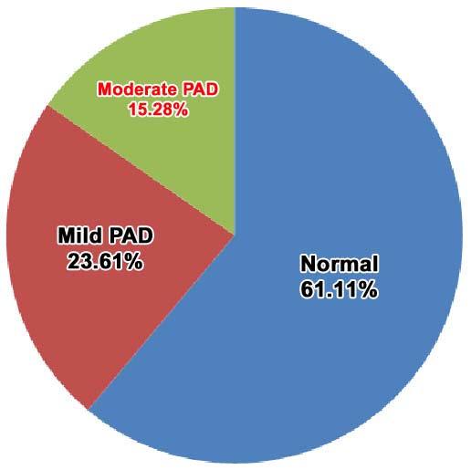
Out of 144 cases studied, 82 cases (56.94%) were females while 62 cases (43.06%) were males.
Mean ABI was 0.968±0.151 and we found low ABI (<0.9) suggestive of PAD in 38.89% of patients. Mild PAD constituted 23.61% and moderate PAD 15.28% (Fig 1). In remaining 61.11% cases ABI was more than 0.9.
Maximum cases ie, 41.67% cases were in the age group of 65 to 74 years of age. The mean age was 63.51 ±8.096 years. Table 1 depicts correlation of ABI with age. It was observed that the group of patients aged >75 years shows significant association with ABI as the difference has P value <0.05.
Among various risk factors for ischemic stroke, hypertension was present in 50% cases and diabetes mellitus in 33.33% cases. Other risk factors were history of previous stroke in 34.72% cases, history of ischemic heart disease (IHD) in 65.3% cases. Also 27.8% were smoker and 16.67% were alcoholic. Significant association was noted between low ABI and some of these risk factors in our present study. (Table 2).
Patients with higher NIHSS score on admission
Table 1 — Distribution of ABI in different age groups
Age groupNo ofABI <0.9ABI >0.9P value patients(PAD) (Normal) 45-54246180.742 55-645018320.773 65-746026340.731 >7510640.046
Table 2 — Different risk factors and clinical and laboratory parameters in PAD and normal patients
Risk factors ABI <0.9ABI >0.9 P-value (PAD)(Normal)
Hypertension38 340.005
Diabetes Mellitus2820 0.898 h/o IHD4252 0.037
NIHSS score7.89±2.696.25±1.89 0.033 Waist Circumference80.75±5.8378.7±5.42 0.962
LDL158.79±33.69 137.06±26.76 0.0033
HDL43.75±9.1250.98±8.64 0.316 Triglyceride 167.29±26.66136.82±16.47 <0.0001
were more in ABI<0.9 group and mean NIHSS significantly correlated with ABI. Waist circumference was higher in patients with PAD, compared to normal. Patients with dyslipidaemia were more common in low ABI group and a significant association was found between mean values of LDL, Triglyceride and ABI. (Table 2).
A logistic regression analysis was performed to study the effects of significant variables. Having hypertension [Odds ratio (OR) 2.6, 95% confidence interval(CI) 1.30-5.19], previous history of CVA (OR 3.92, 95% CI 1.9-8.09), history of IHD (OR 2.08, 95% CI 0.99-4.35)were more likely to have PAD. Also patients having higher NIHSS score (OR 2.05, 95% CI 1.09-3.01), higher LDL (OR 1.04, 95% CI 1.01-1.07), higher Triglyceride (OR 1.09, 95% CI 1.06-1.14)has higher propensity to PAD (Table 3).
Difference between mean SBP of upper and lower limb of both sides were higher in low ABI group and they showed significant correlation with ABI (P value <0.0001) (Table 4)(Fig 2).
DISCUSSION
Since PAD remains asymptomatic in most individuals, it often remains a neglected part in the evaluation. Presence of PAD can be detected early by measurement of ABI.
We studied 144 ischemic stroke patients. We found low ABI (<0.9) suggestive of PAD in 38.89% of patients, which includes mild PAD (23.61%) and moderate PAD (15.28%). However severe PAD (ABI <0.4) was absent in our study. Also we did not find patients having ABI >1.4. This may be because of the fact that our patient pooll contained less number of elderly people as severe PAD is more common with increasing age. Patients
having low ABI was 52.8% in a study by Weimer et al6 Alvarez Sabin et al showed low ABI in 40.5% patients in their study, where mean value of ABI was 0.92±0.217. Another study result revealed PAD in 26% of ischemic stroke patients conducted by Mohammad Selim Shahi et al 8
In our study greater number of patients ie, 41.67% were between 65 to 74 years of age. The mean age was 63.51 ± 8.096 years. Study done by Ratanakorn D showed the mean age of stroke patients was 63.5±14 years9. Another study showed the mean age as 64.04 ± 12.24 years in patients with normal ABI and 70.48 ± 11.78 years in patients with abnormal ABI5.
As far as gender is concerned amongst 144 cases studied, 82 cases (56.94%) were females while 62 cases (43.06%) were males. Prevalence of PAD was
Table 3 — Logistic regression of significant risk factors and parameters
Risk factors andOdds Ratio 95% CIP value other parameters Hypertension 2.6(1.3-5.19) 0.0041 h/o IHD 2.08(0.99-4.35)
0.0009 Triglyceride1.09(1.06-1.14) <0.0001
Table 4 — Mean Systolic BP in PAD and normal patients
SBPABI <0.9ABI >0.9P value (PAD)(Normal)
Right arm 154.07±13.85145.23±13.81 0.0003
<0.0001
Difference between Right arm and ankle28.21±11.3414.68±3.61 <0.0001
Fig 2 — Mean systolic BP in PAD and normal patients
higher among females in our study which could be attributable to age associated atherosclerosis. Tziomalos K et al and Gronewold J et al in their study also found higher percentage of PAD among females as 62.6% and 52.7% respectively10,11. However study conducted by Mohammad Selim Shahi et al shows different result as it shows male preponderence8
Various risk factors (h/o DM, HTN, IHD, past CVA) and other clinical parameters (NIHSS score, waist circumference, Systolic BP) were evaluated in our study. Hypertension was found in 50% cases and diabetes mellitus was seen in 33.33% cases. Other risk factors were history of previous stroke in 34.72% cases, history of cardiac disease in 65.3% cases. Studies carried out by various previous workers show similar observations. Low ABI has shown significant association with recurrence of vascular events (IHD, recurrent CVA)12
Result of different studies regarding association of PAD and stroke are controversial. Study by Weimer et al showed that patients with PAD had a significantly higher recurrent stroke risk compared to patients having ABI >0.96
Patients with higher NIHSS score were more in ABI<0.9 group and mean NIHSS significantly correlated (p value 0.033)with ABI. Lee et al in their study also found that mean NIHSS was higher among PAD patients compared to normal (p value 0.003)13
One previous study analysed different risk factors using logistic regression and showed that older age, hypertension, history of ischemic heart disease, raised systolic blood pressure, were all significantly associated with stroke14.
Mean systolic BP of both side upper and lower limbs were different in PAD & normal patients and this difference is statistically significant.
Difference between mean SBP of upper and lower limb of both sides were higher in low ABI group and they showed significant correlation with ABI (P value<0.0001). Sharma et alnoted similar result in their study15. Patients with dyslipidaemia were more in low ABI group and ABI showed significant correlation with mean values of different components of lipid profile (LDL, HDL, Triglyceride).
Hence beyond its accuracy in diagnosis of PAD, ABI can be regarded a predictor for the development of atherosclerotic events in future. Different risk factors like HTN, DM, dyslipidaemia were associated with both PAD and CVA. Though our inferences are based on a smaller subset of patients, but it suggests that PAD
is not very uncommon in CVA patients in our country. We recommend further studies, so that their correlation can be explored to a great extent.
1Jameson, Fauci, Kasper, Hauser, Longo, Loscalzo — Cerebrovascular Diseases, In: Smith WS, Jhonston SC, Hemphill JC, III. Harrison’s Principles Of Internal Medicine.20th ed. USA: McGraw Hill Education; 2018: p.3068
2Rothwell PM, Coull AJ, Giles MF, Howard SC, Silver LE, Bull LM. et al — Change in stroke incidence, mortality, case-fatality, severity and risk factors in Oxfordshire, UK from 1981 to 2004 (Oxford Vascular Study). Lancet 2004; 363: 1925-33.
3Nowik M, Dreschler H, Nowacki P — Atherosclerotic plaque instability and ischemic stroke: the role of inflammatory and immunologic factors. NeurolNeurochir Pol 2004; 38: 209214.
4Marinelli MR, Beach KW, Glass MJ, PrimozichJF, Strandness DE — Noninvasive Testing vs Clinical Evaluation of Arterial Disease: A Prospective Study. JAMA 1979; 241: 2031-4.
5Johnston KW, Hosang MY, Andrews DF — Reproducibility of noninvasive vascular laboratory measurements of the peripheral circulation. J Vasc Surg 1987; 6:147-51.
6WeimarC, Goertler M, Rother J, Ringelstein EB, Darius H, Nabavi DG, Kim IH. etal—Systemic Risk Score Evaluation in Ischemic Stroke Patients (SCALA): a prospective cross sectional study in 85 German stroke units. J Neurol 2007; 254: 1562-8.
7Alvarez-Sabín, J, Quintana M, Santamarina E, Maisterra O, Nunez AG — Low ankle-brachial index predicts new vascular events and functional outcome after 1 year in patients with non-cardioembolic stroke: Our experience and review. Eur J Neurol 2013; 21: 100-6.
8Shahi M, Rahman A, Wadud M, Saha U, Ahmed A, Ali Z, et al Association of Ankle Brachial Pressure Index (ABPI) in patients with ischemic stroke : A Case Control Study. ChattagramMaa-O-Shishu Hospital Medical College Journal 2013; 12: 27-33
9Ratanakorn D, Keandoungchun J, Tegeler CH — Prevalence and association between risk factors, stroke subtypes, and abnormal Ankle Brachial Index in acute ischemic stroke. J Stroke Cerebrovasc Dis 2012; 21: 498-503.
10Tziomalos K, Giampatzis V, Bouziana S, Pavlidis A, Spanou M, Papadopoulou M, et al — Predictive value of the Ankle Brachial Index in patients with acute ischemic stroke; Vasa 2014; 43: 55-61.
11Gronewold J, Hermann DM, Lehmann N, Kröger K, Lauterbach K, Berger K, et al — Heinz Nixdorf Recall Study Investigative Group ; Ankle-brachial index predicts stroke in the general population in addition to classical risk factors. Atherosclerosis 2014; 233: 545-50.
12Bilic I, Dzamonja G, Lusic I, Matijaca M, Caljkusic K — Risk Factors And Outcome Differences Between Ischemic And Hemorrhagic Stroke: Acta Clin Croat 2009; 48: 399-403
13Lee DH, Kim J, Lee HS, Cha MJ, Kim YD, Nam HS, etal—Low ankle-brachial index is a predictive factor for initial severity of acute ischemic stroke. Eur J Neurol 2012; 19: 892-8.
14Ovbiagele B — Association of ankle-brachial index level with stroke. J Neurol Sci 2009; 15, 276(1-2): 14-7.
15Sharma N, Gupta A, Priyanka P, Singh R, Gupta R, Sharma D — A Study of Ankle-Brachial Index in Patients Of Stroke; IOSRJDMS 2017; 16: 47-52.
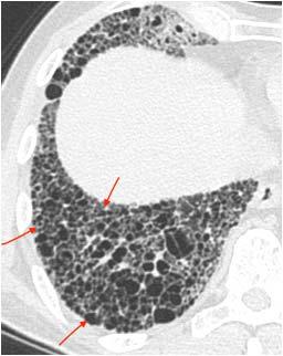
CT scan images of a 70-years old man with 2 years history of progressive dyspnea
Questions :
(1)Name the sign shown by red arrow.
(2)What pattern is shown?
(3)In view of clinical history, what is the most likely diagnosis?
Answers:
(1)Honeycombing — It refers to clustered, thin/thick walled cystic spaces usually of similar diameters (3-10mm, but occasionally larger), in stacks of 2 or more, usually peripheral. It may also present as a single layer.
(2)UIPpattern—HRCT features frequently seen in UIP pattern include honeycombing, traction bronchiectasis/ bronchiolectasis (shown by yellow arrow), which may be seen with the concurrent presence of fine reticulation. The typical distribution of UIP is subpleural with basal predominance, although some upper lobe involvement is common.
(3)Idiopathic pulmonary fibrosis (IPF) is the most likely diagnosis — UIP is the hallmark radiologic pattern of IPF.
Questions:
(1)What is the diagnosis?
(2)What are the differential diagnosis?
(3)What is plasmacytoma?
Answers:
(1)A well-defined expansile osteolytic lesion in anterior aspect of right 5thrib. An image guided biopsy was performed from the lesion. It was plasmacytoma of rib.
(2)The differential diagnosis of osteolytic lesion at this age would include- metastasis, myeloma and lymphoma. Biopsy is must to arrive at a conclusive diagnosis.
(3)Plasmacytomas are plasma cell tumour, localised to bone which have a predisposition for red marrowcontaining axial skeleton. The peak incidence is in 4th to 6th decade. They appear as expansile osteolytic lesion with thinning of cortex and absence of sclerotic reaction.
Picture This by Jankharia, Mumbai, Maharashtra 1MD, DNB (Radiology) 2MD, DMRD (Radiology)
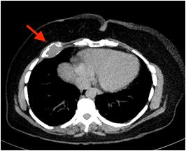
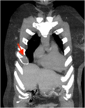
JOURNAL
M Chenniappan1
Series 3 : ECG
“Obvious is obvious, look for unobvious”
This is the ECG of 50year diabetic with intermittent chest pain
Questions:
1.What is obvious?
2.What is unobvious?
3.What is the practical implication?
Answers
1.Obvious : The presence of Right Bundle Branch Block (RBBB), left anterior fascicular block and anterolateral and high lateral pathological Q waves are obvious indicating Antero and high lateral MI.
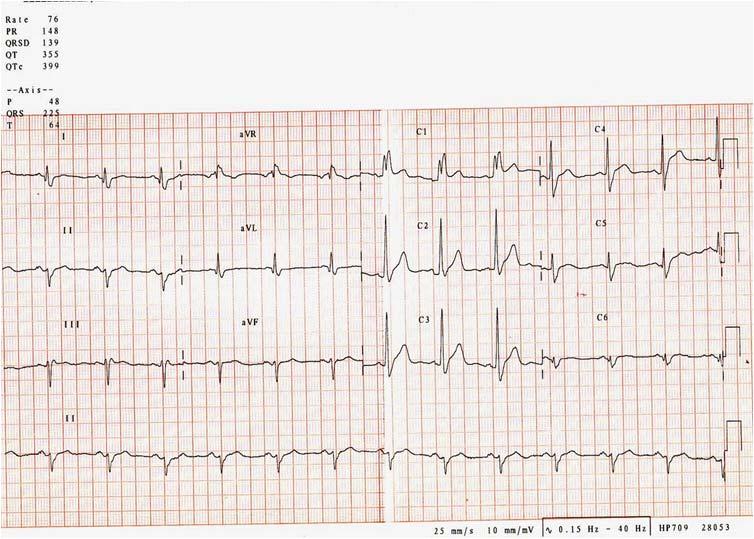
2.Unobvious : Unobvious is associated Posterior Wall Myocardial Infarction (PWMI). Most often it is difficult to diagnose PWMI in the presence of RBBB. One should concentrate on initial R wave in RBBB. In uncomplicated RBBB, in V1 this initial r is due to septal activation occurring from Left to Right and it is narrow
and small – but in the presence of RBBB, the initial R wave becomes tall and broad in V1. In addition, there may be homophasic ST T changes in V1 where ST T are in the same direction as QRS. So, in this ECG, in addition to Anterior Injury and ischemia, patient has old PWMI indicated by Tall and Broad initial R in V1 (Fig 3A).
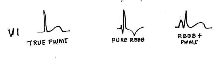
Fig 3A — Showing ECG complex in V1 with PWMI, RBBB and the combination
1Adjunct Professor, Dr MGR Medical University, Tamilnadu; Senior consultant cardiologist, Tamilnadu; Ramakrishna Medical Centre, Apollo Speciality Hospital, Trichy
3.Practical implications : It is always a good practice to look for PWMI in inferior MI whether it is in acute phase or chronic phase in the form of reciprocal ST depression or Tall R in VI respectively. Association of PWMI in addition to IWMI indicates more myocardial involvement and more extensive disease. This ecg illustrates how to diagnose PWMI in the presence of RBBB which masks chronic PWMI.
Soma Saha1, Paresh Bhowmik2, Susmita Deb3
Lymphangioleiomyomatosis (LAM) is a rare multiple cystic lung disease of unknown etiology that traditionally affects young women of child bearing or premenopausal age. It is characterized by proliferation of atypical smooth muscle cells, preferentially along bronchovascular structures that causes progressive respiratory failure. Due to its unusual and nonspecific presenting symptoms, patients often receive missed or delayed diagnosis. Diagnosis is made by a combination of clinical features and computed tomography scanning specially High resolution CT of Thorax (HRCT) or, in cases of doubt, lung biopsy. In patients with rapidly progressive disease, hormone treatment (predominantly progesterone) is tried. The only treatment for severe LAM is currently lung transplantation.
[J Indian Med Assoc 2020; 118(8): 58-61]
Keywords : Lymphangioleiomyomatosis (LAM), HRCT, Progesterone.
Lymphangioleiomyomatosis (LAM) is a rare hamartomatous proliferation of smooth muscle induced in blood vessels and lymphatics in the lung1,2. It extends into the pulmonary interstitium leading to diffuse thin walled cystic lesions and pulmonary hemorrhage and lymph node involvement which may result in chylous effusion3.
LAM is a multisystem disorder that may also result in extrapulmonary manifestations such as angiomyolipomas and lymphatic tumours. It predominantly occurs in premenopausal women but can also present in postmenopausal women.
Clinically, LAM is characterized by progressive dyspnea with exertion, fatigue, chronic cough, wheezing and chest pain, complications of spontaneous pneumothorax and chylothorax; pulmonary function tests (PFTs) usually show an obstructive and/or mixed restrictive/obstructive pattern with air flow limitation and impaired lung diffusion. Chest radiography show diffuse interstitial infiltrates and high resolution computed tomography (HRCT) usually reveals thin walled cystic lesions, distributed in diffuse manner.
There are various therapeutics modalities for LAM with differing efficacy and lung transplantation remains the only therapeutic option for patients with advanced disease.
CASE REPORT
Case 1 :
A 36 year Hindu female of Tripura presented with history of progressive breathlessness on exertion of last 3 year duration. No history of seasonal variation, cough, wheezing, chest pain, paroxysmal nocturnal dyspnea, hemoptysis or
1MBBS. DNB (Medicine), Associate Professor, Department of Medicine, TMC and DR BRAM Teaching Hospital and Corresponding Author
2MBBS. MD (Radio diagnosis), Senior Resident, Department of Radio diagnosis, AGMC
3MBBS, Postgraduate trainee, Department of Medicine, TMC and DR BRAM Teaching Hospital
Received on : 20/07/2020
Accepted on : 25/07/2020
Editor's Comment :
Pulmonary lymphangiomyomatosis (LAM) is a rare cystic lung disease of premenopausal women. HRCT Thorax is a good modalities non-invasive investigation to diagnose LAM. LAM can be treated with progesterone for improvement of Oxygen saturation though there is no structural changes in the lung.
fever. No history of Tuberculosis. She is non-diabetic and non-hypertensive. She was treated as bronchial asthma for the last 3 year.
On examination patient was averagely built and nourished. All the vitals were in normal limits. Patient was hypoxic and cyanosed, but improved with oxygen supplementation. Clubbing was absent. Respiratory system examination was unremarkable except bilateral basal crepitations and diminished vesicular types of breath sounds. Cardiovascular system examination did not reveal any abnormality. Other systems were essentially normal. All the routine blood investigations were not significant. Antinuclear antibody (ANA) and human immunodeficiency virus (HIV) test were negative.
Her chest radiograph was suggestive of bilateral extensive reticulonodular pattern with cystic changes (Fig 1) High resolution computed tomography (CT) thorax (Figs 2&3) showed multiple thin walled cysts scattered bilaterally with areas of interseptal thickening. It also showed mediastinal lymphadenopathy. Spirometry showed obstructive pattern with increased lung volumes and reduced diffusion capacity (DLCO). Arterial blood gas revealed pH 7.35, PaO2 59 mmHg, PaCO2 30.3 mmHg, and O2 saturation 68%. The diagnosis was confirmed on the basis of classical high resolution CT(HRCT) findings. In view of severe hypoxia invasive procedure like bronchoscopy was not done.
The subject was put on oral progesterone with O2 therapy. She showed good improvement in oxygen saturation and was discharged with oral progesterone and domiciliary oxygen supplementation.
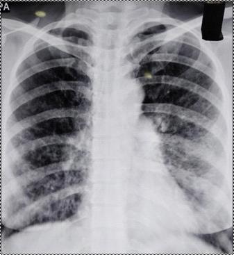
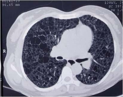
grade fever and progressive shortness of breath with dry cough and mild chest pain on deep inspiration. She had no history of seasonal variation, hemoptysis, paroxysmal nocturnal dyspnea, tuberculosis or such contact. No history of diabetes or hypertension. She is treated as chronic obstructive pulmonary disease for past 8 years. She had history of menorrhagia due to leiomyoma which lead to her hysterectomy 1 year back.
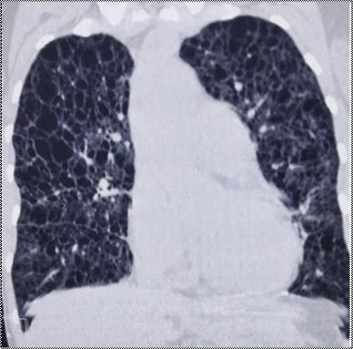
The case was reviewed after 6 months. There was significant improvement in dyspnea, spirometry, and arterial blood gas values. However, radiological features remained stationary with no deterioration. HRCT can often confirm the diagnosis and tissue diagnosis may not be necessary. HRCT findings suggestive of LAM are small, thin walled, air containing cyst ranging from 2-20 mm or more in diameter scattered throughout the lung fields.
Case 2 :
A 44 year old Hindu, smoker female from South Tripura, presented to the casualty department with 3 days of high
General physical examination revealed average Indian built with pallor, bilateral pitting pedal edema, raised jugular venous pressure without clubbing, cyanosis, lymphadenopathy and icterus. Clinically, she had tachycardia, tachypnoea with decreased O2 saturation which improved moderately after oxygen supplementation.
Her respiratory system examination revealed prominent accessory muscles of respiration, diminished chest expansion with bilateral hyperresonant notes of percussion and bilateral coarse crackles with wheeze. Cardiovascular system examination revealed apex beat at 6th intercostal space 3 cm lateral to mid clavicular line with loud P2, low pitched ejection systolic murmur in pulmonary area, with otherwise normal findings. On per abdominal examination, liver was just palpable and soft in consistency with tenderness on palpation and on other organomegaly or palpable masses.
All routine blood investigations were normal other than
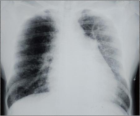
iron deficiency anemia. Anti nuclear antibodies test was negative.
Electrocardiogram showed sinus tachycardia; chest X ray showed bilateral diffuse ground glass opacities with honeycombing and cardiomegaly and straightening of left heart border with fullness of pulmonary conus and bilateral hilar lymphadenopathy(Fig 4). 2D echocardiography confirmed pulmonary hypertension without any evidence of regional motion wall abnormalities and ejection fraction of 67%. PFT showed mixed obstructive and restrictive pattern with increased lung volumes and decreased diffusion capacity. Arterial blood gas (ABG) analysis suggested type I respiratory failure.
HRCT of thorax showed multiple thin walled cysts diffusely in both lung fields suggestive of bilateral cystic lung disease and features of fibrosis in right middle lobe along with bilateral hilar lymphadenopathy and bilateral lower lobe consolidation (Figs 5&6). These classical findings confirmed our diagnosis of LAM. Ultrasound of abdomen did not suggest any mass in liver or kidneys. Thoracoscopy could not be performed because of severe hypoxia and toxic condition of the patient.
Patient was initially treated with bronchodilators and intravenous antibiotics and oxygen therapy, resulting in improvement of her oxygenation status also her later ABG reports.
After recovery, oral pirfenidone and progesterone were started. Her daily oxygen requirement decreased drastically and was discharged with home nocturnal oxygen therapy. On 8 months follow up, her PFTs and ABG showed moderate improvements but radiographic findings however, showed no regression.
LAM is a rare cystic lung disease that usually affects women of child bearing age and may present with developmental delay and cutaneous manifestations like ash leaf macules, shagreen patch and seizures when associated with tuberous sclerosis(30%). But it mostly occurs sporadically1
It may also be seen in postmenopausal women who present with progressive exertional dyspnea, chronic cough and spontaneous pneumothorax (57%) or chylothorax and is frequently associated with renal angiomyolipomas (32%)3
Clinical presentation greatly varies; ranging from chronic cough to hemoptysis(32%) to pleural effusion(12%)3. One study reported 40-80% of LAM patients can have recurrent pneumothorax4. These symptoms usually present in later stages of disease and therefore initially is often mistaken for reactive airway disease. Often these patients are misdiagnosed and treated with bronchodilators5
Pathologically, this condition is characterized by proliferation of immature smooth muscle cells derived from associated lymphatics. These smooth muscle cells infiltrate the walls of alveoli and bronchi with resultant air trapping and impaired gas transfer which may mimic emphysema1,3 Involvement of pulmonary venous vasculature may lead to hemosiderin deposition in lung parenchyma due to recurrent
hemorrhage4. There are close histological parallels between LAM and pulmonary manifestations of tuberous sclerosis, although the hereditary condition effects both sexes and also has cutaneous and cerebral manifestations5
Radiographic findings often vary depending on disease severity and progression. Typically, chest Xrays show hyperinflated lungs due to obstructive nature of disease. Reticular pattern can also be seen in later stages due to coalescence of the cysts. This must be differentiated from Langerhans’ cell histiocytosis, which may also present with similar symptoms and reticular pattern on chest X-ray6. It may also show ground glass appearance from hemosiderosis 7 .
Concomitant with chest X-ray findings, PFTs with LAM is often obstructive or mixed pattern5,7. total lung capacity is often increased and residual volume to total lung capacity(RV/TLC) is also increased. Air flow is limited with decreased forced expiratory volume in first second(FEV1)6
disease progression can be best monitored with diffusion capacity and FEV18
Cysts and bullae can be anatomically detailed on HRCT6,17,18. CT changes can vary from a reticular infiltrate to honeycombing with diffuse cysts depending on disease progression10. The diagnosis can be made with HRCT but in many cases tissue biopsy is obtained by various means. Besides taking tissue biopsy, Visually assisted thoracoscopic surgery(VATS) can also be implemented in cases of spontaneous pneumothorax as well. It can also be used for surgical wedge resection of apex of lung, pleural abrasion or chemical pleurodesis15,16
Diagnosis is confirmed with characteristic immunochemical stain that are specific for smooth muscle cells e.g. actin, desmin as well as melanocytic markers HMB45, HMSA1 and MELAN A11. Amongst them, HMB45 is the gold standard for atypical cells of LAM11
The fact that LAM presents predominantly in premenopausal women1 and is shown to subside after menopause led to numerous studies to determine the role of estrogen in pathogenesis of LAM. However, no association has been established. Hence, there is no definite therapeutic strategies targeting the hormonal receptors9. According to latest evidence, even the first episode of pneumothorax in a case of LAM should be treated with pleurodesis since it opens the possibility of recurrent pneumothorax 3 . Bronchodilators are the mainstay of supportive measures initially. Some patients have been started on sirolimus, everolimus, which provides a median transplant free survival of approximately 29 years from the onset of symptoms and 10 years transplant free survival of 86% 12,13 . these therapeutic options are only disease stabilizing and not curative. Lung transplantation remains the last treatment resort for advanced LAM patients for survival14
The estimated prevalence of LAM is thought to be around one to 2.6 patients in 1,000,000 in the general female population10. Knowledge of LAM as a cause of chronic cough, spontaneous recurrent pneumothorax and especially in young females would be helpful for primary care physician for diagnosis. It is also necessary to understand that early cases are often treated as reactive airway disease or interstitial lung disease mistakenly. HRCT thorax alone can sensitively diagnose the disease, however, tissue biopsy remains the definitive diagnostic test.
The rarity of this cystic disease is a principal cause behind reporting the cases, as well as the efficient diagnosis without biopsy and the salient treatment measures. This case series is aimed to create awareness and better understanding of the disease.
1McCormack FX — Lymphangioleiomyomatosis: A clinical update. Chest 2008; 133: 507-16.
2Jain VV, Gupta OP, Jajoo S, Khiangate B — Recurrent pneumothorax in a young female with pulmonary lymphangiomyomatosis: A case report and overview of literature. J Fam Med Primary Care 2014; 3: 86-8.
3Ryu JH, Moss J, Beck GJ, Lee JC, Brown KK, Chapman JT, et
al — The NHLBI lymphangioleiomyomatosis registry: Characteristics of 230 patients at enrollment. Am J Respir Crit Care Med 2006; 173: 105-11.
4Carrington CB, Cugell DW, Gaensler EA, Marks A, Redding RA, Schaaf JT — Lymphangioleiomyomatosis. Physiologicpathologic-radiologic correlations. Am Rev Respir Dis 1977; 977-95.
5Taylor JR, Ryu J, Colby TV, Raffin TA: Lymphangioleiomyomatosis. N Engl J Med 1990, 323: 1254-60. 10.1056/ NEJM199011013231807.
6Chu SC, Horiba K, Usuki J, et al — Comprehensive evaluation of 35 patients with lymphangioleiomyomatosis. Chest 1999, 115: 1041-52.
7Kitaichi M, Nishimura K, Itoh H Izumi T — Pulmonary lymphangioleiomyomatosis: a report of 46 patients including a clinicopathologic study of prognostic factors. Am J Respir Crit Care Med 1995; 151: 527-33. 10.1164/ ajrccm.151.2.7842216.
8Steagall WK, Taveira-DaSilva AM, Moss J — Clinical and molecular insights into lymphangioleiomyomatosis. Sarcoidosis Vasc Diffuse Lung Dis 2005; 22: 49-66.
9Taveira-DaSilva AM, Steagall WK, Moss J — Lymphangioleiomyomatosis. Cancer Control 2006; 13: 27685. 10.1177/107327480601300405.
10Yamazaki A, Miyamoto H, Futagawa T, et al — An early case of pulmonary lymphangioleiomyomatosis diagnosed by videoassisted thoracoscopic surgery. AnnThoracCardiovascSurg 2005; 11: 405-7.
11Bonetti F, Chiodera P.L, Pea M, Martignoni G, Bosi F, Zamboni G — Transbronchial biopsy in lymphangiomyomatosis of the lung. HMB45 for diagnosis. Am J Surg Pathol 1993; 17: 1092102.
12Oprescu N, McCormack FX, Byrnes S, Kinder BW — Clinical predictors of mortality and cause of death in lymphangioleiomyomatosis: a population-based registry. Lung 2013; 191: 35-42. 10.1007/s00408-012-9419-3
13Huang J, Manning BD — A complex interplay between Akt, TSC2 and the two mTOR complexes. Biochem Soc Trans 2009; 37: 217-22. 10.1042/BST0370217
14Johnson SR — Lymphangioleiomyomatosis. Eur Respir J 2006, 27:1056-65. 10.1183/09031936.06.00113303-2019 Rhee et al. Cureus 11(1): e3938. DOI 10.7759/cureus.3938
15Baumann MH — Management of spontaneous pneumothorax. Clin Chest Med 2006; 27(2): 369–81.
16Baumann MH, Strange C, Heffner JE, et al — Management of spontaneous pneumothorax: an American College of Chest Physicians Delphi consensus statement. Chest 2001; 119(2): 590-602.
17Spiliopoulos K, Tsantsaridou A, Papamichali R, Kimpouri K, Salemis NS, Koukoulis GK, et al— Recurrent spontaneous pneumothorax in a 42 years old woman with pulmonary lymphangioleiomyomatosis: Insights and pitfalls of the surgical treatment. J Clin Med Res 2013; 5: 70-4.
18Kirchner J, Stein A, Viel K, Dietrich CF, Thalhammer A, Schneider M, et al — Pulmonary lymphangioleiomyomatosis: Highresolution CT findings. Eur Radiol 1999; 9: 49-54.
19Baldi S, Papotti M, Valente ML, Rapellino M, Scappaticci E, Corrin B — Pulmonary lymphangioleiomyomatosis in
Tony Ete1, Swapan Saha2, Vanlalmalsawmdawngliana Fanai3, Arun Kumar3, Habung Mobing4, Shakeel Ahamad Khan3, Utpal Kumar3, Narang Naku5, Animesh Mishra6
Unilateral absence of pulmonary artery should be considered in patients presenting with haemoptysis and recurrent respiratory infections. Usually it is a diagnosis of exclusion. Patient may present with non specific symptoms. A high index of suspicion with proper investigations, non invasive as well as invasive, are required for diagnosis and management. The diagnosis is usually confirmed by CT and MRI. Angiography is done only for patients who require embolisation or revascularisation surgery.
[J Indian Med Assoc 2020; 118(8): 62-3]
Key words : Hemoptysis, pulmonary artery, angiography.
absence of a pulmonary artery (UAPA), a rare condition, usually occurs in combination with other cardiovascular conditions like tetralogy of Fallot (TOF) or septal defects. Patients with isolated absence of one pulmonary artery often present with dyspnoea, chest pain, hemoptysis or recurrent chest infections but may be asymptomatic till late adulthood. About 20% of the patients develop inconsequential hemoptysis, although massive hemoptysis is very rare1. Therefore, diagnosis may be difficult due to these nonspecific presentation2, sometimes diagnosed incidentally on chest radiographs. Here we are presenting a case with isolated absent right pulmonary artery with haemoptysis, as clinical presentation.
CASE REPORT
A 37-year gentle man, non hypertensive, non diabetic and non smoker, presented with recurrent episodes of hemoptysis for the last six months, scanty in amount. There was no history of fever, breathing difficulty or recurrent chest infection. No history of pulmonary tuberculosis in the past. Physical examination revealed body temperature of 36.6oC, pulse rate of 90 beats per minute, respiration rate of 16 per minute and blood pressure of 100/70 mmHg. Clinically there were no signs of cyanosis, edema, or clubbing of the fingers. Cardiac and chest auscultation was normal. The electrocardiogram revealed normal sinus rhythm.
Department of Cardiology, North Eastern Indira Gandhi Regional Institute of Health and Medical Sciences, Shillong 793018
1MBBS, MD, DM (Cardiology), Assistant Professor and Corresponding Author
2MBBS, MD, DM (Cardiology), Consultant, Department of Cardiology, Desun Hospital Siliguri
3MBBS, MD, Senior Resident
4MBBS, MD, Senior Resident, Department of Medicine
5MBBS, MS, Assistant Professor, Department of General Surgery
6MBBS, MD, DM (Cardiology), Professor and Head
Received on : 25/06/2020
Accepted on : 02/07/2020
Editor's Comment :
Unilateral absence of pulmonary artery (UAPA) should be considered in patients presenting with hemoptysis and recurrent respiratory tract infections.
A high index of suspicion is required for diagnosis. It is usually a diagnosis of exclusion. Chest radiograph may suggest the diagnosis whereas echocardiography can be used for the evaluation of possible associated cardiac anomalies and assessment of pulmonary hypertension.
The diagnosis is usually confirmed by CT scan and MRI. Angiography is done for patients who require embolisation or revascularization surgery.
Echocardiogram was normal with normal pulmonary artery pressure except for absent right pulmonary artery. A chest radiograph (postero-anterior view) revealed dilated artery (?Main pulmonary) in the right hilum with alveolar infiltrates in right lung field. The right hemi diaphragm was elevated without any cardiac and mediastinal displacement (Fig 1).
Computed tomography (CT) angiogram revealed the absence of the right main pulmonary artery. A focal vascular dilation was detected in right lung possibly representing an aneurysmatic formation or an arteriovenuous fistula (Fig 2 A). Blood was supplied to the right lung by tortuous, dilated arterial branches of indeterminate origin (Fig 2 B).
In view of patient’s symptom and to look for source of blood supply to the right lung, patient was taken for catheterization and pulmonary angio and aortogram was done with an intention to proper decision making for the management. Pulmonary angiogram showed absent right pulmonary artery (Fig 3 A). Selective angiogram of right subclavian artery showed upper zone of right lung is supplied by right vertebral artery and lower zone is supplied by right internal mammary artery (Fig 3 B).
As the patient’s symptom improved thereafter, he was kept under close follow-up on an outpatient basis, with symptomatic
Fig 1 — Chest radiograph (postero-anterior view) shows dilated artery in the right hilum with alveolar infiltrates in right lung field with elevated right hemi diaphragm
and supportive treatment for recurrent haemoptysis.
DISCUSSION
Fig 2 — Transaxial CT angiogram with intravenous contrast in a soft tissue window (A) shows an absent left pulmonary artery (B) tortuous, dilated arterial branches supplying right lung
Fig 3 — (A) Pulmonary angiogram showed absent right pulmonary artery Selective angiogram of right subclavian artery showed upper zone of right lung is supplied by right vertebral artery and lower zone is supplied by right internal mammary artery
The prevalence of UAPA is 1 in 200,000 young adults2 and it can occur in an isolated manner although most cases are associated with other cardiovascular anomalies like tetralogy of fallot2,3. In about two third of cases, isolated UAPA involves the right lung1 Developmental alteration of ventral bud of the ipsilateral 6th aortic arch is thought to be the embryological basis of UAPA 4 . In the affected artery, generally the distal intrapulmonary branches remain intact which can be supplied by collaterals from bronchial, intercostal, internal mammary, subdiaphragmatic, subclavian or even coronary arteries5. Clinical course of many patients with isolated UAPA is benign and a diagnosis is not made until adulthood2. In symptomatic patients, one study showed, chest pain, pleural effusion and recurrent infections to be present in 37% of patients, while dyspnea or exercise intolerance in 40% of patients. Pulmonary hypertension was present in 44% of patients. Hemoptysis occurred in about 20% of patients whereas high-altitude pulmonary edema was seen in approximately 10% of patients1. Hemoptysis is caused by collateral circulations that create high pressures in venous system5. The systemic collaterals usually arise from the bronchial, intercostals, subclavian or subdiaphragmatic arteries1. The diagnosis of UAPA is, in generally, based on history, physical examination and findings on chest radiographs. Pulmonary function test in patients with UAPA is usually unremarkable 6 . CT thorax with contrast enhancement confirms the absence of the affected pulmonary artery. Echocardiography is helpful for exclusion of other cardiac anomalies and pulmonary hypertension. Pulmonary angiography is the gold standard and is usually reserved for patients requiring embolisation or revascularisation surgery7
At present there is no consensus regarding management of patients with UAPA. Some recommends serial echocardiography of asymptomatic patients for the development of pulmonary hypertension8. On the other hand,
revascularization of peripheral branches to the pulmonary hilum can be attempted9. Hemoptysis can be managed with embolization, lobectomy or pneumonectomy10.
In conclusion, UAPA should be considered in patients presenting with haemoptysis and recurrent respiratory infections. Chest radiograph may suggest the diagnosis whereas echocardiography can be used for the evaluation of possible associated cardiac anomalies and assessment of pulmonary hypertension. The diagnosis is usually confirmed by CT and MRI. Angiography is done only for patients who require embolisation or revascularisation surgery.
1Harkel DJT, Blom NA, Ottenkamp J — Isolated unilateral absence of a pulmonary artery: a case report and review of the literature. Chest 2002; 122: 1471-7.
2Bouros D, Pare P, Panagou P, Tsintiris K, Siafakas N — Th e varied manifestation of pulmonary artery agenesis in adulthood. Chest 1995; 108(3): 670-6.
3Presbitero P, Bull C, Haworth SG, de Leval MR – Absent or occult pulmonaryartery. Br Heart J 1984; 52(2): 178-85.
4Thomas P, Reynaud-Gaubert M, Bartoli J–M, Augé A, Garbe L, Giudicelli R, et al — Exsanguinating hemoptysis revealing the absence of left pulmonary artery in an adult. Ann Thorac Surg 2001; 72: 1748-50.
5Kadir IS, Th ekudan J, Dheodar A, Jones MT, Carroll KB — Congenital unilateral pulmonary artery agenesis and aspergilloma. Ann Th orac Surg 2002; 74(6): 2169-71.
6Werber J, Ramilo JL, London R, Harris VJ — Unilateral absence of a pulmonary artery. Chest 1983; 84: 729-32.
7Hayek H, Palomino J, Thammasitboon S— Right pulmonary artery agenesis presenting with uncontrolled asthma in an adult: a case report. J Med Case Rep 2011; 5: 353.
8Turner DR, Vincent JA, Epstein ML — Isolated right pulmonary artery discontinuity. Images Paediatr Cardiol 2000; 4: 24-30.
9Welch K, Hanley F, Johnston T, Cailes C, Shah MJ — Isolated unilateral absence of right proximal pulmonary artery: surgical repair and follow-up. Ann Th orac Surg 2005; 79(4):1399402.
K Mugundhan1, M Sathishkumar, P R Sowmini2, K.Sakthivelayutham2, K Malcolm Jayaraj2, R Viveka Saravanan1
A 12 year old boy presented with flat, dark brown macules of varying size and shape present over anterior aspect of left side of chest above nipple, anterior abdominal wall, lumbosacral and left calf region since birth. He presented with seizures at the age of tweleve years. He had no history of headache, vomiting, fever or any other focal neurological deficit. His Intelligence Quotient was normal.
On dermatological examination, the child showed café au lait spots of varying size and shape over anterior aspect of left side of chest above nipple and anterior abdominal wall(Fig 01). He also showed multiple café au lait spots over lumbosacral region (Fig 02). A large triangular shaped café au lait spot over left calf region (Fig 03). Based on the above clinical findings, it was diagnosed as neurofibromatosis type 1 (NF1). His EEG showed bilateral epileptiform activity. MRI Brain (T2W & FLAIR) showed focal hyperintense lesions at right basal ganglia, left periventricular whitematter and right medial cerebral peduncle suggestive of non neoplastic hamartomas(Fig 04, 05 and 06). X-ray long bones, skull PNS view and Ultrasonogram abdomen were normal.
Neurofibromatosis type 1 is associated with neurological manifestations like seizures, tumors of brain, spinal cord and optic nerve. 60-78% patients with NF1 have hyperintese lesions within the basal ganglia, thalamus, brain stem and cerebellum on MRI brain (T2w & FLAIR) 1 Hyperintese lesions are highly prevalent and characteristic in patients with NF1. MR imaging contributes to a definitive diagnosis of NF12
This case highlights the characteristic MRI findings of neurofibromatosis type1.
REFERENCES
1Walter G Bradley — Neurology in clinical practice 2008;2: 1874-77
1Associate Professor, Department of Neurology, Govt. Stanley Medical College, Chennai 600001
2 Assistant Professor, Department of Neurology, Government Stanley Medical College, Chennai 600001
2Menor, F., Marti-Bonmati, L., Arana, E., Poyatos, C.,et al — 1998, “Neurofibromatosis type1 in children: MR imaging and followup studies of central nervous system findings” Eur J Radiol, Vol.26, PP, 121-131
Rudrajit Paul, Jyotirmoy Pal
Florence Nightingale (1820-1910) was one of the greatest health professionals to have ever adorned the surface of the earth. This year (2020), we are celebrating her 200th birth anniversary.
The birthday of Florence Nightingale (12 May) is celebrated as the international Nurses’ day. Nurses all over the world now take the “Nightingale Pledge” at the beginning of their career. But we think that categorizing Florence Nightingale as only a nurse is a gross injustice to her memory. She was an excellent scholar of medical statistics and also a social reformer. She was also a prolific writer on versatile topics from religion to feminism. In fact, historians say that the full impact of Nightingale on medical history has only recently been uncovered fully.

Florence Nightingale
(This picture is in public domain in its country of Origin)
about sanitation and hygiene in order to make any government health program successful. Such thoughts about female empowerment and public engagement were not prevalent at that time. She also regularly read reports on sanitary conditions of other Indian cities like Ahmedabad and Calcutta and advised the government officials on ways to improve the situation. Nightingale was highly interested in improving public health in India and reducing the impact of epidemics. She was well-versed in all the contemporary events in India. For example, when Kadambini Ganguly started practice as the first female physician of India, Nightingale wrote :
Florence nightingale first came to prominence during the Crimean war when she cared for the British soldiers and helped reduce the death rate in military hospital substantially. She was the first to scientifically demonstrate that simple hygienic measures like handwashing can reduce military wartime mortality significantly. Her moniker, “Lady with the lamp” was coined after description of her activities in “The Times” newspaper and an 1857 poem by Longfellow.
Florence Nightingale was a prolific writer. Collections of her letters are digitized and available online. These letters reveal her interests in various topics. They also reveal her extremely modernistic outlook. For example, in a letter to Dr. Thomas Gillham Hewlett, the Sanitary commissioner of Bombay, she writes that the native women in India must be educated
“Shehasalreadypassedwhatiscalledthefirst licentiate in medicine and surgery examinations and is to go up for the final examination in March next.Thisyounglady,Mrs.Ganguly,marriedafter she made up her mind to become a doctor and hashadone,ifnottwochildrensince.Butshewas absent only thirteen days for her lying-in and did notmiss,Ibelieve,asinglelecture!”
In 1878, when a terrible famine occurred in India, Nightingale criticized the British government severely:
“We do not care for the people of India… Betweenfiveandsixmillionhaveperishedinthis famine…Howcanwerealisewhatthemiseryisof every one of those figures: a living soul, slowly starvingtodeath?”
HIVinfection, first appearing in a big way in the 1980s, took the world by storm. As news of the various aspects of the disease spread in the media, there was a sense of panic among the pundits and the public all over the world. But did HIV actually start in the 1980s, or was it present in a small way much earlier?
On 3rd July, 1981, the New York Times reported: ”Rare Cancer Seen in 41 Homosexuals”. In that article, the “rare cancer” is Kaposi Sarcoma &the cause of the illness is described as “unknown”. This was the first proper coverage of this new disease in the media. After the initial phase of fear and confusion, intense scientific research started and by 1984, the virus was discovered. Was it something very old with a new face (like the coronavirus)? Or was it something absolutely new? Who were the persons first affected with HIV? These questions plagued researchers in the subsequent years.
In the ancient Indian text of CharakaSamhita (300 BCE), a disease called Azokshyam is described. Description of the disease has some similarities with AIDS. But it is almost impossible to say what the exact cause of this disease was. In the absence of more scientific data, scientists can only make a guess.
But what about the 1950s, 1960s or the 1970s? Was HIV infection present then? To answer this question, scientists have found a way. Tissue samples from patients of that time period are sometimes preserved and scientists have tried to do tests for HIV in those samples. The results have often been interesting.
For example, in between August 1972 and July 1973, blood was collected from clinically healthy children in Uganda as part of a study on Cancer. This blood was stored as part of the study and later, in 1985, as the HIV test became available, this blood was tested for HIV antibodies. It was found that 50 of the 75 samples were positive for the antibody (66%). However, in this study, the study subjects were not followed up. Hence, we do not know the fate of those infected.
Next, we will look at the records of some patients before 1980.
Grethe Rask :
Ms Grethe was a Danish Surgeon who started working in a village of modern Congo in 1972. In 1977, she went back to Denmark with some “unknown illness” and died of PCP illness on 12th December of the same year. After she came back to Denmark, she was tested at Copenhagen and found to have almost 0 T cell count. Grethe had also worked for a brief period in Africa in the 1960s. In 1983, one of her colleagues wrote a letter to the Lancet where her illness is described in details. At that time, HIV virus was still not discovered. But later analysis has convinced people that Grethe developed AIDS, probably after exposure to blood and body fluids of her patients in Africa. Later in 1987, her preserved blood samples were tested again in the USA and HIV infection was confirmed. Grethe is considered as one of the first non-African patients of HIV.
Robert Rayford :
Robert Rayford, who was a teenager in Missouri, USA was depicted as the first fatality from HIV in North America. In 1968, he was admitted to the hospital with disseminated Chlamydia infection. He later died in 1969 from severe pneumonia. At that time, the disease was not known but the physician caring for Redford had preserved his tissue samples. The case, due to its unusual features, had generated immense curiosity among the doctors at that time but no solution was forthcoming. These frozen tissues proved invaluable as later, as tests for HIV were discovered, those frozen serum and tissue samples were subjected to advanced tests including PCR and HIV was diagnosed in 1987. This led to the conjecture that HIV infection was probably occurring sporadically in the world for a long time before its emergence as an epidemic in the 1980s. On 11 th March, 1987, the Chicago Tribune ran a headline: “Case Shakes Theories Of Aids Origin”. The case of Rayford is described in details in this article. Question is, how did Rayford get the disease? He had never travelled out of the area. The exact route of transmission is unknown but probably, it is thought, he got the virus from sexual abuse at a young age.
Roed family :
In Norway, Arne VidarRoed was a sailor and truck driver, who became ill in 1968. He had recurrent lung infections, joint pain and edema. He later developed
JOURNAL OF THE INDIAN MEDICAL ASSOCIATION, VOL 118, NO 08, AUGUST 2020
motor dysfunction and dementia and died in 1976, after almost 10 years of living with the virus symptoms. His wife and young daughter also died of similar illness the same year. At that time, the cause of the illness was essentially unknown. Later, from their preserved tissue samples, HIV-1 infection was detected. Later research including genetic analysis found that Mr.Roed probably contracted the infection in the 1960s in Cameroon. The Roed family is probably the first documented family in Europe which was ravaged by the infection.
The HIV organism is said to have originated from Africa. But when was the first case of AIDS in Africa? Nobody can be sure. But historical analysis revealed that probably, the disease originated in and around Kinshasa in Congo. A plasma sample of an African person, which was collected in 1959 in Kinshasa, was later tested and found to be HIV positive. This testing was done as a part of an effort by scientists to trace the origin of the AIDS epidemic. But when did the disease come from Africa to Europe or USA? No one can be sure of an answer and probably, multiple people got infected from different sources around the same time period.
As this brief discussion makes clear, the HIV infection did not start suddenly. The virus was lingering in humans for a long time before exploding into an epidemic. This is not only true for HIV, but also for other infections. Tracing the root of an infection is often necessary to find its source.
How did HIV enter the Homo sapiens species? This topic is still open to speculation. But the general scientific theory is that the virus jumped from apes to humans when humans were exposed to primate blood and body fluids. Why did humans get exposed to primate blood? Because they were hunting bushmeat. And why? Here is where the topic diverges from hardcore scientific discipline to the realm of economics. Many authors argue that the brutal colonial economic policies of Europeans in Africa drove the local people to hunting bushmeat for subsistence and this increase in primate hunting in deep forests may have caused
the emergence of HIV. In Congo, the local people were driven by the colonists into deep forests in search of rubber and these activities increased chances of manape interaction. Then, the railroad network built by the colonial masters for transportation of rubber may have been instrumental in spreading the HIV epidemic throughout Congo.
1Altman LK — Rare Cancer Seen in 41 Homosexuals. The New York Times. [Published 1981 Jul 03; Cited 2020 Mar 12]. [Internet]. Available online from https://www.nytimes.com/ 1981/07/03/us/rare-cancer-seen-in-41-homosexuals.html
2Krishna MR, Reddy SR — Did AIDS viruses originate in India? Retrovirology. 2009; 6(Suppl 2): P74.
3Saxinger WC, Levine PH, Dean AG, et al — Evidence for Exposure to HTLV-III in Uganda Before 1973. Science 1985; 227: 1036-8.
4Bygbjerg IC — AIDS in a Danish surgeon (Zaire, 1976). Lancet. 1983; 1(8330): 925.
5Garry RF — Early case of AIDS in the USA. Nature 1990; 347; 509.
6Hooper E. Sailors and star-bursts, and the arrival of HIV. BMJ 1997; 315: 1689.
7Jonassen T, Stene-Johansen K, Berg ES, et al — Sequence Analysis of HIV-1 Group O from Norwegian Patients Infected in the 1960s. Virology 1997; 231: 43-7.
8Wain-Hobson S — 1959 and all that. Nature 1998; 391: 5312.
HIV in figures : —
Almost 38 million PLHIV globally ( of them 8 million unaware of their status)
Around 2 million new infections per year
Between 770000-1000000 deaths per year from HIV
24.5 million accessing ART
INDIA :
♦ 2.1 million PLHIV
♦ Annual death: 69000
♦ Annual new infection: 88000
Rudrajit Paul
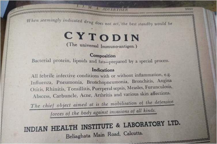
While medical science has bred a lot of spectacular success stories over the years, there have also been a lot of questionable or outright false and misleading business attempts in the name of medicine. For the last two hundred or so years, human beings have tried to conquer infectious diseases by two main weapons: antibiotics and vaccinations. While antibiotics have had measurable success, the story for vaccinations has not been similar. There is a lot of difference between theoretical and practical aspects of vaccination and many vaccines found to be theoretically robust had failed in subsequent field trials.
The figure 1 here shows an advertisement for “universal bacterial antigen” which was marketed in
India more than 75 years ago. The “drug” claimed to have bacterial extracts which would protect against a host of systemic infections (somewhat similar to the claims made by homeopathic drugs). This was an injectable preparation which was claimed to generate protective antibodies against all bacteria! Naturally, such a “miracle cure” for all infections is likely to be bogus but this drug enjoyed some financial success at that time.
In view of the recent coronavirus pandemic, we should be wary of such claims for a successful vaccine. In times of distress, unscrupulous merchants often try to cash in on the vulnerability of the people and peddle questionable remedies.
Partha S Ray1
The novel coronavirus SARS-Cov-2 has claimed the lives of thousands of unfortunate patients in this global pandemic and has infected millions throughout the globe. This pathogen has transgressed all international boundaries and overwhelmed all health systems in both developed and resource limited health setups. The COVID-19 illness has exposed deficits in research and development in respect to novel pathogens, healthcare funding and structure, people’s personal health habits and practices and aspects of lifestyle and human behaviour. This current global crisis has exposed the vulnerability of the human race despite all the developments in the health sector and other human endeavours we are so proud and potentially complacent of. The virology features of this novel pathogen and the way it interacts with the human immune system and the responses that it generates has been a steep learning curve for all. The Neurology of COVID-19 has been protean and we are still learning every day from our patients. The need to organise randomised clinical trials for evidence based therapy has again been reemphasised to save lives and avoid iatrogenic errors and adverse patient outcome. Lives and livelihood for patients and health care providers has been severely threatened in this pandemic. But we must win. [J Indian Med Assoc 2020; 118(8): 69-73]
Keywords : COVID-19 SARS-Cov-2, Neurology, CSF, RT-PCR, MERS, Immunopathogenesis.
the infamous novel coronavirus and the COVID-19 illness that it causes was first diagnosed on December 8, 2019 in Wuhan, central China1 after which it rapidly spread all over the world for the World Health Organisation (WHO) to declare it as a pandemic on 30 January 2020. Till date the Johns Hopkins database 2 shows 6,54,103 dead and 164,82,592 infected. The silver lining in the dark cloud is the recovery number of 95,90,529 individuals. We Indians presently occupy the unenviable position of being third among the total number of confirmed cases (1480) in the world and most alarmingly the sixth position in the number of deaths that of today has crossed the 33,000 mark3. As repeatedly locked downs are re-imposed and new hotspots are identified in India and globally, we continue to hold our breath in great anxiety and fear of life and livelihood. We remain alert in anticipation for the second wave4 and for the lives at risk and the future and long-term ravages on the population and global economy through the novel virus.
1MBBS, DTM&H, MD (Medicine), DNB (Medicine) MRCP (UKNeurology) FRCP (London and Edinburgh), DNB (Neurology) CCST (Neurology & Clinical Neurophysiology), Consultant Neurologist and Neurophysiologist, NHS England and Clinical Lecturer Liverpool University and Post Graduate Medical School, Liverpool and Corresponding author
Received on : 01/08/2020
Accepted on : 00/08/2020
Editor's Comment :
We have all been exposed in regards to our knowledge base in being able to protect humanity against the unknown natural and novel pathogens with whom we cohabit this planet. The witnessed impact of a pandemic on global economy and livelihood should prioritise future health policy planning and further resource allocation to epidemiology and medical research and focus on population positive health initiatives. It is hoped the global community will work cooperatively and collaboratively under the auspices and leadership of the World Health Organisation to promote global health endeavours and be able to predict and manage any future threats to international health better to protect humanity.
Virology :
SARS-CoV occurred in China in 20035 affecting approximately 8000 people with a 10% mortality rate and the Middle East respiratory syndrome (MERS) outbreak in Saudi Arabia in 20126 affecting 2500 individuals with a 35% mortality rate have been harbingers of the current corona virus as it has approximately 80% sequence homology with SARSCoV. Peculiarly there is a 96% homology with a bat coronavirus and 92% with the pangolin coronavirus raising the hypothesis that it arose in animal species and then spread between the animals to infect humans7. The full sequence of SARS-CoV-2 to was published on 7 January 2020 confirming that this was
a beta coronavirus similar to other human coronaviruses responsible for 15% of all cases of acute viral nasopharyngitis(“common cold”). The contagiousness of this coronavirus has made it immensely threatening to the human species as the polybasic cleavage site in the spike protein is a potential determinant of increased transmissibility. This protein helps the virus binds to the angiotensin converting enzyme 2(ACE2) receptor that is also a receptor for SARS-Cov-28. This is followed by proteolytic cleavage of the spike protein by the transmembrane protease (TMPRSS2)9. The ACE2 is expressed abundantly in the lung alveolar cells which causes the primary respiratory presentation although is present in the brain, gut, kidney, gallbladder, testes and adrenal glands enabling the virus the opportunity to become a systemic illness. This contagiousness has given rise to the R0 value10 which indicates the number of people who can be infected by single individual to be as high as 4.7 – 6.6 leading to the number of infected individuals during the early epidemic stage doubling every 2.4 days.
In the preceding MERS and SARS epidemics the experience with neurological manifestations was limited as the total number of cases was approximately 10,500 and the complications were limited. However, with COVID-19 as the numbers affected are so much more, we are observing a broader spectrum of neurological manifestations and the long-term manifestations of the same will also be of interest with longitudinal follow up of recovered individuals and any long term sequalae.
Immunopathogenesis :
Asymptomatic or minor symptomatic SARS-CoV2 infection occurs in approximately 80% individuals (mostly children and young adults). 20% develop varying degrees of severity of manifestations of COVID19. SARS-CoV suppresses type I IFN response and downstream signalling molecules, and this dampening relates to disease severity11. SARS-CoV and SARSCoV-2 use similar strategies to evade the innate immune response which the host precisely calibrates to control viral replication without triggering immune pathogenic injury. Mouse model studies in SARS showed rapid SARS-CoVreplication and delay in IFN1 signalling leading to monocyte-macrophage accumulation, elevated lung cytokine and chemokine levels and associated vascular leakage and pneumonia. The resulting “cytokine storm” decreased the T cell countspreventing T cell response to SARS-CoV infection12. Similarly, in COVID-19 through significant reduction of the T cell subtypes (CD4 + and CD8+) and rise of serum IL-6, IL-10 and TNF alpha led to
adverse ITU outcome. Reduced and delayed IFNgamma production in the lungs potentiated viral injury through inadequate control of viral replication and upregulation of above-mentioned inflammatory cytokines. This raises the hypothesis that immune dysregulation pathways rather than direct viral infection in the lung bed triggers cell injury and similar mechanisms may occur in the central nervous system. IL-6 production from infected neurons of transgenic mouse models of SARS-Cov is seen. Lymphopenia secondary to high circulating cytokines levels is seen.
T cell apoptosis by interaction with its receptor TNFR1 is a mechanism through which TNF alpha and IL-10 preventing T-cell proliferation and causing T cell exhaustion (high levels of PD-1 and Tim-3 exhaustion markers on the T cells) considerably weakens the T cell inflammatory response of SARS-CoV-2 in severe cases. Macrophage activation syndrome in addition blunts the adaptive immune response13. Clinically the high levels of circulating pro-inflammatory cytokines are hypothesised to cause confusion and alter consciousness.
A weakened T cell response would be unable to eliminate virus infected cerebral tissue and cause neurological dysfunction. To better understand the COVID-19 immunopathogenesis CSF cytokine profile, T cell response to SARS-Cov-2 and autopsy of CNS, PNS and muscle tissues are needed. A clear understanding will help guide therapy with IL-6 inhibitors14 and evaluate/contraindicate corticosteroids that dampen the adaptive cellular immune response. All this will need to be staged to the specific immunological stage and state of the host immune response to the virus. We are not used to dealing with viruses in this manner in an acute setting by studying the individual immune profiles of patients in a clinical setting that determines the therapeutic approach as we analyse the immune system better on ITU patients.
Neurological manifestations of COVID 19 :
(A)NeuroinvasionbySARS-CoV-2—
Definitive evidence to support direct viral invasion of the brain includes positive CSF RT-PCR for SARSCov-2, demonstration of intrathecal synthesis of SARSCoV-2 to specific antibodies or detection of SARSCoV-2 antigen or RNA in brain tissue at biopsy or autopsy.
Cases meeting strict criteria as above for direct invasion of the virus into the CNS are rare although several plausible case reports are emergent15-17. In these, the CSF RT-PCR was positive while in others there were inflammatory features consistent with encephalitis on CSF and imaging but no evidence of
direct viral CNS invasion. In others presenting with “akinetic mutism” with nuchal rigidity the PCR was repeatedly negative. There are other case reports of patients with neuropsychiatric symptoms who had detectable NMDA receptor antibodies raising the possibility that SARS-Cov-2 may trigger an autoimmune encephalitis.
The repeated absence of CSF RT-PCR positivity (absence of direct viral invasion) brings in the scope of detection of intrathecal SARS-CoV-2 antibody synthesis or of viral antigen or nucleic acid in brain tissue, which may establish the evidence for viral invasion when the CSF RT-PCR studies are negative.
At post-mortem,the SARS-CoV antigen was detected in brain tissue by immunohistochemistry (IHC) and viral RNA by in situ hybridisation (ISH). Detection of intrathecal antibody synthesisis more sensitive than CSF nucleic acid synthesis for both West Nile virus neuroinvasive disease and enterovirus (EV) – D68 associated with acute flaccid myelitis, this paradigm would increase the diagnostic criteria of direct viral invasion. In EV D68, the site of sample collection from nasopharynx and throat gives early positivity at disease onset while correspondingly the CSF RT-PCR is positive only in a small minority. The sensitivity of nasopharyngeal RT-PCR ishigh and we need more CSF data to evaluate sensitivity of same in CSF.
(B)Post infectious and immune-mediated complications—
Associations of COVID-19 and GBS and GBS variants including Miller Fisher syndrome with characteristic electrophysiology, clinical features, MRI showing caudal nerve root enhancement with characteristic CSF findings in keeping with the diagnosis have been confirmed. The presentations started 5-10 days following COVID-19 symptom onset in this group of five patients with GBS. None of the patients had SARS-CoV-2 in CSF by RT-PCR18. Cases of acute necrotising encephalopathy (ANE) with positive nasopharyngeal RT-PCR and characteristic imaging findings raised the diagnostic possibility and causation 19. Rare cases of ADEM with positive nasopharyngeal RT-PCR and characteristic brain imaging findings have been diagnosed. There are further case reports of acute flaccid myelitis as well.
An autopsy study undertaken on one ADEM subject did not show presence of the virus but otherwise classical pathology of acute haemorrhagic leukoencephalitis 20. The rarity of post COVID-19 possible immune-mediated cases apart from GBS makes the diagnosis less certain. Also, the distinguishing point of ADEM with acute
encephalopathy or encephalitis has made diagnostic certainty less firm. The CSF changes and absence of CSF RT-PCR has led to discussions of alternative aetiology, pathogenesis from COVID -19 infection or cooccurrence as explanations of the presenting symptoms. The cases have also been rapidly worked up and often incompletely investigated through resource limitation/restriction in COVID-19 times leading to diagnostic ambivalences.
(C) other COVID-19 related neurological disorders —
Loss or disturbance of smell and/or taste are well identified symptoms of COVID-19 19 infection. A detailed study from the Wuhan series of 31 patients showed 81% of COVID-19 cases showing smell disorder (46% anosmia, 29% hyposmia and 6% dysosmia) and disorders of taste in 94% (ageusia 45%, hypogeusia 23% and dysgeusia26%); the duration of these was 7.1days21. These findings were reproduced in a multicentre European study where recovery was within eight days.
In transgenic mice expressing the human SARS virus receptor (ACE2) and infected with SARS-Cov there is evidence that the virus enters the CNS through the nasopharyngeal route or infects the cardiorespiratory centre in medulla via the oropharyngeal route. Such evidence of host entry via this pathway has not been confirmed in humans. In some case reports MRI evidence of olfactory bulb contrast enhancement with subsequent normalisation raises this as a distinct possibility. Skeletal muscle injury manifesting as symptoms of muscle pain and raised CK was seen in severe COVID 19 patients and again through inadequate clinical and laboratory workup it is difficult to be certain on these aspects to any further extent of causation.
(D) Neurological complications of systemic COVID-19 —
Initial reports suggested that 36% of patients in Wuhan China had neurological symptoms. The nonlocalising symptoms were ones of dizziness, headache, impaired consciousness, acute strokes, ataxia and seizures. It was also noted that patients with severe pneumonia had higher incidence of CNS disease. Impairment of consciousness was common particularly among the cohort of older (58 ±15 yrs.) versus younger (49± 15yrs) subjects and in patients with commodities including hypertension, diabetes, cancers, cardiac, previous strokes or kidney disease (48% vs 33%; p=0.03). This group also had evidence of systemic inflammation – elevated CRP, D dimer and evidence of hepatic and renal dysfunction. MRI studies
showed evidence of cerebral perfusion disturbances and RT-PCR for SARS-Cov-2 negative in the CSF. In a specific study of five patients with the ARDS and delayed recovery following mechanical ventilation, MRI studies showed enhancement of the wall of the basal brain arteries without enlargement of the vessel wall or stenosis. CSF RT-PCR was negative and treatment with methylprednisolone resulted in marked improvement after 48 to 72 hours. The hypothesis of an endothelialitis rather than vasculitis responsible for the encephalopathy was thus proposed, resulting from direct infection of the endothelial cells by SARS-CoV2. There was associated endothelial inflammation at post-mortem in a variety of organs which however did not include the brain22
In the brain autopsy studies microthrombi, acute infarction, focal parenchymal infiltrates of T lymphocytes, minimal inflammation and slight neuronal loss and no acute hypoxemic ischaemic changes were seen. ACE2 was expressed in the brain capillaries. There was systemic inflammation in all these patients. The second major manifestation of systemic COVID19 is acute cerebrovascular disease. These occurred between 8 and 24 days after onset of COVID-19. The patients had a highly prothrombotic state with exceedingly high D dimer levels, elevated ferritin, detectable lupus anticoagulant, anticardiolipin IgA, antiphospholipid IgA and IgM against B2-glycoprotein1 in varying combinations.
In older patients the ischaemic involvement of predominant large vessels was resultant from the systemic hyperinflammatory state whereas in younger subjects hypercoagulability was causative.
Paediatric patients presenting with the Kawasaki disease like multisystem inflammatory syndrome (MIS) have also been described23
Thus SARS-CoV-2 infects and injures endothelial cells. It is not definitive if this then leads to a vasculopathy through virus induced injury to endothelium or true vasculitis is the main driver of COVID-19 cerebrovascular syndromes.
Detailed vessel wall imaging and neuropathological analysis will help to distinguish. This will have impact on the role of antiplatelet or anticoagulant drugs dependent on the underlying pathophysiological mechanism in each patient. The recommendations for immunomodulatory therapies dependent upon the dysfunction of specific arms of the immune system – cytokines, interleukins,and other immune active substances from monocyte/macrophages and T cells at the specific stage of the COVID-19 illness
dependent on close immunological monitoring in each case with associated comorbidities will need to be trial driven and evidence based24. This has been based on key opinion leader opinion based to serve the best interest of the critically ill patient so far. Hence, the plethora of drugs being recommended so far and possibility of harm if careful selection of pharmacological intervention is not matched to the specific immune dysfunction in the specific patient –“personalised medicine”
COVID-19 has tested us as clinicians who have had to respond expeditiously to save lives of millions globally using the wealth of their experience collaboratively globally including using the social media (Twitter, FB etc) to provide expert opinion on published papers and therapies. Now is the time to design effective trials of therapies to have evidence-based medicine in place and to research virology and immunology25. We also need to follow up the COVID19 patients longitudinally and to investigate them in a structured manner to get the best clinical information from the ravages of a novel virus of apocalypticproportions that nearly brought us to the cliff edge –we surely will win this cliff hanger with global collaboration in research and health care delivery and strategy planning with WHO and National Health Departments.
Acknowledgement :
I wish to thank Prof Igor Koralnik and Prof Kenneth Tyler for inspiring me to write this article following their American Neurology Association webinar on June 26, 2020 on "Neurological Manifestations of COVID-19" and to Prof Koralnik giving me permission to reproduce the Figure and Table from their publication “COVID19: A global Threat to the Nervous system”. Special thanks to Dr Daniel Roe,PhD, Managing Editor Annals of Neurology and Lisa NagyProduction ManagerContent Management and Technology Research, WILEY for giving me copyright clearance.
Annalsof Neurology,Volume: 88,Issue:1,Pages:1-11,Firstpublished:07June2020,DOI:(10.1002/ana.25807)
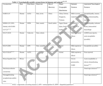
My thanks to Dr Anna S. Nordvig for giving me permission to reproduce 2 Tables from “Potential neurological manifestations of COVID-19” in Neurology Clinical Practice, an American Academy of Neurology publication.
1Wu Z, McGoogan JM. Characteristics of and Important Lessons From the Coronavirus Disease 2019 (COVID-19) Outbreak in China: Summary of a Report of 72?314 Cases From the Chinese Center for Disease Control and Prevention. JAMA. 2020;323(13):1239–1242. doi:10.1001/jama. 2020.2648
2COVID-19 Dashboard by the Center for Systems Science and Engineering (CSSE) at Johns Hopkins University (JHU)https:// coronavirus.jhu.edu/map.html
3Ministry of Health and Family Welfare Govt. of India COVID-19 INDIAhttps://www.mohfw.gov.in/
4Shunqing Xu, Yuanyuan Li — Beware of the second wave of COVID-19,The Lancet,Volume 395, Issue 10233,2020Pages 1321-1322,ISSN 0140-6736,https://doi.org/10.1016/S01406736(20)30845-X.(http://www.sciencedirect.com/science/ article/pii/S014067362030845X)
5WHO Severe Acute Respiratory Syndrome (SARS) - multicountry outbreak - Update 29Situation in China, status of scientific and clinical knowledge14 April 2003https:// www.who.int/csr/don/2003_04_14a/en/
6WHOMiddle East respiratory syndrome coronavirus (MERSCoV)https://www.who.int/emergencies/mers-cov/en/
7Andersen, K.G., Rambaut, A., Lipkin, W.I. et al. The proximal origin of SARS-CoV-2. Nat Med 26, 450–452 (2020). https:// doi.org/10.1038/s41591-020-0820-9
8Jia HP, Look DC, Shi L, et al. ACE2 receptor expression and severe acute respiratory syndrome coronavirus infection depend on differentiation of human airway epithelia. J Virol. 2005;79(23):14614-14621. doi:10.1128/JVI.79.23.1461414621.2005
9Shutoku Matsuyama, Noriyo Nagata, Kazuya Shirato, Miyuki Kawase, Makoto Takeda, Fumihiro TaguchiEfficient Activation of the Severe Acute Respiratory Syndrome Coronavirus Spike Protein by the Transmembrane Protease TMPRSS2Journal of Virology Nov 2010, 84 (24) 12658-12664; DOI: 10.1128/ JVI.01542-10
10Obadia, T., Haneef, R. & Boëlle, P. The R0 package: a toolbox to estimate reproduction numbers for epidemic outbreaks. BMC Med Inform Decis Mak 12, 147 (2012). https://doi.org/10.1186/ 1472-6947-12-147
11Channappanavar R, Perlman S. Pathogenic human coronavirus infections: causes and consequences of cytokine storm and immunopathology. Semin Immunopathol 2017; 39: 529– 539
12Channappanavar R, Fehr AR, Vijay R, et al. Dysregulated type
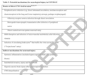
I interferon and inflammatory monocyte-macrophage responses cause lethal pneumonia in SARS-CoV-infected mice. Cell Host Microbe 2016; 19: 181– 193.
13Bryce C, Grimes Z, Pujadas E, et al. Pathophysiology of SARSCoV-2: targeting of endothelial cells renders a complex disease with thrombotic microangiopathy and aberrant immune response. The Mount Sinai COVID-19 autopsy experience. medRxiv 2020. https://doi.org/10.1101/2020.05.18.20099960.
14Zhang Y, Zhong Y, Pan L, Dong J. Treat 2019 novel coronavirus (COVID-19) with IL-6 inhibitor: Are we already that far?. Drug Discov Ther. 2020;14(2):100-102. doi:10.5582/ ddt.2020.03006
15Natoli, S., Oliveira, V., Calabresi, P., Maia, L.F. and Pisani, A. (2020), Does SARS-Cov-2 invade the brain? Translational lessons from animal models. Eur J Neurol. doi:10.1111/ ene.14277
16Li H, Xue Q, Xu X. Involvement of the Nervous System in SARS-CoV-2 Infection. Neurotoxicity Research. 2020 May 13.Li H, Xue Q, Xu X. Involvement of the Nervous System in SARS-CoV-2 Infection. Neurotoxicity Research. 2020 May 13.
17Li, Y-C, Bai, W-Z, Hashikawa, T. The neuroinvasive potential of SARS-CoV2 may play a role in the respiratory failure of COVID-19 patients. J Med Virol. 2020; 92: 552– 555. https:// doi.org/10.1002/jmv.25728
18Carrillo-Larco RM, Altez-Fernandez C, Ravaglia S and Vizcarra JA. COVID-19 and Guillain-Barre Syndrome: a systematic review of case reports Wellcome Open Res 2020, 5:107 (https://doi.org/10.12688/wellcomeopenres.15987.1)
19Poyiadji N, Shahin G, Noujaim D, et al. COVID-19-associated acute hemorrhagic necrotizing encephalopathy: CT and MRI features. Radiology 2020;201187. https://doi.org/10.1148/ radiol.2020201187:201187.
20Reichard RR, Kashani KB, Boire NA, et al. Neuropathology of COVID-19: a spectrum of vascular and acute disseminated encephalomyelitis (ADEM)-like pathology. Acta Neuropathol 2020. https://doi.org/10.1007/s00401-020-02166-2.
21Lechien JR, Chiesa-Estomba CM, De Siati DR, et al. Olfactory and gustatory dysfunctions as a clinical presentation of mildto-moderate forms of the coronavirus disease (COVID-19): a multicenter European study. Eur Arch Otorhinolaryngol 2020. https://doi.org/10.1007/s00405-020-05965-1.
22Varga Z, Flammer AJ, Steiger P, et al. Endothelial cell infection and endotheliitis in COVID-19. Lancet 2020; 395: 1417– 1418.
23Jones VG, Mills M, Suarez D, et al. COVID-19 and Kawasaki disease: novel virus and novel case. Hosp Pediatr 2020; 10: 537– 540. https://doi.org/10.1542/hpeds.2020-0123.
24COVID-19 Treatment Guidelines Panel. Coronavirus Disease 2019 (COVID-19) Treatment Guidelines. National Institutes of Health. Available at https:// www.covid19treatmentguidelines.nih.gov/.
25Shamsoddin E. A COVID-19 pandemic guideline in evidence-
Shambo Samrat Samajdar1, Santanu K Tripathi2
Development of anti-COVID 19 therapeutics is generally centered on principles of drug repurposing. Drug repurposing (also called drug repositioning, re-profiling or re-tasking) is a way to identify new indications or uses for already approved drugs1. To compress the timeline for drug development it is the only way to search drugs for managing COVID 19. It is difficult to afford time for a new drug development now in this pandemic situation as it may cost minimum 10-12 years. Starting from hydroxychloroquine, remdesvir, lopinavir-ritonavir and favipiravir; all these agents were actually approved for some other indications but now used for COVID 19 management. Glenmark pharmaceuticalafter receiving manufacturing and marketing approval from India’s drug regulator, had launched on June 20, 2020, first oral Favipiravir for the treatment of mild to moderate COVID-19 infected patients.
Favipiravir – Mechanism of Action :
Favipiravir was approved in Japan in March 2014, for treatment of new or re-emerging influenza virus infections. Favipiravir is a prodrug which is converted into T-705-ribosyl triphosphate (T-705RTP) in vivo and inhibits viral RNA polymerase selectively 2 . It is postulated that favipiravir is a broad spectrum anti RNA viral agent. The EC50 of favipiravir for COVID-19 in vitro is 61.88 µM2; whereas EC50 of remedsvir is 1.76 µM3 and hydroxychloroquine is 0.724. Larger the value of EC50 generally suggests lesser potency of the drug. This is why we need a very high dose of favipiravir for its anti-viral effects.
Evidences with Favipiravir :
One prospective, multicenter, open-label, randomized controlled trial5 was conducted in Wuhan,
1MD, DM Resident in Clinical Pharmacology, School of Tropical Medicine, Kolkata 700073 and corresponding author
2MD, DM (Clinical Pharmacology), Professor & Head, Department of Clinical & Experimental Pharmacology, School of Tropical Medicine, Kolkata 700073
Received on : 03/08/2020
Accepted on : 10/08/2020
China in February, 2020; to compare the efficacy and safety of favipiravir and umifenovir in COVID 19 pneumonia patients. In this trial clinical recovery rate at 7 days or the end of treatment was considered as primary outcome. favipiravirgroup and umifenovir group were assigned with 120 patients each. 7 day’s clinical recovery rate was 55.86% in the umifenovir group and 71.43% in thefavipiravir group (P = 0.0199). Without comorbidities COVID-19 patients and COVID-19 patients with hypertension and/or diabetes, the time of fever reduction and cough relief in favipiravir group was significantly shorter than that in umifenovir group (both P < 0.001). Regarding secondary outcome like need of auxiliary oxygen therapy or noninvasive mechanical ventilation rate, there were no statistical significant differences. Hyperuricemia, abnormal LFT reports and psychosomatic adverse effects were concerns in favipiravir arm. In this trial effect on viral load was not assessed.
Dosage :
1800 mg twice daily on Day 1 and followed by 800 mg twice daily for Day 2 to day 14.
Need to Vigilant6 :
1.There is a chance to have potential adverse drug – drug interactions with pyrazinamide, repaglinide, theophylline, famciclovir. Need to be cautious while using this drug in back ground of hyperuricemia and altered hepatic function.
2.When oral administration is extremely difficult there is a possibility to prepare the drug suspension by adding water heated to 55 ° C and deliver it to the subject via insertion of a nasogastric tube.
3.Contraindicated in pregnancy or woman who may have pregnancy as this drug is highly toxic to embryo. Screening of pregnancy is an absolute necessity before prescribing favipiravir.
4.The risk of teratogenic effect of favipiravirshould be fully explained to patients. Both the partners should be informedto use contraceptive measures duringthe administration period and for 7 days after the administration.
5.Favipiravir is secreted into semen; the risk
should be elaborately explained when administering to male patients. They should use contraceptive preferably condom during sexual intercourses while taking the medicine and 7 days after stopping favipiravir.
6.Sexual intercourse with pregnant women during taking favipiravir should be prohibited.
7.Increase of plasma level of favipiravir had been seen in patients with impaired hepatic function.
8.Psychoneurotic symptoms such as abnormal behavior after administration of favipiravirwere seen and been reported but the causal relationship is not yet established. Need to be vigilant in this regard. Influenza encephalopathy may be a differential diagnosis. But this vulnerable period requires close attention from family members, which may hamper the classic rule of isolation.
Adverse Effects of Favipiravir :
Major adverse reactions included hyperuricemia, diarrhoea, neutropenia and increase in SGOT and SGPT level. Patients on favipiravir need cautious monitoring and may require urgent de-challenge of the drug and further care if patients develop shock, anaphylaxis, pneumonia, fulminant hepatitis, hepatic dysfunction, jaundice, toxic epidermal necrolysis (TEN), Stevens-Johnson syndrome, acute kidney injury, leucopenia, neutropenia, thrombocytopenia, neurological and psychiatric symptoms (like altered consciousness, behavioral abnormality, delirium, hallucination, delusion, convulsion, etc) and hemorrhagic colitis. Adverse drug reporting form should be filled up and reported to the nearest ADR monitoring center.
Favipiravir and Umifenovir Combination :
Favipiravir and umifenovir Combination is also being tried to see the efficacy on SARS COV 2. Favipiravir prevent viral replication by inhibiting RNA dependent RNA polymerase; whereas umifenovir interacts with viral attachment. Umifenovir7 inhibits fusion between the viral envelope and the cell membrane of the target cell. Viral entry to the target cell would be prevented. So we have to wait to get the clinical trial reports to finally comment on synergistic effect of this combination against SARS COV 2.
Interim Indian Guidelines and Favipiravir : Till date ‘Clinical Management Protocol: COVID19’ by GOI version 4 (dated 27/06/2020) not includes favipiravir as a treatment option8. ‘Standard Treatment Protocol for COVID 19’by Maharashtra government revision 2 Dated-22.06.2020 includes favipiravir. Symptomatic upper respiratory tract infection mild category COVID 19 patients with comorbidity and
moderate category COVID 19 patients were advised to be administered with favipiravir 1800 mg twice daily on day 1 followed by 800 mg BD for 7 days ad if needed can be continued up to maximum 14 days9
Conclusion :
A phase 3 clinical trial is ongoing in Japan with favipiravir, a phase 2 trial is planned to be conducted in the United States and that would enroll approximately 50 patients with COVID-1910. In India, Glenmark pharmaceutical has started a phase 3 trial combining 2 antiviral agents, favipiravir and umifenovir, recently11. We have to wait for the results to have a clear knowledge on the efficacy and safety of this novel drug.
1Ashburn TT, Thor KB — Drug repositioning:identifying and developing new uses for existing drugs. Nat Rev Drug Discov 2004; 3: 673-83.
2Guidance of antiviral drug treatment for COVID-19 1st edition – (26 Feb. 2020)The Japanese Association for Infectious Diseases
3Wang M, Cao R, Zhang L, Yang X, Liu J, Xu M, Shi Z, Hu Z, Zhong W, Xiao G — Remdesivir and chloroquine effectively inhibit the recently emerged novel coronavirus (2019-nCoV) in vitro. Cell Res 2020; 30: 269-71.
4Yao X, Ye F, Zhang M, Cui C, Huang B, Niu P, Liu X, Zhao L, Dong E, Song C, etal— In Vitro Antiviral activity and projection of optimized dosing design of hydroxychloroquine for the treatment of severe acute respiratory syndrome coronavirus 2 (SARS-CoV-2). Clin. Infect. Dis. 2020
5Favipiravir versus Arbidol for COVID-19: A Randomized ClinicalTrial Chang Chen, Jianying Huang, medRxiv preprint doi: https://doi.org/10.1101/2020.03.17.20037432. The copyright holder for this preprint (which was not peerreviewed)
6Favipiravir Prescribing Information – Japan (November 2017 4th version)
7Leneva IA, Russell RJ, Boriskin YS, Hay AJ (February 2009). "Characteristics of arbidol-resistant mutants of influenza virus: implications for the mechanism of anti-influenza action of arbidol". Antiviral Research. 81 (2): 132–40. doi:10.1016/ j.antiviral.2008.10.009. PMID 19028526.
8Clinical Management Protocol: COVID-19Government of IndiaMinistry of Health and Family WelfareDirectorate General of Health Services(EMR Division)Version 4, 27.06.20
9Standard Treatment Protocol for COVID 19 Revision 2Dated22.06.2020 – Government ofMaharashtra
10Fujifilm to start phase II clinical trial of Avigan for COVID-19 patients in US. Reuters. Available at https://www.reuters.com/ article/us-health-coronavirus-fujifilm-avigan/fujifilm-to-startphase-ii-clinical-trial-of-avigan-for-covid-19-patients-in-u-sidUSKCN21R0KF. 2020 Apr 09
11Glenmark to commence new phase 3 clinical trial on combination of two anti-viral drugs favipiravir and umifenovir in hospitalized patients of moderate COVID-19 in India. PR Newswire. Available at https://www.prnewswire.com/in/ news-releases/glenmark-to-commence-new-phase-3clinical-trial-on-combination-of-two-anti-viral-drugsfavipiravir-and-umifenovir-in-hospitalized-patients-ofmoderate-covid-19-in-india-836904730.html. 2020 May 26
1.The following statements are made about the modification of Jones Criteria for Rheumatic fever, 2015. Find out which of them are true and which are false.
a.In these new criteria, parameters are decided based on risk assessment. T/F
b.The levels of ESR or temperature to define it as minor criteria are different for different risk groups. T/F
c.Subclinical carditis is not considered as an inclusion criteria. T/F
d.Polyarthralgia is a minor criterion for all risk groups. T/F
e.Subcutaneous nodule has been removed as a major criterion. T/F
2.What is the level of body temperature for it to be included as a minor criterion in Jones criteria, 2015?
a. > 38.5ºC for both low and high risk population
b. > 38ºC for both risk groups
c. > 38.5ºC for low risk population and = 38ºC for high risk population
d. > 38.5ºC for high risk population and = 38ºC for low risk population
3.The world heart federation developed a screening guideline for rheumatic fever. What is the basis for screening in this guideline?
a.ECG
b.ECG and echocardiography
c.Blood tests and Echocardiography
d.Echocardiography

4.A 27 year old patient presented with occasional fever with arthritis of knee and elbow. He presented to the emergency twice with acutely painful oral ulcers. It was thought that he has Behcet syndrome. Which of the following is an essential criterion in clinical diagnosis of this syndrome?
a.Recurrent oral ulcers
b.Recurrent genital ulcers
c.Pathergy test
d.Recurrent uveitis
5.A 36 year old man presented with acute pain in left wrist with fever. He had so severe pain that he was kept awake at night. He came to the ER with a fever of 103°F. On examination, the left wrist was swollen and difficult to touch. Aspiration of the joint revealed turbid fluid with neutrophil count: 68000/cmm. The patient also showed a macular rash on his trunk. What is the most appropriate treatment for this case?
a.i.v. vancomycin
b.joint surgery
c.i.v. ceftriaxone
d.i.v. levofloxacin
6.A 40 year old woman had high fever with arthritis of fingers in both hands. She was diagnosed to have Chikungunya fever. However, five months after her illness, she presented with persistent stiffness and pain of some of the fingers. What is the ideal treatment in such cases?
a.Physiotherapy
b.Methotrexate
c.Infliximab
d.Paracetamol
(Answer : next page)
Answer:Mediquiz
1.A : T; B : T; C : F; D : F; E : F
In the 2015 modification of the Jones criteria, there is a difference between low risk and high risk groups as far as the parameters are concerned. Low risk population is defined as: A low risk population is one in which cases of acute RF occur in = 2/100 000 school-age children or rheumatic heart disease is diagnosed in = 1/1000 patients at any age during one year (IzabelaSzczygielska et al, 2018). The old criteria are all kept, but there are a few changes. For example, while fever or raised ESR was just mentioned in the old version, now the level of body temperature, or ESR or CRP values to be included as criteria are precisely defined. Also, in the high risk group, polyarthralgia is now included as a major criterion while monoarthralgia is a minor criterion. However, in the low risk group, polyarthralgia is a minor criterion. It is now clearly mentioned that for all risk groups, clinical or subclinical carditis is a major criterion. Subclinical carditis is defined as a lesion which has no clinical findings but echocardiography with Doppler shows valvular lesions. Mitral or aortic valve lesions are considered specific for rheumatic fever.
This is the temperature level for minor criterion in Jones criteria (2015).
3.D
The WHF has developed a screening program involving only echocardiography for rheumatic fever with cardiac involvement.
4.A
According to International clinical criteria for Behcet disease, recurrent oral ulcers (at least three times per year) is an essential criteria for diagnosis.
5.C
This young male has arthritis with purulent effusion, fever and rash. So, gonococcal arthritis should be a strong possibility. In such cases, i.v. penicillin or ceftriaxone are the drugs of choice. Vancomycin does not act against gram negative organisms.
6.B
Post-viral arthritis is quite common in cases of Chikungunya infection. This can sometimes resolve spontaneously although frequently it evolves into inflammatory arthritis akin to rheumatoid arthritis. Thus, DMARDs may be used in such cases. Also, some clinicians prefer a short course of steroids.
[The Editor is not responsible for the views expressed by the correspondents]
SIR, — Non-valvular atrial fibrillation (NVAF) remains an important indication for the usage of anticoagulation therapy in the prevention of stroke. Given the better efficacy and safety, the use of novel oral anticoagulants (NOAC) is preferred over vitamin K antagonists (VKAs). This study observed the trends of usage of NOACs among the physicians in terms of the preferred NOAC and the probable reason for theirpreferences.
This study highlighted the preferred use of dabigatran compared to other NOACs among physicians. Nearly twothirds of the patients were receiving dabigatran followed by apixaban and rivaroxaban with almost equal frequency. According to the study, the major reason for the preferential use of dabigatran over other NOACs was the efficacy and safety of the drug. Various landmark studies have compared the efficacy and safety profile of NOACs with VKAs, though no trials have studied the head to head comparisons between various available NOACs. Literature shows that compared to VKAs, dabigatran and apixaban are superior while rivaroxaban is non-inferior in terms of stroke prevention in patients with NVAF. In terms of safety profile, dabigatran and rivaroxaban have a higher risk of gastrointestinal (GI) bleed while apixaban has similar GI bleeding risk compared to VKA.The risk of haemorrhagic stroke is significantly less for all NOACs when compared with VKAs.
Apart from the safety and efficacy profile, other parameters which could add on to the physician preference include the drug availability, cost, availability of its antidote, and dosing schedule. In this study, these parameters attributed less to their preferences.
The usage of the inappropriately lower dose of a NOAC will defeat the purpose of its superiority over VKA. In this study, it was found that about half of the patients on dabigatran received the inappropriately lower dose. The dosage of the NOACs should be guided by creatinine clearance (CrCl) and other clinical parameters like age, bleeding risk profile, and body weight.
NOACs have a good efficacy and safety margin in preventing stroke and thromboembolic events in patients with NVAF. The patient subset should be appropriately chosen to extend the maximum benefit to the patients. We need to consider bleeding risk profile, renal functions, age, and body weight while choosing the appropriate NOAC and its dosage.
Prof and Head of Cardiology, Gurpreet Singh Wander DAV Medical College, Ludhiana, Punjab 141001
JOURNAL OF THE INDIAN MEDICAL ASSOCIATION, VOL 118, NO 08, AUGUST 2020