Thyroid ultrasound ‌. CFD
06.05.2011 08:08
33
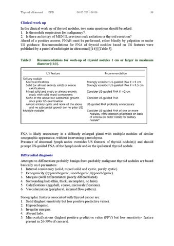
Clinical work up In the clinical work up of thyroid nodules, two main questions should be asked: 1. Is the nodule suspiscious for malignancy?. 2. Is there an history of MEN II, previous neck radiation or thyroid resection? Ahead of a positive answer, FNAB must be performed, either blindly by palpation or under US guidance. Recommendations for FNA of thyroid nodules based on US features were published by a panel of radiologist in ultrasound [(14)] [Table 5]. Table 5
Recommendations for work-up of thyroid nodules 1 cm or larger in maximum diameter [(14)].
FNA is likely unnecesary in a diffusely enlarged gland with multiple nodules of similar sonographic appearance, without intervening parenchyma. Presence of abnormal lymph nodes overrides US features of thyroid nodule(s) and should prompt US-guided FNA of the lymph node and/or the ipsilateral thyroid nodule. Differential diagnosis Attempts to differentiate probably benign from probably malignant thyroid nodules are based basically on 6 parameters: 1. Internal consistency (solid, mixed solid and cystic, purely cystic). 2. Echogenicity (hyperechogenic, isoechogenic, hypoechogenic). 3. Margins (well differentiated, poorly differentiated). 4. Sorrounding halo (thin, thick, incomplete, no halo). 5. Calcifications (eggshell, coarse, microcalcifications). 6. Vascularization (peripheral, internal flow pattern). Sonographic features associated with thyroid cancer are: 1. Solid (highest sensitivity but low positive predictive value). 2. Hypoechogenic. 3. Irregular margins. 4. Absent halo. 5. Microcalcifications (highest positive predictive value (PPV) but low sensitivity- feature present in 26-59% of cancers).