Ultrasound of scrotum
21.06.2011 12:42
34
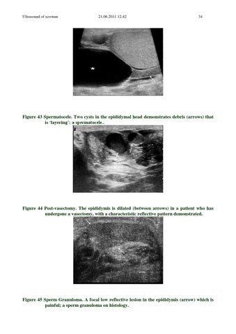
Figure 43 Spermatocele. Two cysts in the epididymal head demonstrates debris (arrows) that is ‘layering’; a spermatocele..
Figure 44 Post-vasectomy. The epididymis is dilated (between arrows) in a patient who has undergone a vasectomy, with a characteristic reflective pattern demonstrated.
Figure 45 Sperm Granuloma. A focal low reflective lesion in the epididymis (arrow) which is painful; a sperm granuloma on histology.