Ultrasound of scrotum
21.06.2011 12:42
24
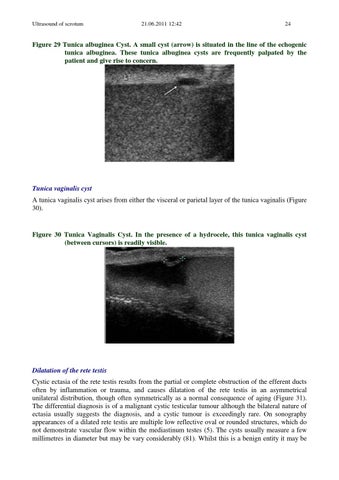
Figure 29 Tunica albuginea Cyst. A small cyst (arrow) is situated in the line of the echogenic tunica albuginea. These tunica albuginea cysts are frequently palpated by the patient and give rise to concern.
Tunica vaginalis cyst A tunica vaginalis cyst arises from either the visceral or parietal layer of the tunica vaginalis (Figure 30).
Figure 30 Tunica Vaginalis Cyst. In the presence of a hydrocele, this tunica vaginalis cyst (between cursors) is readily visible.
Dilatation of the rete testis Cystic ectasia of the rete testis results from the partial or complete obstruction of the efferent ducts often by inflammation or trauma, and causes dilatation of the rete testis in an asymmetrical unilateral distribution, though often symmetrically as a normal consequence of aging (Figure 31). The differential diagnosis is of a malignant cystic testicular tumour although the bilateral nature of ectasia usually suggests the diagnosis, and a cystic tumour is exceedingly rare. On sonography appearances of a dilated rete testis are multiple low reflective oval or rounded structures, which do not demonstrate vascular flow within the mediastinum testes (5). The cysts usually measure a few millimetres in diameter but may be vary considerably (81). Whilst this is a benign entity it may be