Doppler ultrasound of the aorta, inferior vena cava and visceral arteries c
d
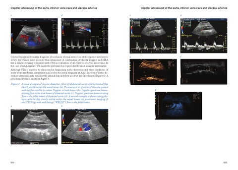
Colour Doppler may enable diagnosis of occlusion of renal arteries or of the superior mesenteric artery, but CTA is more accurate than ultrasound. A combination of duplex Doppler and MRA has a similar accuracy compared with CTA in evaluation of all features of aortic aneurysms. In the case of AAA rupture, CT should be performed as it provides the most accurate assessment. Although CTA is superior to ultrasound in diagnosing aortic dissection and other conditions of acute aortic syndrome, ultrasound may lead to the initial suspicion of AAS. In cases of aortic dissection ultrasound may visualise the intimal flap and flow in a true and false lumen (Figure 4). A pseudoaneurysm is shown in Figure 5. Figure 4 B-mode example of chronic dissection (Dis) of abdominal aorta with the intimal flap clearly visible within the vessel lumen (a). Transverse scan of aorta of the same patient with the flow visible by colour Doppler in both lumina (b). Doppler spectrum demonstrating flow in the true lumen of dissected aorta (c). Doppler spectrum demonstrating flow in the false lumen of dissected aorta (d). A second example is shown using photopic with the flap clearly visible within the vessel lumen (e), panoramic imaging (f) and CEUS (g) with undulating (“WELLE�) flow in the false lumen. a b
664
Doppler ultrasound of the aorta, inferior vena cava and visceral arteries c
d
e
f
g
665