3 minute read
Semi-direct and indirect technique: indications and limitations

from EstrattoVeneziani_EN
by Grupo Asís

20 21
Advertisement

22 23
24 25 26
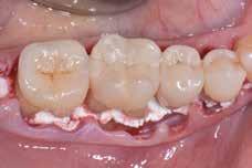
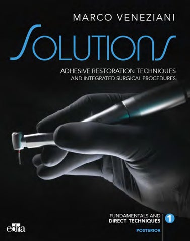
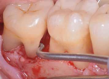
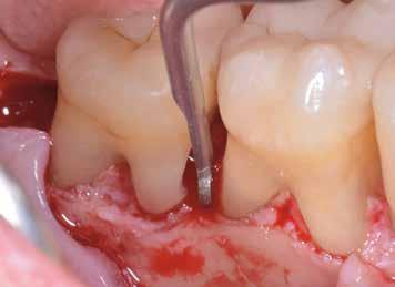
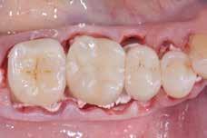
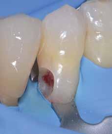
Figures 20-22 Bone recontouring and cavity preparation procedures are performed on the M aspect of the M root of 47 – with similar characteristics to those described above – with the aid of sonic diamond inserts.
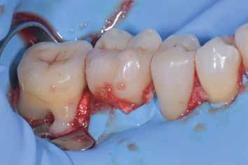
Figure 23 Image of the quadrant after elevating the flaps, osseous-resective surgery, preparation of the external resorption lesions D44, M45, M47. Tooth 46 shows no lesions. The adequate vasoconstriction induced by the anaesthetic with 1:100000 adrenaline, the accuracy of execution, the correct incision of the flaps, the effect of the cut produced by the sonic instruments used for resective surgery made it possible to obtain a clean surgical field with low bleeding, prerequisite to be able to perform intraoperative field isolation.
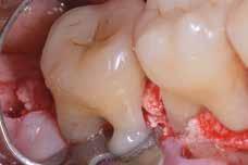
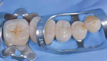
Figures 24-26 One of the most demanding stages of the procedures consists in the isolation of the intraoperative field. According to the method proposed by the Author, a cellulose-based paste isolating material is used (OraSeal Caulking, Ultradent), applied directly on the osseous tissue below the elevated flaps. A premolar clamp is then used for stabilising (Ivory no.1; Fig. 26) on root M of 47 with self-adhesive flowable composite.
27
28 29

30 31 32
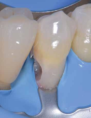
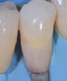
Figures 27-29 The rubber dam is then placed from the second molar to the canine, taking care to make smaller and more distant holes. The “dirty” appearance in the placement stage gives way to a well-isolated and clean field, after thorough washing, which is the essential prerequisite to be able to perform intraoperative adhesive restorations. A supplementary type 212 SA clamp allows the dam to be retracted and the cavity to be adequately exposed at the level of the premolars in the crucial area, i.e. the cervical margin (Fig. 29). Figures 30-32 Pulp exposure, in the absence of a carious lesion, leads to direct capping with light-cured MTA (TheraCal, Bisco) which allows the procedures to be performed with self-etching type adhesive followed by freehand layered composite restoration before the final suture.
Clinical case 37
INVASIVE EXTERNAL CERVICAL RESORPTION IN VITAL TOOTH Solution: ROOT RESECTION AND MAINTENANCE OF VITALITY WITH INTRA-OPERATIVE RESTORATION IN BIOCERAMIC MATERIAL AND COMPOSITE
INVASIVE CERVICAL RESORPTION (ICR) (see remarks on the previous case) A young, 23-year-old male patient, referred by a colleague, diagnosed with massive external resorption of tooth 27.
1 2
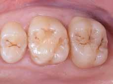
Figure 1 The clinical examination of quadrant 2, and in particular of 27, shows an apparently normal situation with good trophism of the tissues without inflammation and preserved clinical crown.
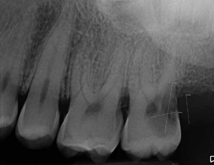
Figure 2 Intraoral radiography, however, shows a very large radiolucent lesion with coronal-apical extension invading the DV root and the supracrestal attachment complex. The lesion is also assumed to invade the pulp tissue, but the tooth is wholly asymptomatic and responds to vitality tests. This is an idiopathic type of invasive external cervical resorption. In light of the extent of the lesion, the tooth appears non-restorable but, given the patient’s young age, his high motivation, the good clinical situation of the teeth on both arches and the absence of spontaneous painful symptoms, a one-stage restorative/surgical approach is opted for. The patient signs the informed consent concerning an unpredictable procedure not based on scientific evidence.