Autopsy and Case Reports 2011; 1(4): 29-37
The macroscopic appearance of the lungs and adjacent lymph nodes was also remarkable. The right lung weighed 410 g (mean reference value, 450 g; range, 360-570 g), and the left lung weighed 390 g (mean reference value, 375 g; range, 325-480 g). We also found small, citrine, bilateral pleural effusion. Paratracheal lymph node enlargement was found in the carinal and peribronchial regions (the lymph nodes in the former measuring up to 5.0 cm), and there were nodules, measuring up to 1,5 cm, in the adjacent connective tissue surrounding the main and segmental bronchi, although only superficially in the peribronchial lung tissue (Figures 4A and 4B). There was no diffuse distribution of nodules in the lung parenchyma. Macroscopic examination revealed that the lesions were dark and, for the most part, firm, some being hard (Figures 4C and 4D). Microscopic examination revealed multiple, sometimes coalescent, oval-shaped nodules, some of which had spiculated margins; the nodules were characterized by severe fibrosis exhibiting thick collagen bands and numerous black-pigmented macrophages (Figures 5 and 6A).
Takayasu V, Lima FR, Campos FPF.
Some areas showed amorphous necrotic material and cholesterol crystal clefts, accompanied by foreign body giant cell reaction and groups of xanthomatous macrophages (Figure 6B). The Ziehl-Neelsen method and the GrocottGomori methenamine-silver stain technique were consecutively used in order to screen for acid-fast bacilli and fungi, and the results were negative. Some foci of osseous metaplasia were seen. Under polarized light, we found no birefringent particles in the lesions. The findings of abundant black pigment, severe fibrosis, and spiculated nodules were suggestive of mixed-dust pneumoconiosis.2 Many hemossiderine-laden macrophages (“heart-failure” cells) were found in intra-alveolar spaces. This finding is probably related to previous episodes of pulmonary edema in a pacient with leftsided heart failure. Adicionally, there were inhaled carbon pigment engulfed by alveolar and intersticial macrophages (pulmonary anthracosis).
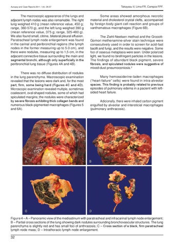
Figure 4 – A – Panoramic view of the mediastinum with paratracheal and infracarinal lymph node enlargement; B – Partial cross sections of the lung showing dark nodules surrounding bronchovascular structures. The lung parenchyma is slightly red and has small foci of anthracosis; C – Cross section of a black, firm paratracheal lymph node mass; D – Intrathoracic lymph node enlargement.
32