4 minute read
Clinical Case Review: Foot Rot in Beef Cattle


By: Lisa Taylor, Jose Valles and Dr. Tom Noffsinger, Production Animal Consultation and thank you to Dr. Dörte Döpfer, MSc, PhD, University of Wisconsin - Madison for your assistance with this clinical case review.
Foot rot, also known as interdigital necrobacilosis or interdigital phlegmon, is an infectious disease affecting the interdigital region of the bovine foot. Foot rot is a prominent cause of lameness in beef cattle which is associated with substantial economic losses for producers due to performance loss and treatment costs.
Foot rot cases are primarily caused by a synergism between Dichelobacter nodosus and Fusobacterium necrophorum. D. nodosus enters and resides in the epithelium when the interdigital skin is damaged by maceration or microtrauma. Protein break-down by D. nodosus facilitates growth and entry of F. necrophorum. In addition, increased inflammatory response and reduced leukocyte function induced by F. necrophorum lead to inflammatory reactions attracting purulent Trueperella pyogenes to the scene when toxins result in pus formation and tissue destruction. Other bacteria species, such as Bacteroides melaninogenicus, Escherichia coli, and Staphylococcus aureus may also be involved. Super foot rot, a particularly acute and rapidly progressing condition, is thought to involve an antibiotic-resistant strain of F. necrophorum.
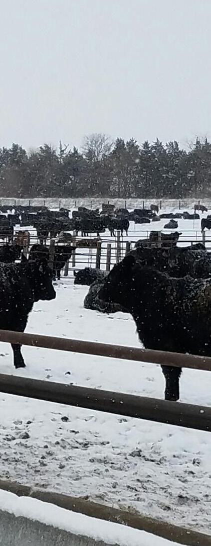
Foot rot outbreaks are sporadic and can affect anywhere from a few animals to more than 25% of the herd. Cattle of all ages are affected, and cases are seen year round and worldwide. Cattle are more susceptible to foot rot infection in hot, humid environments or when housed on high-moisture surfaces, such as pens with high accumulations of manure, urine, or mud.
Foot rot usually affects only one limb. Infected cattle will commonly present sudden lameness in the affected limb. Because foot rot is painful, the animal will become hesitant to move. Other symptoms include decreased appetite and possible fever. In severe cases, animals may go off feed completely.
Simple locomotion observation is not adequate to accurately differentiate foot rot from other lameness diagnoses. The animal must be properly restrained so the affected foot can be lifted and thoroughly examined.
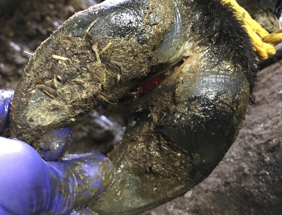
First the foot should be properly washed or wiped clean up to the declaw area. Then the limb and interdigital region should be examined for pain, swelling, redness, lesions, necrotic tissue, and odor. Foot rot cases will present symmetrical swelling throughout the foot.
Three stages of foot rot are differentiated. In stage 1, sud- den onset of swelling and lameness are evident. The skin in the interdigital space may appear red and the inflammation may start to separate the claws (figure 1). Within 72 hours after onset of first signs, the interdigital skin ruptures (stage 2; figures 2, 3). This lesion can extend from the front to the back of the foot and is characterized by protrusion of necrotic tissue and a distinctive foul odor (figure 4). Stage 3 is characterized by formation of additional tissue in the interdigital space, leading to closure of the interdigital lesion. This can result in chronic sequelae such as corns, axial wall fissures and abnormal horn production. In chronic or severe cases, swelling may progress up the leg and infection can penetrate deeper, affecting bones, connective tissue, joints, and tendons.


Early detection of foot rot is key for improved treatment response. Mild cases may be treated topically, but most cases will require systemic antibiotic treatment. Several antibiotics can be used, including oxytetracycline, penicillin, ceftiofur, florfenicol, tulathromycin, and sulfonamides. NSAIDs may also be administered for pain relief.
Affected cattle should be isolated and closely monitored; if recovery does not progress within a few days, additional treatment may be required. In severe cases, surgical treatment or railing the animal may be considered. A veterinarian should be consulted to determine the best treatment protocol given the disease severity, operation type, and withdrawal time.
Preventative measures are essential in avoiding and managing outbreaks of foot rot. Proper ration formulation and consistent bunk management reduces foot rot risk. It is vitally important to keep the surfaces cattle stand on as dry and free of debris as possible as moist pen conditions increase risk of foot rot infections.
Pen surfaces should be scraped regularly to avoid accumulation of urine, feces, mud, and other debris, particularly around bunks and water tanks. Proper drainage and sprinkler usage will help in reducing mud and high moisture areas. Pen surfaces should also be kept free of gravel, rocky soil, and frozen or dried mud and manure, which can cause trauma to the interdigital space of the foot. Flooring surfaces in processing, loading, and unloading areas should be inspected frequently for rough surfaces that may cause disruption of the interdigital skin.
Supplementation of zinc, organic iodide, vitamin A, and vitamin D may aid in prevention of foot rot and other lameness issues. Some operations have utilized copper sulfate or formaldehyde footbaths at the time of unloading, initial processing, or re-implant as a preventative measure. While these footbaths can reduce foot rot incidence, it is important to understand the associated costs and environmental issues. Both commercial and autogenous bacterin vaccines are available for foot rot, but the cost effectiveness of such vaccines has not been clearly demonstrated. General management and biosecurity programs focused on improved health should also be implemented.
References
1. AABP Lameness Committee. 2016. Interdigital Phlegmon (Foot Rot). American Association of Bovine Practitioners. Accessed October 31, 2016, from http://www. aabp.org/Members/resources/AABP%20Footrot.pdf.
2. Currin, J.F., W.D. Whittier, and N. Currin. 2009. Foot Rot in Beef Cattle. Virginia Cooperative Extension Publication 400-310. Accessed October 27, 2016, from https:// pubs.ext.vt.edu/400/400-310/400-310.html.
3. Mülling, C., D. Döpfer, T. Edwards, C. Larson, D. Tomlinson, and M. Branine. 2014. Cattle Lameness: Identification, Prevention and Control of Claw Lesions, First Edition, Zinpro Corporation, p. 52-55.
4. Dewell, G. and J. Shearer. 2009. Foot Rot in Beef Cattle. Iowa State University Extension PM 1728. Accessed October 27, 2016, from https://store.extension.iastate. edu/Product/Foot-Rot-in-Beef-Cattle-PDF.
5. Greenough, P.R. 2015. Interdigital Phlegmon (Footrot, Foul in the Foot) in Cattle, The Merck Veterinary Manual. Accessed October 27, 2016, from http://www.merckvetmanual.com/mvm/musculoskeletal_system/lameness_in_ cattle/interdigital_phlegmon_footrot_foul_in_the_foot_ in_cattle.html.
6. Step, D.L., B. Whitworth, E.J. Giedt, and D. Lalman. Foot Rot in Cattle. Oklahoma Cooperative Extension Service ANSI-3355. Accessed October 27, 2016, from http:// pods.dasnr.okstate.edu/docushare/dsweb/Get/Document-2023/ANSI-3355web.pdf.
7. Zoetis. Bovine Footrot. Accessed October 27, 2016, from https://www.zoetisus.com/conditions/beef/bovine-footrot.aspx.


