Required Practical
1.4
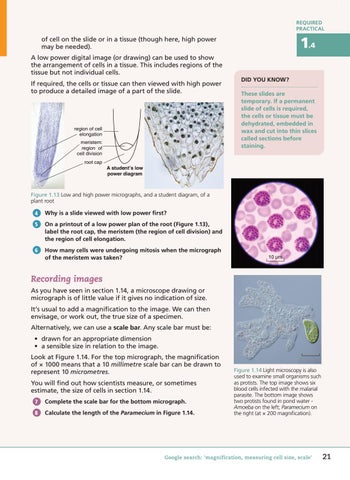
of cell on the slide or in a tissue (though here, high power may be needed). A low power digital image (or drawing) can be used to show the arrangement of cells in a tissue. This includes regions of the tissue but not individual cells. If required, the cells or tissue can then viewed with high power to produce a detailed image of a part of the slide.
region of cell elongation meristem: region of cell division root cap
Did you know? These slides are temporary. If a permanent slide of cells is required, the cells or tissue must be dehydrated, embedded in wax and cut into thin slices called sections before staining.
A student’s low power diagram
Figure 1.13 Low and high power micrographs, and a student diagram, of a plant root 4
Why is a slide viewed with low power first?
5
On a printout of a low power plan of the root (Figure 1.13), label the root cap, the meristem (the region of cell division) and the region of cell elongation.
6
How many cells were undergoing mitosis when the micrograph of the meristem was taken?
10 µm
Recording images As you have seen in section 1.14, a microscope drawing or micrograph is of little value if it gives no indication of size. It’s usual to add a magnification to the image. We can then envisage, or work out, the true size of a specimen. Alternatively, we can use a scale bar. Any scale bar must be: • drawn for an appropriate dimension • a sensible size in relation to the image. Look at Figure 1.14. For the top micrograph, the magnification of × 1000 means that a 10 millimetre scale bar can be drawn to represent 10 micrometres. You will find out how scientists measure, or sometimes estimate, the size of cells in section 1.14. 7
Complete the scale bar for the bottom micrograph.
8
Calculate the length of the Paramecium in Figure 1.14.
Figure 1.14 Light microscopy is also used to examine small organisms such as protists. The top image shows six blood cells infected with the malarial parasite. The bottom image shows two protists found in pond water Amoeba on the left; Paramecium on the right (at × 200 magnification).
Google search: 'magnification, measuring cell size, scale'
75047_P012_047.indd 21
21
5/20/16 10:07 AM
