Clinical Features, Debates & Research / Débats, recherche et articles cliniques (continued from page 22)
Epidemiology and Prevalence KD occurs most commonly in men, young people and manual labourers3,4. The reported prevalence is between 1.2 and 2.5%5-7.
present with boggy synovitis of the radiocarpal joint. Range of motion is usually decreased and grip strength is commonly reduced. Imaging Radiographs, bone scintigraphy, magnetic resonance imaging (MRI), and computed tomography (CT) are useful for assessment.
Etiology and Risk Factors The etiology of KD remains unclear. Mechanical, anatomic and systemic mechanisms have all been implicated.
Figure 3
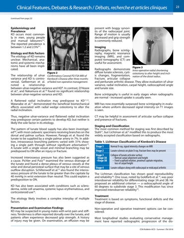
Figure 2
The relationship of ulnar Confirmatory Coronal PD FSA MRI of variance and KD is contro- Kienbock’s Disease after three months versial. Gelberman et al. of failed non-operative management. noted a relationship between ulnar-negative variance and KD8. In contrast, D’Hoore et al.9, and Nakamura et al.10 found no significant relationship between ulnar-negative variance and KD. A flattened radial inclination may predispose to KD11,12. Watanabe et al.12 demonstrated the beneficial biomechanical effects associated with radial wedge osteotomy to alter the radial inclination. Thus, negative ulnar-variance and flattened radial inclination may predispose certain patients to develop KD, but neither is likely the sole factor in its etiology. The pattern of lunate blood supply has also been investigated4,8, with most cadaveric specimens receiving branches on the dorsal and palmar surface. However, Panagis et al. found the lunate to be supplied by a single palmar artery in 7%. In addition, intraosseous branching patterns varied with 31% showing a single path through without significant arborisation13. A lunate with a single vessel and minimal branching may be predisposed to ON after an injury or fracture. Increased interosseous pressure has also been suggested as a cause. Pichler and Putz14 examined the venous drainage of the lunate and found a dense plexus of venous vessels at the volar and dorsal periosteal surface that may be a site of venous outflow disruption. Schiltenwolf et al.15 also found the interosseous pressure of the lunate to be greater than the capitate by 40 mmHg in wrist extension than neutral. This could explain a predisposition to ON. KD has also been associated with conditions such as scleroderma, sickle cell anaemia, systemic lupus erythematosus, and corticosteroid use16. The etiology likely involves a complex interplay of multiple factors. Presentation and Examination Findings KD may be suspected in young males with wrist pain and stiffness. Tenderness is often reported dorsally over the lunate, and patients often experience decreased grip strength. A history of injury may be given. On examination, an effusion may be
Radiographs demonstrate Intra-operative radial shortening diffuse lunate sclerosis, cyst- osteotomy to alter height and incliic changes, fragmentation, nation of the distal radius. fracture, articular collapse and perilunate arthritic disease. They allow evaluation of ulnar variance, radial inclination, carpal height, radioscaphoid angle and lunate size. Bone scintigraphy is useful in early stages when radiographs are normal - increased uptake is usually seen. MRI has now essentially surpassed bone scintigraphy in evaluation where uniform decreased signal intensity on T1 images is seen. CT may be helpful in assessment of articular surface collapse and presence of fractures. Staging and Classification The most common method for staging was first described by Stahl17, but Lichtman et al.4 modified this to produce the most widely accepted classification based on radiographs. Table 1. Lichtman Classification of Kienbock’s Disease Stage 1 Stage 2 Stage 3 •Stage 3A •Stage 3B Stage 4
Normal X-ray, signal intensity changes on MRI Lunate sclerosis on plain X-ray, fracture lines may be present Collapse of lunate articular surface • Normal carpal alignment and height • Fixed scaphoid rotation, proximal capitate migration, loss of carpal height Lunate collapse along with radiocarpal or midcarpal arthritis
The Lichtman classification has shown good reproducibility and reliability18. One issue, noted by Goldfarb et al.19, was poor interobserver reliability for differentiating stage 3A and 3B. He proposed an additional criterion — a radioscaphoid angle of 60 degrees to subdivide stage 3. This modification has since improved interobserver reliability19. Treatment Treatment is based on symptoms, functional deficits and the stage of disease. Non-operative and operative treatment options can be considered. Most longitudinal studies evaluating conservative management have reported radiographic progression of the disCOA Bulletin ACO - Summer / Été 2018
23