The Women’s Center is open Monday – Friday 8 am – 4:30 pm. Evening hours are also available.
Convenient parking is located close to the facility.
Most major health insurance plans are accepted.

The Women’s Center is open Monday – Friday 8 am – 4:30 pm. Evening hours are also available.
Convenient parking is located close to the facility.
Most major health insurance plans are accepted.

The Women’s Health Center at St. Francis Hospital & Heart Center is a state-of-the-art imaging center committed to women’s health and education.
The radiologists at the center are each board certified through the American College of Radiology. Our dedicated team of physicians, nurses and technologists are experts in the latest techniques that provide the earliest diagnosis of many women’s health issues. We have recently completed an expansion and renovation and are pleased to offer the latest technology available, along with a high level of comfort, convenience and patient-focused care.
Diagnostic and interventional modalities:
• Pre-operative studies including SCOUT®, Hologic® LOCalizer™ and Magseed® localizations prior to surgical excision.
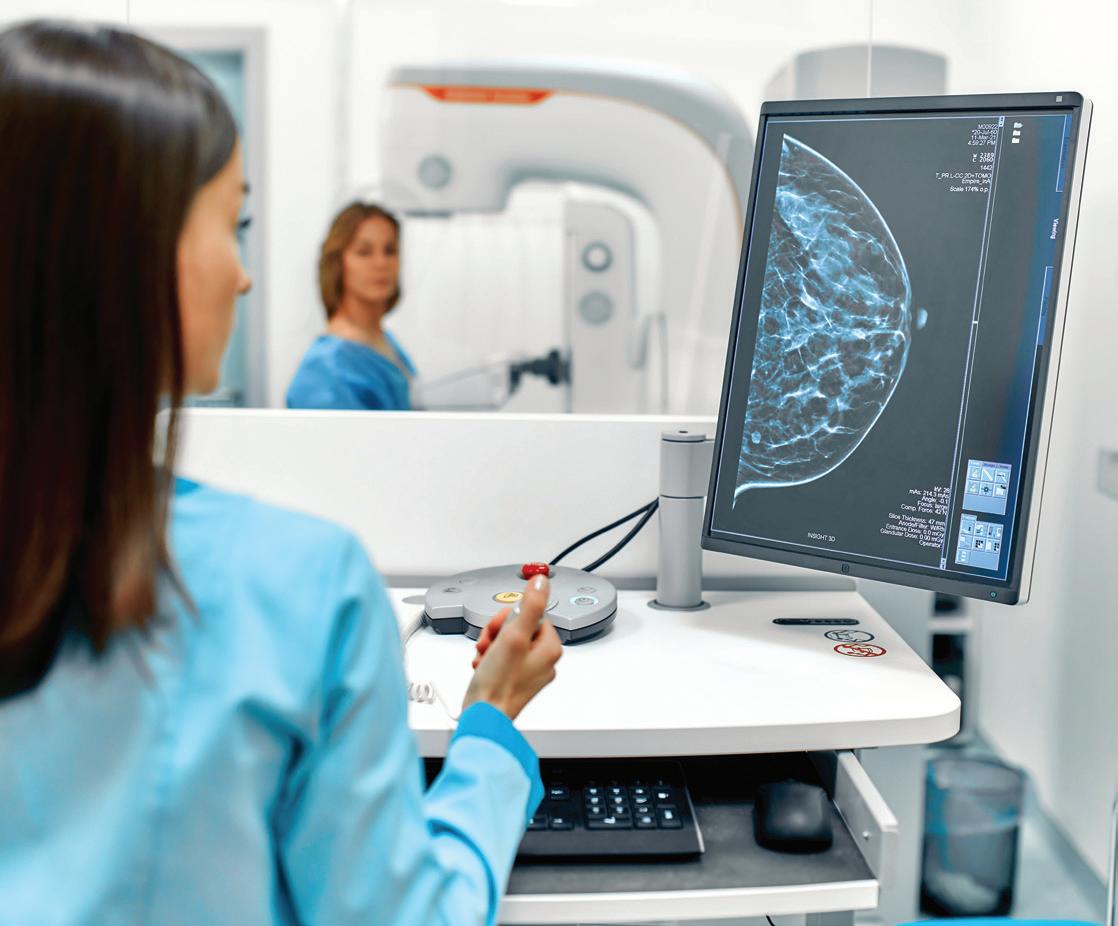
• Digital breast tomosynthesis (3D mammography) with computer-aided detection (CAD).
• Stereotactic core biopsy.
• Breast MRI and MRI-guided breast biopsy.
• Ultrasound including breast, pelvic and sonohysterography (saline hysterograms).
• Ultrasound core biopsy.
• Bone densitometry.
• Whole-body composition.


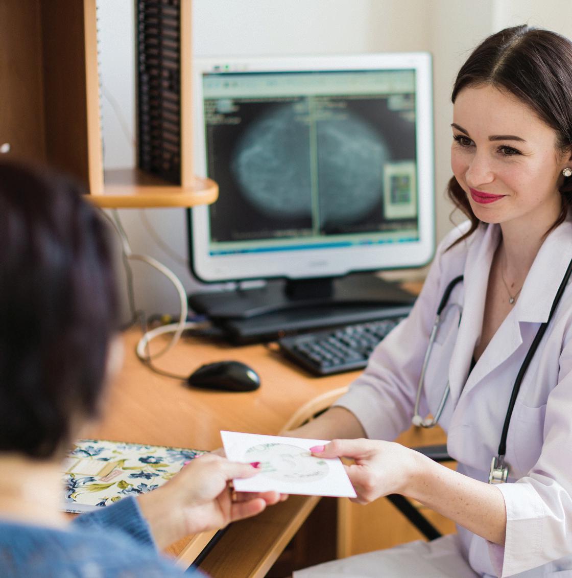
At the Women’s Health Center, we are committed to treating each patient with dignity and respect. We understand that patients may experience fear or anxiety when undergoing a medical examination. That is why we provide same-day results for mammography and breast ultrasounds. If an abnormality is found, our physicians will discuss a number of options for further analysis, including stereotactic or ultrasound guided needle core biopsy, or MRI evaluation.
The Women’s Center at St. Francis Hospital & Heart Center is a Breast Imaging Center of Excellence, designated by the American College of Radiology and accredited in digital breast tomosynthesis (3D mammography), stereotactic breast biopsy with Affirm, breast ultrasound, ultrasound guided breast biopsy and gynecological ultrasound services.

As a Pink Ribbon Facility, a distinction awarded to providers utilizing the most advanced mammography technologies, skilled clinicians are compliant with the latest screening recommendations. The Center is also a SCOUT® Wire-Free provider for demonstrating excellence in breast tumor localization.
Localization prior to surgical procedure: SCOUT
We evaluate and diagnose several women’s health issues:
• Breast cancer
• Lump/mass in breast
• Mastitis
• Fibrocystic changes
• Carcinoma in situ
• Mammographic microcalcifications
• Malignant neoplasm of nipple and areola
• Implant leakage
• Malignant neoplasm of male breast and gynecomastia
• Osteoporosis
• Osteopenia
• Osteopenia
Localization is a procedure which involves placement of a reflector, a tiny device about the size of a grain of rice into the breast prior to surgery. The system then uses safe, nonradioactive radar waves to detect the reflector’s location within the breast. The reflector is then activated in the operating room when your surgeon uses the SCOUT system to locate and remove both the tumor and the reflector.
Biopsy: If your mammogram or breast ultrasound has revealed an abnormality, your Women’s Center physician may recommend an ultrasound, stereotactic, or MRIguided breast biopsy.
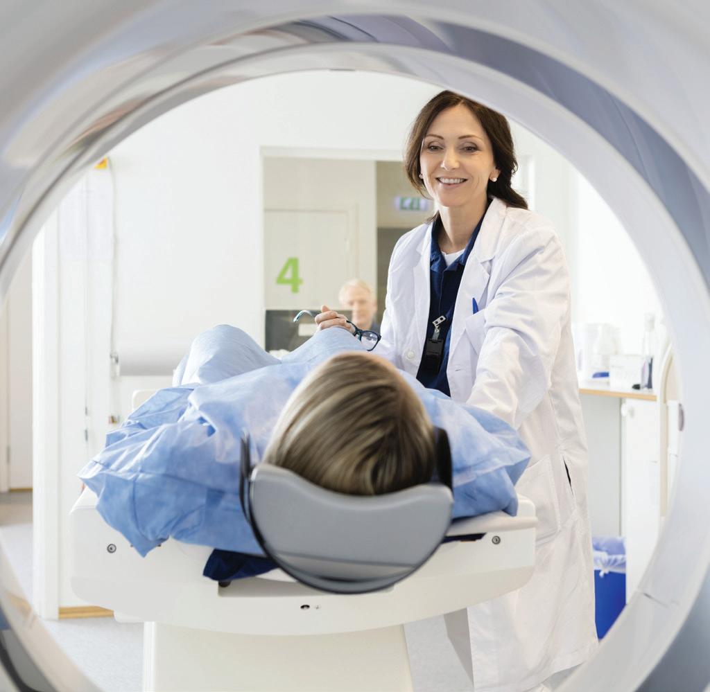
Breast MRI: Breast magnetic resonance imaging (MRI) is a sensitive and painless technique for diagnosing early breast cancer. It also helps determine the extent of the cancer in patients who have been diagnosed with breast cancer, or those with a strong family history of breast carcinoma. Breast MRI is also used in the evaluation of possible breast implant rupture.
MRI-guided breast biopsy: Magnetic resonance imaging biopsy is a highly accurate way to evaluate suspicious tissue in areas of the breast which are sometimes only seen on the MRI images.
Digital Breast Tomosynthesis (3D-Mammography) with Computer-Aided Detection (CAD): This procedure uses low doses of X-rays to look at the breast tissue and detects cancers that may be too small for you or your doctor to feel. This technology affords sharp images with thin sections through the breast through computer generated imaging. Starting at age 40 (or earlier if you have a family history of breast cancer) you should have a mammogram every year. Early detection is the best defense.
Stereotactic Breast Biopsy: Also called X-ray-guided breast biopsy, this procedure permits precise threedimensional positioning of a needle to obtain tissue samples. This is based on an area of concern previously noted on the mammogram.
Ultrasound (Breast/Pelvis/Sonohysterography):
Ultrasonography is a non-invasive imaging technique that uses high-frequency sound waves for viewing the breast or pelvis. Ultrasound can distinguish a cyst from a solid lesion, which may rule out the need for a biopsy. Ultrasonography can also evaluate pelvic pain, abnormal vaginal bleeding, fibroids, and ovarian cysts or masses.
Ultrasound breast biopsy: This is a highly accurate way to evaluate palpable and non-palpable suspicious masses that are visible on ultrasound and then with ultrasound guidance the radiologist can biopsy the area of concern.
Bone density testing: A bone densitometry scan is a simple, painless examination that detects osteoporosis. When a woman goes through menopause, reduced hormone levels increase the risk of osteoporosis, a disease which leads to bone fragility and weakness. If left untreated, osteoporosis can progress painlessly until a bone breaks. These fractures occur typically in the hip, spine and wrist.