13 minute read
What is the Prevalence of Temporomandibular Joint (TMJ) Condylar Osteoarthrosis in an Asymptomatic Oral and Maxillofacial Surgery Department Population?


What is the Prevalence of Temporomandibular Joint (TMJ) Condylar Osteoarthrosis in an Asymptomatic Oral and Maxillofacial Surgery Department Population?
John Vorrasi, D.D.S.; Laura Mendomo Mvomo, B.S.
ABSTRACT
The aim of the present study is to determine the prevalence and demographics of temporomandibular joint (TMJ) condylar changes in the asymptomatic general population of a hospital-based oral and maxillofacial surgery (OMFS) practice. We retrospectively reviewed cone-beam computed tomography (CBCT) scans of 272 TMJs from 136 consecutive patients to detect osteophyte, subchondral cyst, sclerosis, cortical erosions and flattening. While gender did not appear to play a significant role in TMJ pathology, age was positively correlated with prevalence of condylar changes. Patients 78 years and older were significantly more likely to have osteophyte, cyst, sclerosis and breakdown than were their younger counterparts. Flattening was the most common osseous change in our patient cohort.
Temporomandibular joint disorders (TMD) are common musculoskeletal disease processes that affect roughly 5% of the population, according to the National Health Survey.[1] Up to 12% of individuals report at least one clinical symptom of TMD.[2] The development of these clinical signs is multifactorial, and has been associated with hormonal influence.[3,4] It has been suggested that upregulation of estrogen receptors may be associated with TMJ degeneration, though nothing has definitely been proven. Also, there is no evidence osseous changes occur more frequently in females compared to male counterparts.
Signs and symptoms of TMDs can vary, ranging from musculature disorders and pain to osteoarthritis. Literature supports that anatomic variations can lead to dysfunction, but there is little evidence that the most common age for TMD correlates to the time point of the most notable osseous changes. Osseous changes in bone architecture may accompany symptoms. In theory, more bone changes, degeneration or breakdown should be reflected in a similar age and gender category as the most common symptoms.
TMJ osteoarthritis may be present in up to 40% of older adults, but may only be detectable on roughly 10% of plain radiographs or panoramic films.[5] Previous investigations have highlighted osseous changes in patients with symptomatic TMD using in-office cone-beam computed tomography (CBCT).[6] The evolution and accessibility of CBCT technology in the past 10 to15 years have met or surpassed medical-grade CT imaging and can accurately display TMJ changes.
The primary aim of this study was to identify the prevalence of TMJ condylar changes in asymptomatic consecutive patients in an oral and maxillofacial population and examine how this correlates to the demographics for TMD. To date, this is the only study reviewing CBCT of asymptomatic patients for TMJ bony changes to approximate prevalence of changes in a population.
Methods
This retrospective cohort study was approved by the University of Rochester Research Subjects Review Board. We reviewed the Sirona CBCT scans (Dentsply, North Carolina) of 272 TMJs from 136 patients at the University of Rochester Department of OMFS. Clinic visits spanned from August 2016 to January 2017. We included patients between 18 and 95 years old, and excluded any patient who previously had a TMJ or orthognathic surgery, any craniofacial anomalies, TMJ or condylar trauma, any autoimmune, systemic, metabolic or infectious process involving the TMJ. The scans were anonymized, then stratified by gender (male, female) and by age (18-37 years old; 38-57 years old; 58-77 years old; 78-99 years old).
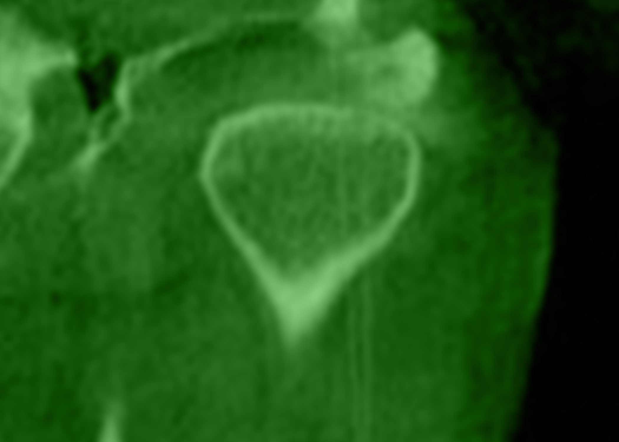
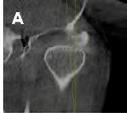
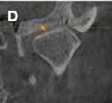
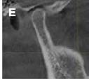
We screened CBCT imaging for TMJ osteophyte, subchondral cyst, sclerosis, surface erosion and flattening, as outlined by Cömert Kiliç et al., using Sidexis software (Dentsply, North Carolina) (Figure 1).[7] Presence of changes was labelled as “1.” Absence was “0.” Severity was not graded, rather just as presence or absence. All TMJs were reviewed by a single source (LM). Data were then analyzed using PRISM linear regression software (GraphPad, California). All comparisons in this study were analyzed by unpaired t-tests for prevalence of TMDs for age considerations over the different age groups and Anova for subgroup analysis. A p value of 95% or .05 was considered significant, as the observations would not be due to chance and correlated. The statistical measurements were used to validate correlations between CBCT findings and several categories, including age and gender. Areas of significance where p values are <0.05 are denoted in Figure 2. This is considered statistically significant.


Results
Of the 272 TMJs reviewed, the most prevalent TMJ change observed was flattening (141 TMJs, 52%), followed by surface erosion (60 TMJs, 22%), osteophyte (19 TMJs, 7%), sclerosis (9 TMJs, 3%) and subcortical cysts (2 TMJs, 1%) (Figure 1).
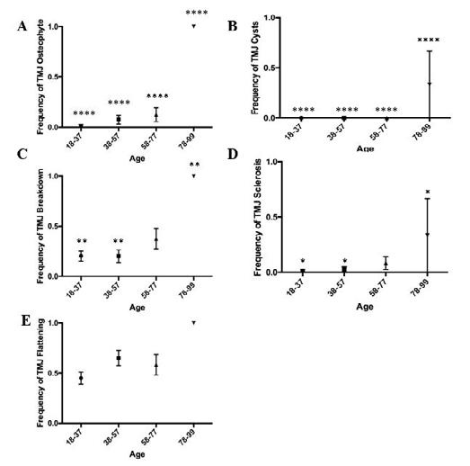
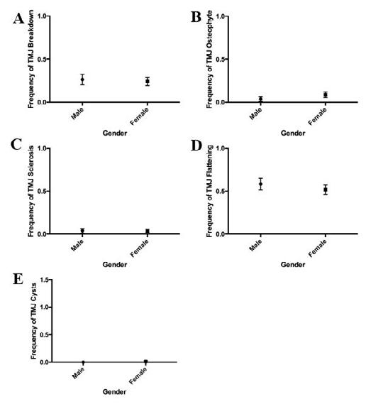
While age was not associated with TMJ flattening (p=0.07), older patients had a higher incidence of osteophyte (p<0.001), subcortical cysts (p< 0.001), surface erosions (p<0.01) and sclerosis (p<0.05) than did younger patients (Figure 2). These findings are statistically significant and are not due to chance. This difference was most significant between patients above 78 years of age and patients younger than 57 years old. Of the patients over 78, all exhibited osteophyte and joint breakdown, whereas only 10% of patients under 57 years of age exhibited osteophyte formation, and about 30% exhibited surface erosion. Approximately 30% of patients over 78 years of age had TMJ cysts and sclerosis, while nearly no patient under 57 exhibited these conditions. Gender was not associated with TMJ flattening, sclerosis, erosions, osteophyte or cysts (p=0.42, p=0.99, p=0.73, p=0.23, p=0.32) (Figure 3). Range distribution of condylar changes and demographics are also presented graphically.
Discussion
Although osseous changes do not correlate with symptomatic degenerative joint disease, TMDs have been shown to incorporate articular disease processes. Our retrospective study shows that condylar changes—breakdown, osteophyte, cysts and sclerosis—are more prevalent in older adults than they are in younger adults, but do not seem to have a gender predilection. Figure 2 reveals that frequency of bony changes in the condyle increases with age, and the most common finding is condylar flattening. Interestingly, there is no correlation between gender and condylar changes at all age groupings. Figure 3 is a graphical representation of gender comparison at all age groups. Since there was no statistically significant category of condylar change frequency at all ages, we did not further study specific age groups individually. These findings are consistent with other studies that have found that as patients get older, they are more at risk for TMD.[8,9] We did not, however, find any sexual dimorphism in bone changes between males and females. This may be due to our sample size (n=136) and the fact that our subjects came from our OMFS clinic, which may already reflect a high-risk population. To confirm prevalence, we would need a larger sample population.
Considering the previously mentioned exclusion factors for skeletal abnormalities and TMJ surgery, our OMFS clinic receives roughly 5,000 outpatient visits over six months. Our overall consecutive subject number of 136 represents 2.7% of our extrapolated clinic population. With about 80 patients of the 136 (58.8%) showing some osseous irregularity, roughly 2,900 patients may show some irregularity of bony changes in the TMJ without symptoms.
Knee and hip osteoarthritis present a global burden, with prevalence measured to be 3.8% and 0.85%, respectively.[10] These conditions have considerable financial and healthcare implications. Utilizing in-office CBCT to examine TMJ changes could potentially provide more insight as a screening component for patients to correlate changes in other joints throughout the body. Further investigations would be required to correlate TMJ radiographic findings with other joint changes and the chronology. Age has been shown to be strongly correlated with increased knee and hip osteoarthritis, similar to our study. Our study reveals a high prevalence of osseous changes with age and not with gender.
Given the high-risk potential of menopausal and post-menopausal osteoporosis, we were anticipating a higher female gender prevalence of osseous changes, erosions and osteophytes. Future investigations may help in assessing TMJ osteoarthritis in comparison to other joint and systemic osteoarthritis. Office CBCT of the maxillofacial skeleton may show initial stages of osteoarthritis or bone degeneration.
Nah in 2012 highlighted the prevalence of bony changes in symptomatic TMD patients using CBCT.[6] Two hundred and twenty patients were reviewed, and the most common finding of bony changes was sclerosis, in 30% of patients reviewed, followed by surface erosion (29.3%). There was no gender correlation with TMJ changes or demographics reviewed for TMD patients in the review; however, they did note that 65.9% of patients with surface erosion had pain as a chief complaint.
A similar study by Cevidanes in 2010 reviewed TMJ changes in a symptomatic group with osteophyte formation and surface erosions at 40% of their sample.[11] Additional shape and deformation were examined by both of these studies; however, our review could be expanded in future studies. Our investigation revealed considerable bony changes, specifically, erosions (22%) in a general review questioning the ability to definitely associate “symptomatic” TMD surface erosions.
Talaat et al., reviewed patients[12] with TMD and non-TMD via CBCT in office and found significantly more joint space in non-TMD patients. Joint flattening was documented to be prevalent in TMD joints; however, according to our study, flattening is a common finding with asymptomatic TMJs as well. We typically obtain MRI imaging for symptomatic TMD patients and disc derangement, therefore, additional CBCT imaging may be unnecessary and only for academic purposes. Future studies will need to review larger populations of TMD and non-TMD patients, potentially using CBCT imaging to further assess clinical correlations and demographics.
The increased prevalence of TMD in older adults may have some implications for treatment of neck pain and conditions such as depression and anxiety. Neck pain, a prevalent comorbid condition of TMD, has been studied in rehabilitation-induced motor activity changes (Fougeront et al, 2018).[13] Understanding which populations are more at risk for TMDs can, thus, improve diagnosis and treatment outcomes in musculoskeletal therapeutics for neck pain.
Depression and anxiety are often associated with TMD. [14] Studies have shown that selective serotonin reuptake inhibitors (SSRIs) can induce bruxism or teeth-grinding, which further exacerbates some TMD symptoms. This could be a factor in force-based osseous changes in symptomatic TMD patients, similar to osteoarthritis in knees and hips. This is outside the scope of this article, and is a topic for a future investigation. The prevalence of TMD in certain demographics can help physicians better gauge appropriate treatment for patients suffering from anxiety and depression. SSRIs may have more undesired TMJ side effects in older adults compared to younger adults. Additional studies can be used to identify severity of condylar disease within patient populations or compare asymptomatic to symptomatic cohorts.
Conclusion
Changes within the TMJ in asymptomatic individuals appear to have a high prevalence and are strongly associated with age more so than gender. These changes do not appear to reflect the longterm effects of TMD derangements as compared with age and gender of patients with TMJ osteoarthrosis, although previous studies have shown higher bone erosions and reduced joint space with symptomatic patients. Future investigations may be used to incorporate TMJ changes as a systemic health indicator.
Queries about this article can be sent to Dr. Vorrasi at john_vorrasi@urmc.rochester.edu.
REFERENCES
1. Ghali GE, Miloro M, Waite PD et al. Peterson’s Principles of Oral and Maxillofacial Surgery. 3rd Edition. Shelton (CT) Pmph USA. 2012.
2. Lipton JA, Ship JA, Larach-Robinson D. Estimated prevalence and distribution of reported orofacial pain in the United States. J Am Dent Assoc 1993; 124:115-21.
3. Abubaker AO, Raslan WF, Sotereanos GC. Estrogen and progesterone receptors in the temporomandibular joint discs of symptomatic and asymptomatic persons: a preliminary study. J Oral and Maxillofac Surg 1993; 51:1096-100.
4. Aufdemorte TB, Van Sickels JE, Dolwick MF, et al. Estrogen receptors in the temporomandibular joint of the baboon (Papio cynocephalus): an autoradiographic study. Oral Surg Oral Med Oral Path 1986; 61: 307-14.
5. Neville BW, Damm DD, Allen CM, Bouquot JE. Oral and Maxillofacial Pathology. 2nd ed. Philadelphia: Saunders, 2002.
6. Nah K. Condylar bony changes in patients with temporomandibular disorders: a CBCT study. Imaging Science in Dentistry 2012; 42:249-53.
7. Cömert Kiliç S, Kiliç N, Sümbüllü MA. Temporomandibular joint osteoarthritis: cone beam computed tomography findings, clinical features, and correlations. Int J Oral Maxillofac Surg 2015 Oct;44(10):1268-74.
8. Yadav S, Yang Y, Dutra EH, Robinson JL, Wadhwa S. Temporomandibular joint disorders in older adults. J Am Geriatr Soc 2018;66(6):1213–1217.
9. Johansson A, et al. Gender difference in symptoms related to temporomandibular disorders in a population of 50-year-old subjects. J Orofac Pain 2003;17:29-35.
10. Cross M, Smith E, Hoy D, Nolte S, Ackerman I, Fransen M, Bridgett L, Williams S, Guillemin F, Hill CL, Laslett L, Jones G, Cicuttini F, Osborne R, Vos T, Buchbinder R, Woolf A, March L. The global burden of hip and knee osteoarthritis: estimates from the global burden of disease 2010 study. Ann Rheum Dis. 2014 Jul;73(7):1323-30.
11. Cevidanes LH, Hajati AK, Paniagua B, Lim PF, Walker DG, et al. Qualification of condylar resorption in temporomandibular joint osteoarthritis. Oral Surg Oral Med Oral Pathol Oral Radiol Endod 2010;110:110-117.
12. Talaat W, Al Bayatti S, Al Kawas S. CBCT analysis of bony changes associated with temporomandibular disorders. Cranio 2016 Mar;34(2):88-94.
13. Fougeront N, Fleiter B. Temporomandibular disorder and comorbid neck pain: facts and hypotheses regarding pain-induced and rehabilitation-induced motor activity changes. Can J Physiol Pharmacol 2018 Nov;96(11):1051-1059.
14. Rajan R, Sun YM. Reevaluating antidepressant selection in patients with bruxism and temporomandibular joint disorder. J Psychiatr Pract 2017 May;23(3):173-179.

John Vorrasi, D.D.S., is program director, associate professor, Department of Oral and Maxillofacial Surgery, University of Rochester Medical Center, Eastman Institute for Oral Health, Rochester, NY.
Laura Mendomo Mvomo, B.S., is a nueroscience graduate of the University of Rochester, NY.


