
15 minute read
Understanding Persistent Bleeding in Children

Understanding Persistent Bleeding in Children
A Review of the Literature and Report of Three Cases
Bret Lesavoy, D.M.D.; Christine Wang, D.D.S.; Richard Yoon, D.D.S.
ABSTRACT
This article addresses a rare postoperative complication of persistent bleeding and “liver clot” coagulum following placement of a permanent tooth preformed metal crown, an extraction of a carious permanent molar and an accelerated exfoliating primary molar in young patient who was medically compromised. Mild bleeding following dental procedures or tooth exfoliation is not an unusual occurrence; however, persistent bleeding is an uncommon event for patients. A review of literature revealed 15 reports of liver clots following adult dental procedures, but no documented cases of liver clot formation in young patients. The purpose of this article was to document its presence in the pediatric and adolescent population.
The “liver clot” is a rare occurrence that stems from abnormal coagulation and prolonged bleeding. Venous hemorrhage, characterized by slow-oozing and darkly pigmented red blood, can lead to a hemoglobin-rich currant jelly-like clot in areas where there may be difficulty controlling bleeding with pressure alone.[1] Mild bleeding following dental procedures or during tooth exfoliation is not an unusual occurrence; however, persistent bleeding and oozing is an uncommon event for patients without any known risk factors.
Hemorrhage is typically limited in blood vessel injury by a hemostatic response, which includes vasoconstriction, primary hemostasis from platelet activation, secondary hemostasis by deposition of insoluble fibrin by the proteolytic coagulation cascade and, lastly, by fibrinolysis and anti-thrombotic events.[2,3] A disruption in this process—specifically with regards to secondary hemostasis—leads to incomplete fibrin clotting, which has been shown to be associated with formation of a liver clot.[3]
Bleeding that occurs within 24 hours of insult is known as intermediate hemorrhage and can be due to intrinsic trauma from enamel or bone spicules, infection or presence of a foreign body, such as dental material preventing normal coagulation pathways.[4] Other risk factors for liver clot formation or prolonged bleeding include a lack of constricting vasculature, liver disease, antiplatelet agents and/or anticoagulants.
Previous literature documents cases of liver clots in the dental setting; however, no reports have been found to date in the pediatric and adolescent population. The purpose of the following report is to present three dissimilar cases of liver clot formation and to analyze potential contributing factors unique to its occurrence in young patients. The first case describes a liver clot that developed following placement of a permanent tooth preformed metal crown (PMC); the second, following the extraction of a nonrestorable permanent molar; the third, in conjunction with a mobile primary molar for a medically complex patient taking enoxaparin.
Case One
A 7-year-old female presented to the Children’s Hospital emergency department (ED) in the evening hours with a chief complaint of persistent bleeding from a crown placed the day prior. Bleeding had not subsided since early that morning, and the child reported pain in the lower right quadrant of her mouth. The child had not taken in any food or fluids since the previous night due to discomfort. A dose of over-the-counter ibuprofen was given to the patient the night before presenting to the ED. Past medical history review revealed no significant findings with no known drug or other allergies.
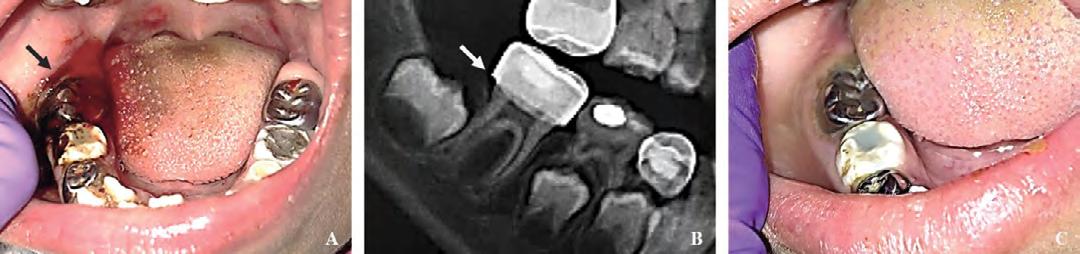
A large liver-like clot was removed in order to perform a clinical examination and ice water-soaked gauze was placed on the site with gentle pressure without improvement. Extraoral evaluation was not significant. Removal of the gauze revealed continuous oozing of heme localized to the gingival sulcus of the mandibular right permanent first molar (Figure 1A), which had a PMC placed one day prior. The radiograph (Figure 1B) revealed normal dental development for chronological age, multiple restorations on the permanent and primary dentition, and no radiographic radiolucency indicating signs of pathology. Proximity was noted on the distal margin of the PMC on the mandibular right permanent first molar to the adjacent tooth follicle.
Local anesthetic infiltration in the form of 2% lidocaine with 1:100,000 epinephrine was administered surrounding the affected area, and the gingival sulcus was curetted and irrigated with saline solution to remove any local irritants. The gingival sulcus was packed with a gelatin matrix and the gingival tissues cauterized. Hemostasis was achieved, and the patient was instructed to provide continuous biting pressure (Figure 1C). The patient was discharged and received follow-up care in the outpatient dental clinic the next day, at which time she was exhibiting absence of bleeding complications and normal healing gingival tissues.
Case Two
An 11-year-old female presented to the Children’s Hospital ED in the evening hours with a chief complaint of constant bleeding and upper left quadrant pain after routine extraction of the permanent maxillary left first molar seven hours prior that day. Since the procedure, the patient had been unable to take in any food, fluids or analgesic medication due to bleeding and discomfort. A review of the patient’s medical history revealed well-controlled asthma and no other reported medical conditions or allergies.
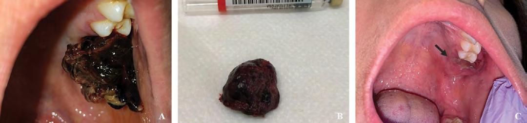
Initial presentation of the patient showed the child sitting comfortably, lightly holding gauze with her anterior teeth. Extraoral evaluation was not significant. Intraoral evaluation revealed generalized dental caries, heme pooling and accumulation in the oral cavity, and a large jelly-like clot protruding from the maxillary left permanent first molar region (Figure 2A).
Supraperiosteal infiltration local anesthesia in the form of 2% lidocaine with 1:100,000 epinephrine was administered at the extraction site. The clot was removed, and the socket was curetted and irrigated with saline solution (Figure 2B). Examination of the socket revealed visualization of the unerupted crown of the permanent maxillary left second molar serving as the distal wall of the socket. After 30 minutes of finger pressure, small loose clots formed but were not adhering, and bleeding continued. The socket was curetted and irrigated, and an absorbable gelatin dental sponge was gently placed in the extraction site. Hemostasis was achieved with finger gauze pressure. The patient was discharged and followed up at the outpatient dental clinic the next day with no residual pain or persistent bleeding issues (Figure 2C).
Case Three
A 6-year-old female with short gut syndrome, cholestatic liver disease, dependent on chronic total parenteral nutrition and later found to suffer from liver cirrhosis presented with a chief complaint of constant gingival bleeding without pain in the upper right quadrant. The patient was being managed at the Children’s Hospital as an inpatient and was taking enoxaparin to manage portal vein thrombosis. Her mother reported no history of dental restorations or extractions in the area. Initial presentation of the patient showed the child jaundiced but lying comfortably bedside.
Intraoral examination revealed moderate plaque and calculus accumulation, no signs of dental caries, a Grade II mobile primary maxillary left first molar, and a Grade III mobile primary maxillary right first molar, which was encompassed by coagulum (Figure 3A). The patient’s last dose of enoxaparin had been administered roughly 12 hours prior. At this time, the clot was removed with gauze, the tooth was irrigated with saline, and firm gauze pressure was intermittently applied (due to acute stress reaction) for 10 minutes until hemostasis was achieved.

Gingival bleeding returned within 24 hours of the patient resuming enoxaparin, resulting in a jelly-like clot coagulum that was again visualized encompassing the primary maxillary right first molar. Enoxaparin was again temporarily halted. Due to the patient’s medical complexities and acute stress reaction, definitive treatment was postponed until the following day when the patient was scheduled for a liver biopsy.
Hemostasis was again achieved bedside with sterile saline irrigation and gauze pressure. Under anesthesia, a periapical radiograph of the primary maxillary right first molar was obtained and revealed an advanced eruption pattern (Figure 3B). Supraperiosteal infiltration local anesthesia in the form of 2% lidocaine with 1:100,000 epinephrine was administered and the mobile tooth delivered. Exuberant granulation tissue was excised, absorbable gelatin dental sponge was placed, and hemostasis achieved (Figure 3C). On follow-up, examination showed the extraction site was hemostatic and healing well.
Discussion
Bleeding is a common sequela of dental procedures that is generally self-limiting. Extensive bleeding may lead to formation of a liver clot, as described in these cases. Treatment of a liver clot includes removal with high-volume suction and curettage, irrigation and direct pressure, which allows a healthy organized blood coagulum to form in order to stop hemorrhagic or persistent bleeding.[4]
While the conservative approach to treatment is, often, all that is required in management of liver clots, additional hemostatic treatment modalities could be considered. The formation of a natural blood clot is dependent on the presence of platelets and the interaction with coagulation factors to form a fibrin matrix.[5] In the event of inadequate hemostasis, from irrigation and direct pressure alone, adjunctive procedures and topical hemostatic agents can be considered to facilitate proper care for the patient. Adjunctive procedures include sutures, passive and active hemostatic agents, and lasers, to name a few. Sutures act as a mechanical method to seal off blood vessels and manage bleeding, which in the right circumstance can be a relatively easy and conservative supplemental approach to treatment. Topical hemostatic agents, including collagen, cellulose, gelatins and polysaccharide spheres, alternatively, act passively by forming a physical platform, which coheres to the site of injury, activates the extrinsic clotting pathway and provides a matrix where platelets can collect to form a clot.[6]
Because passive hemostasis relies on fibrin production to achieve hemostasis, topical hemostatic agents are only appropriate for use in patients who have an intact coagulation cascade.[6] Topical thrombin products, instead, act as active hemostatic agents to form the basis of a fibrin clot by influencing the coagulation cascade, promoting the conversion of fibrinogen to fibrin.[7] Overall hemostasis may be improved by simultaneous use of these passive and active hemostatic agents.
Further, as wide use of lasers in dentistry has become commonplace, diode lasers, in particular, have become known as a heat-generating device that can be utilized to provide hemostasis of soft tissues, among other uses. Diode lasers, which produce laser wavelengths ranging between 810 nm and 980 nm, have been used in dentistry since the mid-1990s and have advanced significantly since their implementation in the field.[8] Diode laser wavelengths differ from erbium lasers, which have an approximate wavelength of 3,000 nm, and CO2 lasers which have an approximate wavelength of 10,000 nm. Photo-thermal coagulation with lasers primarily occurs from denaturation of soft-tissue proteins and constriction of blood and lymphatic vessels at increased temperatures. Diode lasers have been shown to be significantly more effective with coagulation over extended volumes compared to erbium or CO2 laser wavelengths.[9]
In the first case described, a unique formation of a liver clot developed following placement of a PMC; in the second, following an extraction; and in the third, the result of repeated irritation from an exfoliating tooth on a medically complex patient taking enoxaparin. Although the preceding events of liver clot formations in these cases were different, similarities are noted. In all three cases, the children were in the mixed dentition phase, and there were dental follicles or unerupted teeth directly adjacent to the sites of uncontrolled bleeding. As a tooth develops, the dental follicle surrounds the enamel organ and dental papilla. It provides nutrition to the developing tooth and has a rich blood supply in order to do so. This rich blood supply may have been a contributing factor to the formation of the liver clot in these cases.[10]
Hemorrhage following seating of a PMC is uncommon due to closure of the soft tissue at the gingival sulcus. However, a sharp edge of a crown may act as constant irritation, preventing normal clotting and healing. The radiograph of the first case reveals the PMC was also near the dental follicle of the permanent right mandibular second molar and may have impinged on the vasculature, causing persistent bleeding. In the second case, lack of pressure may have been the main contributing factor for the abnormal clotting. However, during extraction of the first permanent molar, there may have been possible traumatic impingement of the adjacent dental follicle, causing persistent bleeding. The unerupted crown also may have been an inadequate surface for clots to adhere. In the third case, the repeated trauma of an exfoliating tooth crown meant constant reinjury to the surrounding tissue and, possibly, the erupting tooth follicle.
It is also important to understand and note any possible underlying conditions in patients that may lead to uncontrolled bleeding. A complete blood count (CBC) and coagulation study were ordered to rule out blood dyscrasias in all three cases. The patient in Case Three had increased risk factors for bleeding due to the enoxaparin the patient required to manage portal vein thrombosis, as well as the impact of the patient’s liver dysfunction on her coagulation cascade. Enoxaparin is a low-molecular weight heparin that disrupts the coagulation cascade and has been shown to cause elevated partial thromboplastin time (PTT). Prolongation of PTT, prothrombin time (PT), international normalized ratio (INR), as well as mild thrombocytopenia, is also common in individuals with liver disease.[11]
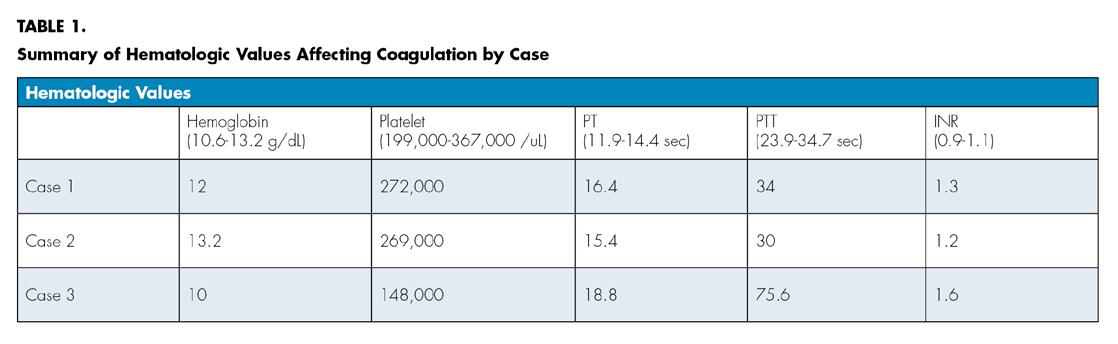
Although they had uncomplicated medical histories, the patients in the first and second cases also showed slightly elevated PT and INR levels, which may have predisposed them to prolonged bleeding (Table 1).
Adverse events after dental procedures are uncommon; however, it is important to recognize and address postoperative problems that may arise. Young patients in the mixed dentition may have an added risk of forming a liver clot if dental follicles are in close proximity to the area being manipulated. Identifying these cases and taking caution around these sites may minimize the risk of persistent bleeding and liver clot formation. While most liver clots can be treated by conventional curettage, irrigation and controlled pressure, adjunctive hemostatic agents and matrices may also be effective in management.
The authors thank Drs. Liza Idelchik and Annie Zhao for providing clinical photos for cases One and Three. They report no conflict of interest in the preparation of this paper. Queries about this article can be sent to Dr. Lesavoy at drbret@lesavoydental.com.
REFERENCES
1. Druckman RF, Fowler EB, Breault LG. Post-surgical hemorrhage: Formation of a “liver clot” secondary to periodontal plastic surgery. J Contemp Dent Pract 2001;2(2): 62-71.
2. Bakutra G, Vishnoi S, Chandran S, Barot V. Liver Clot: A reactionary haemorrhage - Case report. Natl J Integr Res Med 2015;6(1):116-8.
3. Nair MB, Shashikumar P. “Liver Clot” after periodontal plastic surgery. J Int Clin Dent Res Organ 2019;11(2):106-09.
4. Pandya D, Manohar B, Mathur LK, Shankarapillai R. “Liver clot” - A rare periodontal postsurgical complication. Indian J Dent Res 2012;23(3):419-22.
5. Hadjipanayi E, Kuhn PH, Moog P, Bauer AT, Kuekrek H, et al. The Fibrin Matrix Regulates Angiogenic Responses within the Hemostatic Microenvironment through Biochemical Control. PLOS ONE 2015;10(8).
6. Samudrala S. Topical hemostatic agents in surgery: a surgeon’s perspective. AORN J 2008;88(3):S2-S11.
7. Achneck HE, Sileshi B, Jamiolkowski RM, Albala DM, Shapiro ML, Lawson JH. A comprehensive review of topical hemostatic agents: efficacy and recommendations for use. Ann Surg 2010;251(2):217-228.
8. Desiate A, Cantore S, Tullo D, Profetta G, Grassi FR, Ballini A. 980 nm diode lasers in oral and facial practice: current state of the science and art. Int J Med Sci 2009;6(6):358-64.
9. Willems PWA, Vanderton WP, Verdaasdonk RM, van Swol CFP, Jansen GH. Contact laserassisted neuroendoscopy can be performed safely by using pretreated ‘black’ fibre tips: Experimental data. Lasers in Surgery and Medicine 2001:28(4):324-9.
10. Kuyama K, Iwai S, Ogura N, Eda T, Kondoh T, Yamamoto H. Histopathological and immunohistochemical study of the characteristics of dental follicle. J Hard Tissue Biol 2012;21(3):237-44.
11. Intagliata NM. Concepts and controversies in haemostasis and thrombosis associated with liver disease: Proceedings of the 7th international coagulation in liver disease conference. Thromb Haemost 2018;118(8):1491-506.

Bret Lesavoy, D.M.D., is a private practitioner in Pennsylvania and New Jersey. He is a former postdoctoral residency fellow in pediatric dentistry at Columbia University Medical Center, New York, NY.

Christine Wang, D.D.S., is a private practitioner in Connecticut. She is a former postdoctoral residency fellow in pediatric dentistry at Columbia University Medical Center, New York, NY.

Richard Yoon, D.D.S., is an associate professor of dental medicine, Section of Growth and Development, College of Dental Medicine, at Columbia University Medical Center, New York, NY.
