11 minute read
An Unusual Presentation of Langerhans Cell Histiocytosis: Case


An Unusual Presentation of Langerhans Cell Histiocytosis:
Case Report and Review of the Literature
Aaron E. Yancoskie, D.D.S.; Ghazeleh Peiravani, D.D.S.; Jordana S. Rothenberg, D.D.S.; Anthony S. Alessi, D.M.D., M.D.; Heather Z. Fugazy, D.D.S.
ABSTRACT
Langerhans cell histiocytosis (LCH) is a destructive process that may involve both hard and soft tissues. While rare, it has a predilection for the oral and maxillofacial region. LCH occurs across the age range yet is often diagnosed in the pediatric population. The dentigerous cyst (DC) is the most common developmental odontogenic cyst. It typically presents in young adult patients as a solitary lesion associated with impacted third molars. Here we present a case of LCH occurring in the wall of a DC in a 12-year-old male.
The dentigerous cyst (DC), also known as the follicular cyst, is the most common developmental odontogenic cyst, representing up to approximately 25% of all gnathic cysts.[1-5] DCs typically present in the second to fourth decades, and demonstrate a modest predilection for men compared to women.[1-5]
Radiographically, DCs show a unilocular radiolucency associated with the crown of an impacted tooth, most commonly a mandibular 3rd molar.[1-5] The histopathological findings include a fibrovascular connective tissue wall with a non-keratinized epithelial lining.[1-5] In the absence of a substantial inflammatory infiltrate, the non-keratinized epithelial lining is thin and shows a flat interface with the connective tissue wall.[1-3]
The DC is often an incidental radiographic finding; however, the potential for significant expansion exists.[6] In such cases, clinical features may include displacement of the regional anatomy (e.g., teeth and the inferior alveolar nerve canal), cortical expansion, facial asymmetry and, uncommonly, paresthesia and pain.[1-3] In rare instances, cortical expansion and thinning may lead to elevated risk for pathologic fracture of the jaw.
Definitive management comprises complete excision.[2] Extremely rare cases of transformation to odontogenic tumors and squamous cell carcinoma have been reported.[1-5] However, no reports have documented a DC in association with Langerhans cell histiocytosis (LCH).
Case Report
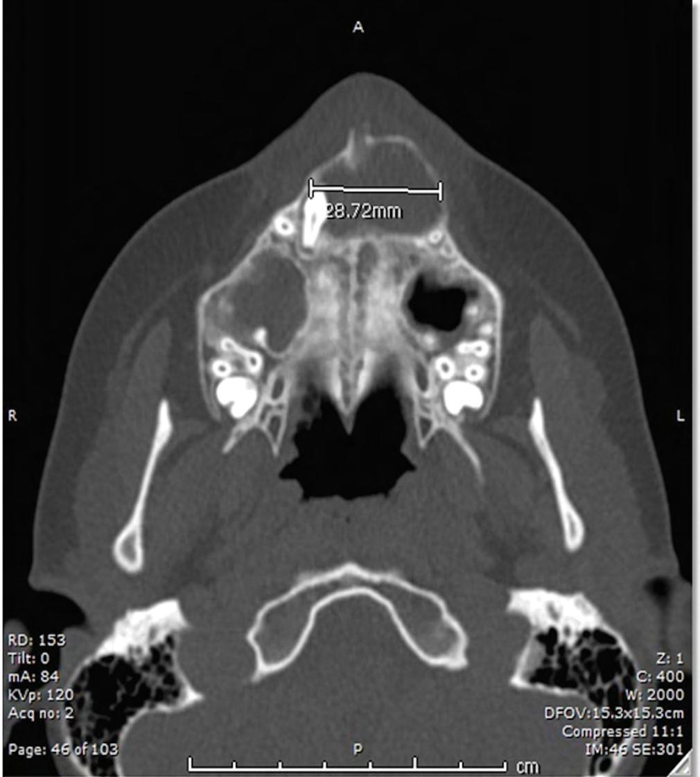
A 12-year-old male with no known medical diagnoses presented to oral and maxillofacial surgery for evaluation of an impacted right maxillary canine. CBCT demonstrated a 2.9 x 2.0 cm well-circumscribed unilocular radiolucency in the anterior maxilla associated with tooth #6 (Figure 1). An incisional biopsy was performed. A general pathologist diagnosed the specimen as a developmental cyst, commenting on focal characteristics of Langerhans cell histiocytosis. The impacted canine was extracted, and the cyst enucleated under general anesthesia.

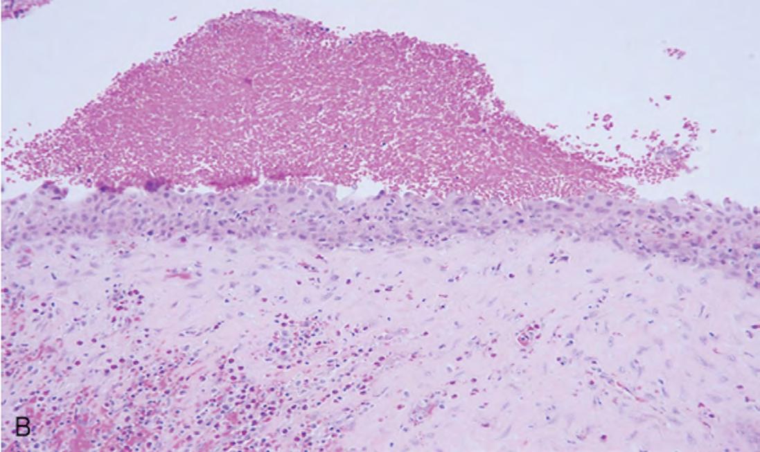
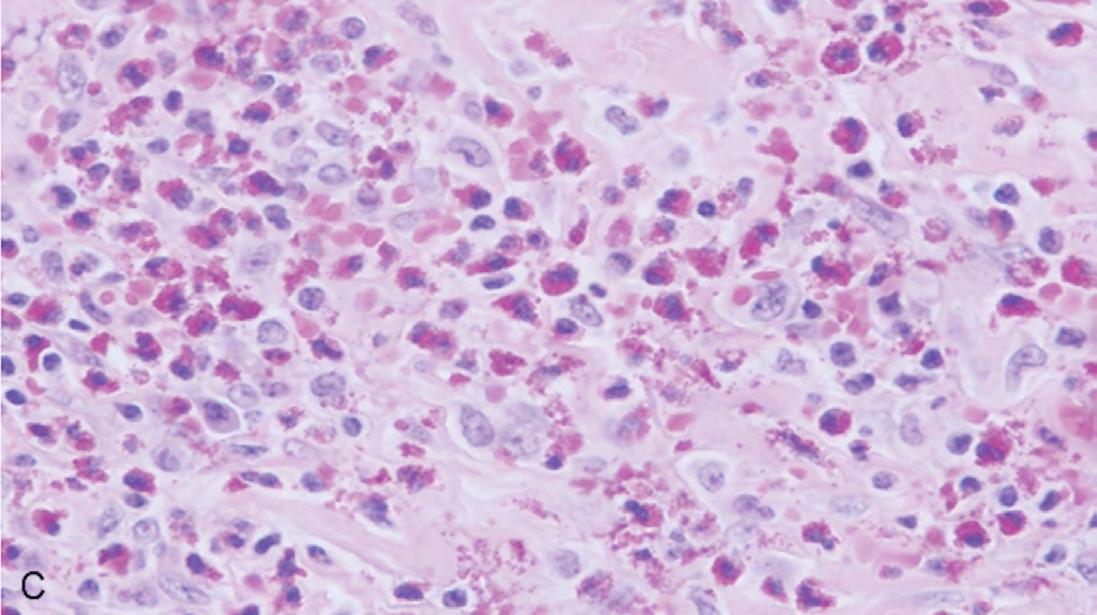
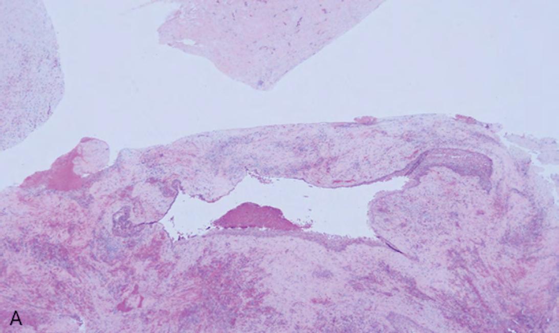
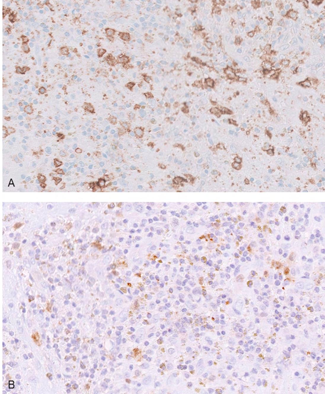
The gross specimen was submitted to oral and maxillofacial pathology. It consisted of multiple pieces of tan-red soft and hard tissue measuring 3.5 x 2.5 x 2.0 cm in aggregate. Microscopic review showed a cystic lining of non-keratinized stratified squamous epithelium with an inflamed fibrovascular connective tissue wall (Figures 2A,B). The inflammatory infiltrate included plasma cells, lymphocytes, numerous eosinophils and a population of histiocytoid cells possessing a central depression (Figure 2C). The histiocytoid cells stained positively for antibodies directed against CD1a (Figure 3A) and S-100 (Figure 3B), confirming the diagnosis of LCH. Molecular analysis for BRAFV600E mutation was negative.
The patient was referred to pediatric hematology and oncology for management. Additional radiographic studies confirmed that disease was limited to the maxilla. Initial treatment included a six-week regimen of vinblastine and prednisone. Minimal response was observed, and further treatment comprised six cycles of clofarabine. Management was complicated by brief hospitalizations for a neutropenic fever and, later, an episode of herpes zoster of the right buttock. Following his second chemotherapeutic regimen, the patient has been free of disease for 36 months.
Discussion and Review of the Literature
LCH is a disease characterized by destruction of bone and soft tissue by infiltrates of dendritic-like cells positive for S-100, CD207, CD1a and histiocytic markers.[7] It is a rare disease that occurs in both the pediatric and adult populations. The incidence in childhood is approximately one case in 200,000 per year.[7] The rate in the adult population is suspected to be similar; however, epidemiological information is not well-documented in comparison to pediatric data.[7-8]
There has been a long-term debate as to whether LCH represents a reactive or neoplastic process. B-Raf proto-oncogene (BRAF) is located on chromosome 7 and encodes a protein involved in several cellular processes, including cell division and differentiation.[9] Mutations in this gene have been identified in melanoma, thyroid carcinoma, adenocarcinoma of the lung and hairy cell leukemia, as well as several other neoplastic processes.[9] The discovery of BRAF gene mutation in approximately 50% of cases and ERK pathway (a cell signaling pathway regulated by BRAF protein) activation in 100% of cases provides support that LCH indeed represents a true neoplasm.[9-10]
The clinical presentation demonstrates substantial heterogeneity, ranging from isolated unifocal lesions in soft or hard tissue to multiorgan involvement.[7-8] The bones of the skull are the most commonly involved osseous sites.[2,11,12] Clinical manifestations vary and may include pain, local swelling and loss of organ function.[2,8] Radiographic studies of bone involvement show single or multiple clearly circumscribed radiolucencies.[1] Involvement of the alveolar bone in dentate patients may produce a tooth “floating-in-air” appearance.[2] Histopathological findings include a proliferation of Langerhans-like cells, accompanied by lymphocytes, eosinophils, plasma cells and multinucleated giant cells in varying numbers.[7-8,10-11] The Langerhans-like cells possess indented nuclei and delicate chromatin,[13] yet cannot be distinguished from other histiocytic cells by standard light microscopy.[1-2] With appropriate clinical correlation, positive immunohistochemical staining for antibodies directed against S-100, CD1a and/or CD207 is specific for the diagnosis.[1-2,8,11,13]
Treatment of LCH ranges from observation, curettage or steroid injection for unifocal disease, to cytotoxic chemotherapeutics and targeted therapies for multi-focal and multi-system disease.[2,7-8,14]The prognosis for LCH depends upon the number and sites of involvement.[8] Prognosis is excellent for patients with disease involving only a single site, yet worsens with increased number of foci and organ systems.[1-2,8] Involvement of high-risk organs (liver, spleen and bone marrow) portend a poor prognosis.[7-8,11] Recurrence is reported in approximately one-third of all patients diagnosed with LCH.[8] The relative survival rate at five years for children is 90% and 70% for adults.[8]
While this is the first reported case of LCH occurring in the wall of a DC, LCH occurring in the wall of a periapical inflammatory cyst has been described in the literature. Rawal and colleagues presented a case of LCH associated with a periapical inflammatory cyst in a 42-year-old female. This patient presented with pain localized to the residual roots of tooth #19.[15] A 1.25 cm, well-circumscribed, unilocular radiolucency possessing a sclerotic border was identified around the apex of the roots. Similar to the case presented here, microscopic examination showed a cystic lining and an associated connective tissue wall possessing the typical LCH histopathogical findings, including CD1a positive histiocytoid cells. The patient was treated with curettage and demonstrated an absence of recurrence two years postoperatively.[15]
Concomitance of LCH with other neoplastic entities has been documented.[8] However, DCs are not considered neoplastic, but are thought to represent a reactive process,[1-6] in which expansion and growth are driven by fluid movement.[2] Select odontogenic cysts, including odontogenic keratocysts and calcifying odontogenic cysts, are known to harbor mutations, raising the question of whether they should be considered neoplasms.[5,16-17] However, underlying genetic aberrations have yet to be identified in DCs. It is probably the case that these two events, development of a DC and LCH, occurred as distinct processes.
A wide diversity of diseases may present with unilocular radiolucencies associated with impacted teeth. It is critical that these specimens be submitted for microscopic review, as some proportion will represent aggressive entities.
Queries about this article can be sent to Dr. Yancoskie at aaron.yancoskie@ touro.edu.
REFERENCES
1. Wenig BM, Childers ELB, Richardson MS, Seethala RR, Thompson LDR. Atlas of Nontumor Pathology: Non-neoplastic disease of the head and neck. Fascicle 11, 1st series. Washington, DC: American Registry of Pathology, Armed Forces Institute of Pathology; 2017.
2. Neville BW, Damm DD, Allen CM, Chi AC. Oral and Maxillofacial Pathology. 4th ed. St. Louis: Elsevier; 2016.
3. Robinson RA. Diagnosing the most common odontogenic cystic and osseous lesions of the jaws for the practicing pathologist. Mod Pathol 2017;30:S96-103.
4. Zhang LL, Yang R, Zhang L, Li W, MacDonald-Jankowski D, Poh CF. Dentigerous cyst: a retrospective clinicopathological analysis of 2082 dentigerous cysts in British Columbia, Canada. Int J Oral Maxillofac Surg 2010;39:878–82.
5. EA Bilodeau, BM Collins. Odontogenic cysts and neoplasms. Surgical Pathology Clinics 2017;10:177-222.
6. Farah CS, Savage NW. Pericoronal radiolucencies and the significance of early detection. Aust Dent J 2002;47:262-265.
7. Allen CE, Beverley PCL, Collin M, Diamond EL, Egeler RM, Ginhoux F, Glass C, Minkov M, Rollins BJ, van Halteren A. The coming of age of Langerhans cell histiocytosis. Nat Immunol 2020;21:1-7.
8. Rodriguz-Galinodo C, Allen CE. Langerhans cell histiocytosis. Blood 2020; 135(16):1319-1331.
9. Gene [Internet]. Bethesda (MD): National Library of Medicine (US), National Center for Biotechnology Information; [1988] – . Gene ID: 673, Homo sapiens B-Raf proto-oncogene (BRAF); [cited 2022 Sept 21]. Available from: https://www.ncbi.nlm.nih.gov/gene/673.
10. Badalian-Very G, Vergilio JA, Degar BA, MacConaill LE, Brandner B, Calicchio ML, Kuo FC, Ligon AH, Stevenson KE, Kehoe SM, Garraway LA, Hahn WC, Meyerson M, Fleming MD, Rollins BJ. Recurrent BRAF mutations in Langerhans cell histiocytosis. Blood 2010;116(11):1919-23.
11. Felstead AM, Main BG, Thomas SJ, Hughes CW. Recurrent Langerhans cell histiocytosis of the mandible. Br J Oral Maxillofac Surg 2013;51:254-265.
12. Bedran NC, Carlos R, Benevenuto de Andrade BA, Bueno APS, Romanach MJ, Milito CB. Clinicopathological and immunohistochemical study of head and neck Langerhans cell histiocytosis from Latin America. Head Neck Pathol 2017;12:431-39.
13. Sholl LM, Hornick JL, Pinkus JK, Pinkus GS, Padera RF. Immunohistochemical analysis of Langerin in Langerhans cell histiocytosis and pulmonary inflammatory and infectious diseases. Am J Surg Pathol 2007;31:947–952.
14. Kobayashi M, Tojo A. Langerhans cell histiocytosis in adults: advances in pathophysiology and treatment. Cancer Sci 2018;109(12):3707-3713.
15. Rawal YB, Anderson KM, Mincer HH, Rawal SY, Tortorich AL. Langerhans cell histiocytosis in a periapical cyst: a wolf in a sheep’s clothing? J Tenn Dent Assoc 2008 Fall;88(4):26-8; quiz 29-30.
16. Sekine S, Sato S, Takata T, Fukuda Y, Ishida T, Kishino M, Shibata T, Kanai Y, Hirohashi S. Beta-catenin mutations are frequent in calcifying odontogenic cysts, but rare in ameloblastomas. Am J Pathol 2003;163(5):1707-12.
17. Yukimori A, Oikawa Y, Morita K, Nguyen CTK, Harada H, Yamaguch S, Kayamori K, Yamaguchi A, Ikeda T, Sakamoto K. Genetic basis of calcifying cystic odontogenic tumors. PLoS One 2017;12(6):e0180224.


Aaron E. Yancoskie, D.D.S., is an attending pathologist, Westchester Medical Center Department of Pathology and Laboratory Medicine, and assistant dean for academic affairs and associate professor of dental Medicine, Touro College of Dental Medicine at New York Medical College, Hawthorne, NY.
Ghazeleh Peiravani, D.D.S., is in private practice in Houston, TX.
Jordana S. Rothenberg, D.D.S., is in private practice in Freeport, NY.
Anthony S. Alessi, D.M.D., M.D., is an attending surgeon, Westchester Medical Center and assistant professor New York Medical College, Department of Dental Medicine, Valhalla, NY.
Heather Z. Fugazy, D.D.S., is an attending surgeon, Phelps Memorial Hospital and assistant professor, New York Medical College Department of Dental Medicine, NY, NY.