31 minute read
Managing Perforating Internal Root Resorption Nonsurgically


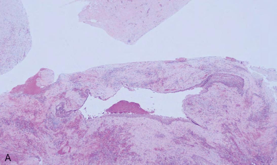
tenderness on the buccal gingiva around tooth #10. The tooth was sensitive to percussion and biting. A small, fluctuant intraoral swelling was noted near the apex of tooth #10. The tooth did not respond to vitality testing. Radiographic examination revealed a large perforating internal resorptive defect towards the apical third of the root (Figure 1A).
Managing Perforating Internal Root Resorption Nonsurgically
A Report of Two Cases and Review of the Literature
Joseph C. Stern, D.D.S.
ABSTRACT
Aim: To describe the use of bioceramics in the nonsurgical treatment of perforating internal root resorption.
Summary: Confusion over the subject of root resorption seems ubiquitous in the general dental community. Because of the several subheadings, such as internal, external cervical and apical, and because of a variety of causes for each, such as trauma, pressure, infection, inflammation and systemic, the confusion can be understandable. It seems that this confusion arises primarily from the nomenclature describing the location and etiology of root resorption. This case report deals with perforating internal root resorption (IRR), its causes, diagnosis and successful nonsurgical treatment, effectively using cone-beam computed tomography to visualize the extent of the lesion and bioceramics to fill the defect. The differential diagnosis between internal and external root resorption is examined, as well as a review of the literature.
Root resorption is defined as the loss of dental hard tissues as a result of clastic activities, which can occur as a pathologic or physiologic process depending on the location and timing of the resorptive process.[1] While most internal and external resorption is pathologic in nature, the resorption associated with the primary dentition is most often a normal physiologic process.[2]
The primary theory for what initiates internal root resorption (IRR) is multinucleated giant cells located in the granulation tissue that forms in response to infected coronal pulp tissue. These odontoclasts are believed to be responsible for the resorption of the lining of the pulp space. A second theory suggests that the granulation tissue arises from the vascular system, outside of the pulp space. Damage and/or loss of the predentin and odontoblastic layer must occur prior to the resorptive process.[3] Trauma is suspected as an initiating cause, possibly supported by continuous stimulation from infection. Iatrogenic causes of continued inflammatory excitation of the coronal pulp include overheating the tooth.[4-8]
IRR is insidious and often progresses without symptoms. Pain and/or swelling may not occur until the process perforates the root, at which time the prognosis for a successful outcome becomes more questionable. As long as the apical portion of the pulp retains vital tissue, the resorptive process continues. Another requirement for continued resorption is bacterial infection, as a microbial stimulus is required for the continuation of IRR.[7]
When and/if the pulp tissue becomes necrotic, before perforation, the process can be self-limiting. Usually, internal root resorption is first observed at a routine radiographic exam. Because it begins in the pulp space, the lesion is contiguous with the space. It can be confirmed if two acute-angled radiographs, taken from extreme mesial and distal positions show no separation between the lesion and the pulp space. Cone-beam computed tomography (CBCT) can help make this differential diagnosis between external and internal resorption.
One of the treatment options for large lesions associated with perforating IRR has been a surgical approach. This is based on the literature that suggests that periapical lesions of greater than 5 mm have a poor prognosis with conventional, nonsurgical endodontic treatment.[9-10] Given advances in disinfecting and sealing the root canal space, it is time to question the surgical approach as the primary treatment based on the lesion size alone, whether the lesion is associated with the periapex or a perforating resorptive defect.
There is nothing magical about a 5 mm lesion that it should portend a negative future outcome. Rather, we should look more closely during the diagnostic phase at other biologic factors that can influence future results. All-size lesions can have successful outcomes when planned and treated properly. Our treatment of IRR follows the standard protocol for nonsurgical endodontics. The root canal space is debrided and decontaminated to the apical constriction and, subsequently, filled. Interrupting the vital tissue pathway at the apex arrests the resorptive process. Extra care, mechanically and chemically, may be in order to remove tissue from the undercut areas that are created by the resorptive process. Creating a straight-line access to the resorptive defect is often not feasible, as this would require removal of more dentin, further weakening the root structure.
Our technique in these two cases differs from the 17 cases reported previously in the literature (Table 1). We use a calcium silicate sealer to both fill the perforating defect and seal the gutta-percha simultaneously, thus saving the time of an extra visit.


Case One
A 41-year-old male presented for treatment with a chief complaint of pain and swelling adjacent to tooth #10. The patient reported a history of trauma when he was a teenager. The area had not bothered him until this recent development of pain and swelling. Clinical exam revealed
The CBCT (Veraviewepocs 3D R100; J. Morita) showed significant alveolar bone loss adjacent to the resorptive defect, enveloping the entire mesial side of the apical half of the root of tooth #10 and extending to the root of tooth #9 (Figures 1B,C). A diagnosis of pulpal necrosis with acute apical abscess was made. All options of treatment were discussed with the patient, including extraction and replacement with an implant or bridge. The patient was motivated to try to retain this tooth with root canal therapy and repair of internal resorptive defect, rather than have it extracted. Informed consent was obtained from the patient.
First Visit
The patient was anesthetized with 1.7 mL 4% articaine 1:100000 epinephrine (Septocaine; Septodont, New Castle, DE) via labial infiltration. Rubber dam isolation was achieved, and the tooth was accessed within a #2 surgical length carbide round bur. A necrotic pulp was encountered. Working length was established with a Root ZX apex locator (Morita, Tokyo, Japan). The canal was instrumented up to a size 35 .04 Vortex Blue rotary file (Dentsply Tulsa Dental, Johnson City, TN). Care was taken that the files passed through the resorptive defect and entered the apical portion of the canal.
The canal was irrigated with 5.25% sodium hypochlorite. The EndoActivator (Dentsply, Tulsa, OK) was used to sonically agitate the irrigant in the canal to ensure thorough disinfection of the resorptive defect. The canal was dried with paper points, and calcium hydroxide (Ultracal XS, Ultradent Products Inc, South Jordan, UT) was syringed into the canal and the defect. The tooth was temporarily restored with Cavit (3M ESPE, Neuss, Germany)

Figure 1.
A. Preoperative periapical radiograph of tooth #10 showing internal root resorption in apical third of root. There is significant alveolar bone loss adjacent to defect.
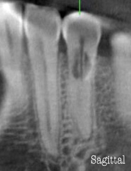
B. Sagittal CBCT image of tooth #10 showing internal resorptive defect perforating on mesial aspect of root. Note adjacent alveolar bone loss extending proximally to tooth #9.
C. Axial CBCT image of internal resorptive defect perforating on mesial aspect of root. There is thin layer of circumferential dentin remaining and extensive alveolar bone loss adjacent to defect.
D. Periapical radiograph showing gutta-percha cone fit. Gutta-percha cone passes through resorptive defect to contact apical portion of root canal.
E. Immediate postoperative periapical radiograph of tooth #10 once root canal was completed and resorptive defect was restored.
F. Two-year follow-up showing complete healing of radiolucency adjacent to defect and reestablishment of PDL.
G. Two-year follow-up CBCT. Coronal slice showing complete healing of radiolucency adjacent to defect and reestablishment of PDL.
H. Five-year follow-up radiograph. Patient is completely asymptomatic.A. Preoperative periapical radiograph of tooth #10 showing internal root resorption in apical third of root. There is significant alveolar bone loss adjacent to defect.
B. Sagittal CBCT image of tooth #10 showing internal resorptive defect perforating on mesial aspect of root. Note adjacent alveolar bone loss extending proximally to tooth #9.
C. Axial CBCT image of internal resorptive defect perforating on mesial aspect of root. There is thin layer of circumferential dentin remaining and extensive alveolar bone loss adjacent to defect.
D. Periapical radiograph showing gutta-percha cone fit. Gutta-percha cone passes through resorptive defect to contact apical portion of root canal.
E. Immediate postoperative periapical radiograph of tooth #10 once root canal was completed and resorptive defect was restored.
F. Two-year follow-up showing complete healing of radiolucency adjacent to defect and reestablishment of PDL.
G. Two-year follow-up CBCT. Coronal slice showing complete healing of radiolucency adjacent to defect and reestablishment of PDL.
H. Five-year follow-up radiograph. Patient is completely asymptomatic.
Second Visit
The patient returned after two weeks for completion of endodontic treatment. He reported that all symptoms had subsided. Clinical examination revealed that the swelling had resolved. Calcium hydroxide was removed from the canal with instrumentation, irrigation and activation with the EndoActivator. The canal was dried with paper points, and excess irrigant was removed using surgical suction with a micro-tip. A master gutta-percha cone was placed to length and confirmed by radiographic exam (Figure 1D).
The canal was coated with EndoSequence BC (bioceramic) sealer (Brasseler USA, Savannah, GA) to allow for suffi-
cient amounts of sealer to fill the resorptive defect, and was then obturated with gutta-percha and BC sealer using the technique of warm vertical condensation (Figure 1E). The lingual access opening was restored with TPH Spectra ST composite (Dentsply Sirona, Charlotte, NC) and the patient was put on a recall schedule to monitor healing.
At the one-year, two-year and five-year recall visits, the patient was completely asymptomatic, and radiographically showed complete healing with full restoration of the bone and lamina dura adjacent to the resorptive defect (Figures 1F,H). The patient was very satisfied with the result, as he was able to get significant time out of a tooth that was originally planned for extraction.

Figure 2.
A. Preoperative periapical radiograph of tooth #19. Visualization of periapical radiolucency associated with mesial root is possible. Root also appears to be calcified in middle and apical thirds.
B. Sagittal CBCT slice showing internal resorption in apical portion of mesial root. Visualization of resorptive defect perforating on distal aspect of mesial root and significant periapical pathology extending close to furcation is possible. Defect and extent of pathology was not visualized on periapical radiograph.
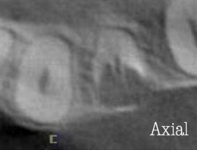
C. Axial CBCT slice showing resorptive defect encapsulating both MB and ML canals and perforating on distal aspect of mesial root. There is significant bone loss adjacent to perforating defect.
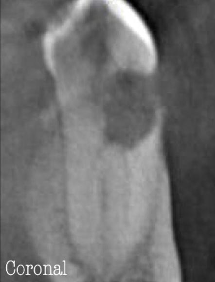
D. Coronal CBCT slice showing internal resorptive defect encapsulating both mesiobuccal and mesiolingual canals.
E. Immediate postop radiograph after completion of endodontic treatment.
F,G. Coronal and axial CBCT slice at 16-month follow-up. Note resorptive defect filled with bioceramic sealer and complete healing of adjacent bone and reestablishment of PDL. Patient returned at 16-month point for endodontic treatment of tooth #19.
H. Three-year follow-up radiograph. Patient remained completely asymptomatic on tooth#19.
Case Two
A 56-year-old male presented with a chief complaint of vague discomfort in the left mandible. The patient reported that this discomfort had been present on and off for more than six months, but recently the pain had worsened. Clinical examination revealed pain to percussion on tooth #19. No swelling was noted, and the tooth was not sensitive to palpation or biting.
Radiographic and CBCT (Veraviewepocs 3D R100; J. Morita) examination revealed a crowned tooth #19 with perforating internal root resorption near the apical end of the mesial root with associated periapical pathology extending distally to encompass the distal root and coronally toward the furcation (Figures 2A-D). A diagnosis of pulpal necrosis with symptomatic apical periodontitis was made. A discussion was had regarding the prognosis for treating this tooth with root canal therapy. Alternative options of extraction and implant or bridge placement were discussed. As finances were an issue, the patient opted to have the tooth endodontically treated rather than extracted. Informed consent was obtained from the patient.
First Visit
The patient was anesthetized with 1.7 mL 3% mepivicaine (Carbocaine, Dentsply Pharmaceutical, York, PA) administered via left inferior alveolar nerve block and 1.7 mL 4% articaine 1:100000 epinephrine (Septocaine; Septodont, New Castle, DE) via local buccal infiltration. Rubber dam isolation was achieved, and the tooth was accessed through the porcelain-fused-tometal (PFM) crown with a combination of a round diamond bur and a #2 surgical length carbide round bur. A necrotic pulp was encountered. Working length was established with a Root ZX apex locator (Morita, Tokyo, Japan).
The canals were instrumented up to a size 35 .04 Vortex Blue rotary file (Dentsply Tulsa Dental, Johnson City, TN) and irrigated with 5.25% sodium hypochlorite. The EndoActivator (Dentsply, Tulsa, OK) was used to sonically agitate the irrigant in the canal to ensure thorough disinfection of the resorptive defect. The canals were dried with paper points, and calcium hydroxide (Ultracal XS, Ultradent Products Inc, South Jordan, UT) was syringed into the canals and the defect. The tooth was temporarily restored with Cavit (3M ESPE, Neuss, Germany).
Second Visit
The patient returned after three weeks for completion of endodontic treatment. He reported that all symptoms had subsided. Calcium hydroxide was removed from the canals with instrumentation, irrigation and activation with the EndoActivator. The canals were dried with paper points, and excess irrigant was removed using a surgical suction with a micro-tip. The canals were coated with BC sealer to allow for sufficient amounts of sealer to fill the resorptive defect and were then obturated with gutta-percha and BC (bioceramic) sealer using the technique of warm vertical condensation (Figure 2E). The occlusal access opening was restored with TPH Spectra ST composite (Dentsply Sirona, Charlotte, NC) and the patient was put on a recall schedule to monitor healing.
The patient returned at the 16-month point for endodontic treatment of tooth #18. At a 16-month recall visit, the patient was completely asymptomatic on tooth #19. and radiographic/CBCT examination revealed complete healing of the lesion adjacent to the resorptive defect on tooth #19 (Figures 2 F,G). At the three-year recall, the patient was asymptomatic on both teeth #18 and #19.
Discussion
As this is an uncommon phenomenon, the exact cause of IRR has not been fully determined. The literature suggests trauma related to pulpitis, pulpotomy,[11] cracked tooth, tooth transplantation, restorative procedures, orthodontic treatment and herpes zoster virus,[12] leading to pulpal necrosis. Trauma can also be iatrogenically related to heat when drilling without adequate coolant. The trauma, regardless of its source, disrupts the predentin layer, predisposing the denuded root surface to invasion by multinucleated giant cells.[4]
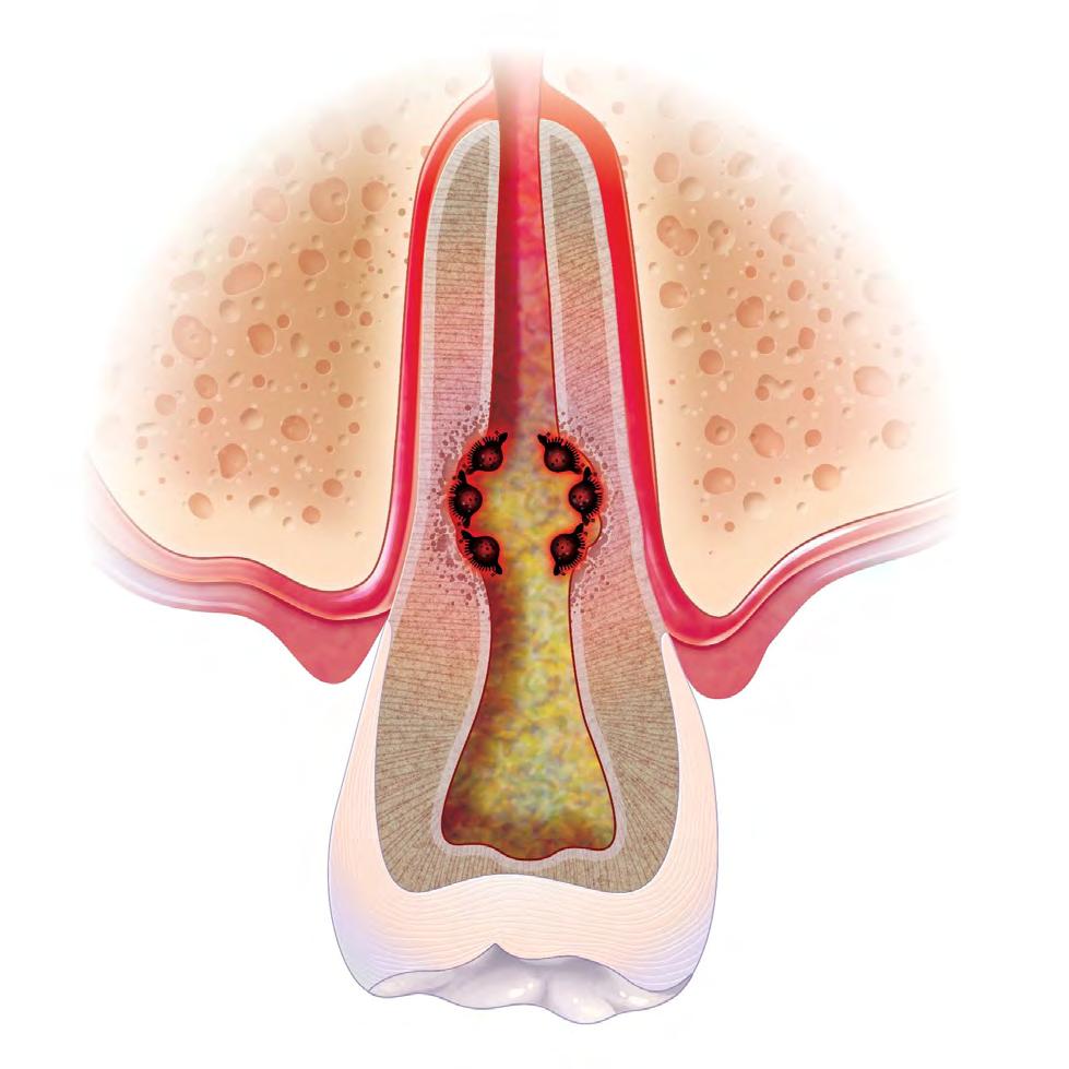
For IRR to progress, the pulp tissue apical to the resorptive lesion must have vital tissue to provide nutrition to the viable clastic cells, while the infected necrotic coronal pulp tissue provides the irritant and stimulus for those clastic cells to continue to resorb the adjacent dentin. (Figure 3).[5-8] As the resorptive lesion progresses it can eventually perforate into the adjacent periodontal ligament causing an extraradicular lesion (Figures 1, 2).

If the pulp tissue becomes completely necrotic, the resorptive process will halt, as there is no longer any vital tissue to supply the clastic cells with the nutrients needed for the resorptive process to continue. As the pulp continues to degenerate, bacteria will usually infect the entire root canal system, resulting in apical periodontitis, which can become contiguous with the extraradicular lesion if the IRR perforation has occurred. This lesion may also migrate coronally through the periodontal ligament to the gingival sulcus, forming a pseudo-periodontal pocket.
It is interesting to note that internal resorption can be thought of as a misplaced periapical lesion found inside the root canal rather than at the apex. Both are caused by the presence of bacteria and the triggering of resorptive cells. Both form in a symmetrical manner. However, the periapical lesion has surrounding vital tissue, which allows these lesions to grow in size, unlike IRR, which is self-limiting. Because active IRR requires a pulp space that is partially vital and partially necrotic, vitality testing is unreliable. One cannot be sure whether the pulp tissue at the time of diagnosis is necrotic and, therefore, the resorptive process is arrested, or vital tissue remains, and the IRR is ongoing (Figure 3). Regardless, if perforation has occurred, the external lesion has a life of its own and treatment is essential.
One of our challenges with teeth that develop IRR is the compromised remaining root structure that predisposes the tooth to future fracture. We must also deal with the perforating defect into the periodontal ligament that creates significant bone destruction. Control of infection is much more difficult when there is a perforation. If the tooth is deemed restorable and has a reasonable prognosis, nonsurgical root canal treatment is the treatment of choice.
The goal should be to preserve tooth structure during treatment to avoid further weakening an already compromised tooth.[8]Most of these defects are located in the middle or apical thirds of the root and the perforation is thus incased within the alveolar bone. It is important to use a biocompatible material to seal the defect, as well as to contribute to the regeneration of the bone and periodontal ligament. If, however, the defect is located in the cervical region of the tooth, and the tooth is deemed restorable, it might be better to restore it with a resin composite or glass ionomer restoration, given that these materials might strengthen an already weakened tooth structure, preventing potential snap-off of the coronal tooth structure. This is not an issue when the resorption is located more apically in the root.
Both cases presented here are examples of perforating internal resorption located in the apical third of the root, which were treated exclusively with an internal approach and sealed with the same biocompatible bioceramic material that was simultaneously sealing the gutta-percha in place. In all the previous nonsurgical treated cases in the literature, the MTA was used first, as an additional step to seal the defect prior to filling the canal. These 17 previously reported cases fall into one of the four classical treatment categories:
• Long-term calcium hydroxide treatment to trigger hard-tissue formation.
• Surgical repair with MTA.
• Nonsurgical internal repair with MTA.
• Regenerative endodontic procedure using an MTA plug.
Treatment for IRR includes removal of the infected and inflamed pulp tissue throughout the pulp space. The canal, both apical and coronal to the resorptive defect, must be thoroughly disinfected, using irrigant activation, which allows for vigorous agitation of irrigant solutions to reach the often undercut, untouched and hard-to-reach resorptive areas of the root canal.[13-16] The irrigants of choice for this disinfection are sodium hypochlorite and Ethylenediaminetetraacetic acid (EDTA).[17-21] If the perforation is large, it might be advisable to use lower concentrations of sodium hypochlorite or an alternative irrigant, such as chlorhexidine. If the resorption is active, there can be excessive bleeding upon access, making visualization difficult. Thorough disinfection and packing of the root canal space with calcium hydroxide is the best way to achieve hemostasis between visits.[22-26]
One of the treatment modalities discussed in the literature used long-term calcium hydroxide in the canal for extended periods of time until healing of the periodontium was achieved. This was then followed by either conventional obturation of the root canal with gutta-percha and sealer[27] or an MTA (mineral trioxide aggregate) plug with[28] or without gutta-percha.[29]
The most common treatment for IRR discussed in the literature was the use of MTA to seal perforating resorptive defects with the use of calcium hydroxide as a shortterm inter-visit medicament.[30-37] The primary reason for the use of MTA is its biocompatibility. Not only does MTA react favorably when it contacts vital tissue, it also can provide an impervious seal. One study utilized Biodentine (Septodont, Saint-Maur-des-Fosses, France) as an alternative calcium silicate cement as the material of choice to seal the perforating lesion.[38]
Two reported cases used a surgical approach, in which the lesion is accessed and restored surgically with a bioceramic material such as MTA.[39-40] If conventional nonsurgical methods fail, there is always an option to then surgically treat the resorption.[34]
A novel approach of utilizing regenerative endodontic treatment to treat these cases has also been reported.[41-43] These cases are unique in that the canals are not filled by conventional techniques but, rather, an MTA plug is placed on top of a blood clot in the canal. Historically, it was not advised to use standard gutta-percha and cement with perforating internal resorption, as this did not provide an adequate seal.[7] With the advent of bioceramic (calcium silicate) sealers, a more standard nonsurgical, internal approach can be used to restore these defects,[44] as our two cases have shown. A standard two-visit approach can be utilized with calcium hydroxide used as a short-term medicament only.
Bioceramics such as BC sealer have qualities which make it an ideal sealer for repairing a perforating internal root resorption. As a bioactive material, BC sealer has the ability to create a hydroxyapatite layer when in contact with tissue fluid. This allows the material to be highly biocompatible, osteoinductive and osteoconductive.
BC sealer, a newer generation hydraulic cement, uses nanotechnology to reduce the particle size to a mixture of nano and microparticles and, hence, gives it the ability to flow much better in filling the irregular sizes and shapes of the internal resorptive defect. Unlike MTA, BC sealer cement can set in the presence of tissue fluids, where no
additional setting time is needed.[45-46] These sealers can be utilized with either warm vertical condensation or with a single-cone technique and hydraulic condensation.[47-50] Bioceramic sealers further help fill these spaces by expanding slightly while setting and once set, will not resorb easily.[51-55]
While external root resorption comes in many forms, such as transient surface resorption, pressure resorption, external inflammatory root resorption, invasive cervical root resorption and replacement resorption (ankylosis), internal root resorption is uniquely different. The differential diagnosis is made by taking multiple radiographs at different angles.[56,57] Utilizing the buccal object rule, a lesion of internal origin will remain close to the canal regardless of the angle, while a lesion of external origin will move away from the canal depending on the angle of the radiograph (Figures 4 A,B).


Additionally, with IRR, the outline of the root canal is usually distorted and appears contiguous with the resorptive defect, while with external resorption, the root canal outline appears normal and can usually be seen running through the radiolucent resorptive defect, as there remains a thin layer of dentin separating the canal from the resorptive area[56-57] (Figures 5 A-D). The radiographic appearance of IRR is a fairly uniform radiolucent enlargement of the root canal. There would only be alveolar bone loss adjacent to the resorption if the resorption perforates into the PDL. The best and most accurate tool we have for diagnosing IRR and determining the path of the perforating lesion is cone-beam computed tomography (CBCT). It is best to use a limited field of view (FOV), as opposed to the larger FOV used with other disciplines in dentistry. A smaller FOV increases image resolution, while at the same time providing a lower effective radiation dose to the patient. It is worth noting that in Case Two, one cannot visualize the resorptive defect from just looking at the periapical radiograph. It has been shown in countless studies that CBCT gives a more accurate diagnosis and better visualization of periapical pathology.[58-60]
It is worth noting that despite the sizeable perforations and concomitant bone resorptions that rendered the canal architecture most challenging from a mechanical perspective, successful outcomes were achieved in both cases. Given these structural hazards created by the IRR, one can safely assume that the disinfection and subsequent filling of the canals were less than ideal.
Why, then, the success? The key is probably due to a thorough and detailed diagnostic workup utilizing CBCT that confirmed IRR. If the lesions were from a necrotic pulp, with external root biofilm, or if the multinucleated giant cells that were resorbing hard tissue had their origin
in the periodontal ligament and not the pulp space, success would probably not have been attainable nonsurgically. A larger sample size and longer-term follow-ups of 10 or more years would help to better assess the treatment outcomes for perforating IRR.
Conclusion
Two cases of extensive perforating internal root resorption, treated nonsurgically, are presented. A discussion of the biologic process of IRR, along with the need for an accurate diagnosis, shows that a nonsurgical approach in these situations should be the primary treatment plan. With the advent of bioceramic sealers, we now have the opportunity to treat perforating internal resorption with a more standard endodontic approach. The use of CBCT in the diagnostic planning phase is of critical importance in visualizing the full extent of the lesion.
Queries about this article can be sent to Dr. Stern at jstern5819@gmail.com.
REFERENCES
1. Patel S, Ford TP. Is the resorption external or internal? Dent Update 2007;34(4):218-229. doi:10.12968/denu.2007.34.4.218.
2. Harokopakis-Hajishengallis E. Physiologic root resorption in primary teeth: molecular and histological events. J Oral Sci 2007;49(1):1-12. doi:10.2334/josnusd.49.1.
3. Wedenberg C, Zetterqvist L. Internal resorption in human teeth—a histological, scanning electron microscopic, and enzyme histochemical study. J Endod 1987;13(6):255-259. doi:10.1016/S0099-2399(87)80041-9.
4. Tronstad L. Root resorption—etiology, terminology and clinical manifestations. Endod Dent Traumatol 1988;4(6):241-252. doi:10.1111/j.1600-9657.1988.tb00642.x.
5. Calişkan MK, Türkün M. Prognosis of permanent teeth with internal resorption: a clinical review. Endod Dent Traumatol 1997;13(2):75-81. doi:10.1111/j.1600-9657.1997.tb00014.x.
6. Fuss Z, Tsesis I, Lin S. Root resorption—diagnosis, classification and treatment choices based on stimulation factors. Dent Traumatol 2003;19(4):175-182. doi:10.1034/j.16009657.2003.00192.x.
7. Haapasalo M, Endal U. Internal inflammatory root resorption: the unknown resorption of the tooth. Endodontic Topics 2006;14(1), 60–79. https://doi.org/10.1111/j.1601-1546.2008.00226.x.
8. Patel S, Ricucci D, Durak C, Tay F. Internal root resorption: a review. J Endod 2010;36(7):11071121. doi:10.1016/j.joen.2010.03.014.
9. Wang N, Knight K, Dao T, Friedman S. Treatment outcome in endodontics—The Toronto Study. Phases I and II: apical surgery. J Endod 2004 Nov;30(11):751-61. doi: 10.1097/01. don.0000137633.30679.74. PMID: 15505504.https://doi.org/10.1097/01.don.0000137633.30679.74.
10. Ricucci D, Russo J, Rutberg M, Burleson JA, Spångberg LS. A prospective cohort study of endodontic treatments of 1,369 root canals: results after 5 years. Oral Surg Oral Med Oral Pathol Oral Radiol Endod 2011 Dec;112(6):825-42. doi: 10.1016/j.tripleo.2011.08.003. PMID: 22099859.
11. Cabrini RL, Manfredi EE. Internal resorption of dentine; histopathologic control of eight cases after pulp amputation and capping with calcium hydroxide. Oral Surg Oral Med Oral Pathol 1957 Jan;10(1):90-6. doi: 10.1016/s0030-4220(57)80120-0. PMID: 13400487.
12. Solomon CS, Coffiner MO, Chalfin HE. Herpes zoster revisited: implicated in root resorption. Journal of Endodontics 1986;12(5), 210–213. https://doi.org/10.1016/s0099-2399(86)80157-1.
13. Weller RN, Brady JM, Bernier WE. Efficacy of ultrasonic cleaning. J Endod 1980 Sep;6(9):7403. doi: 10.1016/S0099-2399(80)80185-3. PMID: 6935384.
14. Jensen SA, Walker TL, Hutter JW, Nicoll BK. Comparison of the cleaning efficacy of passive sonic activation and passive ultrasonic activation after hand instrumentation in molar root canals. J Endod 1999 Nov;25(11):735-8. doi: 10.1016/S0099-2399(99)80120-4. PMID: 10726540.
15. Blanken J, De Moor RJ, Meire M, Verdaasdonk R. Laser induced explosive vapor and cavitation resulting in effective irrigation of the root canal. Part 1: a visualization study. Lasers Surg Me. 2009 Sep;41(7):514-9. doi: 10.1002/lsm.20798. PMID: 19639622.
16. Boutsioukis C, Lambrianidis T, Kastrinakis E. Irrigant flow within a prepared root canal using various flow rates: a computational fluid dynamics study. Int Endod J 2009 Feb;42(2):14455. doi: 10.1111/j.1365-2591.2008.01503.x. PMID: 19134043.
17. Yamada RS, Armas A, Goldman M, Lin PS. A scanning electron microscopic comparison of a high-volume final flush with several irrigating solutions: Part 3. J Endod 1983 Apr;9(4):13742. doi: 10.1016/S0099-2399(83)80032-6. PMID: 6406635.
18. Calt S, Serper A. Time-dependent effects of EDTA on dentin structures. J Endod 2002 Jan;28(1):17-9. doi: 10.1097/00004770-200201000-00004. PMID: 11806642.
19. Zehnder M. Root canal irrigants. J Endod 2006 May;32(5):389-98. doi: 10.1016/j.joen.2005.09.014. PMID: 16631834.
20. Mohammadi Z. Sodium hypochlorite in endodontics: an update review. Int Dent J 2008 Dec;58(6):329-41. doi: 10.1111/j.1875-595x.2008.tb00354.x. PMID: 19145794.
21. Haapasalo M, Shen Y, Qian W, Gao Y. Irrigation in endodontics. Dent Clin North Am 2010 Apr;54(2):291-312. doi: 10.1016/j.cden.2009.12.001. PMID: 20433979.
22. Sjögren U, Figdor D, Spångberg L, Sundqvist G. The antimicrobial effect of calcium hydroxide as a short-term intracanal dressing. Int Endod J 1991 May;24(3):119-25. doi: 10.1111/ j.1365-2591.1991.tb00117.x. PMID: 1778624.
23. Safavi KE, Nichols FC. Effect of calcium hydroxide on bacterial lipopolysaccharide. J Endo 1993 Feb;19(2):76-8. doi: 10.1016/S0099-2399(06)81199-4. PMID: 8509740.
24. Siqueira JF Jr, Lopes HP. Mechanisms of antimicrobial activity of calcium hydroxide: a critical review. Int Endod J 1999 Sep;32(5):361-9. doi: 10.1046/j.1365-2591.1999.00275.x. PMID: 10551109.
25. Peters LB, van Winkelhoff AJ, Buijs JF, Wesselink PR. Effects of instrumentation, irrigation and dressing with calcium hydroxide on infection in pulpless teeth with periapical bone lesions. Int Endod J 2002 Jan;35(1):13-21. doi: 10.1046/j.0143-2885.2001.00447.x. PMID: 11858203.
26. Siqueira JF Jr, Guimarães-Pinto T, Rôças IN. Effects of chemomechanical preparation with 2.5% sodium hypochlorite and intracanal medication with calcium hydroxide on cultivable bacteria in infected root canals. J Endod 2007 Jul;33(7):800-5. doi: 10.1016/j.joen.2006.11.023. PMID: 17804315.
27. Benenati FW. Treatment of a mandibular molar with perforating internal resorption. J Endod 2001 Jul;27(7):474-5. doi: 10.1097/00004770-200107000-00010. PMID: 11504000.
28. Nunes E, Silveira FF, Soares JA, Duarte MA, Soares SM. Treatment of perforating internal root resorption with MTA: a case report. J Oral Sci 2012 Mar;54(1):127-31. doi: 10.2334/josnusd.54.127. PMID: 22466897.
29. Ebeleseder KA, Kqiku L. Arrest and calcification repair of internal root resorption with a novel treatment approach: report of two cases. Dent Traumatol 2015 Aug;31(4):332-7. doi: 10.1111/edt.12171. Epub 2015 Mar 15. PMID: 25771955.
30. Meire M, De Moor R. Mineral trioxide aggregate repair of a perforating internal resorption in a mandibular molar. J Endod 2008 Feb;34(2):220-3. doi: 10.1016/j.joen.2007.11.011. PMID: 18215687.
31. Jacobovitz M, de Lima RK. Treatment of inflammatory internal root resorption with mineral trioxide aggregate: a case report. Int Endod J 2008 Oct;41(10):905-12. doi: 10.1111/j.13652591.2008.01412.x. Epub 2008 Aug 11. PMID: 18699793.
32. Brito-Júnior M, Quintino AF, Camilo CC, Normanha JA, Faria-e-Silva AL. Nonsurgical endodontic management using MTA for perforative defect of internal root resorption: report of a long-term follow-up. Oral Surg Oral Med Oral Pathol Oral Radiol Endod 2010 Dec;110(6):784-8. doi: 10.1016/j.tripleo.2010.07.008. PMID: 21112535.
33. Takita T, Tsurumachi T, Ogiso B. Endodontic treatment of a maxillary lateral incisor with a perforating internal resorption by using cone beam computed tomography as a diagnostic aid: a case report. Quintessence Int 2011 Oct;42(9):745-52. PMID: 21909499.
34. Bhuva B, Barnes JJ, Patel S. The use of limited cone beam computed tomography in the diagnosis and management of a case of perforating internal root resorption. Int Endod J 2011 Aug;44(8):777-86. doi: 10.1111/j.1365-2591.2011.01870.x. Epub 2011 Mar 4. PMID: 21371054.
35. Kothari HJ, Kumar R. Endodontic management of a mandibular second premolar with perforating internal resorption by using MTA and cone beam computed tomography as a diagnostic aid. J Conserv Dent 2013 Jul;16(4):380-4. doi: 10.4103/0972-0707.114343. PMID: 23956546; PMCID: PMC3740655.
36. Mittal S, Kumar T, Mittal S, Sharma J. Internal root resorption: an endodontic challenge: a case series. J Conserv Dent 2014 Nov;17(6):590-3. doi: 10.4103/0972-0707.144612. PMID: 25506152; PMCID: PMC4252938.
37. Bendyk-Szeffer M, Łagocka R, Trusewicz M, Lipski M, Buczkowska-Radlińska J. Perforating internal root resorption repaired with mineral trioxide aggregate caused complete resolution of odontogenic sinus mucositis: a case report. J Endod 2015 Feb;41(2):274-8. doi: 10.1016/j.joen.2014.10.007. Epub 2014 Nov 22. PMID: 25459569.
38. Borkar S, de Noronha de Ataide I. Management of a massive resorptive lesion with multiple perforations in a molar: case report. J Endod 2015 May;41(5):753-8. doi: 10.1016/j. joen.2014.12.022. Epub 2015 Feb 26. PMID: 25728818.
39. Hsien HC, Cheng YA, Lee YL, Lan WH, Lin CP. Repair of perforating internal resorption with mineral trioxide aggregate: a case report. J Endod 2003 Aug;29(8):538-9. doi: 10.1097/00004770-200308000-00011. PMID: 12929703.
40. Altundasar E, Demir B. Management of a perforating internal resorptive defect with mineral trioxide aggregate: a case report. J Endod 2009 Oct;35(10):1441-4. doi: 10.1016/j. joen.2009.06.017. PMID: 19801247.
41. Saoud TM, Mistry S, Kahler B, Sigurdsson A, Lin LM. Regenerative endodontic procedures for traumatized teeth after horizontal root fracture, avulsion, and perforating root resorption. J Endod 2016 Oct;42(10):1476-82. doi: 10.1016/j.joen.2016.04.028. Epub 2016 Aug 26. PMID: 27576211.
42. Kaval ME, Güneri P, Çalışkan MK. Regenerative endodontic treatment of perforated internal root resorption: a case report. Int Endod J 2018 Jan;51(1):128-137. doi: 10.1111/iej.12784. Epub 2017 May 29. PMID: 28439906.
43. Arnold M. Reparative endodontic treatment of a perforating internal inflammatory root resorption: a case report. J Endod 2021 Jan;47(1):146-155. doi: 10.1016/j.joen.2020.09.022. Epub 2020 Oct 14. PMID: 33065177.
44. Main C, Mirzayan N, Shabahang S, Torabinejad M. Repair of root perforations using mineral trioxide aggregate: a long-term study. J Endod 2004 Feb;30(2):80-3. doi: 10.1097/00004770200402000-00004. PMID: 14977301.
45. Zhang H, Shen Y, Ruse ND, Haapasalo M. Antibacterial activity of endodontic sealers by modified direct contact test against Enterococcus faecalis. J Endod 2009 Jul;35(7):1051-5. doi: 10.1016/j.joen.2009.04.022. PMID: 19567333.
46. Camilleri J, Formosa L, Damidot D. The setting characteristics of MTA Plus in different environmental conditions. Int Endod J 2013 Sep;46(9):831-40. doi: 10.1111/iej.12068. Epub 2013 Feb 26. PMID: 23441890.
47. Barbosa FO, Gusman H, Pimenta de Araújo MC. A comparative study on the frequency, location, and direction of accessory canals filled with the hydraulic vertical condensation and continuous wave of condensation techniques. J Endod 2009 Mar;35(3):397-400. doi: 10.1016/j.joen.2008.12.009. PMID: 19249603.
48. Al-Haddad A, Che Ab Aziz ZA. Bioceramic-based root canal sealers: a review. Int J Biomater 2016;2016:9753210. doi: 10.1155/2016/9753210. Epub 2016 May 3. PMID: 27242904; PMCID: PMC4868912.
49. Kharouf N, Hemmerlé J, Haikel Y, Mancino D. Technical quality of root canal filling in preclinical training at Strasbourg University using two teaching protocols. Eur J Dent 2019 Oct;13(4):521-526. doi: 10.1055/s-0039-1698848. Epub 2019 Dec 31. PMID: 31891969; PMCID: PMC6938450.
50. Zavattini A, Knight A, Foschi F, Mannocci F. Outcome of root canal treatments using a new calcium silicate root canal sealer: a non-randomized clinical trial. J Clin Med 2020 Mar 13;9(3):782. doi: 10.3390/jcm9030782. PMID: 32183124; PMCID: PMC7141324.
51. Zhang W, Li Z, Peng B. Ex vivo cytotoxicity of a new calcium silicate-based canal filling material. Int Endod J 2010 Sep;43(9):769-74. doi: 10.1111/j.1365-2591.2010.01733.x. Epub 2010 Jun 8. PMID: 20546044.
52. Ma J, Shen Y, Stojicic S, Haapasalo M. Biocompatibility of two novel root repair materials. J Endod 2011 Jun;37(6):793-8. doi: 10.1016/j.joen.2011.02.029. Epub 2011 Apr 13. PMID: 21787491.
53. Chen I, Karabucak B, Wang C, Wang HG, Koyama E, Kohli MR, Nah HD, Kim S. Healing after root-end microsurgery by using mineral trioxide aggregate and a new calcium silicate-based bioceramic material as root-end filling materials in dogs. J Endod 2015 Mar;41(3):389-99. doi: 10.1016/j.joen.2014.11.005. Epub 2015 Jan 14. PMID: 25596728; PMCID: PMC4415855.
54. Silva Almeida LH, Moraes RR, Morgental RD, Pappen FG. Are premixed calcium silicate-based endodontic sealers comparable to conventional materials? A systematic review of in vitro studies. J Endod 2017 Apr;43(4):527-535. doi: 10.1016/j.joen.2016.11.019. Epub 2017 Feb 16. PMID: 28216270.
55. Safi C, Kohli MR, Kratchman SI, Setzer FC, Karabucak B. Outcome of endodontic microsurgery using mineral trioxide aggregate or root repair material as root-end filling material: a randomized controlled trial with cone-beam computed tomographic evaluation. J Endod 2019 Jul;45(7):831-839. doi: 10.1016/j.joen.2019.03.014. Epub 2019 May 9. PMID: 31078325.
56. Gartner AH, Mack T, Somerlott RG, Walsh LC. Differential diagnosis of internal and external root resorption. J Endod 1976 Nov;2(11):329-34. doi: 10.1016/S0099-2399(76)80071-4. PMID: 1069086.
57. Gulabivala K, Searson LJ. Clinical diagnosis of internal resorption: an exception to the rule. Int Endod J 1995 Sep;28(5):255-60. doi: 10.1111/j.1365-2591.1995.tb00310.x. PMID: 8626208.
58. Lofthag-Hansen S, Huumonen S, Gröndahl K, Gröndahl HG. Limited cone-beam CT and intraoral radiography for the diagnosis of periapical pathology. Oral Surg Oral Med Oral Pathol Oral Radiol Endod 2007 Jan;103(1):114-9. doi: 10.1016/j.tripleo.2006.01.001. Epub 2006 Apr 24. PMID: 17178504.
59. Ee J, Fayad MI, Johnson BR. Comparison of endodontic diagnosis and treatment planning decisions using cone-beam volumetric tomography versus periapical radiography. J Endod 2014 Jul;40(7):910-6. doi: 10.1016/j.joen.2014.03.002. Epub 2014 Apr 16. PMID: 24935534.
60. Rodríguez G, Abella F, Durán-Sindreu F, Patel S, Roig M. Influence of cone-beam computed tomography in clinical decision making among specialists. J Endod 2017 Feb;43(2):194-199. doi: 10.1016/j.joen.2016.10.012. PMID: 28132707.