17 minute read
Diagnostic Detail


Diagnostic Detail
Understanding the Impact of Tooth Rotation in Correcting Mucogingival Defects
Trevor F. Simmonds, D.D.S.; Stephanie M. Chu, D.M.D.; Mike Roig, D.M.D.; Kristian T. Poventud, D.M.D.
ABSTRACT
Introduction: The subepithelial connective tissue graft (S-CTG) is the gold standard in achieving root coverage, but it is not without limitations. The acellular dermal matrix (ADM) is an alternative source that can compensate for the S-CTG’s limitations.
Body: Severe recession type 1 (RT1) defects were treated with a combined approach of autogenous and allogeneic soft-tissue grafting, optimizing the benefits of both types of materials to achieve maximum root coverage (MRC).
Conclusion: Identifying the anatomical limitations of gingival recession as it relates to tooth rotation and its impact on vascular supply will best prepare both the clinician and patient for the expected outcome.
In the 2017 World Workshop, the American Academy of Periodontology and European Federation of Periodontology defined gingival tissue recession as the apical shift of the gingival margin in respect to the cemento-enamel junction (CEJ), often associated with attachment loss. The etiology remains unknown; however, several predisposing factors have been suggested, such as toothbrush trauma, intrasulcular restorative margins, periodontal phenotype and secondary to orthodontic tooth movement.[1]
The main indications for periodontal plastic surgery include correcting gingival recession, unsatisfactory esthetics, dentinal hypersensitivity, increased chance of developing plaque retention, gingival inflammation, root caries, and/or tooth abrasion.[2] It has been reported in the literature that the prevalence of gingival recession tends to increase with age. According to Albandar et al., 37.8% of people ages 30 to 39 have gingival recession, and this percentage increases to 90.4% in individuals 80 to 90 years of age.[3]
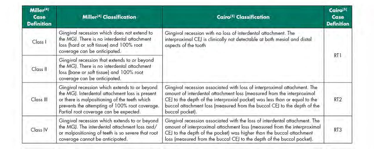
In 1985, Miller[4] proposed a gingival recession classification system that measured the severity (as graded I-IV) of recession utilizing the amount of remaining keratinized tissue (KT) in relation to the mucogingival junction (MGJ) and the presence or absence of interproximal bone loss. In 2011, Cairo[5] and coworkers proposed a new gingival recession classification system (as graded 1-3) based on the level of interproximal clinical attachment level (CAL) in reference to direct facial or lingual CAL. There are some noticeable departures with the newer system: KT width in relationship to the MGJ is not a metric; and there is no prognostic value attached to the diagnostic recession type. A comparison of the two systems is illustrated in Table 1.
According to Chambrone and Tatakis,[6] the S-CTG is the gold standard for Miller I-II (currently known as RT1[7]) defects in terms of achieving a gain in root coverage (RC), CAL and KT width. However, this technique is not without its limitations, as it requires not only the preparation of a recipient bed but the additional preparation of a donor site in order to obtain the connective-tissue graft, which has been
linked to greater patient discomfort and longer surgical procedure time.[8,9] Another limitation to this technique is the finite amount of graft material obtained from harvesting, potentially limiting the amount of root coverage possible.
An alternative allogeneic graft source that circumvents limitations encountered with autogenous grafts is the acellular dermal matrix (ADM). The proprietary processing method removes the epidermis and cells that can lead to graft rejection while preserving the dermal matrix components and vascular scaffold.[10,11]
It is well accepted that both allogeneic and autogenous graft material sources have clinically satisfactory success rates in terms of achieving root coverage.[12] However, specific clinical indications warrant different clinical approaches, and a key factor in determining what treatment approaches to use is the diagnosis of the recession category. As such, the purpose of this article is to highlight the importance of recognizing subtleties that may shift a RT1 into an RT2 category and, as such, affect the predictability of expectations for maximum versus complete root coverage.
Clinical Presentation
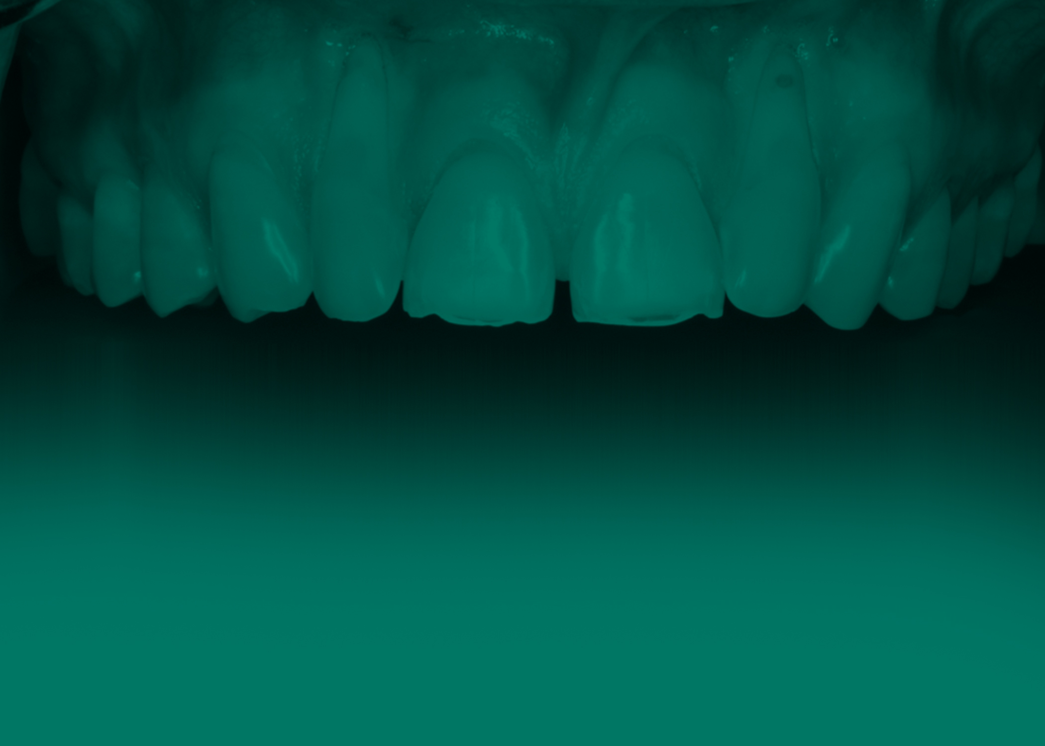
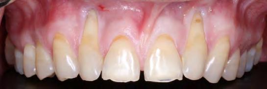
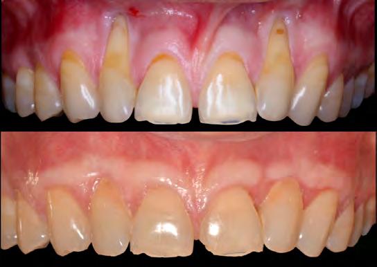
A 36-year-old male presented to the Manhattan Veterans Affairs Postgraduate Department of Periodontics with a chief complaint of dentinal hypersensitivity and “long teeth.” The patient’s medical history was significant for prior substance abuse. Clinical evaluation revealed mild gingival recession on both maxillary canines and central incisors, with severe recession present on the maxillary lateral inci-sors and a narrow zone of keratinized gingiva measuring approximately 1 mm (Figure 1). The patient’s phenotype was characterized as thick-scalloped, and the recession across teeth #6 through #11 was initially classified as RT1 Class A-2, with no loss of interproximal attachment. None of the teeth involved had pre-existing restorations or noncarious cervical lesions (NCCLs).
The patient’s oral hygiene and plaque control were excellent. The suspected primary etiology for gingival recession was potential factitial habits related to a history of substance abuse.
The goal of the treatment was three-fold: to re-establish harmony across all gingival zeniths; augment the zone of keratinized gingiva; and mitigate dentinal hypersensitivity.
Case Management
The patient provided written consent for the procedures and was informed that due to the severity of the defects, two mucogingival surgical procedures were likely. The first of the two procedures consisted of an ADM graft (PerioDerm Musculoskeletal Transplant Foundation, Edison, NJ) via a tunneling approach. The surgical sites were anesthetized using 2% lidocaine HCl with 1:100,000 epinephrine via local infiltration. Teeth #6 through #11 received an ADM soft-tissue graft via tunneling.[13] This technique was deemed most appropriate clinically due to the wide extent of tissue harvest that would be required, limiting comorbidity associated with such.
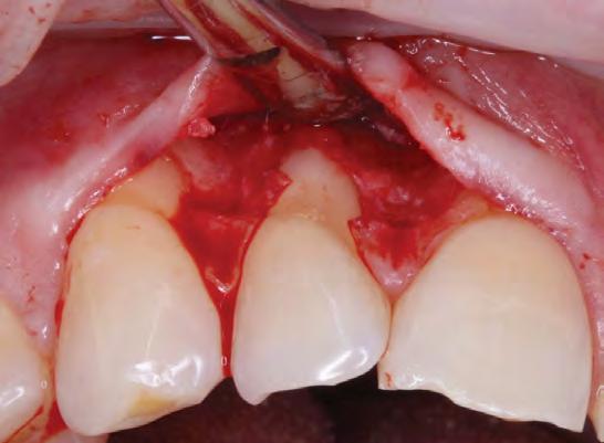
The exposed root surfaces were scaled and root planed. In accordance with Allen’s minimally invasive protocol,[14] one tunnel was prepared via sulcular incisions with an endcutting intrasulcular knife, (Allen end-cutting intrasulcular knife, black line. Hu-Friedy Manufacturing Co., Chicago, IL) followed by fullthickness dissection past the MGJ, extending to one tooth laterally in each direction beyond the gingival defects for adequate tunnel passivity and mobilization.
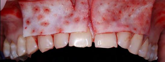
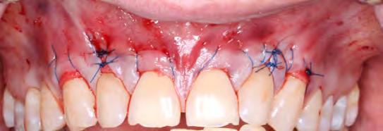
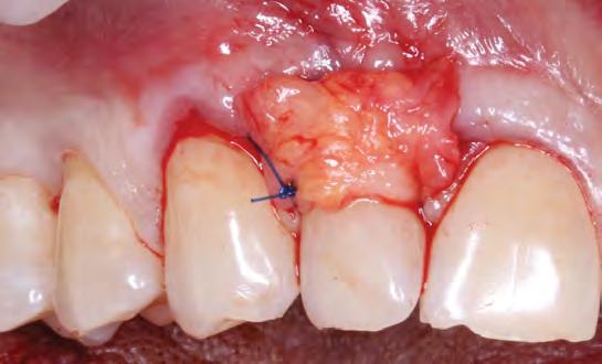
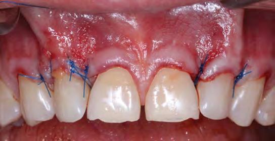
The root surfaces were then treated with 24% EDTA gel (PrefGel Institut Straumann AG, Basel, Switzerland). Two acellular dermal matrix grafts (1x4 cm, 0.89-1.65 mm thickness) were hydrated in sterile saline and introduced through the tunnel, according to manufacturer’s directions (Figure 2). Two sets of continuous 6-0 polypropylene sling sutures from teeth #6 through #8 and #9 through #11 were used to coronally position the flap and the marginal tissue around tooth #7 and tooth #10. Additional interrupted 6-0 polypropylene sutures were used to approximate the mid-facial tissue on tooth #7 and tooth #10 in accordance with the laterally closed tunnel technique[15] (Figure 3).
Postoperative antibiotics were dispensed (amoxicillin 500mg q8h, 7 days), and the sutures were removed at postoperative week three. The patient was recalled after one, two and four weeks and bimonthly for up to five months for clinical examination and oral hygiene reinforcement.
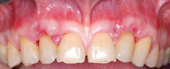
Teeth #7 and #10 still exhibited about 4 mm of exposed root surface after the tunneling procedure (Figure 4) and, thus, a secondary surgery was performed to attempt further root coverage utilizing two coronally advanced flaps in an envelope design, extending to one tooth adjacent mesially and distally, with an S-CTG (Figures 5,6). The flaps were then secured with interrupted sutures in 6-0 polypropylene (Figure 7). A S-CTG was determined the most appropriate graft at this time, as the surgical field was decreased from six teeth to only two after the results from the first surgical procedure. A smaller surgical site allows for a more feasible obtainment and application of a S-CTG that can be of the appropriate dimension to cover the recipient site.
Clinical Outcomes
Healing after Surgery #1: ADM and Tunnel
A clinically normal gingival appearance was noted at five months postoperatively; and probing depths ranged from 1 mm to 2 mm across all anterior teeth. The free gingival margin was located 4 mm apical from the mid-buccal CEJ of both lateral incisors, 1 mm apical from the CEJ on the canines, and at the level of the CEJ on the central incisors (Figure 4).
Healing after Surgery #2: SCTG and CAF
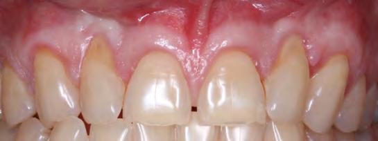
Follow-up at postoperative week two showed satisfactory early healing with mildly delayed healing in the papillary areas around teeth #7 and #10 (Figure 8). There was a noted increase in the width and thickness of keratinized tissue and 80% RC of both laterals and CRC of the central incisors and canines (Table 2). Long-term healing showed relapse of the lateral incisors of 3 mm to 4 mm but overall drastic improvement when comparing the preoperative situation to the final results (Figure 9).
Discussion
An allograft was chosen as the primary donor source, as there were multiple defects initially involved. Employing a tunnel design over multiple sites allows for passive flap mobilization while preserving optimal vascularization of the dermal graft without disturbing any delicate papillary tissue.[16] After the results of the first surgery, the defect type was converted from multiple to single. As such, a combined S-CTG and CAF was chosen for the second surgical approach, as the amount of graft needed was now attainable with palatal harvesting techniques. The quantified results are shown in Table 2.
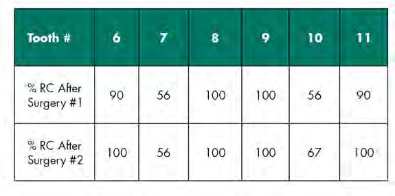
The results of the first tunneling procedure allow the authors to report 100% RC for the two central incisors and 90% RC on the canines. The lateral incisors both gained 56% RC, leaving about 4 mm of exposed root. This could be attributed to certain surgical details, such as leaving the ADM exposed over sites #6, #7, #10, #11 and flap tension. Short-term healing after the second procedure of SCTG + CAF resulted in 100% RC for the canines and 80% RC for the lateral incisors. However, long-term follow-up revealed relapse of the lateral incisors: #7 displayed 56% RC, and #10 displayed 67% RC. As all teeth involved in treatment were initially classified into RT1, one should expect 100% RC across all teeth based on results reported in the literature. However, a closer inspection reveals that the lateral incisors were rotated mesio-buccally, thus warranting a diagnosis of RT2, corresponding to a Miller Class III recession-type defect.[4]
The current classification system put forth by the 2017 World Workshop supports the system proposed by Cairo,[5] especially as multiple authors have brought to light potential disadvantages of the Miller classification system, such as vague criteria for diagnosis regarding the MGJ level and its lack of both exhaustiveness and simplicity.[17]
However, Miller attached a prognostic value to his classification system. As such, the class of the recession is the predictive factor for the surgeon to anticipate complete or partial root coverage. However, one must consider that the original Miller classification system was published in 1985, during a time when the free gingival graft (in either a one-step or two-step procedure) was much more prevalent. Since that time, more recent advances in surgical techniques and materials have led to an increased percentage and reliability of RC and CRC.[18]
Thus, using a classification system from 1985 juxtaposed with current concepts and techniques may translate into a discrepancy in what one can expect in terms of the case’s final result and initial classification. As such, the 2011 Cairo system was proposed as one that circumvents the drawbacks of Miller’s but uses the level of interproximal clinical attachment as its only criterion. Therefore, when prognosticating surgical results in cases of gingival recession over rotated teeth, one is left with the original 1985 system, which cites a likelihood of 50% RC. A prognosis of 50% RC is likely due to the “dead space” between the exposed root surface and blood supply derived from the alveolus and periodontal ligament, all factors that compromise nourishment of the donor graft.
Taking all the aforementioned into consideration, the results of the lateral incisors, averaging 64.5% RC and ranging from 57% to 72% RC, are well above what can be expected for a Miller III defect.
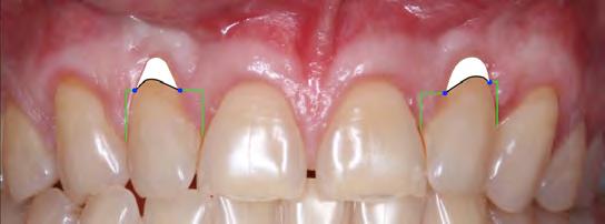
According to Zucchelli et al,[20] a rotated tooth will result in a malpositioned contact point on either the distal or mesial papilla, depending on the direction of rotation. This change in contact point in relation to the level of the CEJ correlates to a decrease in the papilla height, which, ultimately, offers less vascularity and nutritional source to the coronally advanced tissue and donor graft. In these situations, even though there is no appreciable interproximal bone loss, the expectation for complete root coverage (CRC) is not realistic, as the blood supply cannot reach the entire coronal aspect of the anatomic CEJ. Thus, it is more clinically pragmatic and realistic to calculate the maximum root-coverage level (MRC) instead of CRC and set the former as the goal.[20]
The authors of this paper have calculated the predetermined location of root coverage as the MRC to be expected after the second S-CTG + CAF surgery (Figure 10). The predetermination of the level of root coverage achievable is calculated as an imaginary line connecting two reference points provided by the mesial and distal transitional line angles in conjunction with the papillae height. It may seem that the papillary tissue is intact and interproximal CAL is adequate; however, ultimately, the limited prognosis put forth by Miller in 1985 is validated in that only about 50% RC is predictable.
Conclusions
In any clinician’s practice, it is imperative to identify the parameters that comprise “success” and “failure.” In a case presented as such, the authors hope to convey that success may not always be as successful as one thinks. A cursory look at this case would reveal an incorrect diagnosis and a subsequently incorrect prognosis. Should one apply the diagnosis of a RT1 for all affected teeth, surely the case would be deemed a failure. But taking into account all the nuances of the case presentation, one would realize that the lateral incisors are in fact RT2. As such, the results average 61.5% RC (and range from 56-67% RC), above what can be expected for such a defect and certainly successful along these definitions.
The authors report no conflicts of interest related to this case report. Queries about this article can be directed to Dr. Simmonds at Trevor.simmonds@va.gov.
REFERENCES
1. Cortellini P, Bissada N. Mucogingival conditions in the natural dentition: Narrative review, case definitions, and diagnostic considerations. J Periodontol 2018;89(Suppl 1): S204–S213.
2. Wennström J, Pini Prato GP. Mucogingival therapy. In: Lindhe J, Karring T, Lang NP, eds. Clinical Periodontology and Implant Dentistry. Copenhagen: Munksgaard; 1997: 569-591.
3. Albandar JM, Kingman A. Gingival recession, gingival bleeding, ad dental calculus in adults 30 years of age and older in the United States, 1988-1994. J Periodontal 1999;70: 30-43.
4. Miller PD. A classification of marginal tissue recession. Int J Periodontics Restorative Dent 1985; 5(2): 8-13.
5. Cairo F, Nieri M, Cincinelli S, Mervelt J, Pagliaro U. The interproximal clinical attachment level to classify gingival recessions and predict root coverage outcomes: an explorative and reliability study. J Clin Periodontol 2011; 38: 661–666.
6. Chambrone L, Tatakis D. Periodontal soft tissue root coverage procedures: a systematic review from the AAP Regeneration Workshop. J Periodontol 2015; 86 (2S): S8-S51.
7. Cortellini P, Bissada N. Mucogingival conditions in the natural dentition: Narrative review, case definitions, and diagnostic considerations. J Periodontol 2018;89(Suppl 1): S204–S213.
8. Del Pizzo M, Modica F, Bethaz N, Priotto P, Romagnoli R. The connective tissue graft: a comparative clinical evaluation of wound healing at the palatal donor site. A preliminary study. J Clin Periodontol 2002; 29: 848–854.
9. Griffin T, Cheung W, Zavras A, Damoulis P. Postoperative complications following gingival augmentation procedures. J Periodontol 2006;77: 2070-2079.
10. Wong A, Schönmeyr B, Singh P, Carlson D, Li S, Mehrara B. Plast. Reconstr Surg 2008; 121: 1144-1152.
11. Zucchelli G, Mounssif I. Periodontal plastic surgery. Periodontology 2000 2015; 68: 333-368.
12. Cairo F, Pagliaro U, Nieri M. Treatment of gingival recession with coronally advanced flap procedures: a systematic review. J Clin Periodontol 2008; 35 (Suppl. 8): 136–162.
13. Allen AL. Use of the supraperiosteal envelope in soft tissue grafting for root coverage. I. Rationale and technique. Int J Periodontics Restorative Dent 1994; 14: 217-227.
14. Allen EP. Subpapillary continuous sling suturing method for soft tissue grafting with the tunneling technique. Int J Periodontics Restorative Dent 2010; 30: 479-485.
15. Sculean A, Allen EP. The laterally closed tunnel for the treatment of deep isolated mandibular recessions: surgical technique and a report of 24 cases. Int J Periodontics Restorative Dent 2018; 38(4): 479-487.
16. Tavelli L, Barootchi S, Nguyen T, Tattan M, Ravidà A, Wang HL. Efficacy of tunnel technique in the treatment of localized and multiple gingival recessions: a systematic review and metanalysis. J Periodontol 2018; 89: 1075-1090.
17. Pini-Prato G. The Miller classification of gingival recession: limits and drawbacks. J Clin Periodontol 2011; 38: 243–245.
18. Chambrone L, Tatakis D. Periodontal soft tissue root coverage procedures: a systematic review from the AAP Regeneration Workshop. Journal Periodontol 2015; 86 (2S): S8-S51.
19. El Chaar E, Oshman S, Danesh-Sani S, Abed P, Castaño A. Increasing contact between soft tissue graft and blood supply. A technique for managing connective tissue grafting over prominent root. Journal of Cosmetic Dentistry 2016; 32(3): 52-66.
20. Zucchelli G, Testori T, DeSanctis M. Clinical and anatomical factors limiting treatment outcomes of gingival recession: A new method to predetermine the line of root coverage. J Periodontol 2006; 77: 714-721.


Trevor F. Simmonds, D.D.S., is director and assistant chief, Department of Postgraduate Periodontics, Veterans Affairs Manhattan Campus, New York, NY.
Stephanie M. Chu, D.M.D., is a member of the Department of Postgraduate Periodontics, Veterans Affairs Manhattan Campus, New York, NY, and in private practice in New York, NY.
Mike Roig, D.M.D., is in private practice in Miami, FL.
Kristian T. Poventud, D.M.D., is in private practice in Miami, FL.