17 minute read
Oral Manifestations of Metastatic Diseases


Oral Manifestations of Metastatic Diseases
A Clinical and Histologic Analysis of Seven Cases
Zeming Zheng, D.D.S.; Elizabeth Philipone, D.M.D.
ABSTRACT
Metastatic diseases comprise 1% to 3% of malignant oral neoplasms, with nearly one-third of patients unaware of having a primary cancer.[1-4,9] This retrospective analysis identified all oral lesions diagnosed as metastatic disease in the Columbia College of Dental Medicine Department of Oral Pathology’s oral biopsy database from 2015-2020 and recorded patient demographics, clinical presentation and diagnosis, origin of the primary, and whether the patient had known history of the disease. From 17,500 total cases, seven cases were identified as metastatic disease and described in detail. Clinicians’ recognition and awareness of these lesions’ often evasive, ambiguous oral presentations may help facilitate diagnosis, treatment and prognosis.
Metastatic disease to the oral cavity is uncommon, representing only 1% to 3% of all malignant oral neoplasms. These metastatic lesions can affect the bones, soft tissues or both.[1-3] However, lesions are two-times more likely to affect bone than oral soft tissue.[4,5] In particular, the molar region of the mandible is the most commonly affected osseous location.[1-3,6,7] With regards to soft tissue, the gingiva is the most common site for occurrence.[2-5,8] Oral metastases are more common in males and occur most frequently in middle-aged and older adults.
Metastasis to the oral cavity usually suggests end-stage disease, with a poor prognosis of 3.7- to 8.25-month survival time.[4,8-10] Oral metastases develop late in the disease process, with the majority of patients already having an established diagnosis of the malignancy.[9] However, in up to 30% of patients, the malignancy remains undetected until it presents in the oral cavity.[2,4,9]
Clinical presentation of metastatic disease in the oral cavity is dependent on the site of involvement. Patients with metastatic disease to the jaws report symptoms that include pain, swelling, paresthesia, numbness, toothache and loose teeth.[1,5,10,11] These signs and symptoms are nonspecific and can often mimic other common inflammatory conditions, such as periodontal disease or osteomyelitis.[12,13] Unfortunately, extracting the tooth in these cases may further promote metastasis of the cancer. Chronic inflammation, in itself, has also been linked to metastasis. Inflammatory chemokines may promote tumor cell proliferation and dissemination, and many common oral immunoinflammatory processes are associated with an increased chance of malignant transformation.[14]
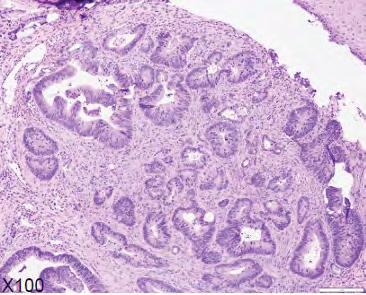
Radiographic presentation of jaw metastases is often described as ill-defined or “moth-eaten” radiolucency, but metastatic prostate and breast carcinomas may present as radiopaque or mixed lesions. Periodontal ligament space widening, fracture and cortical erosion can also be observed.[15]
For soft-tissue lesions, an exophytic nodular mass is the most common clinical presentation.[15] These lesions often resemble commonly occurring benign or reactive lesions, such as pyogenic granuloma, peripheral giant cell granuloma or fibrous epulis.[8]
In this retrospective analysis, we present epidemiologic and diagnostic data on a cohort of patients whose oral biopsy reports confirmed manifestations of metastatic diseases of known and unknown primary origins.
Methods
This study received approval from the institutional review board. The Department of Oral Pathology’s biopsy service database was searched from 2015 to 2020 to identify all biopsied oral lesions that were diagnosed as metastatic disease. Recorded data included patient sex and age at diagnosis, location of biopsy, description of clinical presentation, clinical diagnosis, histologic/biopsy diagnosis and whether the patient had a prior known history of the disease.
Results
Through the search of the oral biopsy database, out of 17,500 total cases, seven cases were histopathologically identified metastatic disease. These patients included five females and two males from 44 to 88 years old, with a mean age of 67.6 years at diagnosis. Three cases represented intraosseous lesions, all of which involved the left mandible. Three cases manifested in the soft tissue, including the maxillary gingiva, mandibular gingiva and mandibular retromolar area. One case involved both soft and hard tissue—presenting as a mandibular radiolucency with adjacent vestibular soft-tissue swelling (Table 1).
The intraosseous metastases presented in various ways, including “a destructive lytic lesion” and an unresolving periapical radiolucency. For two of the cases, the clinical diagnoses of metastatic breast carcinoma and metastatic prostate cancer were correct. These cases presented with clinical descriptions of a “destructive lytic lesion of the left mandible” and “pain and unresolving periapical radiolucency #19, #20,” respectively. Both patients were known to have metastatic disease to other sites prior to the oral biopsies. In one case that presented as a “persistent swelling of left posterior mandible #19,” an incorrect clinical diagnosis of reactive periostitis post root canal therapy was suspected. Upon biopsy this case was diagnosed as metastatic lung carcinoma. The patient was unaware of having lung cancer (Table 1).
As for the three metastases presenting in the oral soft tissues, two cases included the correct clinical diagnosis on the provided differential. The remaining case had incorrect clinical diagnosis of pyogenic granuloma.
For the one case that presented with involvement of bone and adjacent soft tissue, the clinical presentation was of a firm vestibular swelling with adjacent periapical radiolucency that was unresponsive to root canal therapy. A persistent infection was favored by the submitting clinician, with a comment to rule out neoplasm (Table 1).
In five of the seven cases, the patient was known to have a prior diagnosis of malignancy. Thus, in the remaining two cases, the initial manifestation of the disease was within the oral cavity and diagnosed at the time of the biopsy.
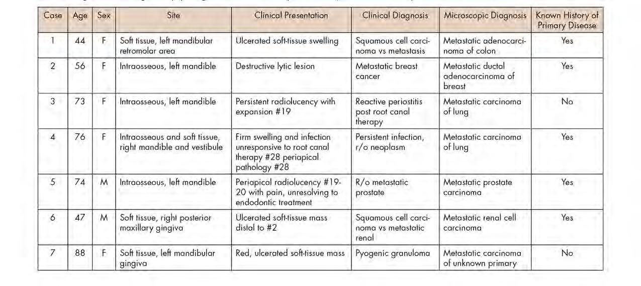
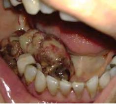
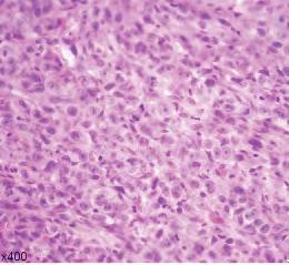
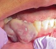
Figure 1. Representative clinical photos, radiographic images and histologic H&E slides of metastatic diseases presented.
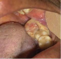


The microscopic diagnoses of these seven cases included colon adenocarcinoma (one case), ductal breast adenocarcinoma (one case), lung carcinoma (two cases), prostate carcinoma (one case) and renal cell carcinoma (one case).
The seventh case could not be further classified from the oral biopsy sample and was, therefore, diagnosed as poorly differentiated metastatic disease of unknown primary. This patient was subsequently found to have multiple metastatic deposits throughout the body, including the brain.
Table 1 presents a summary of recorded data, including patient sex and age at diagnosis, location of biopsy, description of clinical presentation, clinical diagnosis, histologic/ biopsy diagnosis and whether the patient had a prior known history of the disease. Figure 1 presents representative clinical, histologic and radiographic images of these cases.
Discussion
In this retrospective study, we identified seven cases with oral metastases out of a total of 17,500 biopsy reports (0.046%) over the past five years. This is consistent with the literature reporting that metastatic disease presenting in the oral cavity is very rare.[1]
In terms of site distribution, all three cases of bony metastases presented in the mandible, which is the most commonly affected bony location.[1-3,6,7] For the three cases of soft-tissue metastases, the gingiva was the most common site. This is, again, consistent with what is cited in the literature as the most commonly affected soft-tissue location.[2-5,8] Ninety percent of oral metastases are reported to present in the bone.[1] However, we found an equal distribution of cases presenting in the soft tissue (n=3) and in bone
(n=3). One case presented in both the bone and adjacent soft tissue. Murillo et al. also reported a slight variation in distribution, with a predominance of mixed metastatic presentation, followed by soft-tissue and then bony lesions.[10]
Figure 1. Representative clinical photos, radiographic images and histologic H&E slides of metastatic diseases presented.

Hirshberg et al. found that gender distribution differed for metastases presenting in bone vs. soft tissue, with an equal gender distribution in jaw bone metastases and 2:1 male predilection for soft-tissue metastases.[4] Although Murillo et al. found that there was a male predominance for all metastases, they stated that oral metastases are often found in similar proportions in male and female patients.[10]
Aniceto et al. reported a female-to-male ratio of 3:2 in a similar study of nine oral metastases presenting in the mandible, with breast adenocarcinoma being the most common primary tumor. Our results may be more in alignment with the findings of Aniceto et al. because of the similar cohort size with all bony metastases presenting in the mandible and a case of invasive ductal carcinoma of the breast that rarely presents in men. Although lung and colon cancer are both more common in men, the two cases of lung cancer and one case of colon cancer in our study presented in females, which may have further contributed to the female predilection.[16]
Although oral metastases can affect patients of all ages, the greatest prevalence occurs around 50 to 60 years of age:
51.5 years in men and 47.1 years in women; 52 years for bony metastases and 42 years for soft-tissue metastases.[4] In this study, patients ranged from 44 to 88 years old, with a mean age of 67.6 years at diagnosis: 60.5 years for males and 70 years for females; 67.7 years for bony metastases and 56.3 years for soft-tissue metastases. Therefore, in general, our cohort was slightly older in age.
The common sites of primary disease metastasizing to the oral cavity include breast, lung, kidney, liver and thyroid gland.[1-3,7,17] In the U.S., breast cancer is the most common primary malignancy to metastasize to the oral cavity in females, followed by lung and colorectal carcinoma. In males, prostate cancer is most common, followed by lung, colorectal and bladder cancer.[18,19] Sites of the primary disease in our patients included colon (n=1), breast (n=1), lung (n=2), prostate (n=1) and kidney (n=1). These were all among the most commonly reported sites of primary disease metastasizing to the oral cavity in the U.S. Lung cancer most often metastasizes to the gingiva, but in our case, presented in the mandible and mandibular vestibule.[21] Breast cancer mostly has been reported to more frequently metastasize to the gingiva, but in our study was found in the mandible.[21] The majority of prostate (80% to 90%) and colorectal cancer have been sited to metastasize to the bone, particularly the mandible, as consistent with our findings.[22,23]
Although oral metastases usually suggest advanced stage disease that has been previously diagnosed elsewhere in the body, in 22% to 30% of patients, the oral cavity is the initial site of diagnosis.[2,4,9] Two patients (28.6%) did not have prior history of a primary cancer before the oral biopsy.

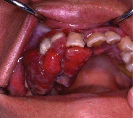
It is important to note that oral metastases can mimic more common reactive lesions, infections, or benign tumors, and without a history of primary malignancy, an oral metastatic lesion would not be clinically suspected (Figure 2). Similar presentations and differential diagno-ses have been reported in the literature as was provided by our group of submitting clinicians.[1,5,8,10,11] In patients with known primary disease, it may be helpful for clinicians to recognize some clinically differentiating signs of metastatic disease, such as a rapidly evolving lesion; tendency to bleed; changes caused by tumor growth, such as root resorption; ulcers; and necrosis.[23] Even then, oral metastases can remain a challenge to diagnose with only clinical signs and symptoms.
Despite the poor prognosis associated with oral metastatic disease, newer treatment options, such as anti-angiogenic agents, for example, Bevacizumab (anti-VEGF), have shown promise in treating metastatic colorectal cancer, non-small cell lung cancer, and renal cell carcinoma. Finally, new developments in next-generation sequencing could develop more personalized treatment plans that target specific mutations and offer better prognosis for patients.[24,25]
Conclusion
Metastatic diseases can present at various sites within the oral cavity in males and females of all ages. Their clinical presentation is often evasive and ambiguous, which complicates diagnosis, treatment and, potentially, prognosis. It is important for clinicians to be aware of the variety of possible clinical presentations of metastatic disease to the oral cavity and to be cognizant that the oral lesion is the initial presentation of the cancer in up to 30% of patients.
The authors report they have no conflicts of interest to disclose. Their research was supported by a College of Dental Medicine Summer Research Fellowship and received approval from the Columbia University institutional review board. Queries about his article can be directed to Zeming Zheng at zz2640@cumc. columbia.edu.
REFERENCES
1. Meyer I, Shklar G. Malignant tumors metastatic to the mouth and jaws. Oral Surg Oral Med Oral Pathol 1965;20:350–62. doi: 10.1016/0030-4220(65)90167-2.
2. Zachariades N. Neoplasms metastatic to the mouth, jaws and surrounding tissues. J Cranio Maxillofac Surg 1989;17(6):283–90. doi: 10.1016/s1010-5182(89)80098-8.
3. Hirshberg A, Leibovich P, Buchner A. Metastatic tumors to the jawbones: Analysis of 390 cases. J Oral Pathol Med 1994;23(8):337–41. doi: 10.1111/j.1600-0714.1994.tb00072.x.
4. Hirshberg A, Shnaiderman-Shapiro A, Kaplan I, Berger R. Metastatic tumours to the oral cavity—pathogenesis and analysis of 673 cases. Oral Oncol 2008;44(8):743-752. doi: 10.1016/j. oraloncology.2007.09.012.
5. D’Silva NJ, Summerlin DJ, Abdelsayed R, et al. Metastatic tumors to the jaws: Retrospective study of 101 cases. Oral Sur Oral Medic Oral Pathol Oral Radiol and Endod 2006;98(2):205206. doi: 10.1016/j.tripleo.2004.06.047.
6. Bodner L, Sion-Vardy N, Geffen DB, Nash M. Metastatic tumors to the jaws: a report of eight new cases. Med Oral Pathol Oral Cir Bucal 2006 Mar 1;11(2):E132-5.
7. Stypulkowska J, Bartkowski S, Pana’s M, Zaleska M. Metastatic tumors to the jaws and oral cavity. J Oral Maxillofac Surg 1979 Nov;37(11):805-8.
8. Allon I, Pessing A, Kaplan I, Allon DM, Hirshberg A. Metastatic tumors to the gingiva and the presence of teeth as a contributing factor: a literature analysis. J Periodontol 2014 Jan;85(1):132-9. doi: 10.1902/jop.2013.130118.
9. van der Waal RI, Buter J, van der Waal I. Oral metastases: report of 24 cases. Br J Oral Maxillofac Surg 2003 Feb;41(1):3-6. doi: 10.1016/s0266-4356(02)00301-7.
10. Murillo J, Bagan JV, Hens E, Diaz JM, Leopoldo M. Tumors metastasizing to the oral cavity: a study of 16 cases. J Oral Maxillofac Surg 2013 Sep;71(9):1545-51. doi: 10.1016/j. joms.2013.03.017.
11. McClure SA, Movahed R, Salama A, Ord RA. Maxillofacial metastases: a retrospective review of one institution’s 15-year experience. J Oral Maxillofac Surg 2013 Jan;71(1):178-88. doi: 10.1016/j.joms.2012.04.009.
12. Kumar G, Manjunatha B. Metastatic tumors to the jaws and oral cavity. J Oral Maxillofac Pathol 2013;17(1):71-75. doi:10.4103/0973-029X.110737.
13. Poulias E, Melakopoulos I, Tosios K. Metastatic breast carcinoma in the mandible presenting as a periodontal abscess: a case report. J Med Case Reports 2011;5:e1-e5. doi: 10.1186/17521947-5-265.
14. Hirshberg A, Berger R, Allon I, Kaplan I. Metastatic tumors to the jaws and mouth. Head Neck Pathol 2014 Dec;8(4):463-74. doi: 10.1007/s12105-014-0591-z.
15. Neville B, Damm DD, Allen C, Chi A. Bone Pathology. In: Saunders WB, Elsevier, eds. Oral and Maxillofacial Pathology. Missouri; 2016:572-631.
16. Ancieto GS, Penin AG, Pages RD, Moreno JJM. Tumors metastatic to the mandible: Analysis of nine cases and review of the literature. J Oral Maxillofac Surg 1990 Mar 1;48(3):246-51. doi: 10.1016/0278-2391(90)90388-I.
17. Antunes AA, Antunes AP. Gnathic bone metastasis: a retrospective study of 10 cases. Braz J Otorhinolaryngol 2008 Jul-Aug;74(4):561-5. doi: 10.1016/s1808-8694(15)30603-0.
18. Siegel R, Naishadham D, Jemal A. Cancer statistics, 2013. CA Cancer J Clin 2013 Jan;63(1):1130. doi: 10.3322/caac.21166.
19. Coleman R, Costa L, Saad F, Cook R, Hadji P, Terpos E, Garnero P, Brown J, Body JJ, Smith M, Lee KA, Major P, Dimopoulos M, Lipton A. Consensus on the utility of bone markers in the malignant bone disease setting. Crit Rev Oncol Hematol 2011 Dec;80(3):411-32. doi: 10.1016/j.critrevonc.2011.02.005.
20. Irani S. Metastasis to the oral soft tissues: A review of 412 cases. J Int Soc Prev Community Den 2016;6(5):393-401. doi:10.4103/2231-0762.192935.
21. Hasheminasab M, Karimi A, Kardoust MP, Kosari F, Asadi A. Metastasis of a prostate adenocarcinoma to mandible: A case report and review of literature. Clin Case Rep 2020;8:2063–66. doi: 10.1002/ccr3.3065.
22. Kameta A, Oda T, Ogura I, et al. Metastasis of colon adenocarcinoma to the mandible: A case report. Oral Science International 2017;14(1):22-25. doi:10.1016/s1348-8643(16)30023-4.
23. Nakamura E, Shimizu M, Itoh T, Manabe T. Secondary tumors of the pancreas: clinicopathological study of 103 autopsy cases of Japanese patients. Pathol Int 2001 Sep;51(9):686-90. doi: 10.1046/j.1440-1827.2001.01258.x.
24. Damodaran S, Berger MF, Roychowdhury S. Clinical tumor sequencing: opportunities and challenges for precision cancer medicine. Am Soc Clin Oncol Educ Book 2015:e175-82. doi: 10.14694/EdBook_AM.2015.35.e175.
25. Hyman DM, Solit DB, Arcila ME, Cheng DT, Sabbatini P, Baselga J, Berger MF, Ladanyi M. Precision medicine at Memorial Sloan Kettering Cancer Center: clinical next-generation sequencing enabling next-generation targeted therapy trials. Drug Discov Today 2015 Dec;20(12):1422-8. doi: 10.1016/j.drudis.2015.08.005.


Zeming Zheng, D.D.S., is a first-year endodontics resident at Columbia University College of Dental Medicine (CDM), New York, New York.
Elizabeth Philipone, D.M.D., is associate professor of dental medicine in pathology and cell biology, Columbia University College of Dental Medicine (CDM), New York, New York.