8 minute read
Successful Treatment of a ‘Pedo-Endo’ Lesion in a Primary First Molar

Successful Treatment of a ‘Pedo-Endo’ Lesion in a Primary First Molar
Case Report
Wayne W. Maibaum, M.A., D.M.D.; Steven R. Spitzer, D.D.S.
ABSTRACT
Patients with a mixed dentition can present for dental treatment with a nonvital primary tooth. The treatment of choice is often extraction, with the subsequent space presenting a cosmetic issue or requiring a space maintainer to preserve arch integrity for the erupting permanent teeth. Presented is a nonsurgical alternative treatment that saves the tooth, is less invasive, and may be preferred by some patients and their families. It is a procedure that may often be overlooked but can be included in the armamentarium of general dentists, pediatric dentists and endodontists.
Pulp therapy in primary teeth is a common dental treatment modality performed by general dentists, pediatric dentists and endodontists. The aim is to maintain a functional tooth and arch integrity in a growing child.[1] Pulpotomy is the treatment of choice when the pulp is vital and the diagnosis is reversible pulpitis. When the diagnosis is irreversible pulpitis or the pulp is necrotic, pulpectomy can be performed. This can allow the tooth to be retained instead of performing an extraction with the possible need for a space maintainer.[2]
Factors determining the choice between pulpectomy and extraction include the age of the child, caries risk, practitioner preference, restorability of the tooth involved, pathologic root resorption and the patient’s medical history.[3,4]
Premature loss of primary teeth can result in overeruption of opposing teeth, mesial drifting of teeth distal to the lost tooth and distal drifting of teeth mesial to the lost tooth.[5] Asymmetry of the dental arch may also occur.[6]
Although there is not much controversy regarding the need for space maintenance after the early loss of a primary second molar, the need for a space maintainer after the early loss of a primary first molar is debatable.[7] There are several factors to consider when assessing the need for clinical management of space loss when a primary first molar is prematurely lost.[8] Although the current case report presents endodontic treatment of a primary first molar, the technique can be applied to other teeth in the primary arch.
Case Report
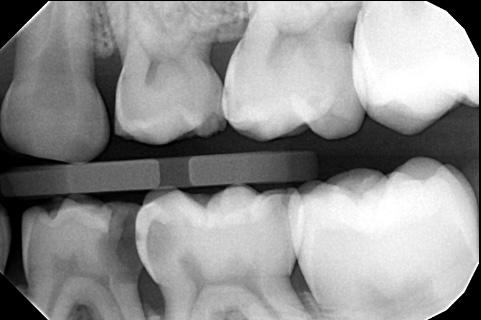
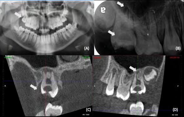
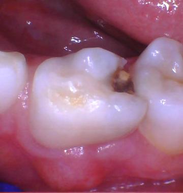
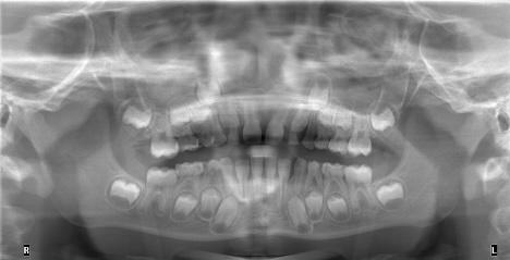
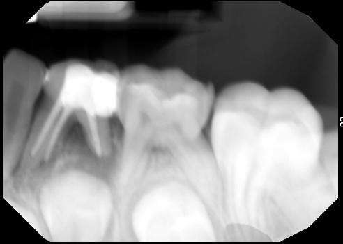
A 7-year-old male child presented to a private office for a routine exam and cleaning. Examination revealed multiple cavities, with a deep open carious lesion on the distal of #L. #L also exhibited a fluctuant swelling on the buccal gingiva near the furcation (Figure 1). A bitewing radiograph showed an apparent carious exposure in #L (Figure 2), and a panoramic radiograph suggested incipient bone loss in the furca (Figure 3). Extraction was considered, but since an endodontist was available in the practice, a consult was scheduled to see if the tooth was salvageable. The endodontist decided to try to save the tooth at the time of the consultation.

After gaining consent from the patient’s mother, tooth #L was adequately anesthetized with an infiltration injection of 1 carpule of lidocaine 2% with 1:100,000 epinephrine, which in a young child can have the same effect as a mental injection. A rubber dam was placed and secured on tooth #K with a #2A rubber dam clamp. Decay was excavated and the pulp was found to be nonvital.
Three canals were located. They were instrumented to the size of the Primary (25.07, red) Wave One Gold reciprocating file (Dentsply Sirona, Johnson City, TN) to a length of about 16 mm. The canals were irrigated with sodium hypochlorite, dried with paper points and filled with zinc oxide eugenol (Dentonics, Monroe, NC) using a vertical condensation method by tamping the filling material down with a moist cotton pellet.
The tooth was restored with IRM intermediate restorative material (Dentsply Sirona, Charlotte, NC), and a postoperative periapical radiograph was taken (Figure 4). In addition, a small incision was made in the buccal swelling for decompression, and the sinus tract was gently curetted with a small spoon excavator.
The patient was very cooperative and tolerated the procedure well. He was discharged, and his mother was told to return to the office to continue his restorative work with the general dentist. She was also advised to contact the office if the patient had any problem with the treated tooth.
The patient did not return to complete his recommended treatment plan. However, at the urging of the endodontist, he returned one year later to check tooth #L. The IRM temporary filling was mostly intact, but the tooth was never properly restored. The existing caries was never treated. However, the soft tissue around #L healed well.
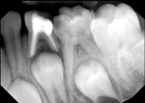
A one-year follow-up periapical radiograph of #L was taken (Figure 5). The film showed continued eruption of the succedaneous tooth #21, along with some resorption of the apices and some filling-in of bone in the furcation area of tooth #L. The patient’s mother reported that her son had no problem with the endodontically treated tooth. She was advised that tooth #K might need similar treatment.
Discussion
When a child presents to the dental office with a nonvital pulp in a primary tooth, a choice must be made whether to extract or treat it endodontically. Extraction can be a traumatic procedure for the patient and/or the patient’s family. Extraction often involves concern for space maintenance of the extraction site to preserve the integrity of the arch as the patient grows. The placement of a space maintainer requires additional visits, often necessitating maintenance of the space maintainer itself. It is reasonable to expect that space maintainers may need replacement or repair during treatment.[9] As such, primary teeth have been described as “the best space maintainers.”[10]
Different root canal filling materials have been tried to complete root canal therapy on primary teeth. The criteria for an ideal filling material includes the following: has antibacterial properties, resorbs at the same rate as the roots, lacks harm to the periapical area or succedaneous teeth, fills the canals easily, adheres to the walls of the canals, resorbs if extruded beyond the apex, is radio-opaque, doesn’t cause discoloration of the tooth.[11,12] Since 1930, zinc oxide eugenol has been the conventional root canal filling material for primary teeth pulpectomy.[13]
Conclusion
Presented is a case describing the endodontic treatment of a nonvital primary tooth. A one-year follow-up demonstrates an asymptomatic tooth with normal visual and radiographic findings in an 8.5-year-old patient. Since the exfoliation of primary mandibular first molars normally occurs between the ages of 9 and 11 years, the case appears to be successful. It is presented as a viable alternative to extraction.
Queries about this article can be sent to Dr. Maibaum at WMaibaum@aol.com.
REFERENCES
1. Ritwik P. A review of pulp therapy for primary and immature permanent teeth. J Calif Dent Assoc 2013 Aug;41(8):585-595.
2. Sebourn S, et al. Pulpectomy versus extraction for the treatment of nonvital primary second molars: a retrospective chart review. J Clin Ped Dent 2020;44(5): 302-306.
3. Hargreaves KM. Cohen’s pathways of the pulp. Eleventh ed. St. Louis, Missouri: Elsevier; 2016:e1-e44.
4. Dunston B, Coll JA. A survey of primary tooth pulp therapy as taught in US dental schools and practiced by diplomates of the American Board of Pediatric Dentistry. Ped Dent 2008;30(1):42-48.
5. Dean JA, et al. Dentistry for the child and adolescent. Ninth ed. Maryland Heights, Missouri: Mosby; 2010:553-572.
6. DaBell J, Huang GJ. Evidence indicates minimal short-term space loss after premature loss of primary first molars: a critical summary of Tunison. JADA 2010;141(1):77-78.
7. Tunison W, et al. Dental arch space changes following premature loss of primary first molars: a systematic review. Ped Dent 2008;30(4):297-302.
8. Mosharrafian S, et al. Clinical evaluation for space maintainer after unilateral loss of primary first molar in the early mixed dentition stage. Int J Dent 2021; Dec:1-6.
9. Ahmad AJ, et al. Methods of space maintenance for premature loss of a primary molar: a review. Eur Arch Ped Dent 2018;19:311-320.
10. Kratunova E, Silva D. Pulp therapy for primary and immature permanent teeth: an overview. Gen Dent 2018;66(6):30-38.
11. Garcia-Godoy F. Evaluation of an iodoform paste in root canal therapy for infected primary teeth. ASDC J Dent for Child 1987;54:30-34.
12. Rifkin A. A simple, effective, safe technique for the root canal treatment of abscessed primary teeth. ASDC J Dent for Child 1980;47:435-441.
13. Najjar RS, et al. A comparison of calcium hydroxide/iodoform paste and zinc oxide eugenol as root filling materials for pulpectomy in primary teeth: a systematic review and metaanalysis. Clin Exp Dent Res 2019;5:294-310.

Wayne W. Maibaum, M.A., D.M.D., FAGD, is a retired life member of the ADA. He lives in Danbury, CT.

Steven R. Spitzer, D.D.S., is a board-eligible endodontist. He is in private practice in Warwick, NY.