16 minute read
Treatment Options for Pre-eruptive Intracoronal Resorption of Permanent Teeth A Report of Two Cases


Treatment Options for Pre-eruptive Intracoronal Resorption of Permanent Teeth A Report of Two Cases
Dhaval Shah, D.D.S.; Bret Lesavoy, D.M.D.; Richard Yoon, D.D.S.
ABSTRACT
Pre-eruptive intracoronal resorption (PEIR) is a condition found in 2% to 8% of cases, initially seen as a radiolucent area beneath the dentino-enamel junction in unerupted teeth. This case series highlights pre-eruptive intracoronal resorption (PEIR) diagnosis and diverse management approaches for two presentations: one with symptomatic maxillary second molar PEIR and another with incidentally discovered mandibular second molar PEIR.
Often confused with traditional dental caries due to its radiolucent appearance, it was previously termed “pre-eruptive” caries. Differentiating between “progressive” and “nonprogressive” forms is crucial. Treatment options depend on radiographic extent, lesion progression and clinical symptoms.
Pre-eruptive intracoronal resorption (PEIR) refers to a dentinal defect occurring just below the dentin-enamel junction in the occlusal aspect of an unerupted tooth.[1,2] Typically discovered incidentally during routine dental radiographs, such as panoramic, periapical and bite-wing radiographs, PEIR exhibits radiolucent characteristics resembling dental caries or coronal resorption in unerupted teeth.[3] These lesions, commonly located in coronal dentin, can appear as single or multiple entities in both maxillary and mandibular arches.[4]
Notably, teeth affected by PEIR often lack detectable defects or pathways for bacterial ingress on the outer enamel surface, with minimal clinical distinctions observed between affected and adjacent/contralateral teeth.[5]
The etiology of PEIR remains uncertain, given the tooth’s crypt encasement during development, making it unlikely to be infected by oral cariogenic microorganisms.[2] Local factors, such as adjacent abutting teeth or ectopic positioning, can exert pressure, prompting resorptive cells to invade dentin through enamel fissures or the cementoenamel junction.[6] Histological examinations of soft tissue in most cases reveal signs of resorption, marked by resorptive cells (macrophages, osteoclasts and multinucleated giant cells) and scalloped lesion borders. Importantly, there is no evidence of microbial invasion, dental caries or pulpal degeneration in PEIR-affected teeth.[4]
Pre-eruptive intracoronal resorption lesions are categorized as either progressive (developing) or non-progressive (static). Non-progressive lesions, characterized by their static and asymptomatic nature, often prompt a conservative approach. Practitioners may opt to allow the teeth to naturally erupt into the oral cavity, while actively monitoring for radiographic signs of progression before considering treatment.
Conversely, urgent treatment is recommended for progressive lesions, given their advancing and symptomatic characteristics. Utilizing periodic radiographic examinations to classify the lesion as non-progressive or progressive plays a crucial role in facilitating appropriate treatment planning.[7]
Pre-eruptive intracoronal resorption lesions exhibit a prevalence ranging from 2% to 8% depending on the subject and 0.6% to 2% based on the tooth and radiograph type, with notable impact on mandibular first premolars, second permanent molars and third permanent molars.[1] Typically, individuals experience involvement of a single tooth, and approximately half of PEIR lesions extend beyond two-thirds of dentin thickness. Notably, no discernible associations have been identified between PEIR and factors such as sex, race, medical conditions, systemic factors or fluoride supplementation.[6]
The aim of this case series is to delineate the diagnosis of PEIR in permanent teeth and present diverse treatment options, guided by clinical and radiographic manifestations. The initial case details a patient with PEIR in an unerupted maxillary permanent second molar, identified through pronounced clinical signs and symptoms. The subsequent case outlines the incidental radiographic discovery of PEIR in an unerupted mandibular permanent second molar.
Case One
In December 2022, a 9-year-old female sought treatment at Children’s Hospital of New York-Presbyterian Emergency Department, a large academic medical center in New York, NY, for acute dental pain on the right side and mild facial swelling. Her medical history included a recent episode of right-sided Bell’s palsy in July 2022, attributed to Lyme IgM infection; it had been successfully managed with doxycycline.
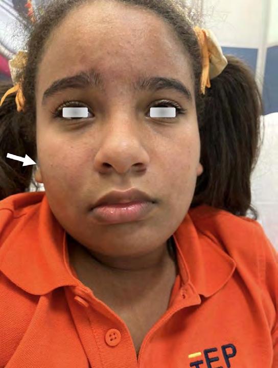
Dental examination revealed mild, right-sided facial swelling, subtle asymmetry and no apparent distress (Figure 1). No erythema, trismus or lymphadenopathy was evident. Intraoral examination showed no visible caries, abscess, swelling, drainage or floor-of-mouth elevation. Partial eruption of the right mandibular second permanent molar with localized moderate gingival inflammation was observed.
Based on clinical presentation, pericoronitis of the partially erupted mandibular permanent right second molar was diagnosed. Pain management included alternating Tylenol and ibuprofen, along with continuing the prescribed amoxicillin regimen. Follow-up at the ambulatory dental clinic for further evaluation and radiographs was recommended.
At the ambulatory dental clinic the next day, a thorough evaluation was conducted, revealing no apparent changes in the patient’s extraoral presentation compared to the previous day. Intraoral examination identified gingival pain upon palpation around the unerupted right maxillary permanent second molar and tenderness to percussion and palpation of the partially erupted right mandibular permanent second molar. Clinical examination indicated well-perfused, pink and stippled attached gingiva along the alveolar ridge distal to the right maxillary permanent first molar.
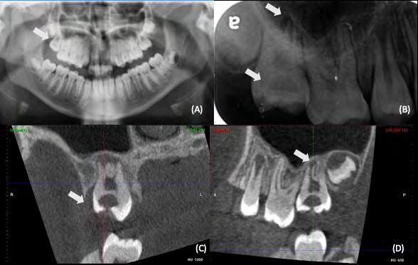
Panoramic and periapical radiographs displayed an unerupted maxillary permanent right second molar with a well-defined radiolucent area in the coronal portion, extending apical of the occlusal dentino-enamel junction and approximating the pulp (Figures 2A, B). Additionally, the periapical radiograph revealed a periapical radiolucency along the developing mesial and palatal root. A preliminary diagnosis of pre-eruptive intracoronal resorption with periapical involvement was established, prompting a referral for cone beam-computed tomography (CBCT) to confirm the diagnosis. Meanwhile, the patient was advised to use over-the-counter Tylenol and ibuprofen as needed for pain relief and to complete the earlier prescribed amoxicillin regimen.

During the interval before the scheduled CBCT appointment, the patient revisited the ambulatory dental clinic after two days, reporting escalating symptoms, which now included nocturnal pain and reduced food intake. Extraoral examination demonstrated persistent, mild facial swelling on the right side; pain upon palpation near the right zygomatic arch and coronoid process; and a newly developed limited opening of approximately 15 cm (partial trismus). Intraoral examination revealed continued pain upon palpation posterior to the right maxillary permanent first molar. Additionally, the patient maintained localized mild gingival inflammation at the right permanent mandibular second molar.
Subsequently, expedited CBCT and radiographic evaluation affirmed the diagnosis of pre-eruptive intracoronal resorption in the right maxillary permanent second molar. Imaging depicted a developing, unerupted follicle with absent enamel at the central crown and communication between the pulp and the alveolar bone encasing the tooth (Figures 2C, D). Open apices were observed on the maxillary permanent right second molar. An endodontic consultation confirmed immature root formation. Considering the lesion’s proximity to the pulp and its unfavorable prognosis, surgical extraction of the maxillary permanent right second molar was recommended after consultation with the endodontist.
Extraction of the maxillary permanent right second molar took place in an outpatient setting. The tooth was exposed using a 15 blade, elevators and forceps. Microscopic examination identified curved fragments of soft tissue characterized by fibrous connective tissue containing focal rests of odontogenic epithelium. Additionally, spicules of hard tissue and focal inflammatory cells were observed. The histologic diagnosis confirmed dental follicular tissue in the right posterior maxilla.
Extraction of the maxillary permanent right second molar took place in an outpatient setting. The tooth was exposed using a 15 blade, elevators and forceps. Microscopic examination identified curved fragments of soft tissue characterized by fibrous connective tissue containing focal rests of odontogenic epithelium. Additionally, spicules of hard tissue and focal inflammatory cells were observed. The histologic diagnosis confirmed dental follicular tissue in the right posterior maxilla.
During the patient’s follow-up appointment, no signs or symptoms of odontogenic infection or pain were detected. Extraoral examination demonstrated the resolution of swelling, with the absence of pain and tenderness upon palpation of the right zygomatic and coronoid processes. Additionally, there were no indications of trismus, and the inferior border of the mandible was palpable.
Intraoral examination revealed the absence of vestibular swelling or fistula-like lesions on the gingiva, indicating apparent resolution of pericoronitis associated with the right mandibular second permanent molar. Clinical examination indicated well-perfused, pink, stippled attached gingiva along the alveolar ridge distal to the right maxillary permanent first molar, with mild plaque-induced gingivitis. Moreover, the right mandibular permanent second molar exhibited increased eruption compared to six months prior, with no overlying gingiva.
Radiographic evaluation revealed no abnormalities, including the absence of radiolucencies suggestive of caries or pathology. The anatomy of the maxillary right permanent first molar, alveolar bone and supporting tissues appeared normal (Figure 3).
Case Two
A 16-year-old male, in good health with no notable medical history or known food/drug allergies, has been a longstanding patient. He denies any prior dental injuries but has a history of restorations in both primary and permanent dentition.
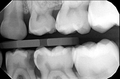
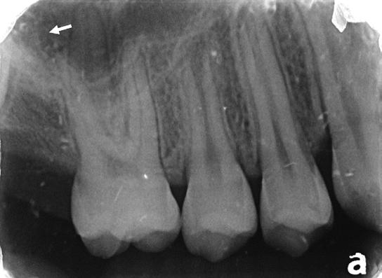
In August 2015, the patient underwent extraction of the maxillary left primary first molar due to dental caries and an acute odontogenic infection. A panoramic radiograph from the same month revealed a well-circumscribed radiolucent lesion in the coronal portion of the unerupted left mandibular permanent second molar, extending below the occlusal dentino-enamel junction (Figure 4A). Despite the absence of signs or symptoms and no significant pathology in the lower left quadrant, the lesion has been regularly monitored through periodic radiographs.

Since 2015, the patient has been on a six-month recall schedule. A bitewing radiograph from December 2020 revealed an erupted mandibular left second permanent molar; the lesion size and shape were consistent with the radiograph from five years earlier (Figure 4B). Subsequent bitewing radiographs from March 2022 (Figure 4C) and February 2023 (Figure 4D) show no discernible changes in the clinical or radiographic appearance. There is no apparent progression in the size or location of the lesion across the film series.
The patient consistently attends periodic examinations for oral hygiene maintenance and remains under active monitoring due to the non-progressing and asymptomatic nature of the lesion.
Discussion
The initial case involved a progressive and symptomatic pre-eruptive intracoronal resorption affecting the maxillary right second permanent molar. Currently, there is no established treatment protocol for pre-eruptive intracoronal resorption. Existing literature proposes a conservative approach involving monitoring the tooth until complete eruption. Periodic radiographic assessments facilitate active monitoring, enabling the evaluation of lesion-size progression. Treatment intervention, such as restorative treatment or surgical measures, is considered upon eruption, contingent on the identification of lesion-size progression, advancement to the pulp, and/or the occurrence of tooth fracture.[8]
In most instances, the identification of pre-eruptive intracoronal resorption in a developing, unerupted tooth is coincidental during routine radiographic assessment. Atypical resorption typically progresses gradually until the crown emerges into the oral cavity. Following eruption, the invasion of cariogenic microorganisms into the resorbed dentin area accelerates lesion growth, rendering it more conspicuous.[9]
Despite an unknown etiology, several theories, including ectopic tooth position, have been proposed. Localized pressure induces damage to the unmineralized tooth layer, initiating intracoronal resorption. Currently, it is hypothesized that such factors (i.e., localized pressure via ectopic eruption) lead to an interruption of crown formation, allowing the invasion of resorptive cells into forming dentin.[10]
Several case reports recommend surgical exposure of the unerupted tooth to halt the resorptive process and prevent progression to the pulp through early restorative treatment. Glass ionomer restorative materials are frequently suggested postsurgical exposure due to their advantageous properties, including minimal tooth preparation, moisture tolerance and fluoride release.[8,11,12]
In one case report involving pre-eruptive intracoronal resorption of an unerupted mandibular permanent second molar, surgical retraction of the gingival tissue covering the unerupted tooth was performed. Caries-like tissue was mechanically removed and substituted with glass ionomer material. Post-eruption, the glass ionomer interim restoration was subsequently replaced with a more durable material. A six-month follow-up indicated no clinical symptoms and normal ongoing root development.[13]
While surgical gingival retraction is a viable option, its efficacy is limited in providing adequate exposure for an unerupted tooth. This limitation increases the likelihood of incomplete lesion removal, potentially resulting in acute dental pain and sensitivity after tooth eruption into the oral cavity. Immature teeth exhibit dentinal tubules that are more permeable than those in post-eruptive mature teeth, heightening the risk of swift bacterial progression into or near the pulp, ultimately causing irritability in the affected tooth. In such cases, the restorations may need replacement following eruption, and vital pulp therapy—such as excavation and partial pulpotomy—might be necessary to achieve a more favorable prognosis.[13]
If vital pulp therapy and restoration prove unsuccessful for a previously affected tooth, several alternatives can be considered. Depending on the root development and eruption status at the time of treatment failure, apexification or nonsurgical root canal treatment may be viable options. In cases where symptoms persist despite subsequent restorations and/or systemic symptoms emerge, extraction becomes a feasible choice to eliminate the source of odontogenic infection.[14]
While literature on the subject is limited, there are scarce reports of extracting permanent teeth affected by pre-eruptive intracoronal resorption due to systemic infection. In a case study involving a 2-year-old male with leftsided facial cellulitis initially diagnosed as pericoronitis, persistent symptoms of systemic infection led to further assessment by pediatric dentistry. Clinical and radiographic evaluation revealed PEIR extending to the pulp of the partially erupted left mandibular primary second molar. Due to a hopeless prognosis, the tooth was extracted.[15]
Similar to the case presented in this series, where a progressive lesion resulted in systemic signs and symptoms, including right-sided facial cellulitis secondary to PEIR, the most appropriate treatment option was determined to be the extraction of the affected unerupted maxillary right second molar.
It is advisable to seek timely orthodontic consultation for potential treatment planning involving molar substitution. In this approach, the adjacent third molar can be orthodontically shifted into the position previously occupied by the extracted molar. Additionally, an alternative and novel option, not previously reported in existing literature or studies of this kind, involves auto-transplantation of the adjacent molar. This procedure is considered viable once the molar in question has completed two-thirds of its root development.[14]
Despite various potential treatment interventions, monitoring the lesion remains an option if it is static and asymptomatic. A prior case report documented the active monitoring of a patient with non-progressive and asymptomatic pre-eruptive intracoronal resorption (PEIR) in a permanent mandibular left second molar over nine years. Due to the lesion’s non-progressive and asymptomatic nature, coupled with the patient’s low caries risk and regular oral hygiene maintenance, post-eruption monitoring involved minimal intervention, specifically, sealant placement.[7]

In another case, a patient underwent regular followups for a PEIR lesion on a left maxillary first premolar. After seven years of active monitoring, restorative treatment intervention was planned due to the lesion’s progressive nature, increased caries risk and a higher likelihood of tooth structure loss.[16]
Similarly, the second case in this series has been consistently monitored. As the radiolucency remains static, and no signs or symptoms are evident, no treatment has been administered, and the mandibular left second permanent molar has been monitored for eight years. Considering the potential for lesion progression, however, ongoing monitoring for radiographic and/or clinical signs and the possible need for treatment intervention continues as outlined previously.
Conclusion
The initial case necessitated the extraction of the right unerupted second maxillary molar, driven by its progressive nature and the manifestation of systemic signs and symptoms, notably, right-sided facial cellulitis stemming from a PEIR-induced odontogenic infection. Conversely, the second case remains under active monitoring due to its nonprogressing and asymptomatic characteristics.
The approach to treating PEIR is contingent on factors such as lesion size, the progressive/static nature of the tooth, clinical symptoms, and the eruption status and root development. Close inspection of periodic radiographs is important for early detection and proper management. Advances in diagnostic techniques and dental materials have improved outcomes for teeth affected by PEIR. An interdisciplinary approach, incorporating specialties like endodontics, orthodontics, and oral and maxillofacial surgery, is imperative to optimize outcomes for PEIR.
The authors declare no financial, economic or professional interests that may influence positions presented in this case series. Queries about this article can be sent to Dr. Shah at dhavalshah1996@hotmail.com.
REFERENCES
1. Zilberman U, Milevski I, Yegorov D, Smith P. A 3000-year-old case of an unusual dental lesion: pre-eruptive intracoronal resorption. Arch Oral Biol 2019;97:97-101.
2. Seow WK. Pre-eruptive intracoronal resorption as an entity of occult caries. Pediatr Dent 2000;22:370-375.
3. Seow WK, Hackley D. Pre-eruptive resorption of dentin in the primary and permanent dentitions: case reports and literature review. Pediatr Dent 1996;18(1):67-71.
4. Counihan KP, O’Connell AC. Case report: pre-eruptive intracoronal radiolucencies revisited. Eur Arch Paediatr Dent 2012;13(4):221-226.
5. Hata H, Abe M, Mayanagi H. Multiple lesions of intracoronal resorption of permanent teeth in the developing dentition: a case report. Pediatr Dent 2007;29(5):420-425.
6. Seow WK, Lu PC, McAllan LH. Prevalence of pre-eruptive intracoronal dentin defects from panoramic radiographs. Pediatr Dent 1999;21:332-339.
7. Manmontri C, Mahasantipiya PM, Chompu-Inwai P. Pre-eruptive Intracoronal radiolucencies: detection and nine years monitoring with a series of dental radiographs. Case Rep Dent 2017;2017:6261407.
8. Le VNT, Kim JG, Yang YM, Lee DW. Treatment of pre-eruptive intracoronal resorption: a systematic review and case report. J Dent Sci 2020;15(3):373-382.
9. Czarnecki G, Morrow M, Peters M, Hu J. Pre-eruptive intracoronal resorption of a permanent first molar. J Dent Child 2014;81:151-155.
10. Al-Batayneh OB, Al-Tawashi EK. Pre-eruptive intracoronal resorption of dentine: a review of etiology, diagnosis, and management. Eur Arch Paediatr Dent 2020;21:1-11.
11. Donly KJ, Segura A. Fluoride release and caries inhibition associated with a resin-modified glass-ionomer cement at varying fluoride loading doses. Am J Dent 2002;15:8-10.
12. Spierer WA, Fuks AB. Pre-eruptive intracoronal resorption: controversies and treatment options. J Clin Pediatr Dent 2014;38(4): 326-328.
13. Davidovich E, Kreiner B, Peretz B. Treatment of severe pre-eruptive intracoronal resorption of a permanent second molar. Pediatr Dent 2005;27(1):74-77.
14. Alon E, Amato R, Ptak D. Pre-eruptive intracoronal resorption (PEIR): a case report. J Endod 2023;39(2):224-228.
15. Morgan N, Smart G. Pre-eruptive intracoronal resorption defect of erupting primary molar leading to facial cellulitis: a case report. Int J Paediatr Dent 2022;33(2):178-180.
16. Currell SD, Cakar T. Incidental observation of pre-eruptive intracoronal resorption after seven years. Aust Dent J 2019;64(4):376-379.

Dhaval Shah, D.D.S., is a private practitioner in Lodi, NJ, and a former postdoctoral resident in pediatric dentistry at New York-Presbyterian and Columbia University College of Medicine, New York, NY.

Bret Lesavoy, D.M.D., is a private practitioner in Allentown, PA, and assistant clinical professor, University of Pennsylvania, School of Dental Medicine, Philadelphia, PA.

Richard Yoon, D.D.S., is professor of dental medicine, Columbia University Medical Center, New York, NY.