Volume 04 / Issue 04 / December 2016
Page 24
boa.ac.uk
JTO Features
The use of 3D printing in paediatric orthopaedics for pre-operative planning and bespoke therapeutics Jim Ballard & Daniel Crawford
Paediatric orthopaedic surgeons regularly carry out a large number of procedures on unusual, congenital and acquired deformities. Traditional radiographic techniques are often employed to plan such surgery. The 3D printed models have been used to improve diagnosis and enhance pre-operative planning in a number of procedures. This article highlights the application of 3D printing applied to three paediatric cases from the Royal Belfast Hospital for Sick Children. Developmental Dysplasia of the Hip A late presenting dysplastic left hip in which the hip was not dislocated but contained within
a high volume acetabular socket. The patient presented at six years old with a leg length discrepancy and discomfort after playing sport.
the anatomy, a CT and MRI scan were performed to determine the volume of the acetabulum and the shape of the femoral head.
An open reduction, capsulorrhaphy and Pemberton osteotomy was performed for containment. The operation was successful, although the acetabulum was still enlarged. It was decided that the patient required further pelvic surgery. However, given the complexity of
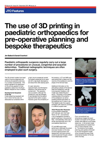
Additional information on the articulating surfaces of the femoral head and acetabulum was felt to be required; therefore, a 3D model was created (Figures 1 and 2). This gives a much greater understanding of the anatomy of the acetabulum as well as enabling artificial articulation of the scale joint as if it were in situ. This helped to plan the surgical correction with maximum coverage, in a difficult surgical case1. Tibial Physeal Bar Resection A young patient presented with knee pain and mild fixed flexion of 10o. Conventional x-ray investigations showed a growth arrest bar in the proximal tibia. Coincidentally, there was a similar asymptomatic bar in the contralateral distal femur.
Jim Ballard
Daniel Crawford
CT and MRI scans were performed to delineate the extent and exact location of the bars so that surgical excision and replacement with bone cement could be planned through a direct transmetaphyseal window.
Figure 1: Close up of degraded acetabulum showing articulating surface in white resin to represent bony anatomy
Figure 2: Proximal femoral head along with dysplastic left hip to accurately assess volume of acetabulum and femoral head condition
From conventional two dimensional images the exact location and extent of the growth arrests were difficult to accurately determine. A 3D printed model was requested and created from the CT scans (Figures 3 and 4). The anatomy and surgical planning were greatly enhanced by the 3D transparent model, which showed the exact three dimensional location of the bar within the tibia.