
9 minute read
Thoracolumbar fracture stability and the difficulties of classification systems

Alexander Durst and Sashin Ahuja
Alexander Durst is currently a Senior Spinal Fellow at the Welsh Centre for Spinal Surgery and Trauma, in Cardiff. He was the first Spinal Trainee Interface Group Fellow for Health Education England. He subsequently completed a fellowship in Complex Spine Surgery at the University of Alberta, in Edmonton, Canada. His interests include spinal trauma and surgical education. He is an East of England T&O rotation alumnus.
Sashin Ahuja is a Consultant Orthopaedic Spinal Surgeon at Cardiff and Vale University Health Board since 2003. His practice covers the full spectrum of spinal disorders including paediatric deformity. He has worked on the executive boards of British Scoliosis Society (BSS), British Association of Spine Surgeons (BASS) and AO Spine. Sashin is the Chair of UKSSB (2021-2024), Co-Chair of the Spine ODEP & BC and has been Past President of BASS (20192021), AO Spine UK & I Council Chair (2017-2020). He is an expert advisor to NICE and MHRA.
Although the topic of fractures comprises a significant proportion of orthopaedic practice, and as such should be an area of common ground amongst us, in the spine it can produce apprehension. This can be partly attributed to controversies in the classification and management of spinal fractures, as well as the potentially devastating clinical implications associated with neurological injury. Even when discussing thoracolumbar fractures, a subject which is potentially less complicated than in the cervical spine, these disagreements can persist. As with most things in orthopaedics, reverting to basic principles and ‘keeping it simple’ can be a helpful method of understanding. We aim to highlight the important points from existing classification systems to help make them useful to clinical practice, future exam delegates and even our non-spinal colleagues examining them. We also hope to nix some unhelpful concepts in the process.
A historical perspective of thoracolumbar fractures
Until Wilhelm Röntgen discovered X-Rays in 1895, spinal fractures were classified clinically; based on whether or not they had neurological compromise. In a case series of 244 patients treated from 1864 through 1905 by Herbett Burrell (Boston City Hospital), an evolution in clinical assessment and treatment was demonstrated. Initially, he did not consider injuries without paralysis to be spinal in nature, however he had re-evaluated this by 1900.
Burrell classified his patients into complete, incomplete or no paralysis groups, with mortality approaching 80% with paralysis. The majority were treated in a plaster jacket, with a few willing paralysed patients treated additionally with laminectomy, durotomy and removal of bone fragments. Neurological recovery was not consistent, and Burrell consequently advocated delayed surgery, since “an injury which has involved destruction of the cord is already done, and will get no worse in a few hours.”
In the period that followed, the development of spinal fracture management, as with much of modern orthopaedics, was strongly linked to early 20th century warfare. The first radiological series of spinal fractures was published by the Austrian (Nazi party member), Lorenz Böhler (Figure 1), of calcaneal angle fame. In his 1929 paper, “Technik der Knochenbruchbehandlung, vol. VIII” (English: Fracture Treatment, volume VIII) he suggested the importance of mechanism of injury, neurological status (paralysed or not) and fracture reduction. He described six types of fractures based on mechanism: compression, flexion, extension, lateral flexion, shear and torsion.

Figure 1: Lorenz Böhler: 1885 – 1973. Copyright 2005 Springer-Verlag London Limited.
In 1931, one of the fathers of British orthopaedics published his own series and classification. Reginald Watson Jones (Figure 2), who was later knighted due to his war efforts as a consultant to the Royal Air Force, believed thoracolumbar fractures were mostly pure flexion injuries. He treated them with hyperextension and plaster jacket immobilisation for four months. Anatomical reduction was allegedly almost always achievable and maintained, producing ‘excellent’ functional results.
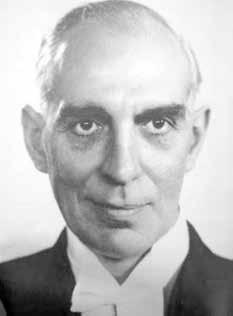
Figure 2: Sir Reginald Watson Jones: 1902 – 1972. Watson Jones hyphenated his names in 1939, to differentiate himself from other Joneses, becoming Reginald Watson Watson-Jones. Watson was his mother’s maiden name. Copyright 2005 Springer-Verlag London Limited.
These results were not found to be reproducible by EA Nicoll (Mansfield; Figure 3), who was commissioned by the Miners’ Welfare Commission, to tour North America in 1947 to research the treatment of traumatic paraplegia. In a series of 166 coal miners, Nicoll classified thoracolumbar fractures as stable or unstable, based on disruption of the interspinous ligament. As with Watson Jones, he believed most fractures had a flexion mechanism, likely due to the confined hunched positions miners worked in. However, his belief that most simple wedge fractures were stable challenged the zeitgeist so radically, that in public, good-humoured, back-and-forth lectures (including SICOT 1950, Paris) Watson-Jones and he comparatively criticised each other’s methods and results. Both presented a slide of one of their successes with a subsequent blank slide annotated saying, ’Nicoll’s method’ or ’Watson-Jones’ method’, respectively.

Figure 3: E.A. Nicoll: 1903 – 1993. Copyright 2005 Springer-Verlag London Limited.
Nicoll’s work had enormous implications. At the time, a miner with a spinal injury was written off as a permanent cripple, unable to work again. However, 80% of Nicoll’s patients returned to hard manual labour in coal pits, and his report was accepted by Aneurin Bevan, the Minister of Health and founder of the NHS. Nicoll’s friend and colleague, Frank Holdsworth (Figure 4), who accompanied him on the North American tour, subsequently opened the Lodge Moore spinal injuries unit in Sheffield in 1954. Building on previous anatomical and biomechanical concepts, Holdsworth classified all spinal fractures according to four mechanisms of injury: flexion, flexion with rotation, extension and compression (Figure 5). Holdsworth also advocated a two-column model of the spine. His anterior column supported compressive loads, whilst the posterior column (consisting of everything dorsal to the posterior longitudinal ligament [PLL]) resisted tensile loads. He considered posterior column failure as the main determinant of instability in the spine. >>
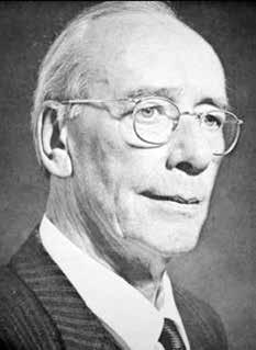
Figure 4: Sir Frank Wild Holdsworth: 1904 – 1969. Copyright 2005 Springer-Verlag London Limited.

Figure 5: Drawings from Holdsworth’s posthumously published classic paper, demonstrating anatomy and mechanism of injury. Copyright 1970 by The Journal of Bone and Joint Surgery, Incorporated.
The three column concept (an examiner’s favourite)
Francis Denis’ (Minnesota; 1984) often misquoted (and mispronounced, as he was a Frenchman who moved to the USA!) review of 412 thoracolumbar fractures built on Holdsworth’s concept of posterior ligamentous rupture, however he did not believe that this alone was enough to produce instability. He recognised stability as a spectrum. His proposed middle column consisted of the posterior wall of the vertebral body, posterior annulus fibrosus and PLL. He did not mention halves or thirds of the vertebral body/disc. This may have been inferred from his drawings and quoted biomechanical work by Panjabi. He did not say that involvement of the middle column equalled spinal instability. Rather, his conceptual model aimed to explain mechanism of injury (as per Holdsworth) according to the modes of failure of the middle column. As this has seemingly brought confusion, subsequent classifications have ignored three-columns, and all in essence follow Holdsworth’s focus on mechanism and anatomy.
The Arbeitsgemeinschaft für Osteosynthesefragen (AO; English: Working group for bone fusion issues), or Magerl (1994), classification follows Holdsworth’s two-column model; with a compression resistant solid anterior column and tension resistant hollow posterior column of neural canal. It subdivides fractures by mechanism into compression (A-type), distraction (B-type) and rotation (C-type), in similar alphanumerical fashion to the AO long bone classification, with increasingly progressive instability. As with the other AO classifications, it can be complex to use for the non-initiated, but the principles remain clear: mechanism of trauma has a correlation to stability.
A new favourite classification amongst examiners
In 2005, a Delphi process of 40 ‘expert’ surgeons produced the Thoracolumbar Injury Classification and Severity Score (TLICS) based on 1) radiographic morphology, 2) posterior ligamentous integrity and 3) neurologic status, with no confusing mention of two- or threecolumn concepts. The three TLICS parameters essentially combine Holdsworth’s concepts with the pre-radiographic clinical classification of Burrell, producing a score to guide treatment. To truly classify according to TLICS, 1) plain radiographs/ computer tomography [CT], 2) magnetic resonance imaging [MRI] and 3) clinical assessment (by American Spinal Injury Association [ASIA] score) must be collated. A score < 3 is considered stable, not requiring surgery, whereas a score > 5 is considered unstable requiring surgery. Unfortunately, TLICS does not give an answer for injuries with a score of 4 (as stability is a spectrum). These are often the same injuries which produce debate when using other classification systems or treatment algorithms.
A more recent compromise between TLICS and Magerl is the 2013 updated AOSpine Thoracolumbar Injury Classification System, (Figure 6). As with Holdsworth, Magerl and TLICS, a descriptive morphological classification based on mechanism is combined with posterior ligamentous integrity and a neurologic status modifier. Additional modifiers aim to aid surgeon decision making, however as with other classification systems they can confuse the uninitiated. As with other classification systems, we believe its best use is for communication between clinicians.

Figure 6: The simplified ‘AOSpine Thoracolumbar Injury Classification System’ which 43 years on, bears similarities to Holdsworth’s drawings. Copyright by AOSpine International, Switzerland.
In our practice and teaching we believe that understanding a definition of stability (as per White and Panjabi), the importance of mechanism of injury, and how to aptly perform an ASIA neurological exam are keys to dealing with spinal fractures. Grasping these concepts should help surgeons, whether or not they are spinal by subspeciality. Then comes the next slew of controversial questions: what approach and technique by which to fix the fracture?! To brace or not?
Key learning points
1) Spinal stability is a spectrum.
2) Mechanism of injury correlates to stability.
3) Neurologic injury from trauma suggests spinal instability.
4) Neurologic damage occurs at the time of injury.
5) Classification systems should be reproducible communication tools.
Selected references
• Nicoll EA. Fractures of the dorso-lumbar spine. J Bone Joint Surg Br 1949;31B(3):376-94.
• Holdsworth F. Fractures, dislocations, and fracture-dislocations of the spine. J Bone Joint Surg Am 1970;52(8):1534-51.
• Denis F. The three column spine and its significance in the classification of acute thoracolumbar spinal injuries. Spine (Phila Pa 1976) 1983;8(8):817-31.
• White AA and Panjabi MM. Clinical biomechanics of the spine, Vol. xxiii. Philadelphia: Lippincott, 1990; p722.
• Vaccaro AR, Lehman Jr, RA, Hurlbert RJ, Anderson PA, Harris M, Hedlund R, et al. A new classification of thoracolumbar injuries: the importance of injury morphology, the integrity of the posterior ligamentous complex, and neurologic status. Spine (Phila Pa 1976). 2005;30(20):2325-33.
• Vaccaro AR, Oner C, Kepler CK, Dvorak M, Schnake K, Bellabarba C, et al. AOSpine thoracolumbar spine injury classification system: fracture description, neurological status, and key modifiers. Spine (Phila Pa 1976). 2013;38(23):2028-37.
