8 minute read
Cauda equina syndrome - an overview

Sheweidin Aziz and Matthew Newton-Ede
Sheweidin Aziz is a spinal consultant at the University Hospitals of Leicester and is a fellow of the RCSEng. She was one of the first Spinal Training Interface Group (TIG) Fellows in the UK. During her fellowship, she gained experience managing primary spinal tumours, metastatic spine disease, adult degenerative conditions, paediatric deformity, and trauma. She is a keen educator, previously awarded the ‘Outstanding Clinical Teacher’ of the year award by the University of Leicester Medical School.
Advertisement
Matthew Newton-Ede is a spinal surgeon at The Royal Orthopaedic Hospital NHS Trust and Birmingham Children’s Hospital. He specialises in treating a wide range of spinal problems in adults and children including surgery of the neck and he has a particular interest in children’s spinal deformity surgery. He is a fellow of the RCSEng, and a member of the BOA, AO Spine, BASS and BSS.
Cauda equina syndrome (CES) is a clinically diagnosed syndrome resulting from the dysfunction of one or more of the lower sacral nerve roots. Back or leg pain or lower limb motor or sensory changes are often present but are not essential to the diagnosis. The presence of at least any of the following is required, bladder/bowel/sexual dysfunction and reduced sensation in the saddle area. CES cannot be excluded based on clinical signs alone. An emergency MRI scan must be performed even in the absence of CES signs. There are several stages of CES; the damage to nerve roots occurs continuously and progressively. Patients place more emphasis on pain, so clinicians must ask direct questions about red flags. The earlier the diagnosis and management, the better the outcome. There are no safe time thresholds, such as the 48-hour ‘safe’ time window. Both early and delayed surgery result in improved neurological outcomes.
Background
CES is a clinically diagnosed syndrome resulting from the dysfunction of one or more of the lower sacral nerve roots (S2 and caudal). One or more of the following symptoms or signs must be present:
• Bladder and/or bowel dysfunction
• Reduced sensation in the saddle area
• Sexual dysfunction
Back or leg pain or lower limb motor or sensory changes are often present but are not essential to the diagnosis. Nerve root compression is almost always the cause, and MRI scanning is essential for confirmation [1]. CES occurs rarely in community populations and is only confirmed in 19% of those presenting with CES symptoms [2].
Duration of symptoms at presentation varies with a median of 48 hours. The commonest symptoms are sciatica, altered sensation in the saddle area, and micturition dysfunction. Sexual dysfunction is often omitted from the doctor’s enquiry, but when questioned, sexual dysfunction exceeds 90% [3].
Classification
The most useful modern descriptive classification (Lavy) [1]:
1. CES Suspected (CESS): Patients are at risk of developing CES. There is bilateral sciatica or motor/sensory changes in the lower limbs but no sphincteric or perineal sensory changes. Includes anyone at risk of developing CES, e.g. due to a large disc.
2. CES Early (CESE): In addition to CESS, there are perineal sensory changes or micturition symptoms. Bladder, bowel, and sexual functions are preserved.
3. CES Incomplete (CESI): Altered bladder function/sensation with/without perineal sensory changes, sexual or bowel dysfunction.
4. CES with retention (CESR): In addition to CESI, there is painless bladder retention and overflow.
5. CES Complete (CESC): Insensate bladder with overflow incontinence, no perineal, perianal or sexual sensation, and no anal tone.
The historical but useful classification (Tandon) [1]:
1. Type 1: Rapid onset of CES symptoms with no previous history of back problems (quite rare).
2. Type 2: Acute bladder/CES symptoms with back problems and sciatica history (commonest type). Patients are usually young (< 40 years) and have a high Body Mass Index (BMI).
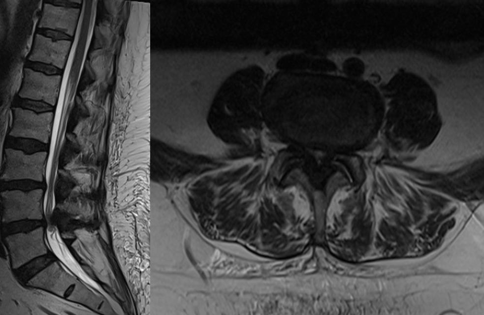
3. Type 3: Longstanding back problems and gradually progressive CES (common in older patients with spinal stenosis – very rare), (Figure 1).

Figure 1: Sagittal and axial MRI T2 weighted images showing a central canal stenosis causing compression of the cauda equina, not requiring emergency decompression due to the absence of cauda equina symptoms and signs.
CESE and CESI are the most time-critical presentations with ‘more to lose’, and surgical decompression should be done as an emergency, especially in CESI. In CESR and CESC, although these are more likely to bear poor outcomes, surgical decompression remains beneficial; however, it appears less clear that it should be performed as an emergency (particularly overnight), although this is controversial with many spinal surgeons averring that all CES cases should be performed as an emergency.
Pathophysiology
CES is usually caused by the compression of nerve roots by lumbar disc herniation. The functional changes induced by compression can be caused by mechanical nerve fibre deformation compression and restriction of nerve root microcirculation, local ischaemia intraneural oedema and resultant Wallerian degeneration [4].
Diagnosis
The diagnosis consists of two critical points:
a) A history and examination consistent with suspicion CES (see above).
b) MRI (Figure 2) or CT [5].
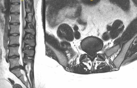
Figure 2: Sagittal and axial MRI T2 weighted images showing a large L5/S1 central disc prolapse causing compression of the cauda equina nerves.
Many patients ignore numbness, placing greater emphasis on pain. Doctors must ask about red flags. Nonetheless, red flag signs, including bilateral sciatica, altered perineal sensation, motor or sensory changes, and bladder/bowel/sexual dysfunction, have a low predictive value (a measure of the times that the value positive or negative is the true value).
The usefulness of particular symptoms and signs:
a) Bilateral sciatica has a sensitivity of 32.4%, PPV 17.2%, and NPV 88.3%.
b) Post-void residual volume (PVR) of ≥ 200 ml has a sensitivity of 94.1%, specificity 66.8%, PPV 29.9% and NPV 98.7% [6].
Patients presenting with CES symptoms should have an emergency MRI scan even in the absence of clinical signs. Changes on PR examination are poorly correlated with findings of CES, and even though a post-micturition bladder scan is more useful, its ‘normality’ does not exclude CES (6% will be missed).
Emergency MRI is part of the triage of the suspected CES patient. It should always be performed locally [7], as early diagnosis offers the best chance of a satisfactory outcome.
Timing of surgery
There is increasing understanding that damage to the cauda equina nerve roots occurs continuously and progressively, and thus, the earlier the surgical decompression, the better the outcome.
There are no safe time thresholds such as the 48-hour ‘safe’ time window described by Ahn [8] because neurologic deterioration in CES can occur rapidly and unpredictably. There are commonly poor outcomes in CES patients leading to a lifetime of disabling, awful symptoms, loss of function and employment.
The British Association of Spine Surgeons (BASS) states that “Nothing is to be gained by delaying surgery and potentially much to be lost”; surgery should be carried out as soon as is practically possible [7].
Removing a large central disc prolapse can be considerably more difficult than a routine discectomy and may require extensive exposure, and midline decompression and interlaminar decompression are the norm. The question of ‘whether to operate in the middle of the night’ with a ‘stable’ CES-I patient remains controversial; however, the authors of this article remind the reader that it is not possible to predict when a patient will progress from CES-I to CESR-R/C and consequently endure a far worse outcome. Emergency surgery is the expected norm [9].
Outcomes of surgical intervention of surgery
Neurological recovery is more likely with early surgical decompression. Patients who have a decompression whilst ‘in CESI’ have a better prognosis than those with CESR/C. Even when there is a delay in presentation with CESR/C, surgery offers improved outcomes. There is always ‘something potentially to be gained’ with decompression [10].
Approximately 75% of all CES cases will eventually have an acceptable urological function; however, often, there is the persistence of back pain and motor and sensory deficits. Of all CES patients, 1 in 5 will have a poor outcome, usually with the need for ongoing treatment, e.g. management of sexual dysfunction, self-catheterisation, colostomy, urological and gynaecological surgery, spinal injuries rehabilitation and psycho-social support. CESR/C patients are likely to have significantly worse long-term outcomes in bladder, bowel and sexual function compared to those with CESI [11].
Medico-legal aspects of cauda equina syndrome
The largest cause of claims is the delay in diagnosis and treatment. According to the National Health Service Resolution Report 2021/2022, orthopaedic surgery is in the top three specialities by volume of claims. From January 2008 to December 2018, NHS resolution received 827 claims for CES; the cost to the NHS was £186m.
Controversies and areas of further research
In patients with large or massive disc prolapses without clinical features of CES, i.e. CESS, these patients can be safetynetted, and emergency surgery is not warranted as they are likely to resolve spontaneously [12].
On the contrary, a small disc with clinical evidence of CES warrants surgical decompression, dependent on the stage.
There remains no agreed radiographic definition for CES. What defines a large disc? What is the crosssectional size? How much compression is needed? Does the presence of CSF exclude CES?
Take home messages
1. All patients with sciatica should be given surveillance advice concerning symptoms of CES (red flag ‘card’).
2. Doctors must enquire concerning red flags.
3. Neurological injury is time-critical.
4. Emergency scan, referral, and surgery.
5. There are no safe time thresholds.
References
References can be found online at www.boa.ac.uk/publications/JTO.
