11 minute read
Management of high-pressure injection injuries


Guang Yim, Jens Roesner, Warren Hammert, Adeline Clement, Patrick Gillespie and Oliver Stone
Major Guang Yim is a Plastic Surgeon currently based in South Wales working as the Major Trauma and Limb Reconstructive Microsurgery Fellow. He has trained in the West Midlands at the prestigious Queen Elizabeth Hospital Birmingham and then undertaken his Speciality Training in the South West of England.
Advertisement
Warren Hammert is Professor of Orthopaedic and Plastic Surgery at Duke University, Durham, NC, USA.
Adeline Clement is a Consultant Orthopaedic Hand and Wrist Surgeon at The Department of Trauma & Orthopaedics, Raigmore Hospital (NHS Highland), Inverness.
Patrick Gillespie is a Consultant Hand Surgeon in the Exeter Hand Unit and Department of Plastic Surgery at the Royal Devon and Exeter Hospital, Exeter.
Oliver Stone is a Consultant Orthopaedic Hand Surgeon and Clinical Lead for Orthopaedic Hand Surgery in the Exeter Hand Unit at the Royal Devon and Exeter Hospital, Exeter.
High pressure injection injuries are defined as puncturing of the dermis by a jet of fluid or air under pressure. When a leak or accidental activation occurs, a pressure of only 100 pounds per square inch (psi) is enough to puncture human dermis. Whilst uncommon, a hand centre in the United Kingdom (UK) is expected to see one to four cases presented per year [1]. If untreated or inadequately treated, the consequences of these injuries are devastating. In this article we provide an overview for clinicians to understand why high-pressure injuries are so damaging and under-recognised. We then provide an outline of how these limb threatening injuries should be managed as surgical emergencies.
Background
High pressure injection injuries are a relatively new phenomena that have emerged since industrialisation. The first case report of a high-pressure injection injury was less than a hundred years ago. Within the English literature, the first report was by Rees in 1937 that involved the high-pressure injection of diesel from an engine injector to the right middle finger [2].
Many of us unknowingly have tools capable of causing significant harm within our own households such as a pressure washer that operate between 1,500 to 4,000 pounds per square inch (psi). Within commercial and industrial settings, paint guns, grease guns and hydraulic hoses are common sources of injury. The pressures that are present within these systems can range from 100 psi for a grease gun to 10,000 psi in hydraulic systems. Some spray guns can deliver 100 litres per minute; this means that even 0.5 seconds of injection can deliver 833 ml of fluid into tissue.
Physics
Fluid is used within hydraulic equipment to multiply the applied force to exert a greater output force through Pascal’s principle.
The ability of high-pressure equipment to inject fluid through skin is a function of:
1. Pressure – Higher pressures increase the ability of the fluid to puncture the skin.
2. Proximity to skin – The further away the aperture is from skin, the greater the air resistance the fluid has to overcome.
3. Aperture size – Smaller aperture sizes lead to higher velocities of fluid as it exits the aperture.
Pathophysiology and mechanism of tissue injury
High pressure injection injuries cause damage and morbidity through three different mechanisms.
1. Direct compressive effects from the presence of large volume of, uncompressible, fluid within small anatomical spaces with little to no room for expansion. This leads to effects analogous to compartment syndrome; ischaemia, venous occlusion and thrombosis due to the presence of foreign material.
2. Chemical toxicity from the noxious chemicals contained within the injected fluid.
3. Secondary infection from fluid that is non-sterile and also the inoculation of environmental organisms into deep tissue.
In the 1970s Kaufman demonstrated that the position of the hand at the point of high-pressure injection influenced the pattern of injuries encountered. Cadaveric Injection of wax at 750psi in the midline over the flexor tendon second and fourth annular pulleys would spread wax in the subcutaneous tissues to completely encircle the digit and the neurovascular structures; due to the thickness of the pulleys. This is shown in Figure 1.


Figure 1: Injection at the level of the annular pulleys leads to fluid spreading in a subcutaneous plane circumferentially around the digit and neurovascular bundles.
If injection occurred in the midline at the level of the joints, where the thin cruciate pulleys lie, then the wax would spread into the flexor tendon sheath in addition to encirclement of the digit (Figure 2).

Figure 2: Injection at the level of the joints leads to fluid spreading into the flexor sheath as well as the subcutaneous plane circumferentially around the digit and neurovascular bundles.
If higher pressures are applied at the level of the joints, intra-articular spread could also occur as shown in Figure 3.
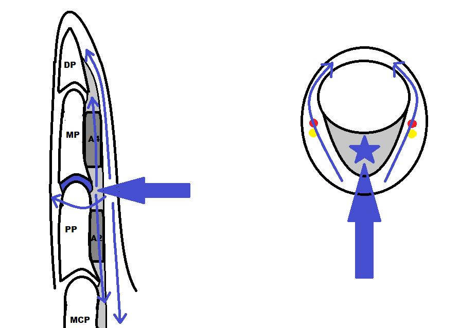
Figure 3: Very high-pressure injection at the level of the joints can lead to fluid spreading into the joint as well as the flexor sheath, the subcutaneous plane circumferentially around the digit and neurovascular bundles.
If para-midline injection occurred, the fluid would also spread to the dorsum and along the dorsal spaces of the finger and hand. If the thumb or little finger tendon sheaths were breeched, the radial and ulnar bursae were respectively contaminated. Injection of the volar palm would lead to contamination of the interosseus muscle spaces, the palmar arch, common digital nerves and also extension to the dorsal spaces of the hand; this is exemplified in Figure 4.

Figure 4: An X ray demonstrating the contamination of the interosseus muscle spaces, the palmar arch, common digital nerves and also extension to the dorsal spaces of the hand after volar injection of into the palm [6].
Reproduced from High-pressure chemical injection injury to the hand: usually underestimated injury with major consequences. BMJ Case Rep. 2019;12(9):e231112. With permission from BMJ Publishing group Ltd.
The composition of the injected material significantly influences the morbidity of these injuries. Table 1 (below) correlates expected amputation rates for the different compositions of material injected.
Table 1: The reported amputation rates according to the type of material injection.
Injected material / Amputation rate
- Solvent based Paint / 80%
- Oil-based Paint, hydrocarbons / 70%
- Grease / 20%
- Water / <20%
Even water alone is still associated with significant morbidity due to flexor tenosynovitis, compartment syndrome and even amputation.
Consequences if untreated or inadequately treated
Common themes in literature through the past century are non-recognition of the severity of the injury by both patients and clinicians. This leads to delayed presentation of patients who do not understand the significance of the injury, when compartment pressures may have been raised for hours. Attending clinicians may lack experience or education in treating such injuries which can lead to a sense of non-urgency in management. This leads to delays in surgical decompression and washout; further compounding the toxic effects of the injected material, increased pressure at the injections site which results in tissue necrosis and longterm morbidity.
If untreated or inadequately treated, the affected areas remain intensely painful as the tissue undergoes necrosis, which often will be complicated by secondary infection. Delays in treatment lead to substantially worse outcomes.
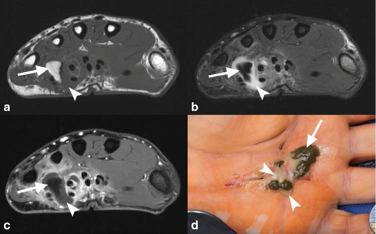
As a result of ischaemia there may be a substantial loss of soft tissues, including amputation if digits are involved. Extensive fibrosis and scarring may lead to contracture limiting the function of the affected limb or digit. If the injected fluid remains in the tissue granulomas of varying sizes will likely form and drainage through sinuses may occur weeks to months after injury as can be seen in Figure 5.

Figure 5: Late MRI findings and clinical appearance of discharging sinuses within the hand after injection of grease into the palm [3].
Reprinted by permission from Skeletal Radiology, High-pressure injection injury of the hand: peculiar MRI features and treatment implications. Skeletal Radiol. 2019;48(2):295-9.
Amputation rates
High pressures are associated with a greater likelihood of amputation. Table 2 shows the reported rates of amputation with increasing injection pressures. When patients presented with altered perfusion at the time of initial assessment, it was associated with amputation [4].
Table 2: The reported amputation rates with increasing injection pressures.
Injection pressure / Amputation rate
<1,000 psi / 19%
1,000 – 7,000 psi / 43%
>7,000 psi / 95-100%
Presentation
The ‘typical’ presentation is a male in their 30s who has an isolated injury of their non-dominant upper limb and this is usually of the index finger. This reflects the manual nature of work associated with the use of the equipment that utilises liquids held in high pressures. However, other more unusual anatomical areas have been reported to have had high pressure injection injuries. These include the face, neck, spinal cord, thorax and even the perineum.
The main circumstances leading to high-pressure injection injuries are inattention, misuse and material failure and this has consistently been reflected across the literature.
The patients feel a sharp sting at the point of injury with immediate swelling. However, as the wounds are pin-point and innocuous little attention is paid to the injury unless they have had prior education. This often leads to delay in presentation of hours to days and is usually due to pain that develops at that time.
Initial assessment and management
Recognition of the injury severity is critical. High-pressure injection injuries should be placed in the same category of urgency as compartment syndrome as lower rates of amputation were seen with decompression and wide debridement <6hrs after injury [5].
A full history of the mechanism, hand position, type of fluid injected, equipment pressure, distance and nozzle type help define the pattern of injury. Occupation, hand dominance, past medical history, allergies, medication status, smoking history and last meal are all relevant to treatment of patients. Patients should be starved for emergency surgery.
The initial clinical examination should start with neurovascular status of the digits and hand before moving onto tendon function. X-rays should be undertaken to assess for joint involvement and the spread of fluid to aid surgical planning. Figure 4 shows the intercompartmental and dorsal spread. Figure 6 shows the clinical appearances.


Figure 6: The clinical appearances of dorsal spread after volar injection of grease into a hand.
Blood tests for, FBC, U&Es, CRP should be undertaken with broad-spectrum intravenous antibiotics started to cover skin organisms and environmental contaminants. Infections are usually poly-microbial and will involve a range of organisms from skin flora to gram-negative bacteria and, if fresh or brackish water is injected, organisms such as Aeromonas or Vibrio may be encountered. It is very important to send samples for microbiology at initial debridement as this is the best guidance for subsequent therapy.
If the fluid injected is toxic, the National Poisons Information Service (Toxbase) advice should be sought to manage systemic effects.
Immediate emergency referral to the relevant surgical specialty (usually a Hand or Upper limb surgeon) should be undertaken to attain surgical decompression less than six hours from injury. The management of these injuries are complex and challenging even for experienced hand surgeons with generally poor outcomes and therefore urgent treatment ideally in a dedicated hand unit may give the best chance of optimal outcome.
Surgical management
The patient should undergo surgery under general anaesthesia +/- Axillary block (by a competent block anaesthetist) with tourniquet control.
The release of the high-pressure injection injuries should be designed along fasciotomy incisions that will be sufficient to ensure full decompression of all neurovascular structures and all muscle compartments. The debridement and washout should aim to remove as much foreign body as possible; balanced against the morbidity of tissue damage. Figure 6 demonstrates the extent of tissue release relative to the initial injury.
The addition of Povidone-Iodine 10%, in cadaveric studies, showed greater removal of latex and oil-based paints compared to saline alone [7]. However there is controversy around its effect on infection rates when compared to saline. In the event of latex or oil-based contaminants which are adherent to tissues it would be reasonable to consider Povidone-Iodine 10% at the initial debridement to aid removal of the contaminant followed by low pressure high volume saline to dilute and remove microbes.
48 hours after the initial decompression and debridement, a second look is required to undertake further debridement and washout of any further non-viable tissue before deciding on the suitability of closure.
Post-operative completion of antibiotics with close supervision of wound care and hand therapy are essential to minimising morbidity and maximising the outcome of patients.
Conclusion
High-pressure fluid injection injuries can be devastating and require prompt diagnosis and treatment or they can result in notable morbidity and loss of function. Lack of recognition from patients and inexperienced clinicians compounds the damage. High injection pressures >7,000psi and oil-based paint are associated with an increased risk of amputation.
If one learning point is to be taken away from this article then it should be that these injuries are surgical emergencies and are often underestimated by patients as well as clinicians. Decompression within six hours from point of injury is associated with a reduction in morbidity.
References
1. Neal NC, Burke FD. High-pressure injection injuries. Injury. 1991;22(6):467-70.
2. Rees C. Penetration of tissue by fuel oil under high pressure from a diesel engine. JAMA . 1937;109:886-7.
3. Collins M, McGauvran A, Elhassan B. Highpressure injection injury of the hand: peculiar MRI features and treatment implications. Skeletal Radiol. 2019;48(2):295-9.
4. Lewis HG, Clarke P, Kneafsey B, Brennen MD. A 10-year review of high-pressure injection injuries to the hand. J Hand Surg Edinb Scotl. 1998;23(4):479-81.
5. Hogan CJ, Ruland RT. High-pressure injection injuries to the upper extremity: a review of the literature. J Orthop Trauma 2006;20(7):503-11.
6. Sharma R, John JR, Sharma RK. Highpressure chemical injection injury to the hand: usually underestimated injury with major consequences. BMJ Case Rep 2019;12(9):e231112.
7. Bascone CM, Sheber B, Dave D, Firriolo JM, Pereira C. Optimal Irrigant in High Pressure Paint Injection Injuries of the Hand. Plast Reconstr Surg Glob Open. 2022;10(1):e4064.