11 minute read
Management of closed extensor tendon injuries

Sarah Turner, Nick Gape and Jonathan Hobby
Sarah Turner has worked at Manchester Foundation Trust for over 20 years as a clinical specialist physiotherapist in hand therapy and has a particular interest in the management of hand trauma.
Advertisement
Nick Gape is an occupational therapist and accredited hand therapist who works at the Cardiff and Vale University Health Board with a special interest in hand trauma.
Jonathan Hobby is a Consultant Orthopaedic and Hand Surgeon at the Hampshire Hospitals, and is the current President of the British Society for Surgery of the Hand.
Injuries to the extensor tendons of the hand and digits are a frequent presentation to the acute hospital setting. They are the most frequent tendon injury (54%) [1], and are predominantly seen in men between the ages of 15 and 59. The majority are closed injuries.
Open injury usually requires further exploration with possible tendon repair but the majority of closed injuries, especially affecting the fingers, can be managed successfully with conservative treatment with the use of splinting and appropriate exercise therapy. The focus of this paper is to update on the management of the most common conditions in extensor zones 1 to 5: mallet finger, closed boutonniere and sagittal band injuries.
Mallet finger
Injury to the terminal extensor tendon in zone 1 is one of the most common hand conditions presenting to Emergency Departments making up almost 6% of all hand injuries. Closed mallets are caused by a sudden flexion (or extension) force to the distal interphalangeal joint (DIPJ), resulting in an extensor lag which is passively, but not actively, correctable. X-rays should always be taken to confirm if the injury involves an avulsion (or impaction [2]) fracture of the dorsal base of the distal phalanx. Patients may present with a chronic mallet deformity some time after injury.
The vast majority of closed injuries can be treated conservatively, however, surgical exploration is always indicated for the less common open mallet injury where the terminal extensor tendon is divided. It may also considered for closed bony mallets where > 30-40% of the DIPJ surface is affected and if there is subluxation of the distal phalanx, a lateral view x-ray taken in extension is essential to confirm the presence of irreducible subluxation. An attempt to treat these injuries with a splint in slight flexion, with reduction confirmed by x-ray, should be made in the first instance [2]. Even in the presence of persistent subluxation the joint can remodel with satisfactory functional outcomes [2].
Management of closed mallet injuries are through application of a splint to maintain DIPJ extension. Various types of splint have been described and include ‘off the shelf’ Stack splints or ones fabricated for a custom fit e.g. moulded foam aluminium (Zimmer splint), or thermoplastic splints made by a specialist hand therapist. There is however no clear evidence for the superiority of one splint type over another [2]. The proximal interphalangeal joint (PIPJ) should be left free to mobilise to full range to prevent stiffness although a longer splint can be fabricated to hold it in a flexed position should a swan neck deformity tendency be present.
There is no consensus of the duration of splint application but the authors advocate it be worn full time for six weeks for avulsion fractures and up to eight weeks for soft tissue mallets; a further two weeks of ‘weaning’ via night splintage is advised. Should the extensor lag recur, a return to full time splintage should be considered for a further 2-3 weeks then the active extension re-assessed.
The outcome of conservative management for acute closed mallets has been reported as less favourable for tendinous injuries, whilst a higher age at time of injury is also associated with poorer outcome [3]. Patients should be always be advised that the result of treatment may not restore full range of DIPJ extension with a small residual lag not being uncommon. There is also usually a dorsal prominence which may be tender and ache in cold weather, although these symptoms improve with time.
A failure of the injury to respond to conservative management should be evaluated as to the effect on the affected digit: persistent DIPJ extensor lags may result in a swan neck deformity in some individuals, especially those with hypermobile PIPJs, which can be functionally debilitating. In the absence of swan-necking, reported pain and the patient’s desire for further intervention should be taken into consideration and the benefits and risks of surgery explained. Surgery may involve a simple trans-articular k-wiring of the DIPJ in extension for six weeks followed by a further four weeks of splintage, or for chronic mallets options include terminal extensor tendon reconstruction and DIPJ arthrodesis. There are a range of more complex surgical techniques described for acute injuries, but there is little robust evidence that these lead to better outcomes and complication rates are higher [2].
Boutonnière injury
The extensor tendon mechanism of the finger is an intricate interplay between the extrinsic and intrinsic muscles. Distal to the metacarpophalangeal joint (MCPJ), the extensor tendon divides in to the central slip, which inserts into the base of the middle phalanx, whilst radial and ulnar lateral bands converge along with the intrinsic muscles to become the terminal tendon that inserts in the distal phalanx.
The term boutonnière (buttonhole) aptly describes the mechanism of this injury of the extensor mechanism at the PIPJ. In the closed injury, the PIPJ is forcefully flexed or dislocated volarly causing the central slip to either avulse from its insertion, or to rupture just proximal to this point in the manner of a mallet injury. The central slip, however, is not the only extensor of the middle phalanx; extension is also possible through the lateral bands, providing their stabilising structures (the triangular ligament and the transverse retinacular ligament) remain intact. If the integrity of these structures is also compromised, then the lateral bands sublux volarly acting instead as a flexor of the PIPJ whilst at the same time hyperextending the DIPJ.
Whilst this typical boutonnière posture may not be present initially, careful assessment using Elson’s or modified Elson’s tests have a high sensitivity in establishing the integrity of the central slip. When the classic deformity is present then Boyes’ test may prove more useful. Early diagnosis and treatment will likely favour a better outcome.
As with mallet injuries, non-surgical management is the mainstay of treatment in this zone of injury. Several treatment approaches have been described but there is little evidence to suggest the superiority of one over another.
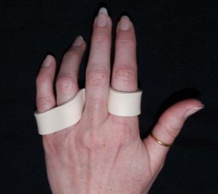
Immobilisation of the PIPJ in full extension for a number of weeks is perhaps the most common approach. Various manufactured and custom-made splints/casts will fulfil this role but whichever is utilised, it is essential that the DIPJ is allowed to mobilise intermittently (Figure 1). This allows the lateral bands to glide, discouraging their volar subluxation, and preventing the tightening of the retinacular ligament, a situation that can quickly become established. Immobilisation for six weeks is suggested [4] in the presence of a purely tendinous injury, although earlier protected mobilisation may be permitted with bony avulsions due to the quicker healing time of bone. Other authors do not differentiate and allow protected movement with a spring-wire Capener splint [5] or within a restricted range [6]. Either will require the supervision and guidance of a hand therapist and progression of mobilisation should be pragmatic, as outcomes, whilst usually favourable, are not always predictable in this zone [4].


Figure 1: Dorsal based extension splint (distal strap removed to allow DIPJ flexion exercises).
From: Boyce, Giddens, Shewring, Tendon Disorders of the Hand & Wrist, Thieme Publishers 2022.
More recently the use of relative motion has been advocated in the treatment of tendon injuries [7]. In this zone, the injured digit is placed in 15-20° relative flexion. This increases the laxity of the flexor digitorum profundus tendon and relaxes lumbrical tension, which in turn decreases the tension on the extensor hood. There may need to be some restriction of the flexion arc initially and potentially a night extension splint. Suitability for this approach requires the patient to pass the pencil test (Figure 2) by actively extending the PIPJ in this relatively flexed position. The central slip cannot realistically heal at the appropriate length with this regimen but this need not matter if the remainder of the mechanism is intact and the potentially deforming forces controlled. The patient is allowed to use their hand almost normally in the Relative Motion Flexion Splint (RMFS) (Figure 3). Reported outcomes in a series of cases studies are encouraging although as with other approaches, there is a paucity of evidence.

Figure 2: Pencil Test.

Figure 3: Relative motion flexion splint (RMFS). From: Boyce, Giddens, Shewring, Tendon Disorders of the Hand & Wrist, Thieme Publishers 2022.
Closed injuries that do not respond to a splinting regimen will require referral to a hand surgeon with a view to extensor tendon reconstruction. Particular attention should be directed to extensor injuries associated with volar or lateral dislocation of the PIP joint. These injuries are uncommon, but may be associated with buttonholing of the condyle through the interval between the central slip and lateral band. This will lead to difficulty in achieving a stable congruent reduction. Early surgical exploration is indicated with open reduction and repair of the injured structures. These injuries are difficult to treat if neglected. Tendon lacerations in this zone, where the central slip and/or lateral bands are involved, will require primary repair. Similar rehabilitation regimens are utilised, although mobilisation is usually initiated at an earlier stage.
Sagittal band injury
Presentation of sagittal band injury may be either acute, with localised pain, swelling and tenderness, or chronic attrition. Acute injuries, frequently the result of either a direct blow including a punch mechanism (so called Boxer’s Knuckle), or a sudden hyperflexion force to the MCPJ, most often affect the radial sided band and the middle finger due to its local anatomical idiosyncrasy. Diagnosis is usually by clinical examination, x-rays may be taken to exclude localised fractures, further investigations are not routinely required but an ultrasound scan can confirm the diagnosis.
Classification by Rayan and Murray [8] is according to the pathological severity; Grade 1 being a contusion or partial tear with no tendon instability; Grade 2 presents with a subluxing central tendon, usually in an ulnar direction off the metacarpal head during MCPJ flexion, relocating centrally when the joint extends. Grade 3 presents with full dislocation of the extensor tendon into the unaffected side intermetacarpal sulcus, the ability to actively extend the joint may be limited or impossible.
Management of the condition varies according to both severity and the time since injury. Evidence is variable about how late conservative management can be effective for patients with complete tendon dislocation [9], injuries of up to three weeks duration are generally accepted to respond better to conservative methods. Outcomes for conservative management are reduced with age, manual labour occupations, and for grade 3 injuries [9]. Chronic presentations, or where tendon dislocation is constant and occurs without pain, swelling or other inflammatory manifestations [8], and also those acute injuries that have failed to respond to less invasive procedures, are likely to require surgical intervention.
Grade 1 sagittal band injuries can be treated adequately in full time neighbour strapping for up to four weeks duration. Grades 2 and 3 require splint application. There is no definitive consensus on which splint is most appropriate with a variety of hand based splints described whose function is to hold the affected MCPJ in a neutral to extended position. The authors favour use of the Relative Motion regime applying the Relative Motion Extension Splint (RMES) to position the affected digit in 15-20° hyperextension relative to the uninjured fingers, so relieving the tension on the extensor complex of said digit via the quadriga effect (Figure 4). There is again no consensus on the splint wear duration but six weeks full time wear and a further two weeks during activity has been advocated.

Figure 4: Relative motion extension splint (RMES). From: Boyce, Giddens, Shewring, Tendon Disorders of the Hand & Wrist, Thieme Publishers 2022.
Light use and finger mobilisation is permitted whilst wearing the splint, recent publications indicate it is well tolerated due to its low profile.
Surgery involves open direct repair of the extensor hood for acute injuries whilst chronic presentations may require reconstruction using tendon grafts to reconstruct the sagittal bands. There are many techniques which create a check rein to centralise the extensor tendon [10]. Post-operative management involves application of the RMES as above.
References
References can be found online at www.boa.ac.uk/publications/JTO.
