Veterinary
A 1-year-old female Yorkshire Terrier (1.7 kg) fractured the right distal radius and ulna (similar to a Colles fracture in humans). Case provided by Randy J Boudrieau, North Grafton, USA
a
b
a
b
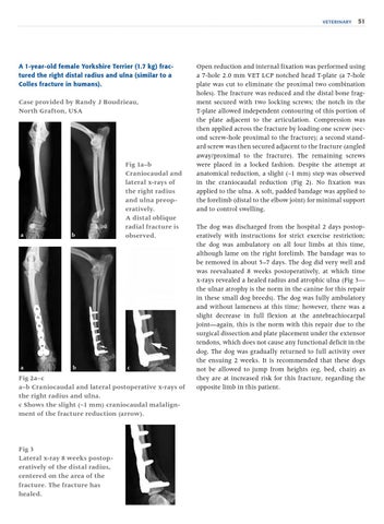
Fig 1a–b Craniocaudal and lateral x-rays of the right radius and ulna preoperatively. A distal oblique radial fracture is observed.
c
Fig 2a–c a–b Craniocaudal and lateral postoperative x-rays of the right radius and ulna. c Shows the slight (~1 mm) craniocaudal malalignment of the fracture reduction (arrow).
Fig 3 Lateral x-ray 8 weeks postoperatively of the distal radius, centered on the area of the fracture. The fracture has healed.
51
Open reduction and internal fixation was performed using a 7-hole 2.0 mm VET LCP notched head T-plate (a 7-hole plate was cut to eliminate the proximal two combination holes). The fracture was reduced and the distal bone fragment secured with two locking screws; the notch in the T-plate allowed independent contouring of this portion of the plate adjacent to the articulation. Compression was then applied across the fracture by loading one screw (second screw-hole proximal to the fracture); a second standard screw was then secured adjacent to the fracture (angled away/proximal to the fracture). The remaining screws were placed in a locked fashion. Despite the attempt at anatomical reduction, a slight (~1 mm) step was observed in the craniocaudal reduction (Fig 2). No fixation was applied to the ulna. A soft, padded bandage was applied to the forelimb (distal to the elbow joint) for minimal support and to control swelling. The dog was discharged from the hospital 2 days postoperatively with instructions for strict exercise restriction; the dog was ambulatory on all four limbs at this time, although lame on the right forelimb. The bandage was to be removed in about 5–7 days. The dog did very well and was reevaluated 8 weeks postoperatively, at which time x-rays revealed a healed radius and atrophic ulna (Fig 3— the ulnar atrophy is the norm in the canine for this repair in these small dog breeds). The dog was fully ambulatory and without lameness at this time; however, there was a slight decrease in full flexion at the antebrachiocarpal joint—again, this is the norm with this repair due to the surgical dissection and plate placement under the extensor tendons, which does not cause any functional deficit in the dog. The dog was gradually returned to full activity over the ensuing 2 weeks. It is recommended that these dogs not be allowed to jump from heights (eg, bed, chair) as they are at increased risk for this fracture, regarding the opposite limb in this patient.