Development and validation of AO fracture classifications | 19
Laurent Audigé
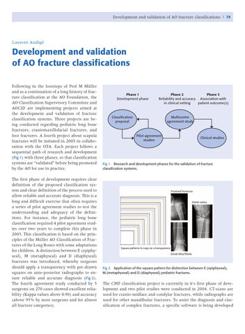
Development and validation of AO fracture classifications Following in the footsteps of Prof M Müller and as a continuation of a long history of fracture classification at the AO Foundation, the AO Classification Supervisory Committee and AOCID are implementing projects aimed at the development and validation of fracture classification systems. Three projects are being conducted regarding pediatric long bone fractures, craniomaxillofacial fractures, and foot fractures. A fourth project about scapula fractures will be initiated in 2005 in collaboration with the OTA. Each project follows a sequential path of research and development ( Fig 1) with three phases, so that classification systems are “validated” before being promoted by the AO for use in practice. The first phase of development requires clear definition of the proposed classification system and clear definition of the process used to allow reliable and accurate diagnosis. This is a long and difficult exercise that often requires a series of pilot agreement studies to test the understanding and adequacy of the definitions. For instance, the pediatric long bone classification required 4 pilot agreement studies over two years to complete this phase in 2003. This classification is based on the principles of the Müller AO Classification of Fractures of the Long Bones with some adaptations for children. A distinction between E (epiphyseal), M (metaphyseal) and D (diaphyseal) fractures was introduced, whereby surgeons should apply a transparency with pre-drawn squares on ante-posterior radiographs to ensure reliable and accurate diagnosis ( Fig 2 ). The fourth agreement study conducted by 5 surgeons on 270 cases showed excellent reliability (Kappa values above 0.90) and accuracy (above 95% by most surgeons and for almost all fracture categories).
Phase 1 Development phase
Phase 2 Phase 3 Reliability and accuracy Association with in clinical setting patient outcome(s)
Classification proposal
Multicentre agreement study
Pilot agreement studies
Clinical studies
Fig 1 Research and development phases for the validation of fracture classification systems.
Proximal humerus
Distal radius
Square patterns to copy on a transparency Distal tibia/fibula
Fig 2 Application of the square pattern for distinction between E (epiphyseal), M (metaphyseal) and D (diaphyseal) pediatric fractures.
The CMF classification project is currently in it‘s first phase of development and two pilot studies were conducted in 2004. CT-scans are used for cranio-midface and condylar fractures, while radiographs are used for other mandibular fractures. To assist the diagnosis and classification of complex fractures, a specific software is being developed