Trauma, upper extremity
11
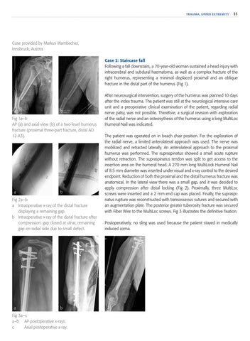
Case provided by Markus Wambacher, Innsbruck, Austria Case 2: Staircase fall Following a fall downstairs, a 70-year-old woman sustained a head injury with intracerebral and subdural haematoma, as well as a complex fracture of the right humerus, representing a minimal displaced proximal and an oblique fracture in the distal part of the humerus (Fig 1).
a
b
Fig 1a–b AP (a) and axial view (b) of a two-level humerus fracture (proximal three-part fracture, distal AO 12-A3).
a
b
Fig 2a–b a Intraoperative x-ray of the distal fracture displaying a remaining gap. b Intraoperative x-ray of the distal fracture after compression: gap closed at ulnar, remaining gap on radial side due to small defect.
a
b
Fig 3a–c a–b AP postoperative x-rays. c Axial postoperative x-ray.
After neurosurgical intervention, surgery of the humerus was planned 10 days after the index trauma. The patient was still at the neurological intensive care unit and a preoperative clinical examination of the patient, regarding radial nerve palsy, was not possible. Therefore, a surgical revision with exploration of the radial nerve and an osteosythesis of the humerus using a long MultiLoc Humeral Nail was indicated. The patient was operated on in beach chair position. For the exploration of the radial nerve, a limited anterolateral approach was used. The nerve was mobilized and retracted laterally. An anterolateral approach to the proximal humerus was performed. The supraspinatus showed a small acute rupture without retraction. The supraspinatus tendon was split to get access to the insertion area on the humeral head. A 270 mm long MultiLock Humeral Nail of 8.5 mm diameter was inserted under visual and x-ray control to the desired endpoint. Reduction of both the proximal and the distal humerus fracture was anatomical. In the lateral view there was a small gap, and it was decided to apply compression after distal locking (Fig 2). Proximally, three MultiLoc screws were inserted and a 2 mm end cap was placed. Finally, the supraspinatus rupture was reconstructed with transosseous sutures and secured with an augmentation plate. The posterior greater tuberosity fracture was secured with Fiber Wire to the MultiLoc screws. Fig 3 illustrates the definitive fixation. Postoperatively, no sling was used because the patient stayed in medically induced coma.
c