The Australasian
Journal of Oral and Maxillofacial Surgery is the official scientific journal of the Australian and New Zealand Association of Oral and Maxillofacial Surgeons
Aim and Scope
The Australasian Journal of Oral and Maxillofacial Surgery is the premier forum for the exchange of information for new and significant research in oral and maxillofacial surgery, promoting the surgical discipline in the Oceanic region.
Oceania comprises 19 countries, spread over one-sixth of the globe, but with Australia and New Zealand being the dominant developed countries.
The Journal comprises peer-reviewed scientific reports, reviews, case reports of rare and unusual conditions, and perspectives; all of value for continuing professional development.
Information for prospective authors, including author guidelines, publication ethics, malpractice statements and patient consent forms are available for download from the Australian and New Zealand Association of Oral and Maxillofacial Surgeons homepage. All correspondence with the Editor is via editorajoms@anzaoms.org
Advertising Information
The Australasian Journal of Oral and Maxillofacial Surgery accepts paid advertisements from companies involved with the surgical discipline. For information on advertising guidelines and rates contact Ms Belinda Mellowes, Executive Officer, Australian and New Zealand Association of Oral and Maxillofacial Surgeons: eo@anzaoms.org Submit advertisements to ajoms@anzaoms.org
Disclaimer
The Australasian Journal of Oral and Maxillofacial Surgery Editors and Editorial Board cannot be held responsible for error or consequence arising from the information contained in the Journal. The views and opinions expressed herein do not necessarily reflect those of the Australian and New Zealand Association of Oral and Maxillofacial Surgeons, AJOMS Editor or Editorial Board. Neither does publication of advertisements constitute any endorsement of the products advertised. © 2026 ANZAOMS

Alastair Goss
• Emeritus Professor of Oral and Maxillofacial Surgery, The University of Adelaide
• Emeritus Consultant Surgeon, The Royal Adelaide Hospital Adelaide, SA, Australia
Andrew Heggie, AM
• Clinical Professor, Department of Paediatrics, The University of Melbourne
• Senior Consultant Oral and Maxillofacial Surgeon, Royal Children’s Hospital of Melbourne
Melbourne, VIC, Australia
Alexander Bobinskas
• Unit Director, Oral and Maxillofacial Surgery, The Canberra Hospital
• Associate Professor, School of Medicine, Australian National University
Canberra, ACT, Australia
Website, Distribution and Advertising
Belinda Mellowes
• Executive Officer, Australian and New Zealand Association of Oral and Maxillofacial Surgeons
Sydney, NSW, Australia
AMPCo Production Editors
Kate Steyn
Laura Teruel
AMPCo Graphic Design
Peter Humphries
Catherine Offler
• Administrative Assistant to the Editor Adelaide, SA, Australia
Business Manager
Dieter Gebauer
• Senior Consultant in Oral and Maxillofacial Surgery, Royal Perth Hospital
• Clinical Associate Professor in Oral and Maxillofacial Surgery, The University of Western Australia
Perth, WA, Australia
Members
Arun Chandu
• Senior Consultant in Oral and Maxillofacial Surgery, Royal Dental Hospital of Melbourne
• Clinical Associate Professor of Oral and Maxillofacial Surgery, The University of Melbourne
Melbourne, VIC, Australia
Nigel Johnson
• Consultant Oral and Maxillofacial Surgeon, Princess Alexandra Hospital
• Associate Professor in Oral and Maxillofacial Surgery, The University of Queensland
Brisbane, QLD, Australia
Paul Sambrook, AM
• Director Oral and Maxillofacial Surgery, The Royal Adelaide Hospital
• Senior Lecturer in Oral and Maxillofacial Surgery, The University of Adelaide
Adelaide, SA, Australia
Darryl Tong
• Professor of Oral and Maxillofacial Surgery, The University of Otago
• Senior Consultant in Oral and Maxillofacial Surgery, Dunedin Public Hospital
Dunedin, New Zealand
Andrew Watkins
• Supervisor of Training, John Hunter Hospital
• Senior Lecturer, Charles Sturt University Newcastle, NSW, Australia
Patrisha Bordbar
• Consultant in Oral and Maxillofacial Surgery, The Royal Childrens Hospital of Melbourne
• IAOMS Executive for the Oceanic Region
Melbourne, VIC, Australia
Ex Officio Members
Nathan Vujcich
• President, Australian and New Zealand Association of Oral and Maxillofacial Surgeons
• Consultant in Oral and Maxillofacial Surgery, Royal Perth and Perth Children’s Hospitals
Perth, WA, Australia
Jasvir Singh
• Immediate Past President, Australian and New Zealand Association of Oral and Maxillofacial Surgeons
• Consultant in Oral and Maxillofacial Surgery
Sydney, NSW, Australia
John Harrison
• Regional Councillor for Oceania, International Association of Oral and Maxillofacial Surgeons
Auckland, New Zealand
Jocelyn Shand
• Clinical Associate Professor, Department of Paediatrics, The University of Melbourne
• Chair, Australian and New Zealand Association of Oral and Maxillofacial Surgeons Research and Education Foundation
• Head of Section of Oral and Maxillofacial Surgery, The Royal Children’s Hospital of Melbourne
• Vice President Elect, International Association of Oral and Maxillofacial Surgeons
Melbourne, VIC, Australia
Large language model performance on a fellowship-level examination in oral and maxillofacial surgery: a novel analysis using open-ended questions and image analysis
Jung P, Bobinskas A, Lewis E, Deane S and Hyam D 12 META-ANALYSIS
Timing, outcomes and biomaterials in the surgical repair of paediatric orbital fractures: a systematic review and meta-analysis
Tang A, Johnston J, McKenzie D, Jeyathevan J and Bordbar P
ARTICLE
Patient factors in the surgical management of maxillofacial trauma: a seven-year multicentre
retrospective review
Goh EZ, Beech N and Johnson NR
32 SCIENTIFIC ARTICLE
Full arch immediate rehabilitation with a definitive prosthesis: a retrospective review of the zero-to-fiveyear outcomes
Bontempo D, Tan P, Khadembaschi D and Gebauer D
40 REVIEW ARTICLE
The Australian Orthopaedic Association National Joint Replacement Registry: insights from 25 years
Lewis PL, Goss AN, Esaian R and Smith PN
43 REVIEW ARTICLE
The establishment of the Australian and New Zealand Association of Oral and Maxillofacial Surgeons
Temporomandibular Joint Replacement Registry: regulations and registry development
Goss A, Burgess M, Moore L, Murdoch B, Mellowes B and Smith P
51 SCIENTIFIC ARTICLE
Early outcomes at 6 weeks following temporomandibular joint arthroscopy
Lockyer JW and Kim SY

Maxillo Facial Imaging (MFI) is Australia's leading dental radiology providersolely focusing on CBCT, OPG and Lat Ceph
The highest quality imaging & workups
Oral & Maxillofacial Radiologists
State-of-the-art imaging technology
Exceptional patient care
Convenient locations
24 hour turn around reporting service*








Temporomandibular joint (TMJ) surgery is a surgical endeavour unique to oral and maxillofacial surgery. This is unlike most other aspects of our scope, as dentoalveolar surgery and complex implant surgery are shared with general and specialist dentistry. Head and neck cancer, facial trauma and reconstruction are shared with other head and neck surgical colleagues.
Often, TMJ surgery has been viewed with some reservation. In the 2012 published history of the first 50 years of our specialty, the authors wrote that “There probably is no other branch of our profession where opinions differ so much about indications and surgical approaches as there are for diseases and abnormalities of the TMJ”.1 Further, “there certainly is a need for some evidence-based treatment in TMJ surgery before the profession jumps on the next bandwagon”.1
Fortunately, these wise words have come to pass. The first step was to understand that the TMJ was a joint similar to other joints in the body, and to treat it orthopaedically. Concurrently there was a need to throw out the dental concept that the TMJ is “unique” and that dysfunction is dictated by the dental occlusion.
Orthopaedics in the last few decades have made significant advances in understanding the biology of joint disease and the role of analysis of biomechanical load and function. From this, they have developed functional joint replacements if simpler treatment methods fail. They have developed careful monitoring of outcomes, both good and bad, by means of quality control registries. This is particularly so in Australia, where the Australian Orthopaedic Association National Joint Replacement Registry (AOANJRR) has documented over 2.4 million cases of joint replacement over the last 20 years.2
There have been parallel developments in TMJ replacement (TMJR), particularly in North America. It has not been a straight line, indeed one of the learning experiences of my life was to go to New Orleans in the mid-1980s to learn how to place the Kent Proplast prosthesis.3 In one week, I saw enough to know that this device was not the answer and indeed supported blocking the importation of the device into Australia. Good call.4 The Kent device failure triggered the foundation of the American Society of TMJ Surgeons forty years ago.5 One of the founders of that group, Dr Lou Mercuri, presented the inaugural John Edwards Memorial Lecture on TMJ replacement at the Australian and New Zealand Association of Oral and Maxillofacial Surgeons (ANZAOMS) (NZ) conference in 2023 and his article was subsequently published in the inaugural volume of the Australasian Journal of Oral and Maxillofacial Surgery (AJOMS).6
ANZAOMS appreciated that it needed to follow the Australian Orthopaedic Association example and set up a TMJR registry. A key step has been the successful achievement of quality assurance status from both the Australian Government and New Zealand Government.7 The significance of this is discussed in the invited articles in this volume.7,8
ANZAOMS has also recently helped set up an Australian and New Zealand TMJ surgeons subspecialist group. This follows the example of our North American colleagues who have recently published an international guideline, which is well worth following.9
Despite the number of articles on TMJR regulation, this May 2026 issue of AJOMS does not disregard the Journal’s primary focus on presenting research performed locally. There are five scientific papers covering Fellowship examination, paediatric orbital reconstruction, complex implant reconstruction, facial fracture and TMJ arthroscopy.
With invited speaker Dr Raquel Guijarro-Martínez (Valencia, Spain)
Christchurch, New Zealand | August 5, 2026 | 1
At the ANZAOMS Annual Scientific Meeting in Adelaide in November 2025, we announced the inauguration of the Editor’s Prize for the best trainee article published in AJOMS. The winners were:
Volume 1 (2024): Dr James McKenzie, New Zealand, with “Metastatic squamous cell carcinoma of the salivary gland: treatment and prognostic factors related to overall survival and recurrence.”
Volume 2 (2025): Dr Anton Sklavos, Victoria, with “What indication criteria are applied to patients receiving total temporomandibular joint replacement? A systematic review”.
These articles contributed much to the success of our Journal. Who will be the winner in 2026?
1 Stoelinga PJW, Williams JL. 50 Years of IAOMS: the development of the specialty. International Association of Oral and Maxillofacial Surgeons. 2012. https://www. iaoms.org/publications/50years/
2 Lewis PL, Gill DR, McAuliffe MJ, et al. 2025 Annual Report: hip, knee and shoulder arthroplasty. Australian Orthopaedic Association National Joint Replacement Registry. AOA, 2025. https://aoanjrr.sahmri.com/ documents/d/guest/aoa-full-report-2025
3 Kent JN, Misiek DJ, Akin RK, et al. J Temporomandibular joint condylar prosthesis: a ten-year report. J Oral Maxillofac Surg 1983; 41(4): 245-254.
I hope you enjoy reading this issue as much as I enjoyed framing it.
Alastair Goss Editor-in-Chief AJOMS
Author contributions
Alastair Goss: Conceptualisation, investigation, original draft, reviewing and editing.
Conflicts of interest
The author declares that they have no conflicts of interest.
doi: 10.63717/2026.MS0072
4 United States Court of Appeal, Eight Circuit. In regard: Temporomandibular Joint (TMJ) Implants Dow Chemical Company, Apelles. No: 95-2886. Decided: May 16, 1997.
5 American Society of TMJ Surgeons. History [website]. https://astmjs.org/history/
6 Mercuri LG. Alloplastic temporomandibular joint replacement: past, present and future considerations. The 2022 ANZAOMS John Edwards Memorial Lecture. AJOMS 2024; 1: 85-92.
7 Lewis PL, Goss AN, Esaian R, Smith PN. The Australian Orthopaedic Association National Joint Replacement Registry: Insights from 25 years. AJOMS 2026; 1: 40-42.
8 Goss A, Burgess M, Moore L, et al. The establishment of the Australian and New Zealand Association of Oral and Maxillofacial Surgeons Temporomandibular Joint Replacement Registry: regulations and registry development. AJOMS 2026; 1: 43-50.
9 Bouloux GF, Chou J, DiFabio V, et al. Management of patients with orofacial pain and temporomandibular disorders. J Oral Maxillofac Surg 2025; 83(9): 1078.e11078.e83.
Jung P (BDS, MBChB)1; Bobinskas A (B Oral Health, Grad Dip Dent, MBBS, FRACDS (OMS), FRCS (OMFS))1; Lewis E (BDS, MBBS, FRACDS (OMS))1; Deane S (BDS, MBBS, FRACDS (OMS))1; Hyam D (BDS, MBBS, FRACDS (OMS))1
Artificial intelligence (AI) is a rapidly growing field that focuses on developing systems capable of processing data and learning to perform tasks that require human-level intelligence. The application of AI in different industries to improve efficiency and innovation is well documented; however, the integration of AI in health care remains slow.1
Large language models (LLMs), a form of generative AI, have received significant attention due to their ability to process vast amounts of information and generate novel human-like answers. This ability has gained interest from both patients and the medical community, due to its potential to change how information is gathered and delivered in the health care setting.2,3
For AI models to be usefully applied in the clinical setting, their performance in medical knowledge recall and clinical reasoning must be comparable with human counterparts. In this context, LLMs such as ChatGPT (OpenAI, San Francisco, USA) have been applied to medical examinations, ranging from undergraduate to specialist level examinations. 4,5 ChatGPT has been shown to perform at a passing, or near passing grade, excelling in information recall in single best answer questions.4-8
More recently, DeepSeek (DeepSeek, Hangzhou, China), an opensource LLM with a focus on technical tasks and logical reasoning, was released. It is freely available and has the potential to be widely adapted; however, its performance has not yet been evaluated in a medical context. In addition, ChatGPT released ChatGPT-4o, a version that includes visual assessments.
In this study, we evaluated the performance of the LLMs, ChatGPT and DeepSeek, by comparing their responses in a fellowship-level oral
Purpose: This study aimed to compare the performance of two large language models (LLMs), ChatGPT and DeepSeek, on the written component of the 2023 Royal Australasian College of Dental Surgeons maxillofacial fellowship examination. Openended questions were used to assess higher-order reasoning in addition to factual recall.
Methods: Thirty-six short-answer questions (30 lower-order and six higher-order) were entered directly into each model. The artificial intelligence (AI) answers and two de-identified human candidate answers were blindly marked by three fellowship examiners using the examination’s 6-point marking rubric. Primary outcomes were question-level pass rate and grade distribution.
Results: DeepSeek passed 34 out of 36 short-answer questions (94.4%), outperforming ChatGPT’s 32 out of 36 (88.9%), a 5.5 % difference. Both models attained identical success on higherorder questions (94.4%) and produced no hallucinations. Both models failed the single image-based question. Inter-rater reliability was assessed with Fleiss’ Kappa, with examiner agreement recorded as moderate (Fleiss’ к = 0.41, P < 0.001).
Conclusions: Current LLMs can generate fellowship-level written answers at a passing standard and may support oral and maxillofacial surgery education, but limited image interpretation and an evolving knowledge base mean they should complement, not replace, expert clinical judgement.
and maxillofacial examination. Unlike previous studies that focused on factual recall with single best answer questions, open-ended
1Department of Oral and Maxillofacial Surgery, The Canberra Hospital, Canberra, ACT, Australia. Corresponding author: Dr Paul Jung ✉ pj.omfs@gmail.com | doi: 10.63717/2026.MS0061
Keywords: artificial intelligence | clinical competence | fellowship examination | large language model | oral and maxillofacial surgery
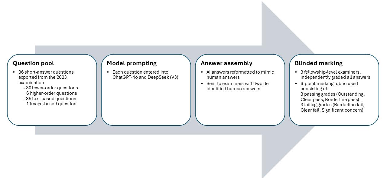
Figure 1. Study workflow for evaluating ChatGPT-4o and DeepSeek (V3) on the 2023 Royal Australasian College of Dental Surgeons oral and maxillofacial fellowship written examination
questions were used to better analyse the LLMs’ ability to perform complex clinical reasoning tasks and higher order judgement.
The complete set of questions from the written component of the 2023 Royal Australasian College of Dental Surgeons maxillofacial fellowship examination was used.
Questions were divided into two types based on Bloom’s taxonomy of cognitive learning objectives:9 lower-order questions, which tested information recall and basic comprehension, and higherorder questions, which tested critical thinking, through the analysis and application of information in clinical reasoning tasks. Of the 36 questions, six were higher-order questions, and 30 were lower-order questions, and of the 36 questions, 35 were text-based and one was image-based. The image-based question was a lower-order question.
To minimise potential sources of bias and ensure consistency, a standardised method of obtaining AI-generated answers was used. New LLM accounts were created to ensure no previous chat history between the user and LLM was present before starting the study.
Each examination question was entered individually into ChatGPT and DeepSeek, in the same format as it appeared to candidates undertaking the examination. A new and independent chat session was created before entering each question, and previous chat sessions were cleared before entering subsequent questions. This controlled approach ensured that responses were generated without influence from prior interactions or residual data, eliminating the possibility of cumulative learning within a single testing session.
The completed ChatGPT and DeepSeek answers were gathered and formatted to match the format of human candidate answers. To facilitate blind marking, two sets of human candidate answers were de-identified and sent together with the LLM compiled answers to examiners for marking.
Both the human and AI answers were assessed and scored by three examiners in a blinded fashion. Examiners were trained fellowshiplevel examiners, following the strict, standardised 6-point scoring rubric used in the actual fellowship examination (Figure 1).
A Fleiss’ Kappa measure was used to measure inter-examiner reliability.
This study was approved by the ACT Health Human Research Ethics Committee (2024.LRE.00004).
A total of 72 responses from LLMs were generated, with 36 responses from ChatGPT and 36 responses from DeepSeek. These responses were independently marked by three examiners using a 6-point performance rubric, each resulting in 108 responses, and a total of 216 outcome results.
All questions were interpreted correctly by both LLMs and all responses addressed the questions posed directly. There were no hallucinations (blatant factual errors or fictional references) observed.
1. Overall grade distribution for ChatGPT and DeepSeek on 36 fellowship-level short-answer questions
Of the 36 questions submitted to ChatGPT, 88.9% were answered at a passing grade and 11.1% were given a failing grade (Table 1, Figure 2a).
DeepSeek achieved a 5.5% higher overall pass rate than ChatGPT, with 94.4% answered at a passing grade and 5.6% scored at a failing grade (Table 1, Figure 2b).
Performance stratified by cognitive level
ChatGPT and DeepSeek achieved matching overall performance on higher-order questions, with 94.4% of responses marked at a passing grade (Table 2). Within the higher-order questions, ChatGPT produced a higher proportion of “outstanding” results than DeepSeek (50% vs 44.4%), and a lower proportion of “borderline pass” results (5.6% vs 11.1%). The proportion of “clear pass” (38.9%) and “borderline fail” (5.6%) grades was the same for both models.
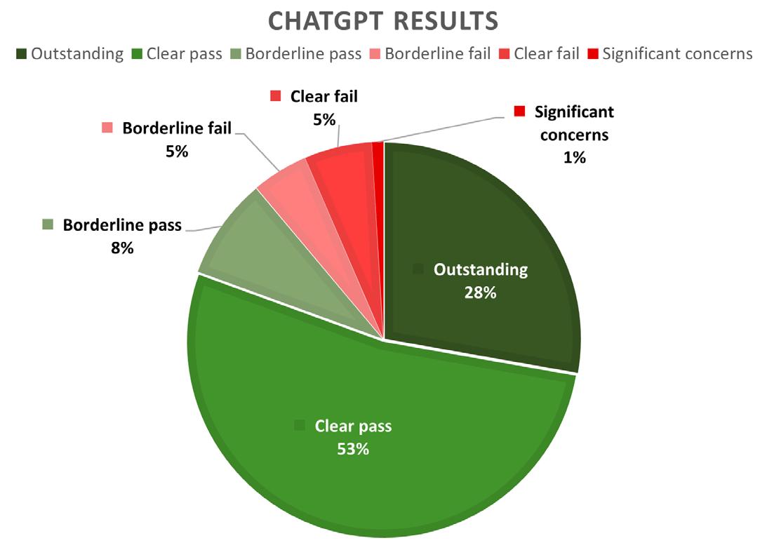
In the lower-order questions, DeepSeek had a higher overall pass rate than ChatGPT (94.4% vs 87.8%) with marginally higher frequencies in each of the three passing categories.
Of the 36 questions, 35 were text-based and one was image-based (requiring anatomical labelling of a microscopic depiction of a nerve fibre). In this image-based task, ChatGPT’s answer was assessed as “borderline fail” by two examiners (66.6%) and as a “significant concern” (33.3%) by one examiner. DeepSeek’s answer was marked as “clear fail” (100%) by all three examiners. As a result, both models received failing grades for this question.
The Fleiss’ Kappa measure was used to assess inter-rater reliability, which was recorded as moderate (Fleiss’ к = 0.41). This result was statistically significant (P < 0.001).
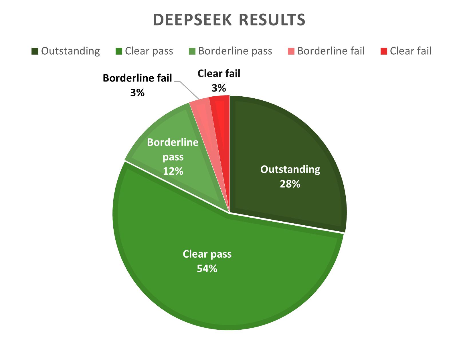
2. Grade distribution for ChatGPT and DeepSeek stratified by cognitive level (Bloom’s taxonomy9)
AI has become increasingly integrated in medical education and clinical practice. LLMs have become widely accessible to both patients and clinicians; however, LLM responses can be outdated or inaccurate, creating the potential for users to place unwarranted trust in their responses. Given their increasing use, thorough evaluation of their performance is essential to determine their reliability and guide development of appropriate safeguards to ensure LLM use remains safe and clinically responsible.
Recent studies assessing LLM performance in medical education have focused on single best answer questions. This has ranged from undergraduate dentistry examinations in maxillofacial surgery, to specialist examinations in ear, nose and throat surgery and plastic surgery.6-8 Although these studies demonstrated that LLMs could achieve a near passing or passing grade, they also identified a key limitation. LLMs performed well on lower-order questions based on information recall but showed a weaker performance on higherorder questions that required interpretation, clinical reasoning and information synthesis.4-8
Our study builds on this foundation by introducing several novel elements. We assessed LLM performance using open-ended questions, which are more reflective of real-world clinical reasoning and decision-making. This allows for a more accurate assessment of higher-order thinking than multiple-choice questions.
Using this question format, both ChatGPT and DeepSeek achieved a passing grade in this study. In keeping with prior research, our study shows that both LLMs performed well on information recall questions, with 87.8% (ChatGPT) and 94.4% (DeepSeek) of lowerorder questions answered correctly. However, in contrast to previous studies, performance in higher-order questions was comparably high, with 94.4% of these questions being answered at a passing level by both models. Several factors likely contributed to this improvement in performance. Firstly, advancements in model architecture and LLMs being trained on progressively larger and more up-to-date datasets have contributed to more efficient and accurate responses. In addition to this, the growing adoption of LLMs by medical professionals has significantly increased feedback, consequently improving model responses to be more clinically relevant.10
Despite these advancements, limitations were observed in some responses. In clinical scenarios where evidence is controversial or evolving, neither model could consistently provide up-to-date or definitive responses. For example, when asked to compare management options of odontogenic keratocysts, both ChatGPT and DeepSeek listed several appropriate options, namely marsupialisation, peripheral ostectomy, use of adjunctive Carnoy’s solution and cryotherapy. However, neither model mentioned more contemporary adjuncts such as the use of 5-fluorouracil (5-FU), and the transition from traditional to modified Carnoy’s solution. Both models also failed to include surgical resection as a treatment option, a long-established and well documented treatment modality, particularly indicated in complex, recurrent or multilocular lesions.11
This omission of adjunctive 5-FU or modified Carnoy’s solution could be attributed to the reliance of current LLMs on historical training data. ChatGPT-4o, for example, has a knowledge cut-off of October 2023, and DeepSeek’s training data was last updated in July 2024.12,13. As a result, neither model may reliably include emerging treatments or the most recent guidelines in their responses.
In contrast, the failure to include surgical resection as a management option cannot be explained solely by outdated training data or the absence of contemporary evidence. Its role is clearly supported in the surgical literature dating back several decades, before any of the model cut-off dates. This omission demonstrates a broader limitation in current LLMs, where there is a tendency to default to conservative or commonly cited management options.
A further limitation observed in this study was the generalised nature of responses, which lacked the nuance required for individualised patient care, particularly in this example, where treatment decisions depended on variables, such as lesion extent, recurrence pattern and other patient-specific factors.
These limitations highlight that although these tools can effectively support medical education, they are not yet suitable for guiding clinical management or making autonomous decisions. Although these personalised, patient-specific considerations are not necessarily required for written examinations, they are for direct clinical care, therefore these findings highlight the importance of using LLMs as supportive adjuncts rather than replacements in health education and clinical decision-making.
The difference in performance seen between ChatGPT and DeepSeek in this study could be attributed to the difference in recency of training data. Although ChatGPT has performed strongly in multiple benchmark studies, DeepSeek outperformed ChatGPT in this comparative study. Another contributing factor could be a difference in model architecture — specifically, DeepSeek’s mixture of experts framework, compared with ChatGPT’s generative pre-trained transformer model. This is the first study evaluating DeepSeek’s performance in the surgical examination context in Australia, and its comparable performance suggests its potential as a viable alternative.
To our knowledge, this is the first study to incorporate an imagebased question when evaluating LLMs in an Australian surgical examination context. In the image-based question included in this study (labelling the anatomical components of a nerve), both ChatGPT and DeepSeek produced incorrect answers, resulting in a failing grade. This highlights a current limitation of LLMs such as ChatGPT and DeepSeek, which are largely text-based systems. In this study, ChatGPT and DeepSeek have not demonstrated the capability to interpret visual data. However, this assessment was based on a single question, which limits its generalisability.
In conclusion, although LLMs can provide reliable general information to a specialist certification level, their current limitations in image interpretation, up-to-date evidence and lack of personalised
treatment advice suggest that they are not ready for clinical use. Their strong performance in both lower-order and higher-order questions in this study substantiates their value as educational aids. However, the observed omissions in contemporary management options, reliance on historical training data and tendency to default to commonly cited management options, highlight the need for cautious interpretation of their outputs. Continued refinement and validation will be required to ensure their safe and effective integration in health care practice.
Author contributions
Jung T: Methodology, data collection, data analysis, draft writing, reviewing and editing.
Bobinskas A: Conceptualisation, data analysis, review of manuscript, supervision.
Lewis E: Data analysis.
Deane S: Data analysis.
Hyam D: Methodology, data analysis, supervision.
1 Amisha, Malik P, Pathania M, Rathaur VK. Overview of artificial intelligence in medicine. J Family Med Prim Care 2019; 8(7): 23282331.
2 De Menezes Torres LM, de Morais EF, Almeida F, et al. The impact of the large language model ChatGPT in oral and maxillofacial surgery: a systematic review. Br J Oral Maxillofac Surg 2025; 63(5): 357362.
3 Dave T, Athaluri SA, Singh S. ChatGPT in medicine: an overview of its applications, advantages, limitations, future prospects, and ethical considerations. Front Artif Intell 2023; 6: 1169595.
4 Gilson A, Safranek CW, Huang T, et al. How does ChatGPT perform on the United States Medical Licensing Examination (USMLE)? The implications of Large Language Models for medical education and knowledge assessment. JMIR Med Educ 2023; 9: e45312.
Permission was granted from the Royal Australasian College of Dental Surgeons (RACDS) for the use of the written component of the 2023 Oral and Maxillofacial Surgery Fellowship Examination in this study.
All authors declare that they have no conflicts of interest.
Data sharing
The data underlying this article are available for sharing upon reasonable request to the corresponding author.
5 Long C, Lowe K, Zhang J, et al. A novel evaluation model for assessing ChatGPT on otolaryngology–head and neck surgery certification examinations: performance study. JMIR Med Educ 2024; 10: e49970.
6 Hoch C, Wollenberg B, Luers JC, et al. ChatGPT’s quiz skills in different otolaryngology subspecialties: an analysis of 2,576 singlechoice and multiplechoice boardcertification preparation questions. Eur Arch Otorhinolaryngol 2023; 280: 42714178.
7 Gupta R, Herzog I, Park J, et al. Performance of ChatGPT on the plastic surgery inservice training examination. Aesthet Surg J 2023; 43(12): NP1078-NP1082.
8 Quah B, Yong W, Lai WM, et al. Performance of large language models in oral and maxillofacial surgery examinations. Int J Oral Maxillofac Surg 2024; 53: 881886.
9 Adams NE. Bloom’s taxonomy of cognitive learning objectives. J Med Libr Assoc 2015; 103(3): 152153.
10 Singhal K, Azizi S, Tu T, et al. Large language models encode clinical knowledge. Nature 2023; 620(7972): 172180.
11 Al-Moraissi EA, Dahan AA, Alwadeai MS, et al. What surgical treatment has the lowest recurrence rate following the management of keratocystic odontogenic tumor? A large systematic review and meta-analysis. J Craniomaxilllofac Surg 2017; 45(1): 131-144.
12 OpenAI. Model release notes: Updates to GPT4o in ChatGPT [Internet]. San Francisco (CA): OpenAI; 2025 Jan 29 [cited 2025 Jun 15]. https://help.openai.com/ en/articles/9624314-model-release-notes
13 DeepSeek. Response to user query about data update timeline [Internet]. 2024 [cited 2025 Jun 15]. https://arxiv. org/abs/2412.19437
A
BDS)
;
J
;
PhD)
Orbital floor fractures have been considered in scientific literature since 1889.1 In 1957, Smith and Regan characterised orbital blowout fractures as fractures of the internal orbital wall in the setting of an intact orbital rim.2 The hydraulic mechanism by which a blunt trauma to the globe raises intra-orbital pressure was proposed as the underlying cause for this fracture pattern. Orbital fracture patterns in children differ from those in adults due to the differences in the craniofacial skeletal ratio, and the histological composition of the facial bones. Soll and Poley first described the paediatric trapdoor fracture in 1965, identifying it as a variant of orbital blowout fractures characterised by a hinge and recoil mechanism of the fractured bones, leading to entrapment of the periorbital soft tissues within the fracture site.3 Despite this early observation, several decades passed before age-related differences in fracture patterns and the clinical features of white-eye syndrome were elucidated.4,5 The latter is characterised by nausea, vomiting, orbital pain, restricted upward gaze, diplopia and occasionally bradycardia. Signs of a fracture on imaging may be absent along with lack of ecchymosis.6
The surgical urgency required to treat this distinct patient population is well established.7 Timing of paediatric orbital fracture surgery has been reported as immediate (0–48 hours), early (3–14 days), and late (beyond 15 days).6 Emergency intervention of < 24 hours has been advocated particularly for patients exhibiting symptoms of white-eye syndrome.5,8-10 Surgical repair of these fractures involves the release of trapped periorbital soft tissue and muscle, using a biocompatible material.6,11 The material barrier prevents re-entrapment of tissues, compared with the restoration of orbital shape and volume for a displaced orbital wall fracture using a more rigid biomaterial. There is no single universally accepted biomaterial for use in the paediatric population. Biomaterials include autogenous grafts, allografts, xenografts and resorbable and nonresorbable alloplasts.9,11 An important consideration for the paediatric population is that the biomaterial must not interfere with growth and development of the facial skeleton.6,12,13
Purpose: This systematic review and meta-analysis set out to evaluate surgical outcomes in paediatric orbital fracture repair in relation to timing of the intervention and biomaterial choice.
Methods: The study adhered to Preferred Reporting Items for Systematic reviews and Meta-Analyses (PRISMA) guidelines and involved a search of the PubMed, Embase and Cochrane Library databases. Twenty-five studies (all retrospective case series) out of 809 articles met the inclusion criteria for reporting pre- and post-operative outcomes, biomaterials used and time to surgery in patients ≤ 18 years old. Methods, including random effects meta-regression with logit transformation, were used for analysis of surgical outcomes.
Results: Surgical intervention improved symptoms of diplopia and restriction of ocular motility. Surgical intervention within 72 hours was associated with decreased negative outcomes (persistence of diplopia, restricted ocular motility and enophthalmos) compared with > 72 hours. Overall, 57.3% (274/478) of biomaterials used was non-resorbable and 42.7% (204/478) was resorbable. Medpor was the most common recorded non-resorbable implant used. A portion of patients (49) did not have an implant placed. Due to the heterogenous nature of materials used (23 different types recorded), the meta-analysis did not identify a statistically significant association between biomaterial used and outcome. When no implant was used, increased negative outcomes were noted; however, this was not statistically significant due to the small number of patients.
Conclusion: The meta-analysis supports the case for operating on paediatric orbital fractures within at least 72 hours of injury.
1Oral and Craniomaxillofacial Surgery, Epworth HealthCare, Melbourne, VIC, Australia; 2Faculty of Medicine and Health Sciences, University of Auckland, Auckland, New Zealand; 3Office for Research, Epworth HealthCare, Melbourne, VIC, Australia; 4Department of Health Sciences and Biostatistics, Swinburne University of Technology, Melbourne, VIC, Australia; 5Oral and Maxillofacial Surgery, Faculty of Medicine Dentistry and Health Sciences, The University of Melbourne, Melbourne, VIC, Australia.
Corresponding author: Adrian Tang ✉ adrian.tang@dhsv.org.au | doi: 10.63717/2026.MS0065
There have been no published trials on the optimal management of paediatric orbital fractures. This, along with the relative paucity of publications on paediatric orbital fractures, small patient numbers and the non-uniformity and interchangeability in the literature of describing the fracture types (trapdoor, blowout, linear, displaced, simple, isolated, entrapped, non-entrapped) managed, likely contributes to the variability observed in the literature regarding management of these injuries.12,14-17 This systematic review and meta-analysis evaluates the overall surgical management of paediatric orbital fractures, focusing on the optimal intervention time and commonly used biomaterials.
This study was performed in accordance with the PRISMA (Preferred Reporting Items for Systematic reviews and Meta-Analysis) guidelines.18 No protocol was published before commencement in 2016. A comprehensive search was conducted online using three scientific literature databases (MEDLINE/PubMed, Embase and Cochrane Library) and was completed in May 2025. The search strategy is listed below:
1: Orbital and (floor OR wall OR blowout) and (children OR paediatric) and (fracture OR trauma OR injury OR defect);
2: Orbital and (floor OR wall OR blowout) and (children OR paediatric) and (reconstruction OR repair);
1 and 2.
Study selection inclusion criteria:
patients aged ≤ 18 years old, with five or more participants;
diagnosis of any of the following orbital fractures: isolated floor, floor extending into one or more orbital walls (not roof), and complex fractures including the naso–ethmoidal and/or zygomatic complex;
description of surgical approach and evaluation of any biomaterial;
use of pre- and/or post-operative imaging (computed tomography [CT] or magnetic resonance imaging [MRI]);
results of pre- and post-operative ophthalmological examination: diplopia, restriction of ocular motility, enophthalmos, nausea and vomiting;
documented time to surgery.
Randomised controlled trials, prospective studies and retrospective case series were considered. Case reports, letters to the editors, editorial comments and review papers were excluded. There was no restriction placed on the date of publication, and studies were to be in the English language. The search and selection were performed by two independent evaluators (JJo and AT), with any discrepancies resolved by a third evaluator (JJe).
All papers were retrospective case series, and the quality of papers was assessed using the JBI critical appraisal tool for case series (JBI SUMARI, Adelaide, Australia).19
Full text publications were obtained for all studies that fulfilled the inclusion criteria. Information extracted from the studies included the year of study, number of patients having surgery, age (mean,
standard deviation and range), follow-up period (mean and range), type of orbital floor fracture, biomaterials used, and pre- and postoperative ophthalmologic signs (diplopia, restriction of ocular motility, enophthalmos, nausea and vomiting). Publications mainly included a heterogenous mix of paediatric blowout fractures, combining both patients with trapdoor fractures leading to entrapment, and patients with orbital wall fracture defects requiring contour restoration. No authors were contacted for any unpublished data. Extracted data was collated in Excel 2023 (Microsoft Corporation Redmond, WA, USA).
Data analysed for overall surgical outcomes comprised total number of patients and number of patients with ophthalmologic signs present pre- and post-operatively, if reported by more than five studies. Pre- versus post-binomial data analysis was undertaken using Comprehensive Meta Analysis (CMA) version 4 (Biostat Inc. Englewood, NJ, 2024). For each study, four subtotals were entered into the software (the number of patients with ophthalmologic signs present at pre- and at post- [++], the number of patients with signs present at pre- but not post- [+-], the number of patients with signs absent at pre- but present at post- [-+] of which there were none, and the number of patients with signs absent at pre- and absent at post[--]). Odds ratios were calculated using CMA for each study.
CMA was used to examine the percentage of post-operative negative outcomes (persistence of diplopia, ocular motility restriction and enophthalmos). Studies were subdivided by time to surgery:
< 72 hours;
72 hours to 2 weeks; and
> 2 weeks.
For hypothesis generation, further subgroup analysis was conducted within the < 72 hour group, analysing patients who had surgery within ≤ 24 hours, or between 24 and 72 hours. A second subgroup analysis compared patients who waited < 72 hours with patients who waited ≥ 72 hours for surgery. Time to surgery data were analysed using meta-regression with Stata version 19 (Stata Corporation, College Station, TX, 2025).20
A random effects model, with logit transformation of the postoperative negative outcomes, was applied.21 I2 was used to measure the percentage of variability in effects estimate that was due to study heterogeneity, while prediction intervals were used to estimate the predicted range of effect sizes.22,23 Publication bias was assessed by funnel plots, generated using CMA. Statistical significance was set at 0.05 level, 2-tailed. Confidence intervals of 95% were reported throughout. Due to limited information on the type of biomaterials and associated outcomes, analysis of the latter was restricted to overall numbers aggregated across studies employing binary logistic regression, using Stata.
The database search yielded 1331 different publications. Of these, 522 duplicates were excluded, leaving 809 article titles and abstracts to be studied. A further 637 failed to meet the inclusion criteria for
publication type. A total of 172 full text articles were analysed and only 25 papers met the inclusion criteria (Figure 1). All 25 articles were retrospective case series (Oxford Centre for Evidence-Based Medicine, Level IV evidence).24
A total of 677 patient cases were reported across 25 papers published between 1998 and 2025. Data collected spanned from 1985 to 2023 and involved data over a range of 3 to 18 years. The follow-up period for each study varied from three months to five years. The mean age of patients across all the studies was 11.9 years. There were interchangeability and non-uniformity in the description of fracture types (blowout, trapdoor, linear, simple, isolated) and the majority of studies combined data for all fracture types. Only six papers described patients with purely trapdoor fractures. Table 1 summarises the characteristics of the papers included in the review.
Table 2 reports on the ophthalmological signs pre- and post-surgery in each paper with some papers reporting only one outcome measure, whereas others reported all of them (diplopia, enophthalmos, nausea and vomiting, and restriction of ocular motility). Only five papers actively measured signs of pre- and post-enophthalmos. A metaanalysis was performed to assess the overall effect of surgery on symptoms of diplopia and restriction of ocular motility.
Pre-surgery diplopia was present in approximately 77% (400/522) of total cases but only 27% (107/400) persisted post-surgery (studies with pre- and post-surgery data) at the final follow-up appointment. Restriction in ocular motility was seen in 84% (377/450) of total cases pre-surgery but it persisted in 21% (58/272) post-surgery (studies with pre- and post-surgery). Nausea and vomiting symptoms had 100% recovery post-surgery.
Diplopia was reported in 21 studies both pre- and post-operatively, whereas 16 studies reported both pre- and post-operative motility restriction. There appeared to be substantial heterogeneity across these studies (I2, 42% for diplopia, 42% for restriction) (additional material (funnel plots) is available from the author).
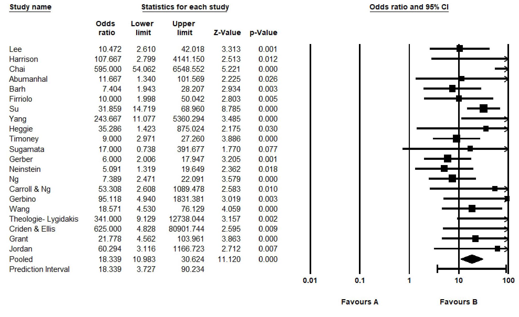
All studies were intra-individual comparisons. All the individual study effects were statistically significant regarding diplopia, except for the study by Sugamata and colleagues, the results of which narrowly failed to achieve statistical significance (odds ratio [OR], 17.00; 95% confidence interval [CI], 0.74–391.68; P = 0.077).36 Overall, the pooled odds ratio was statistically significant (OR, 18.34; 95% CI, 10.98–30.62; P < 0.001) (Figure 2).
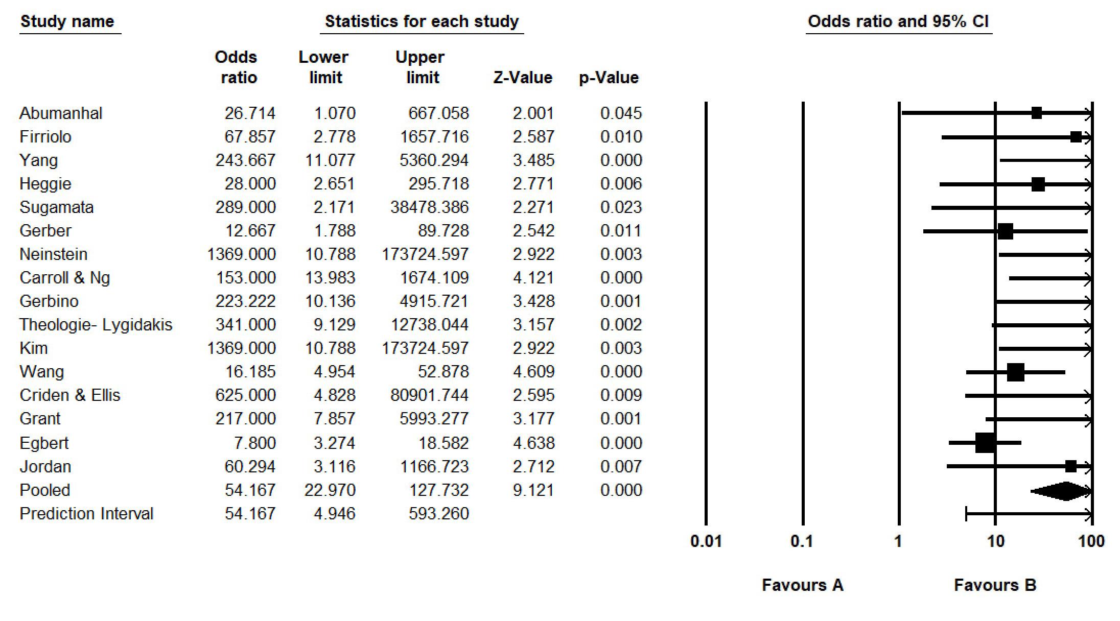
All the individual study effects were statistically significant for ocular motility restriction (Figure 2). The pooled odds ratio was statistically significant (OR, 54.17; 95% CI, 22.97–127.73; P < 0.001) (Figure 3). Both results indicate that there was improvement in both diplopia and ocular motility restriction following surgery.
Meta-analysis was performed to determine outcomes (persistence of diplopia, restriction of ocular motility or enophthalmos) with increased time to surgery. Across all 25 studies, 131 patients were operated on
within 24 hours from injury, 81 patients between 24 and 72 hours, 117 patients between 72 hours and 2 weeks, and 84 patients after 2 weeks. Notably, the remaining 232 patients lacked a reported time frame to surgery. Seventeen studies reported clear outcomes within defined timeframes and were included in the analysis of surgical timing associated with intervention outcomes.
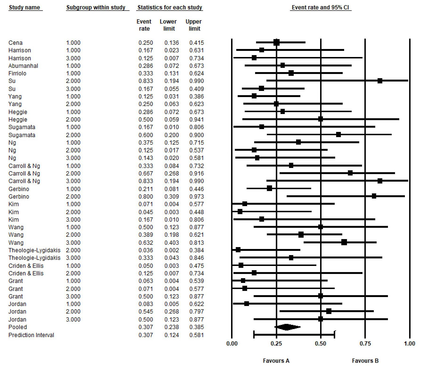
The overall proportion of negative events, taking timeframe into account, was 0.307 or 30.7% (95% CI, 0.24–0.39) (Figure 4). The overall I2 was 28%. There did not appear to be potential publication bias (additional material (funnel plots) is available from the author).
As shown in Table 3, the proportion of negative post-operative outcomes (persistence of diplopia, ocular motility restriction and enophthalmos) was lowest for the < 72-hour group (proportion = 0.19), increasing for the 72-hours-to-2-week group (0.42), then dropping slightly for the > 2 week-group (0.39).
Meta-regression, employing < 72 hours as the reference category, showed that there was a statistically significant increase in proportion of post-operative negative outcome (P = 0.036) if operated on between 72 hours and 2 weeks (Table 3). There was also an increase in proportion of negative outcomes at > 2 weeks, compared with < 72 hours, although the difference narrowly failed to achieve statistical significance (P = 0.053). A subgroup analysis comparing < 72 hours with ≥ 72 hours was statistically significant (P = 0.012). A subgroup analysis of < 72 hours subdivided into ≤ 24 hours versus 24–72 hours employing ≤ 24 hours as the reference category was not statistically significant (P = 0.474). Although these results need to be
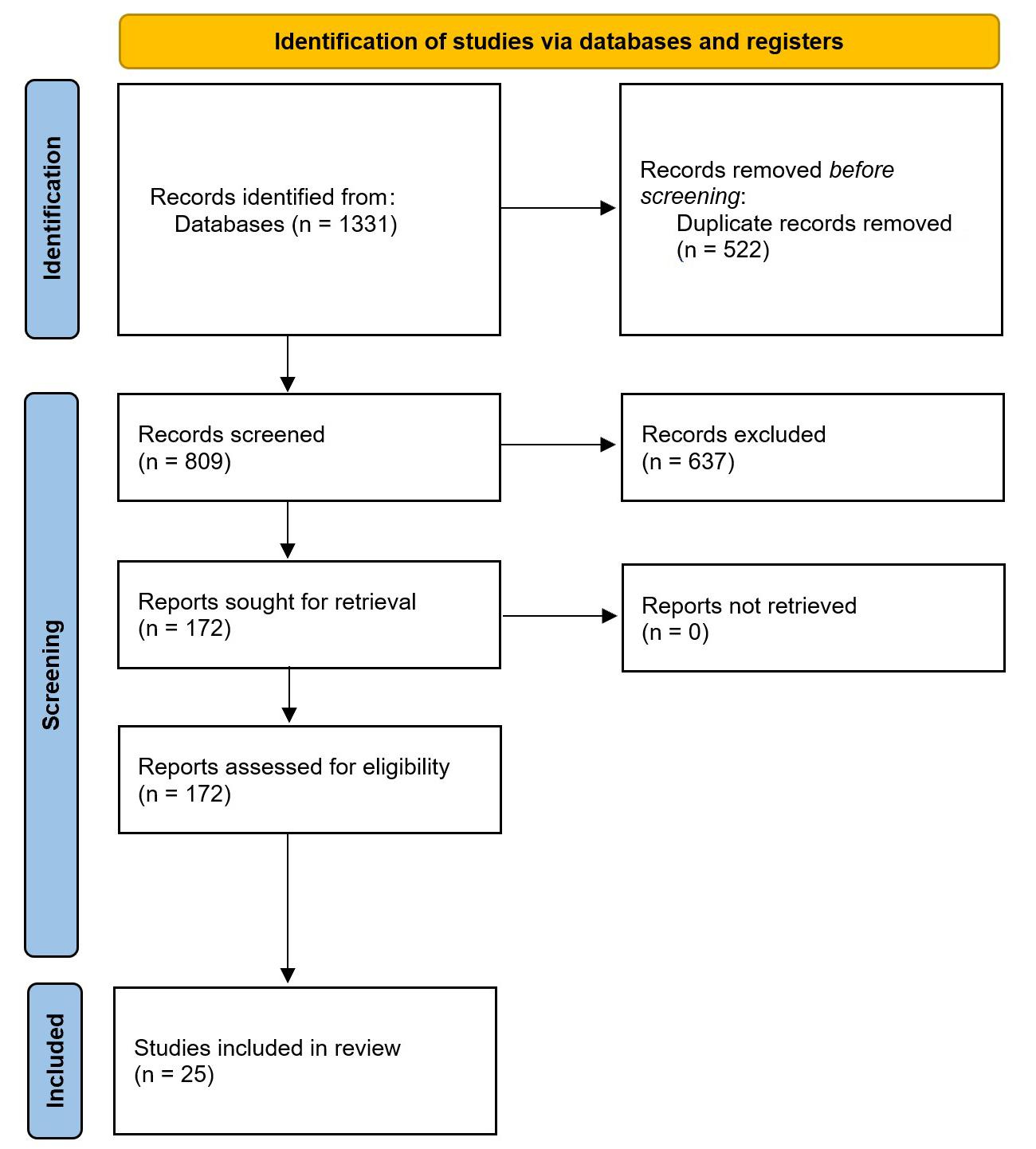
Figure 1. Preferred Reporting Items for Systematic reviews and Meta-Analyses (PRISMA) study selection flow diagram
Author
Lee et al25
Boniao et al26
Cena et al27
Harrison et al28
Chai et al29
Abumanhal et al30
Bahr et al31
Firriolo et al32
Su et al33
Yang et al34
Heggie et al6
Timoney et al35
Sugamata et al36
(9)
Gerber et al37 2013 20 6–17 (13.5)
Neinstein et al38
Ng et al39
Carroll & Ng40
Gerbino et al10
Kim et al41
Wang et al42
Medpor (Stryker, Kalamazoo, MI, USA), titanium, Rapidsorb (DePuy Synthes, West Chester, PA, USA), Osteomesh (Osteopore, Singapore), Polymax (Depuy Synthes, West Chester, PA, USA), Macropore (Macropore Biosurgery, San Diego, CA, USA), no implant
Resorbable implant, Lyoplant (B.Braun, Aesculap, Tutsingen, Germany) or similar, titanium, no implant
Polyesterurethane (Neuropatch) (B.Braun, Meslungen, Germany)
polycaprolactone, silicone sheet, X ray plate, titanium, no implant
Medpor, SYNPOR (DePuy Synthes, West Chester, PA, USA), gelfoam
Medpor, hydroxyapatite, RapidSorb
Gelfilm (Pfizer, New York, NY, USA)
(B.Braun, Melsungen, Germany)
titanium, bioresorbable mesh, no implant
Supramid, Polymax, no implant
(B.Braun, Melsungen, Germany), Lactosorb (Biomet, Jacksonville, FL, USA), Heterologous Bone
(Continues on page 16)
Table 1. Summary of included studies
(Continues from page 15)
TheologieLygidakis et al43
Criden & Ellis44
et al5
Abbreviations: SD = standard deviation. + = intervention or follow-up times beyond the stated number, but unspecified.
* Description of data only for 12 surgical patients with entrapment.
** Exclusion of 3 patients >18 years of age.
replicated on larger samples, < 72 hours appears to exhibit a lower proportion of negative outcomes than ≥ 72 hours.
A summary of the biomaterials used across the 25 papers is presented in Table 4. Several papers did not clearly define the type and/or numbers of biomaterials used, and this is reflected within the table.
There were 23 distinct biomaterials implanted and, of these, 42.7% (204/478) was resorbable (including autogenous bone), whereas 57.3% (274/478) was non-resorbable. Medpor was the most commonly used non-resorbable material (141/274, 51.5%). Poly (L)lactide-co-glycolic acid was the most common resorbable material (45/204, 22.1%) followed by polydiaxanone sheet (PDS) and bone (both 23/204, 11.3%).
A total of 49 patients had surgery and did not have any implant placed. Of these, 27 (55.1%; 95% CI, 40.2–69.3%) patients had a documented outcome, and of these, 10 (37.0%; 95% CI, 19.4–57.6%) had persistence of pre-operative symptoms.
A summary of the biomaterials from the 17 papers used in the analysis of surgical timing associated with intervention outcome is given in Table 5. Studies in which the biomaterials were unspecified had the lowest percentage of negative outcomes, and studies with no implant placed had the highest. Due to uncertainty of the biomaterial used when unspecified, statistical analysis was restricted to resorbable, non-resorbable and no implant, as shown in Table 6 There was no statistically significant difference in outcomes between non-resorbable and resorbable (P = 0.410), and non-resorbable and no implant (P = 0.617).
This systematic review and meta-analysis critically examine the optimal timing for surgical intervention in paediatric orbital floor and wall fractures and analyses the biomaterials used.
This meta-analysis confirms the efficacy of surgical repair in improving diplopia and ocular motility in paediatric orbital fractures and reinforces prior evidence that early surgical intervention leads to better clinical outcomes. A statistically significant difference in outcome was identified in this meta-analysis between patients treated within 72 hours, and those treated after 72 hours.
Early surgical intervention for paediatric orbital fractures has been associated with improved recovery of extraocular muscle function compared with delayed repair.46 Egbert and colleagues reported when surgery was performed more than 30 days post-injury, the resolution rates of ocular motility restriction and diplopia were significantly lower.45 Similar findings were described by Jordan and colleagues5 and Gerbino and colleagues10. In paediatric patients, delayed intervention has been associated with soft tissue fibrosis, excessive scarring and early callus formation within the surgical field, all of which complicate fracture reduction and reconstruction.6,7,9,47
Although this meta-analysis did not reveal a statistically significant difference between patients treated in the first 24 hours and those treated between 24 and 72 hours, this may be due to the limited sample size. It should be noted that several authors in this field have advocated for surgical intervention within the first 24 and 48 hours for paediatric trapdoor fractures.5,8-10
In practical terms, delays in presentation and referral remain a significant challenge within the paediatric cohort. Such delays may stem from limited awareness among non-specialist or regional
* Description of data only for 12 surgical patients with entrapment.
** Exclusion of 3 patients >18 years of age.
† Result in parentheses (residual symptom) derived from studies with both pre- and post-surgery data.
centres of the urgency of early surgical intervention and the differences in management protocols between adults and children. Diagnosis can be complicated by the absence of classic clinical signs such as diplopia, enophthalmos or restriction of ocular motility; particularly in younger patients.6,9,11,46 Additional delays may occur due to the need for transfer from regional or rural facilities to specialised paediatric centres, a process that can take several days. Misdiagnosis may also arise when symptoms are attributed to concussion or when reluctance to expose children to ionising radiation postpones appropriate imaging.9 Consequently, some patients are
Table 2. Ophthalmological findings
admitted for observation rather than undergoing timely surgical assessment, resulting in missed opportunities for early intervention and an increased risk of long-term sequelae, including persistent diplopia and ocular motility impairment.6,9,10,36,45
The heterogeneity in paediatric orbital fractures (trapdoor versus wall defect reconstruction) and the variability in clinical presentation, may have contributed to the meta-analysis narrowly failing to demonstrate a statistically significant difference in outcomes between patients treated after two weeks and those operated on within 72 hours of injury.
Abbreviations: CI = confidence interval.
2. Diplopia forest plot
Abbreviations: CI = confidence interval.
The ideal biomaterial used in the management of paediatric orbital fractures must balance biological safety with adaptability to growth. It should be biocompatible and avoid growth restriction. Low infection risk, cost-effectiveness, radiological compatibility and ease of intra-operative handling are also key, ensuring both functional reconstruction and long-term safety in the growing craniofacial skeleton.48,49
Orbital development is characterised by rapid early growth, with approximately 80–90% of adult orbital volume attained by 5–7 years
Figure 3. Restriction of ocular motility forest plot
of age. Thereafter, growth continues at a slower rate, with gradual incremental increases throughout late childhood. By 7–8 years of age, orbital dimensions are nearly complete, and by 12–15 years of age, an estimated 95–97% of adult orbital volume has been achieved, after which only minimal changes occur.50-52
Mechanical stability is a critical characteristic of the ideal biomaterial in adult and paediatric orbital trauma where large defects are treated to restore orbital volume and support peri-orbital content. In children with a trapdoor fracture, the mechanical stability of the biomaterial is less critical. The objective in these cases is to free the peri-orbital
Abbreviations: CI = confidence interval.
contents caught within the trapdoor fracture and reduce the fracture. Therefore, a less rigid biomaterial that does not interfere with growth is suitable in these cases. Indeed, there are some reports of not using any form of implant once the fracture has been reduced in cases of entrapment.53-55 Others support the use of a biocompatible barrier to avoid scar formation and adherence of the peri-orbital tissue within the fracture site during the healing period.6
4. Time to surgery and negative outcomes forest plot
This meta-analysis found a total of 49 patients had undergone surgery without any orbital implant. In 27 of these patients, a documented outcome was recorded and although statistically not significant, 10 of these 27 were noted to have persistence of their pre-operative symptoms. Logistic regression of overall aggregate data, due to paucity of information, found there to be no statistically
≤24 hours, 24–72 hours
≤24 hrs
Table 3. Time to surgery and negative outcomes meta-regression
significant difference between patient outcomes when no implant placed was placed versus cases where non-resorbables were used.
Autogenous bone grafting has historically represented the gold standard for the management of craniofacial defects, with donor sites commonly including the iliac crest, calvaria, maxilla and mandible.56,57 These grafts offer rigidity, biocompatibility, lower infection rates and are believed to cause less disruption along growth sutures. Other autogenous materials such as cartilage, dura and fascia are also used with similar characteristics but lower strength. Autogenous grafts have the disadvantage of donor site morbidity and increased operative time, which can be avoided when allografts are used.48 Autogenous and allografts both share the disadvantage of graft absorption over time, making them less reliable for the long-term correction of enophthalmos where large defects are treated.58,59 This disadvantage has less relevance in the surgical management of the paediatric orbital trapdoor fracture. Autogenous grafts, allografts, xenografts and other resorbable materials were observed in less than half of the patients in this meta-analysis (42.7%).
Alloplastic material offers the advantages of decreased operative time, accessibility and avoiding donor site morbidity. A wide range of alloplastic material, both resorbable and non-resorbable, has been reported for use in orbital trauma. Non-resorbable implants commonly used in the literature for orbital trauma include titanium mesh, porous polyethylene sheets (Medpor), silicone sheets, Teflon, Supramid, Marlex mesh and Prolene. Resorbable materials reported include Gelfilm and resorbable material made of poly-lactic and poly-glycolic acid.58 The disadvantages of these materials include higher infection rate, migration, potential growth restriction if used inappropriately, and in the case of resorbable materials, sterile inflammation.48,60 This meta-analysis observed Medpor to be the most common nonresorbable material used in the 25 studies.
Two of the implants used across this review are no longer commercially available: Lyodura, discontinued in 1996 due to its association with Creutzfeld–Jakob disease; and Gelfilm, production of which ceased in 2021. Although silicone remains commercially available, its use has declined due to the unacceptably high rate of implant-related complications such as cyst formation, infection and extrusion. Supramid is associated with spontaneous intracapsular haemorrhage.61
Surgeons have the option of using autogenous, allograft or alloplastic implants, including resorbable materials for paediatric orbital trauma. A wide range of biomaterials (23 different types) were identified in this review, ranging from autogenous to alloplastic and resorbable materials. There is heterogeneity within the literature regarding the acceptable age for use of non-resorbable alloplastic material, which could potentially impede growth. The Arbeitsgemeinschaft für Osteosynthesefragen (AO) foundation guidelines state that these materials should be avoided in patients under 4 years of age, whereas others support a more conservative approach, with recommendations that patients under 7 years should receive autogenous grafts.48,53 In this review, a predominance of alloplastic materials was observed (57.3% of patients). This may be in part due to the mean age of patients at the time of surgery (11.9 years)
Autograft Bone R 23
Allograft Dura R 26
Xenograft Porcine gelatine (Gelfilm) R 9
Bovine collagen (Lyoplant) R 19
Porcine gelatine (Gelfoam) R n/s
Bioceramic Hydroxyapatite - Coral derived NR n/s
Metal Titanium NR 13
Polymers Silastic NR 14
Porous Polyethylene Sheet (Medpor) NR 141
Polytetrafluoroethylene (Gore-Tex/ Teflon) NR 30
Nylon (Supramid/SupraFoil) NR 75
Xray film (polyester) NR 1
Polycaprolactone R 7
Polyester urethane (Neuropatch) R 8
Polyactide R n/s
Poly L-Lactide- co glycolic acid (Lactosorb/Rapidsorb) R 45
Polydioxanone Sheet (PDS) R 23
“Bioresorbable mesh/implant” R 12 No implant No implant 49
Unspecified Unspecified R 31 Unspecified NR or R 121 Total patients (645*) 647
Abbreviations: R = resorbable; NR = non-resorbable; n/s = not specified. * 2 patients had bilateral implants
Table 4. Biomaterial summary
with near completion of orbital growth. However, the age range of patients studied was 1 to 18 years and alloplastic material use was reported in children as young as 1 year of age. The increasing trend towards use of alloplastic materials is likely due to their advantages with respect to operative time, donor site morbidity, reliability and structural strength as well as flexibility. Although this meta-analysis did not find a statistically significant difference between resorbable and non-resorbable implants and patient outcomes, these results should be interpreted with caution when considering implant choice given that none of the papers using alloplastic materials exclusively, reported on long-term outcomes on facial or orbital asymmetry and growth restriction.34,35,40-42 According to current evidence, alloplastic material should be avoided in the immature orbit. Although autogenous grafts and resorbable materials were used less frequently across the studies, they remain the gold standard in patients with immature orbital growth.
This meta-analysis revealed that, irrespective of the biomaterial used, surgery facilitated an improvement in pre-operative symptoms and signs, consistent with findings reported in the literature.4,40,62
Abbreviations:
This systematic review and meta-analysis included only level IV retrospective review series and was limited by the small number of patients in each publication, together with inconsistent recording of data.
Potential confounders across studies included variability in timing of early and delayed surgery, technique and experience of surgeon, implant selection and the type of orbital fractures treated (trapdoor with or without entrapment, displaced fractures with or without entrapment).
A significant finding of this systematic review and meta-analysis is the identification of the lack of a prospective study in assessing management and outcomes of paediatric orbital floor fractures, and direct comparison of its subsets. Additionally, recognising the limitations of this systematic review and meta-analysis, we propose
a prospective multicentre study, with data collection via a pre-agreed standard proforma to be used at the initial assessment, surgery, and the follow-up appointments. Importantly, the description of fracture type will be standardised to allow for overall and inter-fracture type data comparison.
This systematic review and meta-analysis demonstrated that surgical intervention improved symptoms of diplopia and restriction of ocular motility in paediatric orbital fractures. This systematic review and meta-analysis supports early surgical intervention (within the first 72 hours) in the surgical management of paediatric orbital fractures, irrespective of fracture type.
In operated patients where biomaterials were used to repair the orbital fracture, improvement was recorded in primary outcomes measured, irrespective of the type of biomaterial used. Alloplastic materials were used in most patients, with the meta-analysis
revealing Medpor as the most common material when the type of non-resorbable biomaterial was recorded. Some studies used alloplastic material in children as young as 1 year old. The long-term impacts remain unknown as none of the studies presented long-term data on orbital growth and asymmetry.
Author contributions
Tang A: Conceptualisation, methodology, formal analysis, investigation, data curation, visualisation, writing – original draft, writing – reviewing and editing.
Johnston J: Formal analysis, investigation, data curation.
McKenzie D: Methodology, formal analysis, investigation, visualisation, writing – reviewing and editing, consultation.
Jeyathevan J: Conceptualisation, methodology, formal analysis, investigation, data curation, visualisation, writing – original draft.
Bordbar P: Conceptualisation, methodology, visualisation, writing –reviewing and editing.
1 Lang W. Traumatic enophthalmos with retention of perfect acuity of vision. Trans Ophthalmol Soc UK 1889; 9(1): 41-45.
2 Smith B, Regan WF. Blow-out fracture of the orbit; mechanism and correction of internal orbital fracture. Am J Ophthalmol 1957; 44(6): 733-739.
3 Soll DB, Poley BJ. Trapdoor variety of blowout fracture of the orbital floor. Am J Ophthalmol 1965; 60: 269-272.
4 De Man K, Wijngaarde R, Hes J, et al. Influence of age on the management of blow-out fractures of the orbital floor. Int J Oral Maxillofac Surg 1991; 20(6): 330-336.
5 Jordan DR, Allen LH, White J, et al. Intervention within days for some orbital floor fractures: the white-eyed blowout. Ophthalmic Plas Reconstr Surg 1998; 14(6): 379-390.
6 Heggie AA, Vujcich NJ, Shand JM, et al. Isolated orbital floor fractures in the paediatric patient: case series and review of management. Int J Oral Maxillofac Surg 2015; 44(10): 1250-1254.
7 Grant JH, Patrinely JR, Weiss AH, et al. Trapdoor fracture of the orbit in a pediatric population. Plas Reconstr Surg 2002; 109(2): 482-489; discussion 490-495.
8 Sires BS, Stanley Jr RB, Levine LM. Oculocardiac reflex caused by orbital floor trapdoor fracture: an indication for urgent repair. Arch Ophthalmol 1998; 116(7): 955-956.
9 Wei LA, Durairaj VD. Pediatric orbital floor fractures. J AAPOS 2011; 15(2): 173-180.
10 Gerbino G, Roccia F, Bianchi FA, et al. Surgical management of orbital trapdoor fracture in a pediatric population. J Oral Maxillofac Surg 2010; 68(6): 13101316.
11 Sirichai P, Anderson PJ. Orbital fractures in children: 10 years’ experience from a tertiary centre. Br J Oral Maxillofac Surg 2015; 53(10): 938-942.
12 Koltai PJ, Amjad I, Meyer D, et al. Orbital fractures in children. Arch Otolaryngol Head Neck Surg 1995; 121(12): 1375-1379.
13 Broyles JM, Jones D, Bellamy J, et al. Pediatric orbital floor fractures: outcome analysis of 72 children with orbital floor fractures. Plast Reconstr Surg 2015; 136(4): 822-828.
14 Hink EM, Wei LA, Durairaj VD. Clinical features and treatment of pediatric orbit fractures. Ophthalmic Plast Reconst Surg 2014; 30(2): 124-131.
15 Alcalá-Galiano A, Arribas-García IJ, Martín-Pérez MA, et al. Pediatric facial fractures: children are not just small adults. Radiographics 2008; 28(2): 441-461.
We would like to acknowledge Epworth Knowledge Services, especially Ms. Susie Moreton, for her valuable assistance in conducting the literature search. This research did not receive any specific grant from any funding agencies – public, commercial or not-for-profit sectors.
The authors declare no conflicts of interest.
The data underlying this article are available for sharing upon reasonable request to the corresponding author.
16 Imahara SD, Hopper RA, Wang J, et al. Patterns and outcomes of pediatric facial fractures in the United States: a survey of the National Trauma Data Bank. J Am Coll Surg 2008; 207(5): 710-716.
17 Hatton MP, Watkins LM, Rubin PA. Orbital fractures in children. Ophthalmic Plast Reconst Surg 2001; 17(3): 174-179.
18 Page M, McKenzie JE, Bossuyt PM, et al. The PRISMA 2020 statement: an updated guideline for reporting systematic reviews. BMJ 2021; 372: n71.
19 Munn Z, Barker TH, Moola S, et al. Methodological quality of case series studies: an introduction to the JBI critical appraisal tool. JBI Evid Synth 2020; 18(10): 2127-2133.
20 Baker WL, Michael White C, Cappalleri JC, et al. Understanding heterogeneity in meta-analysis: the role of meta-aggression. Int J Clin Pract 2009; 63(10): 14261434.
21 Nikolakopoulou A, Mavridis D, Salanti G. Demystifying fixed and random effects meta-analysis. Evid Based Ment Health 2014; 17(2): 53-57.
22 Higgins JPT, Thompson SG, Deeks JJ, et al. Measuring inconsistency in meta-analyses. BMJ 2003; 327(7414): 557-560.
23 Borenstein M, Higgins JPT, Hedges LV, et al. Basics of meta-analysis: I2 is not an absolute measure of heterogeneity. Res Synt Methods 2017; 8(1): 5-18.
24 Howick J, Chalmers I, Glasziou P, et al. The 2011 Oxford CEBM Levels of Evidence (Introductory Document). Oxford Centre for Evidence-Based Medicine. https://www.cebm. ox.ac.uk/resources/levels-of-evidence/ocebm-levels-ofevidence
25 Lee JY, Park J, Choi H. Clinical features and radiologic findings of orbital bone remodelling in pediatric patients after orbital fracture correction. J Craniofac Surg 2025; 36(5): 1751-1755.
26 Boniao EL, Gungab A, Lim BXH, et al. Pediatric orbital fractures in Singapore: demographics, etiology, and the role of bioresorbable implants. Front Opthalmol (Lausanne) 2025; 5: 1506445.
27 Cena P, Immacolata R, Roccia F, et al. An 11-year multicentric surgical experience on pediatric orbital floor trapdoor fracture: A World Oral Maxillofacial Trauma (WORMAT) project. J Stomatol Oral Maxillofac Surg 2025; 126(1): 102033.
28 Harrison P, Hajefi S, Green AO, et al. Isolated paediatric orbital fractures: a case series and review of management at a major trauma centre in the UK. Oral Maxillofac Surg 2023; 27(2): 227-233.
29 Chai G, Zhang D, Hua W, et al. Theoretical model of pediatric orbital trapdoor fractures and provisional personalized 3D printing-assisted surgical solution. Bioact Mater 2020; 14(6): 559-567.
30 Abumanhal M, Ben-Cnaan R, Feldman I, et al. Polyester urethane implants for orbital trapdoor fracture repair in children. J Oral Maxillofac Surg 2019; 77(1): 126-131.
31 Bahr A, Swaminathan M, Mukherjee B. Orbital fractures in children: clinical features and management outcomes. J AAPOS 2018; 22(6): 415.e1-415.e7.
32 Firriolo JM, Ontiveros NC, Pike CM, et al. Pediatric orbital floor fractures: Clinical and radiological predictors of tissue entrapment and the effect of operative timing on ocular outcomes. J Craniofac Surg 2017; 28(8): 1966-1971.
33 Su Y, Shen Q, Lin M, et al. Diplopia of pediatric orbital blowout fractures: a retrospective study of 83 patients classified by age groups. Medicine (Baltimore) 2015; 94(4): e477.
34 Yang JW, Woo JE, An JH. Surgical outcomes of orbital trapdoor fracture in children and adolescents. J Craniomaxillofac Surg 2015; 43(4): 444-447.
35 Timoney PJ, Krakauer M, Wilkes BN, et al. Nylon foil (Supramid) orbital implants in pediatric orbital fracture repair. Ophthalmic Plast Reconstr Surg 2014; 30(3): 212-214.
36 Sugamata A, Yoshizawa N, Shimanaka K. Timing of operation for blowout fractures with extraocular muscle entrapment. J Plast Surg Hand Surg 2013; 47(6): 454-457.
37 Gerber B, Kiwanuka P, Dhariwal D. Orbital fractures in children: a review of outcomes. Br J Oral Maxillofac Surg 2013; 51(8): 789-793.
38 Neinstein RM, Phillips JH, Forrest CR. Pediatric orbital floor trapdoor fractures: outcomes and CT-based morphologic assessment of the inferior rectus muscle. J Plast Reconstr Aesthet Surg 2012; 65(7): 869-874.
39 Ng JY, Gangadhara S, Wong W, et al. Paediatric orbital blow-out fractures: surgical outcomes. Asia Pac J Ophthalmol (Phila) 2012; 1(5): 265-269.
40 Carroll SC, Ng SG. Outcomes of orbital blowout fracture surgery in children and adolescents. Br J Ophthalmol 2010; 94(6): 736-739.
41 Kim J, Lee H, Chi M, et al. Endoscope-assisted repair of pediatric trapdoor fractures of the orbital floor: characterization and management. J Craniofac Surg 2010; 21(1): 101-105.
42 Wang N-C, Ma L, Wu S-Y, et al. Orbital blow-out fractures in children: characterization and surgical outcome. Chang Gung Med J 2010; 33(3): 313-320.
43 Theologie-Lygidakis N, Iatrou I, Alexandridis C. Blow-out fractures in children: six years’ experience. Oral Surg Oral Med Oral Pathol Oral Radio Endod 2007; 103(6): 757-763.
44 Criden MR, Ellis FJ. Linear nondisplaced orbital fractures with muscle entrapment. J AAPOS 2007; 11(2): 142-147.
45 Egbert JE, May K, Kersten RC, et al. Pediatric orbital floor fracture: direct extraocular muscle involvement. Ophthalmology 2000; 107(10): 1875-1879.
46 Bansagi ZC, Meyer DR. Internal orbital fractures in the pediatric age group: characterization and management. Ophthalmology 2000; 107(5): 829-836.
47 Kwon JH, Moon JH, Kwon MS, et al. The differences of blowout fracture of the inferior orbital wall between children and adults. Arch Otolaryngol Head Neck Surg 2005; 131(8): 723-727.
48 Vasile VA, Istrate S, Iancu RC, et al. Biocompatible materials for orbital wall reconstruction – an overview. Materials (Basel) 2022; 15(6): 2183.
49 Goodrich JT, Sandler AL, Tepper O. A review of reconstructive materials for use in craniofacial surgery
bone fixation materials, bone substitutes and distractors. Childs Nerv Syst 2012; 28(9): 1577-1588.
50 Bentley RP, Sgouros S, Natarajan K, et al. Normal changes in orbital volume during childhood. J Neurosurg 2002; 96(4): 742-746.
51 Furuta M. Measurement of orbital volume by computed tomography: especially on the growth of the orbit. Jpn J Ophthalmol 2001; 45(6): 600-606.
52 Escaravage GK, Dutton JJ. Age-related changes in the pediatric human orbit on CT. Ophthalmic Plast Reconstr Surg 2013; 29(3): 150-156.
53 Borsuk D. Orbital reconstruction [Internet]. AO Surgery Reference. 2024. https://surgeryreference.aofoundation. org/cmf/pediatric-trauma/midface/orbital-floor/ reconstruction#emergency-treatment
54 Koide R, Ueda T, Takano K, et al. Surgical outcome of blowout fracture: early repair without implants and the usefulness of balloon treatment. Jpn J Ophthalmol 2003; 47(4): 392-397.
55 Jo EJ, Kim JH, Yang HJ. Inferior blow-out fracture reduction using two urinary balloon catheters. Arch Craniofac Surg 2015; 16(3): 114-118.
56 Potter JK, Malmquist M, Ellis E. Biomaterials for reconstruction of the internal orbit. Oral Maxillofac Surg Clin North Am 2012; 24(4): 609-627.
57 Gunarajah DR, Samman N. Biomaterials for repair of orbital floor blowout fractures: a systematic review. J Oral Maxillofac Surg 2013; 71(3): 550-570.
58 Joshi S, Kassira W, Thaller SR. Overview of pediatric orbital fractures. J Craniofac Surg 2011; 22(4): 1330-1332.
59 Ellis E, Tan Y. Assessment of internal orbital reconstructions for pure blowout fractures: cranial bone grafts versus titanium mesh. J Oral Maxillofac Surg 2003; 61(4): 442-453.
60 Chandler DB, Rubin PA. Developments in the understanding and management of pediatric orbital fractures. Int Ophthalmol Clin 2001; 41(4): 87-104.
61 Custer PL, Lind A, Trinkaus AM. Complications of Supramid orbital implants. Ophthalmic Plast Reconstr Surg 2003; 19(1): 62-67.
62 Avashia YJ, Sastry A, Fan KL, et al. Materials used for reconstruction after orbital floor fracture. J Craniofac Surg 2012; 23(7 Suppl 1): 1991-1997.
Goh EZ (BDSc(Hons), MD)1,2,3; Beech N (BSc (Neurosci), MBBS, BDS, MPhil (Surgery), FRACDS(OMS))1,4; Johnson NR (BDSc(Hons), MBBS, MPhil (Surgery), FRACDS(OMS))1,3,4
Maxillofacial trauma has important consequences, including lifethreatening involvement of nearby vital structures such as the airway, brain and cervical spine; functional implications from injury to local sensory or motor structures; and psychological impacts from the visibility of facial defects.1,2 Successful surgical management must consider evolving trends in patient factors that have significant capacity to influence post-operative outcomes.1
Patient factors include demographics, mechanism of injury, fracture pattern and associated injuries. The World Oral and Maxillofacial Trauma project investigating the prospective management of maxillofacial fractures in 14 centres reported a male:female ratio of 3.2:1 and a mean age of 37 years.3 Road traffic accidents was the most common mechanism overall; and was associated with traumatic brain injuries and high-impact fracture patterns (such as naso-orbito-ethmoid [NOE] complex and frontal bone fractures).3-6 The systemic review by Boffano and colleagues of epidemiological studies over three decades also cited road traffic accidents as the main mechanism in America, Africa and Asia, whereas assaults were more prominent in Europe and Oceania.7 Assault (35%) as the most common mechanism of injury was also consistent with the 2024 Pabbati and colleagues scoping review of the aetiology of maxillofacial trauma in Australia.8 Of interest, e-scooter accidents as a mechanism for craniofacial trauma is becoming more prevalent since the local introduction of ride-sharing schemes in 2018, and continues to garner attention as a public health concern.9,10
This study aims to describe trends in patient factors, and to investigate the association between patient factors and post-operative outcomes in the surgical management of maxillofacial trauma. A better understanding of these aspects of maxillofacial trauma surgery will improve planning of individual management and public health prevention strategies, which in turn will facilitate quality care and efficient resource allocation.
Purpose: Maxillofacial fractures have important functional and cosmetic consequences. This study aims to describe trends in patient factors, and to investigate the association between patient factors and post-operative outcomes in the surgical management of maxillofacial trauma.
Methods: Retrospective review of patients aged 18 years or older, surgically treated for maxillofacial fractures at two tertiary hospitals in Australia (2014–2020). Demographics, mechanism of injury, fracture pattern and associated injuries were recorded. Statistical analyses utilised Fisher’s exact test and binary logistic regression.
Results: There were 2702 cases included (mean age, 35 years; 83% male). The most common mechanism of injury was assault (52%). Depression (11%) was the most common comorbidity, with an increasing trend (P = 0.004). The most common fracture pattern was mandibular angle (25%). The most common associated injuries were long bone (4.6%) and brain (4.4%); there was a significant association with mechanism (P = 0.022), with motor vehicle accidents having the most associated injuries. Complication rates were 31% (1-week review) and 33% (6-week review). Sensory deficit of the mandibular nerve was the most common complication. Revisions (5%) were most commonly due to hardware complications (39%). Smoking was a significant risk factor for revisions (P = 0.030) and 1-week complications (P = 0.022).
Conclusions: Ongoing robust longitudinal studies are required to update knowledge and improve patient outcomes.
1Faculty of Medicine, University of Queensland, Brisbane, QLD, Australia; 2Oral and Maxillofacial Department, Royal Brisbane and Women’s Hospital, Brisbane, QLD, Australia; 3Oral and Maxillofacial Department, Princess Alexandra Hospital, Brisbane, QLD, Australia; 4Oral and Maxillofacial Surgery, Sunshine Coast University Hospital, Birtinya, QLD, Australia; 5School of Dentistry, University of Queensland, Brisbane, QLD, Australia.
Corresponding author: Elizabeth Goh ✉ elizabeth.goh@uq.net.au | doi: 10.63717/2026.MS0057
A retrospective review of patient records from two Australian tertiary hospitals was performed. All procedures were performed in compliance with the Declaration of Helsinki. Study approvals, including waiver of consent, was received from the Royal Brisbane & Women’s Hospital Human Research Ethics Committee (LNR/2020/ QRBW/64241); Queensland Government Health Innovation, Investment and Research Office; Metro South Research Office; and the University of Queensland before commencement. Electronic records were available from May 2014 to December 2020 (Hospital 1), and from December 2015 to December 2020 (Hospital 2), and accessed via computerised searches using International Classification of Diseases 10th Revision (ICD-10)11 codes for maxillofacial fracture repair. Inclusion criteria were: age of 18 years or older, and surgical treatment for maxillofacial fractures. Exclusion criteria were: duplicates, age below 18 years, revision surgeries, nontraumatic fractures, non-surgical management. Each traumatic event, even if requiring multiple surgeries, was considered a single case. Deidentified data was entered into Excel (Microsoft, 2023) in pre-defined categories of patient factors (age and sex; medical history; mechanism of injury; fracture pattern; associated injuries) and post-operative outcomes (complications at T1 [1-week review] and T2 [6-week review]; revision surgeries within the study period).
Statistical analysis was performed with R, version 4.3.0, by a blinded external statistician. Associations between categorical variables were assessed with Fisher’s exact test.12 Associations between continuous variables and dichotomous outcome variables were assessed with binary logistic regression. Statistical significance was defined as P < 0.05.
This study was reported according to the Strengthening The Reporting of OBservational studies in Epidemiology (STROBE) checklist (checklist available on request to the corresponding author).
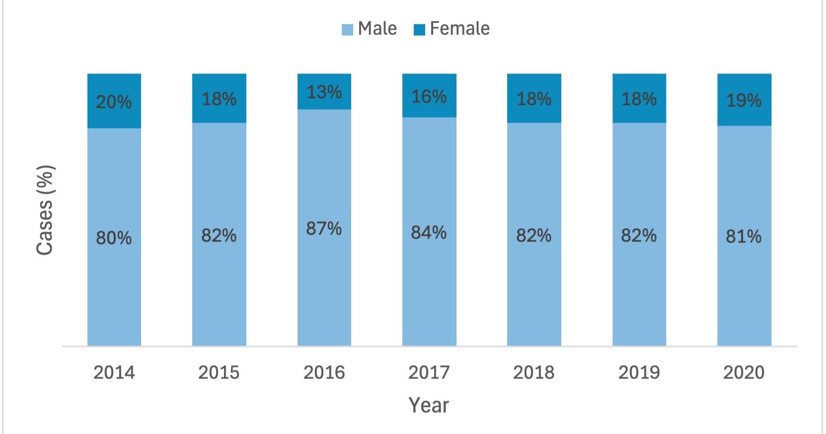
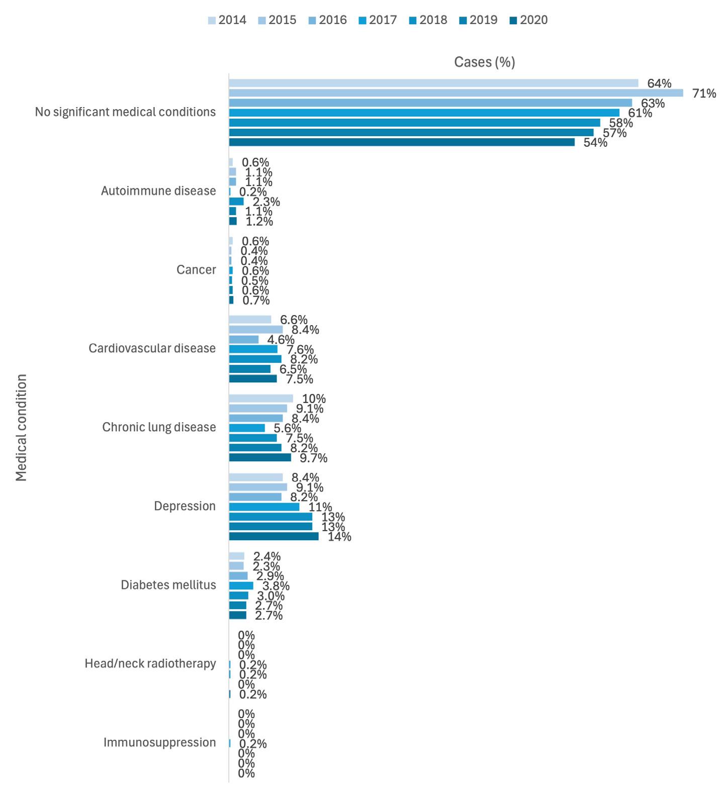
All available records (n=3835) had exclusion criteria applied: duplicates (n=836); age below 18 years (n=175); revision surgeries (n=80); non-traumatic fractures (n=33); non-surgical management (n=9). All remaining cases were included (n=2702).
Patient factors were: age and sex (Figure 1), medical history (Figure 2, Figure 3), mechanism of injury (Figure 4), fracture pattern (Table 1) and associated injuries (Table 2). Annual trends are displayed in the figures. Mean age was 35 years, median was 31 years, and range was 18–90 years. Most cases were male (83%, n=2245).
Most cases had no significant medical conditions (60%, n=1629) and no regular medications (68%, n=1830). Relevant comorbidities were: depression (11%, n=304), chronic lung disease (8.1%, n=218), cardiovascular disease (7.0%, n=189), diabetes mellitus (2.9%, n=79), autoimmune disease (1.1%, n=30), cancer (0.6%; n=15), head/neck radiotherapy (0.1%, n=3) and immunosuppression (<0.1%, n=1). Relevant medications were: anticoagulants/antiplatelets (3.1%, n=85), corticosteroids (2.1%, n=58), non-steroidal anti-inflammatory drugs (1.4%, n=37), antiresorptives (0.6%, n=17) and chemotherapy (0.1%, n=4). For tobacco smoking, 45% (n=1212) were smokers, 49% (n=1326) were non-smokers, and 6.1% (n=164) had no data recorded.
Mechanisms of injury consisted of assault (52%, n=1418), fall (15%, n=402), sports (13%, n=345), motor vehicle accident (4.4%, n=118), pushbike accident (3.6%, n=98), motorbike accident (2.7%, n=73), pedestrian accident (1.1%, n=30), e-scooter accident (0.7%, n=18) and other (7.4%, n=200).
Middle third fractures (54%) were the most common, particularly zygomatico-maxillary complex (ZMC) fractures (23%). For lower third fractures (48%), mandibular angle (25%) and parasymphysis fractures (22%) were the most common. Upper third (1.5%) and panfacial fractures (0.15%) were rare.
Associated injuries were reported in 9.2% (n=249): long bone (4.6%), brain (4.4%), thoracic (3.9%), abdominal (1.4%) and cervical spine (1.4%). There was a significant association with mechanism (P = 0.022), with motor vehicle accidents having the most associated injuries.
Post-operative outcomes were complications at T1 (1-week review) and T2 (6-week review) (Table 3), and revision surgeries (Table 4).
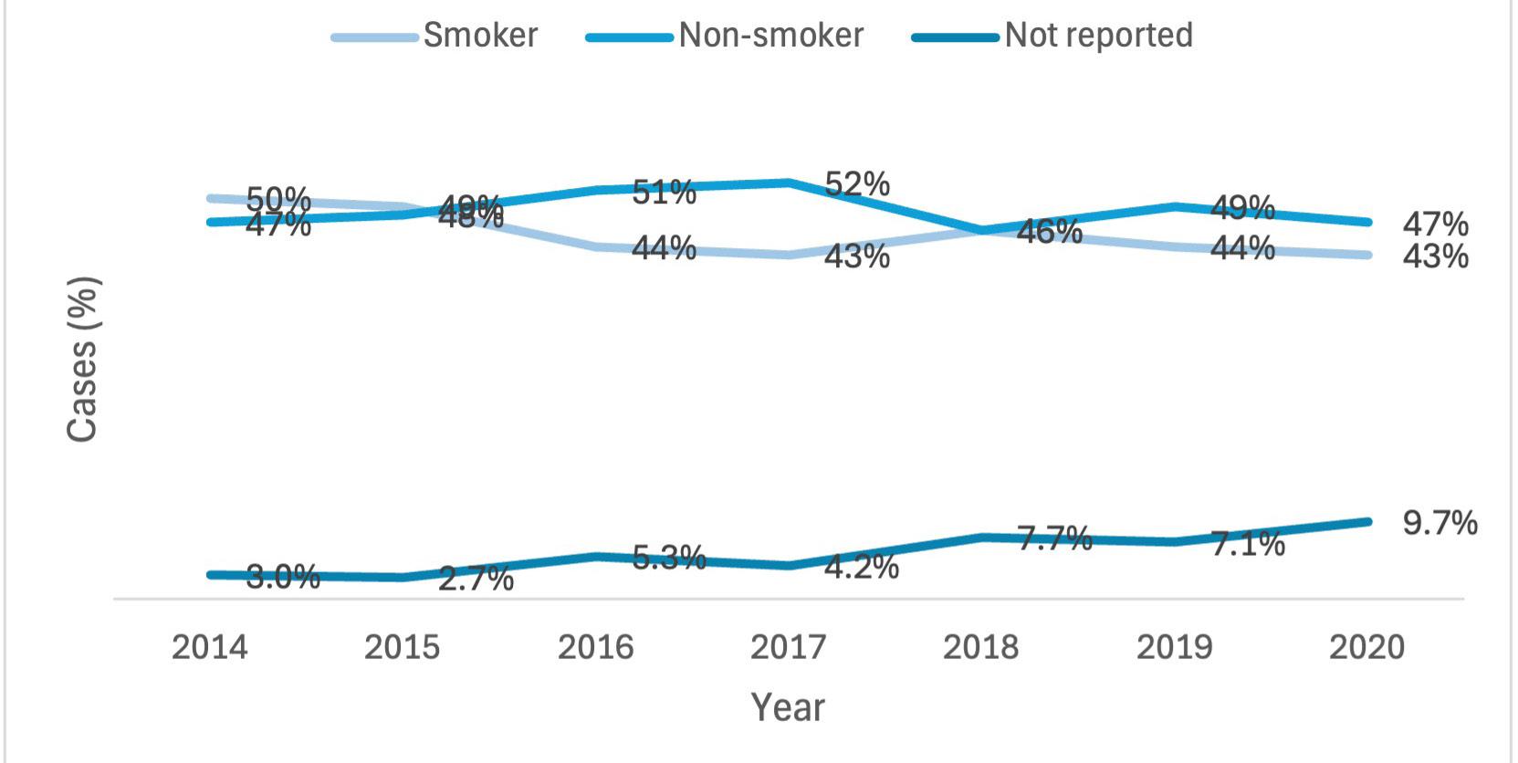
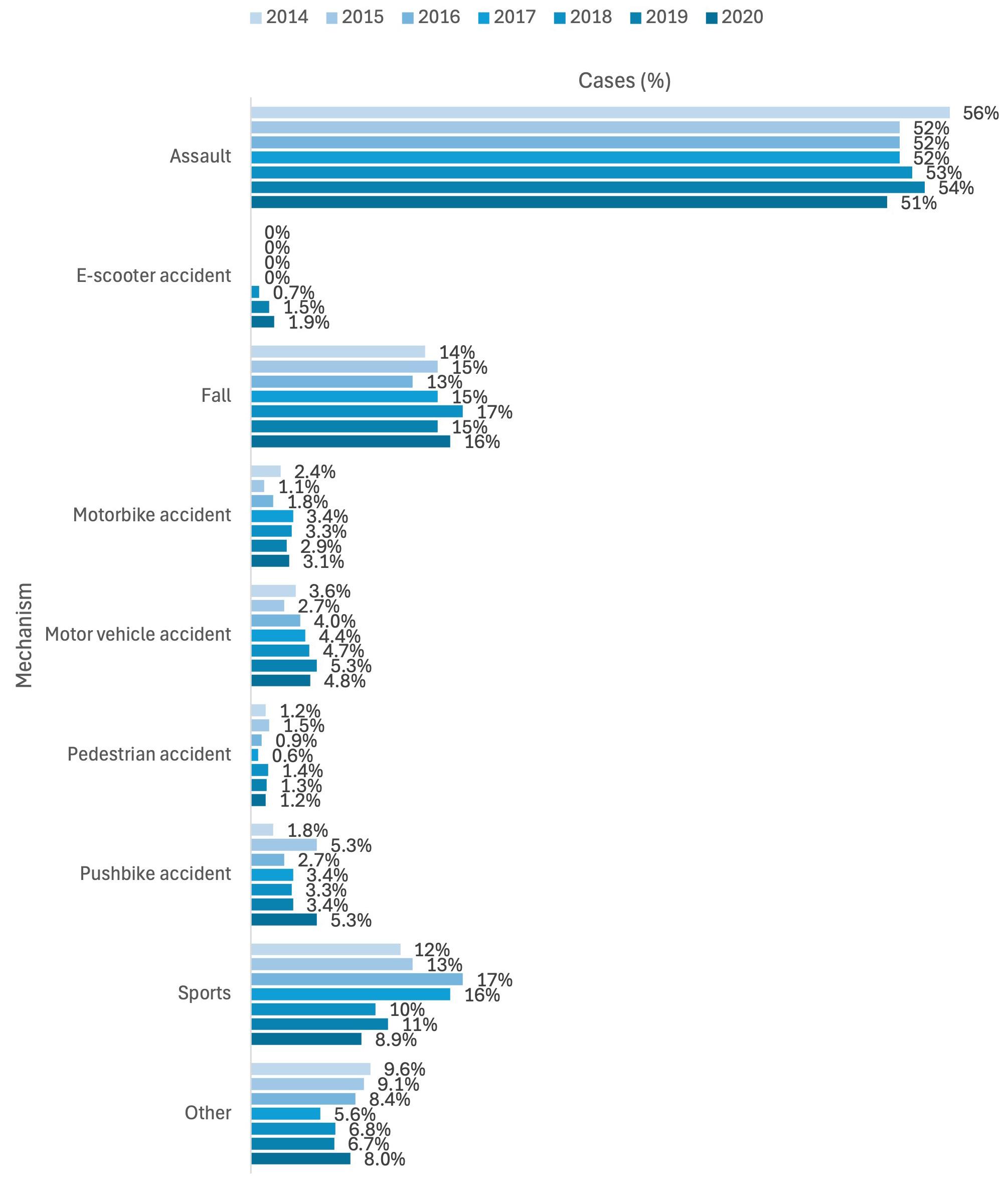
Attendance was 79% (n=2145) at T1 and 42% (n=1147) at T2. Complication rates were 31% (n=655) at T1 and 33% (n=384) at T2, with a total of 759 and 455 complications respectively. The most common complication was sensory deficit of the mandibular nerve (27% at T1 and 22% at T2).
A total of 154 revisions were performed in 137 cases (5.0%), most commonly due to hardware complications (39%). Median timing was:
48 days for first revision (n=137), 128 days for second revision (n=13), 137 days for third revision (n=3) and 257 days for fourth revision (n=1).
Regarding the impact of patient factors on post-operative outcomes, smoking was a significant risk factor, while age, sex and other
n (%) Unilateral, n (%) Bilateral, n (%)
Lower third (mandible) a 1304 (48%) 724 (27%) 580 (21%)
Angle 689 (25%) 660 (24%) 29 (1.1%)
Body 177 (6.6%) 166 (6.1%) 11 (0.4%)
Condyle 369 (14%) 299 (11%) 70 (2.6%)
Coronoid 0 (0%) 0 (0%) 0 (0%)
Parasymphysis 583 (22%) 569 (21%) 14 (0.5%)
Ramus 19 (0.7%) 19 (0.7%) 0 (0%)
Symphysis (total) 75 (2.8%) Not applicable Not applicable
Middle third a 1458 (54%) 1016 (38%) 442 (16%)
Dentoalveolar process (isolated) 16 (0.6%) 9 (0.3%) 7 (0.3%)
Le Fort 1 106 (3.9%) 22 (0.8%) 84 (3.1%)
Le Fort 2 63 (2.3%) 24 (0.9%) 39 (1.4%)
Le Fort 3 42 (1.6%) 17 (0.6%) 25 (0.9%)
Nasal bone 331 (12%) 30 (1.1%) 301 (11%)
Nasomaxillary complex 166 (6.1%) 154 (5.7%) 12 (0.4%)
Naso-orbito-ethmoid complex 29 (1.1%) 11 (0.4%) 18 (0.7%)
Orbital floor b 327 (12%) 325 (12%) 2 (<0.1%)
Orbital medial wall b 76 (2.8%) 76 (2.8%) 0 (0%)
Zygomatic arch (isolated) 133 (4.9%) 131 (4.8%) 2 (<0.1%)
Zygomatico-maxillary complex 632 (23%) 619 (23%) 13 (0.5%)
Upper third (frontal sinus) a 41 (1.5%) 21 (0.8%) 20 (0.7%)
Anterior table 41 (1.5%) 21 (0.8%) 20 (0.7%)
Posterior table c 3 (0.1%) 0 (0%) 3 (0.1%)
a Panfacial fractures (involving all facial thirds) occurred in four cases (0.15%). There were 1226 (45%) cases with lower third fractures only, 1361 (50%) cases with middle third fractures only, and 16 (0.6%) cases with upper third fractures only.
b There were 340 (13%) cases of orbital fractures, consisting of: combined floor and orbital medial wall fractures ( n =63, 2.3%); isolated floor fractures ( n =264; 9.8%); isolated medial wall fractures ( n =13, 0.5%).
c Two of the three cases of posterior table fractures involved the dura.
Table
There were no associated injuries reported for sports injuries or e-scooter accidents.
Table 2. Associated injuries according to mechanism of injury (P = 0.022)
medical history were not significant. Smokers had a significantly higher complication rate (34%) than nonsmokers (28%) at T1 (P = 0.022) but not at T2. There were higher (although non-significant) rates of wound dehiscence at T1 (2.9% smokers, 1.6% non-smokers) and T2 (0.4% smokers, <0.1% non-smokers). There was also no significant association between smoking and infection at T1 (0.7% smokers, 0.8% non-smokers) or T2 (1.6% smokers, 0.9% non-smokers). Smokers had a significantly higher revision rate (6.2%) than nonsmokers (4.2%) (P = 0.030).
This study’s aims were to describe trends in patient factors, and to investigate the association between patient factors and post-operative outcomes in the surgical management of maxillofacial trauma.
Patient factors were age and sex, medical history, mechanism of injury, fracture pattern and associated injuries. The mean age was 35 years, which is higher than previous reports (15–24 years13 and 20–29 years3). As in existing literature,7 most patients were male; however, the male:female ratio in this study was almost 5:1. Annual age/sex trends appeared largely stable.
There was a decreasing trend in fit and healthy patients (those with “no significant medical conditions” declining from 71% in 2015 to 54% in 2020), with a significant increase in depression (8–9% in 2014–2016 to 14% in 2020, P = 0.004). This reflects increasing global trends in depression, including a significant transient increase during the COVID-19 pandemic.14,15 Facial trauma increases risk of depression, and patients with pre-morbid depression are at higher risk of complications.16,17 Early recognition, multidisciplinary involvement and continuity of care in the community are important to improve outcomes.17 Future studies could continue to investigate prevalence trends in depression within maxillofacial trauma patients and the efficacy of peri-operative multidisciplinary approaches in ameliorating risks for adverse outcomes.
Smoking was a significant risk factor for early complications and revisions. Annual trends were inconclusive: the proportion of smokers decreased and the proportion of cases with missing data increased over time, while the proportion of non-smokers remained largely constant. Being a modifiable risk factor, it is important to identify it to facilitate smoking cessation, stratify risk during the consent process and guide treatment options. Vaping is also on the rise, and identification of both smoking and vaping status is important in the pre-operative assessment.18 Future
research could investigate vaping as a risk factor for adverse outcomes.
With regards to mechanism, assault (52%) was the most common, at a much higher rate than previous local reports (35%).8 Also of interest, the local introduction of e-scooter ride-sharing schemes in 2018 has seen a corresponding increase in associated maxillofacial fractures.10 E-scooter injuries remain a public health concern, and further research should target risk factors such as alcohol use and lack of helmet use. COVID-19 lockdowns in 2019–2020 may also have impacted trauma patterns. A retrospective study of 344 emergency presentations noted that falls (44% lockdown, 26% non-lockdown) overtook assault, while sports injuries declined from 17% to 4% during lockdown.19 Similarly, in this study, a drop in sports injuries (8.9% versus 10–11% in previous years) and a rise in pushbike accidents (5.3% versus around 3% in previous years) was noted in 2020. This may be due to school closures and sporting event cancellations in line with social distancing strategies, and increased bicycle traffic on roads as a result of limits on non-essential motor vehicle travel.20 However, there was minimal change in rates of assault, falls and traffic accidents in 2020. Awareness of how evolving trends in injury mechanisms reflect current events continues to underpin preventive strategies.
The most commonly injured facial third was the middle third (54%), with a large range of fracture patterns from ZMC fractures (23%) to isolated dentoalveolar fractures (0.6%). Two rare patterns that tended to occur concurrently were NOE complex (1.1%) and upper third (frontal bone) fractures (1.5%), which share a propensity for high-impact mechanisms and serious sequelae due to their anatomical complexity and proximity to vital structures.6 The mandible was the most commonly fractured bone (48%), which is consistent with previous findings.13
Abbreviations: IMF: inter-maxillary fixation; ORIF: open reduction internal fixation; V1: ophthalmic nerve; V2: maxillary nerve; V3: mandibular nerve.
Associated injuries were significantly linked with injury mechanism. Motor vehicle accidents had the most associated injuries, which is consistent with its high-impact nature. Concomitant traumatic brain injury is of particular interest, due to its proximity to maxillofacial structures and potential for significant morbidity and mortality.4 A 4.4% prevalence was reported here, compared with 8.1% in a previous 10-year retrospective study.21
Post-operative outcomes were complications and revision surgeries. Although complication rates remained similar from T1 (31%) to T2 (33%), attendance rate halved (79% T1, 42% T2), demonstrating the pronounced attrition common in trauma populations. In recognising this, only short-term complications up to 6 weeks (T2) were identified in this retrospective study, many of which often resolve over a longer period of time. A well-known example is sensory deficit of the
mandibular nerve, which was also the most common complication reported (27% at T1, 22% at T2). Regarding revision surgeries (5%), most patients requiring revisions only required one (89%), with almost half being due to hardware complications. For subsequent revisions, non-union became a more prominent indication, which reflects the several months required to allow bony healing to run its course. This study provides an overview of common complications that should be discussed in the consent process and assessed post-operatively. Future work could aim to characterise outcomes specific to different fracture patterns.
This study has several limitations. Firstly, while the overall sample size was large, some subgroups were small (eg, e-scooter accidents, rare complications) or missing data (eg, smoking status, T2 attendance <50%). Also, the study population came from two tertiary hospitals, which care for more complex patients and/or patients from centres without an on-site maxillofacial team. Findings should be cautiously interpreted, especially when generalising to less medically/surgically complex populations. Additionally, revisions were only identified during the study period. Patients whose initial repair was later in the
Table 4. Revision surgeries – indications
study period and required revisions may have had these outside the study period, which would not have been identified. Finally, ICD-10 codes for relevant procedures were used to identify eligible patients, and additional eligible patients may have been missed due to inaccurate coding.
This study illustrates evolving trends in patient factors and their association with adverse post-operative outcomes in maxillofacial trauma. Ongoing robust longitudinal evaluations are required to maintain understanding of this important public health concern.
Goh EZ: Conceptualisation, methodology, investigation, writing –original draft, writing – reviewing and editing.
Beech N: Conceptualisation, methodology, writing – reviewing and editing, consultation.
Johnson NR: Conceptualisation, methodology, writing – reviewing and editing, consultation.
Consent
The authors would like to acknowledge and thank Dr Nicole Milham and Dr Matthew Gilmore for their roles as site contacts; and Dr Andrew Jones from Queensland Cyber Infrastructure Foundation (QCIF) for biostatistical services. This study received funding from the Australian and New Zealand Association of Oral and Maxillofacial Surgeons (ANZAOMS) Research & Education Foundation Grant and Australian Government Research Training Program Scholarship.
All authors declare that they have no conflicts of interest.
The data underlying this article cannot be shared because of limitations imposed by the ethics approval for the study.
1 Vujcich N, Gebauer D. Current and evolving trends in the management of facial fractures. Aust Dent J 2018;63:S35-S47.
2 Sahni V. Psychological impact of facial trauma. Craniomaxillofac Trauma Reconstr 2018;11:15-20.
3 Roccia F, Iocca O, Sobrero F, et al. World Oral and Maxillofacial Trauma (WORMAT) project: A multicenter prospective analysis of epidemiology and patterns of maxillofacial trauma around the world. J Stomatol Oral Maxillofac Surg 2022;123:e849-e857.
4 Goh EZ, Beech N, Johnson NR. Traumatic maxillofacial and brain injuries: A systematic review. Int J Oral Maxillofac Surg 2021;50:P1027-1033.
5 Goh EZ, Bullis S, Beech N, Johnson NR. Surgical management of naso-orbito-ethmoidal fractures: A systematic review. Oral Surg Oral Med Oral Pathol Oral Radiol 2024;138:9-20.
6 Goh EZ, Beech N, Johnson NR. Surgical management of frontal bone and naso-orbitoethmoidal fractures: A seven-year multi-centre retrospective review. Oral Surg Oral Med Oral Pathol Oral Radiol 2024;138:602-607.
7 Boffano P, Kommers SC, Karagozoglu KH, Forouzanfar T. Aetiology of maxillofacial fractures: A review of published studies during the last 30 years. Br J Oral Maxillofac Surg 2014;52:901-906.
8 Pabbati S, Thomson P, Sharma D, Bhandari S. The aetiology of maxillofacial trauma in Australia: A scoping review. Aust Dent J 2024;69:146-156.
9 Goh EZ, Beech N, Johnson N. E-scooters and craniofacial trauma: A systematic review. Craniomaxillofac Trauma Reconstr 2022;16:245-253.
10 Goh EZ, Beech N, Johnson NR. E-scooters and maxillofacial fractures: A seven-year multi-centre retrospective review. Aust Dent J 2023;68:113-119.
11 World Health Organization. ICD-10: International statistical classification of diseases and related health problems: Tenth revision (2nd ed.). 2004.
12 R Core Team. R: A language and environment for statistical computing. R Foundation for Statistical Computing. Vienna (Austria): R Foundation for Statistical Computing, 2023. https://www.R-project.org/ Accessed 2023.
13 Cabalag MS, Wasiak J, Andrew NE, Tang J, Kirby JC, Morgan DJ. Epidemiology and management of maxillofacial fractures in an Australian trauma centre. J Plast Reconstr Aesthet Surg 2014;67:183-189.
14 Moreno-Agostino D, Wu Y-T, Daskalopoulou C, Hasan MT, Huisman M, Prina M. Global trends in the prevalence and incidence of depression: A systematic review and meta-analysis. J Affect Disord 2021;281:235-243.
15 Robinson E, Sutin AR, Daly M, Jones A. A systematic review and meta-analysis of longitudinal cohort studies comparing mental health before versus during the COVID-19 pandemic in 2020. J Affect Disord 2022;296:567-576.
16 Walshaw EG, Taylor R, Anderson J, Sexton P, Parmar JD, Carter LM. The psychological sequelae of maxillofacial trauma: A scoping review of the literature. Br J Oral Maxillofac Surg 2022;60:1303-1320.
17 Dugue D, Taylor GA, Maroney J, Spaniol JR, Ramsey FV, Jones CM. Mind the difference: Characterizing the impact of behavioral health disorders on facial trauma. J Surg Res 2022;271:32-40.
18 Australian Institute of Health and Welfare. National Drug Strategy Household Survey 2022–2023. Australian Institute of Health and Welfare. Canberra (Australia): Australian Institute of Health and Welfare, 2024. https:// www.aihw.gov.au/reports/illicit-use-of-drugs/nationaldrug-strategy-household-survey/contents/about Accessed 2025 Jan 3.
19 Nhongo SS, Sklavos A, Lee K, Chan ST, Austin S. The changing face of maxillofacial trauma during the 2020 COVID-19 lockdowns in Melbourne, Australia. Oral Maxillofac Surg 2023;27:125-130.
20 Sephton B, Mahapatra P, Shenouda M, et al. The effect of COVID-19 on a Major Trauma Network. An analysis of mechanism of injury pattern, referral load and operative case-mix. Injury 2021;52:395-401.
21 Salentijn EG, Peerdeman SM, Boffano P, Van den Bergh B, Forouzanfar T. A ten-year analysis of the traumatic maxillofacial and brain injury patient in Amsterdam: Incidence and aetiology. J Craniomaxillofac Surg 2014;42:705-710.
6-8 AUGUST 2026 / CHRISTCHURCH/OTAUTAHI, NEW ZEALAND
The ANZAOMS 2026 Annual Scientific Meeting will be held in the beautiful Otautahi/Christchurch, New Zealand. The 2026 ASM promises to deliver an engaging and thought-provoking scientific program alongside a dynamic exhibition and social program.

Dr Deepak Kademani USA
Professor Andreas Neff Germany



Bontempo D (MD, DMD, BSc, FRACDS (GDP))1; Tan P (BDSC, Grad Dip Clin Dent, MSD, Grad Cert Pros, FPFA, MRACDS)2; Khadembaschi D (BDSc, MD, MPhil)3; Gebauer D (BDSc, MBBS, MSc, FRACDS (OMS))4,5
Edentulism and a terminal dentition can have profound biopsychosocial consequences on an individual’s quality of life.1 Edentulism impairs oral functions including mastication, phonetics and deglutition, which all have negative sequalae on nutrition and general health.2-4 Tooth loss has an aesthetic and psychosocial toll, and is associated with unhappiness, social isolation and reduced self-esteem.2,4 Rehabilitation with an implantsupported fixed prosthesis helps mitigate the biopsychosocial issues of edentulism and improves patient quality of life.5-8
The literature demonstrates predictable and successful implant and prosthetic outcomes with full arch rehabilitation of the terminal dentition or edentulism with immediate loading protocols.9-20 Full arch immediate loading protocols have cumulative implant and prosthesis survival rates of 90.1%–100% and 93.75%–100% respectively.11,12,18,21 Immediate loading has been shown to minimise the number of surgeries, reduce overall treatment time and reduce the financial burden to patients.8,22,23 Immediate loading with a fixed prosthesis enhances patient satisfaction, comfort, aesthetics and function.2,18,24,25
Conventional immediate loading protocols deliver an initial interim prosthesis within seven days.9 This is replaced with a definitive prosthesis three to six months later.10-13,16,17,24 A provisional prosthesis adds costs and time, and increases the number of interventions required during treatment.25 Immediate loading with a definitive prosthesis can reduce the treatment cost, time and number of appointments.23,26 A recent retrospective analysis compared immediate implant loading with an interim prosthesis versus a definitive prosthesis and demonstrated a comparable level of complications.26
Patient-reported outcome measures (PROMs), including patient satisfaction, should be considered when evaluating treatment success.8,27 In implant dentistry, PROMs describe the patient’s subjective perception of their oral health-related quality of life, function and satisfaction with treatment.25,27
Purpose: To evaluate the implant, prosthetic and patientreported outcome measures (PROMs) of a full arch rehabilitation protocol with immediate implant placement and loading with the definitive prosthesis.
Methods: This study included 170 patients (mean age, 62.5; standard deviation, 10.25 years), 203 prostheses (120 maxilla and 83 mandible) and 1069 implants. Implant and prosthesis survival, prosthesis success, the incidence of prosthetic complications and the PROMs of comfort, aesthetics, function and overall happiness with treatment decision were evaluated. A multivariate Cox proportional hazards regression model was used to evaluate the impact of patient characteristics on implant failure.
Results: Cumulative 3-year implant survival was 95.5% with a mean follow-up of 22 months (standard deviation, 14.95 months). The prosthesis survival and success rates were 91.13% and 87.68% respectively. The incidence of prosthetic complications was 12.32%. Sex (male), smoking status (smoking) and position of the implant (molar) were significantly associated with implant failure (P < 0.05). The PROMs of comfort, aesthetics, function and overall happiness with treatment decision had 96%, 98%, 96% and 94% of patients reporting a score of 5 respectively.
Conclusion: The short term implant and prosthetic outcomes of immediate implant placement and loading with a definitive prosthesis are comparable to other protocols. The PROMs indicate high rates of patient satisfaction with treatment.
1Royal Perth Hospital, Perth, WA, Australia; 2Bayside Dental Specialists, Melbourne, VIC, Australia; 3Fiona Stanley Hospital, Perth, WA, Australia; 4Department of Oral and Maxillofacial Surgery, Royal Perth Hospital, Perth, WA, Australia; 5Department of Human Sciences, Oral Health and Equity, University of Western Australia, Perth, WA, Australia.
Corresponding author: Daniel Bontempo ✉ drdanielbontempo@gmail.com | doi: 10.63717/2026.MS0060
Keywords: dental implants | dental implant therapy | immediate dental implant loading | implant-supported dental prosthesis | osseointegration
Traditional literature focuses on biomechanical, functional and aesthetic goals of care, as determined by the clinician.8 There is a paucity of literature exploring patient satisfaction with full arch immediate implant placement and loading treatment.
The AuDentes All-at-Once protocol describes a full arch rehabilitation technique that involves clinical examination, intraoral scans and photographs, and a cone beam computed tomography (CBCT) scan to prefabricate a customised surgical guide and definitive prothesis that can be inserted at the time of implant placement. This protocol differs from current protocols in that it delivers the definitive prosthesis in a single surgical visit and does not require an interim prosthesis.
The aim of this retrospective analysis is to evaluate the implant, prosthesis and PROMs of patients who received full arch rehabilitation with an immediately loaded implant-supported definitive prosthesis via the AuDentes protocol.
A retrospective cohort study was conducted across multiple private clinics and the AuDentes laboratory in Melbourne, Australia. Ethical approval for this review was obtained from the University of Western Australia Human Research Ethics Committee (2023/ET001031).
Inclusion criteria included all patients who had received full arch rehabilitation with the AuDentes protocol between April 2018 and June 2023 and who gave informed consent to have their data collected for publication. Patients who received treatment were either edentulous or had a terminal dentition and were eligible for full arch rehabilitation with full arch immediate implant loading. Treatment was performed by oral and maxillofacial surgeons, periodontists, prosthodontists and general dentists between April 2018 and June 2023. Implants were placed by 22 different surgeons. Design and manufacturing of the surgical guides and definitive prosthesis were performed by a single prosthodontist in a private purpose-built digital dental laboratory in Melbourne, Australia. Follow-up was performed by 14 different restorative clinicians at three to six months and annually thereafter. Information obtained from the follow-up appointments was provided to the AuDentes laboratory.
Eligible patients were identified by their treating clinicians. Eligibility criteria included a terminal dentition or edentulism, and adequate bone for stable placement of at least four implants. More than 2 mm of buccal bone, 1 mm of palatal/lingual bone, and 2 mm of bone between the inferior alveolar nerve, maxillary sinus and floor of the nasal cavity was considered adequate. A CBCT scan was submitted to the AuDentes laboratory for confirmation of eligibility. Tooth modifications and occlusal adjustments were performed to reorganise and obtain the occlusal scheme with maximal intercuspation in centric occlusion in centric relation.
Data collection consists of intraoral and extraoral photograph series, videos of smiling and talking, intraoral scans or digital impressions and CBCT scans.
and intraoral photographs and videos
The extraoral photograph series consisted of frontal photographs with and without retractors to assess smile line and tooth display and, profile photographs smiling and at rest to assess lip support, competency, facial profile and vertical dimension.
The intraoral photograph series consisted of maxillary and mandibular occlusal photographs, lateral photographs and frontal photographs to assess arch form, spacing, tooth position, canine and molar relationships and wear facets.
Intraoral scans or digital impressions
Intraoral scans were obtained and exported in STL or PLY file formats. Scans captured all present teeth, at least 6 mm of soft tissue in dentate patients and the entire hard palate. The scans were performed at the predetermined vertical dimension, in centric relation with the occlusal scheme organised to ensure centric occlusion and maximal intercuspation.
Physical impressions, taken with alginate or poly-vinyl siloxane, that captured all teeth and 6 mm of soft tissue were digitalised. For patients without an existing removable partial denture, two impressions (the treatment and opposing arches and one bite registration) were obtained. For patients with an existing removable partial denture, impressions of the treatment arch, the opposing arch and a bite registration were obtained. A bite registration was taken.
The CBCT field of view captured the maxilla and mandible ensuring the anterior superior nasal spine was included. Reconstructed slice increments and voxel size were less than 0.25 mm. The CBCT scans were exported in DICOM format as uncompressed axial images.
An individualised smile design, implant and surgical protocol were digitally planned. A virtual wax-up was synthesised from the intraoral and extraoral photograph series, the intraoral scans or digital renditions of physical impressions and the videos. The plan was discussed and confirmed with the patient. The CBCT was used in implant planning. Digital smile design planning was performed with 3Shape Dental System or exocad. Implant planning was performed with AuDentes and determined the position, angulation, depth, platform type and number of implants (four, five, six or seven) plus the need for adjunct procedures. Three surgical guides were designed and fabricated using validated workflows from CoDiagnostix (Straumann), a digital implant platform that allows the fabrication of custom drill guides. The bridge prostheses each contained a customised milled titanium bar framework substructure that supported either milled polymethyl methacrylate (PMMA), printed composite or zirconia suprastructure. The surgical guide and prosthesis quality and
accuracy were verified with a digital model derived from an optical scan that was compared with the original design.
The surgery was performed with an aseptic technique under sedation or a general anaesthetic. Extractions were performed followed by a guided alveoplasty. The osteotomy surgical guide was inserted and anchored to the alveolus with bone pins. The planned implants were placed using a full thickness subperiosteal flap approach. Primary stability of more than 35 N cm was achieved in most sites. The implant systems were Straumann RNI SP (regular neck implants, standard plus) in 79 arches, Straumann TLX SP (tapered tissue level implants, standard plus) in 54 arches, Straumann RNI S (regular neck implants, standard) in 32 arches, Straumann TLX S (tapered tissue level implants, standard) in 15 arches, Straumann BLT (bone level tapered) in 15 arches, Straumann TE (tapered effect) in one arch, Neoss in one arch, and Southern implant IT tapered (internal taper, tapered) in one arch. Four arches had a combination of Straumann RNI SP and RNI S, and one arch had a combination of Straumann BLX and TLX SP implants placed.
Implant abutment positions and fit of the prosthesis substructure were confirmed intraoperatively using a duplicate substructure. The definitive prosthesis was inserted onto the implants without any modifications. Post-surgical management included simple analgesia, 0.2% chlorhexidine glucoronate mouthwash four times daily for 14 days and a soft diet for 3 months.
Data collected included patient demographics, smoking and diabetes status, the location and number of implants placed, timing and location of implant failure and timing and reason for prosthesis failure.
Primary outcome measures were cumulative implant survival, prosthesis survival and success, and PROMs. The incidence of prosthetic complications and prosthesis remakes were secondary outcome measures.
Implants that remained in function, had a primary stability of more than or equal to 35 N cm, and did not require replacement were classified as surviving. The four-to-six-month reverse torque test defines early and late implant failures, with early failures occurring before or at the test. Prosthetic success was defined as a prosthesis in function, without complications or modifications and meeting the patient’s aesthetic expectations. Prosthetic survival was defined as a functional prosthesis requiring modifications. Prosthetic failure was defined by removal and replacement and was attributed to implant, prosthetic or aesthetic failure.
Prosthetic complications were attributed to implant failure necessitating replacement or modification of the prosthesis, aesthetics, a major prosthetic complication defined as a prosthesis fracture necessitating replacement, and a minor prosthetic complication defined as complications that did not necessitate a replacement prosthesis. Minor prosthesis complications included the chipping or detachment of teeth, wear and the loosening or fracture of the prosthetic or occlusal screws. Prosthesis remakes were
attributed to implant failure, prosthetic material failure or aesthetic failure.
PROMs included comfort, aesthetics, function and overall happiness with treatment decision. PROMs were evaluated with a 5-point Likert scale, with 1 being very dissatisfied, 2 being dissatisfied, 3 being neutral, 4 being satisfied and 5 being very satisfied. This is an unvalidated questionnaire.
Risk indicators were established by comparing the primary and secondary outcomes to patient, surgical/implant, and prosthetic data. Patient demographics and medical information included age, sex, smoking and diabetes status. Implant factors included implant number, location, tooth extractions at the time of surgery, and the timing of implant failure. Prosthetic factors include the type of prosthetic complications.
The Statistical Package for Social Sciences (SPSS), version 30 (IBM Corp., USA) was used for statistical analysis. The Kaplan–Meier method based on individual implant fixtures was used to determine cumulative implant survival. The Log-rank test was used to assess for statistical differences between survival curves. To evaluate the effect of risk factors on implant failure, the univariate and multivariate Cox proportional hazards regression model was used to estimate hazard ratios with a 95% confidence interval. The P value for statistical significance was set to < 0.05.
A total of 170 patients, 1069 dental implants and 203 full arch rehabilitations were included in this study. Patient demographics are listed in Table 1. The mean age was 62.5 years, with a median age of 64 years, a standard deviation of 10.25 years and a range of 30 to 84 years. The mean patient follow-up period was 22 months (standard deviation, 14.95 months), range of 3 to 62 months. Twentythree arches were edentulous and 180 were partially dentate before treatment. There was an average of 6.8 extractions per arch with a standard deviation of 4.15 extractions and a range of 0 to 15 extractions.
Implant outcomes
There were 48 implant failures in 29 patient full arch prosthetic rehabilitations, giving a cumulative 3-year implant survival rate of 95.5% (Figure 1). There were 46 early implant failures (95.83%) in 27 patients and two late implant failures (4.17%) in 2 patients. One late failure occurred 18 months after implant placement and the second occurred 42 months after implant placement. Of 48 failures, 18 (37.5%) occurred in smoking patients and 6 of 48 failures (12.5%) occurred in diabetic patients. Location of implant (arch, maxilla or mandible), and/or diabetes were not significantly associated with increased implant failure (P > 0.05).
Non-smoker
Non-diabetic
Full-arch
(20.6)
(79.4)
(41.2)
(36.6)
(22.1)
(39.0) Premolar
(34.2)
(26.8)
Sex (male), smoking status (smoking) and position of the implant (molar ) were significantly associated with implant failure (P < 0.05) (Table 2).
Seven prostheses with complications did not require a remake. Of these, two were secondary to distal molar implant failure, which enabled clinicians to salvage the prosthesis with sectioning and ongoing function with a shortened dental arch. One patient was unhappy with the aesthetics. There were four minor prosthetic complications. One prosthesis required recontouring of the tissue adjacent surface, and the other three prostheses experienced tooth Demographic
Implants placed in the position of an anterior tooth, a premolar tooth or a molar tooth had cumulative 4-year implant survival rates of 98%, 93% and 88% respectively (Figure 2). The increased failure rate by location was statistically significant (P < 0.001). Of the implants that failed, 24 (50%) were in the molar region, 16 (33.33%) in the premolar region and 8 (16.67%) in the incisor region (Table 2).
Prosthetic outcomes
There were 203 full arch prostheses fabricated and inserted using the AuDentes protocol. Of the prostheses, 176 were PMMA bridges, 8 were zirconia bridges and 14 were 3D printed composite bridges.
The prosthesis survival rate was 91.13% with 18 bridges requiring refabrication. The prostheses success rate was 87.68% with 25 having complications. The incidence of prosthetic complications was 12.32%. The reasons for prosthesis failure and complications are outlined in Table 3
The 25 reported complications included prosthesis failure secondary to implant failure, patients unhappy with the aesthetic outcome, major prothesis failure (ie, catastrophic bridge fracture necessitating a remake) and minor prosthetic complications including poor adaptation to the underlying tissues, minor tooth fractures and wear. Of the 18 necessitating a bridge remake, five had no information available on why the bridges were remade.
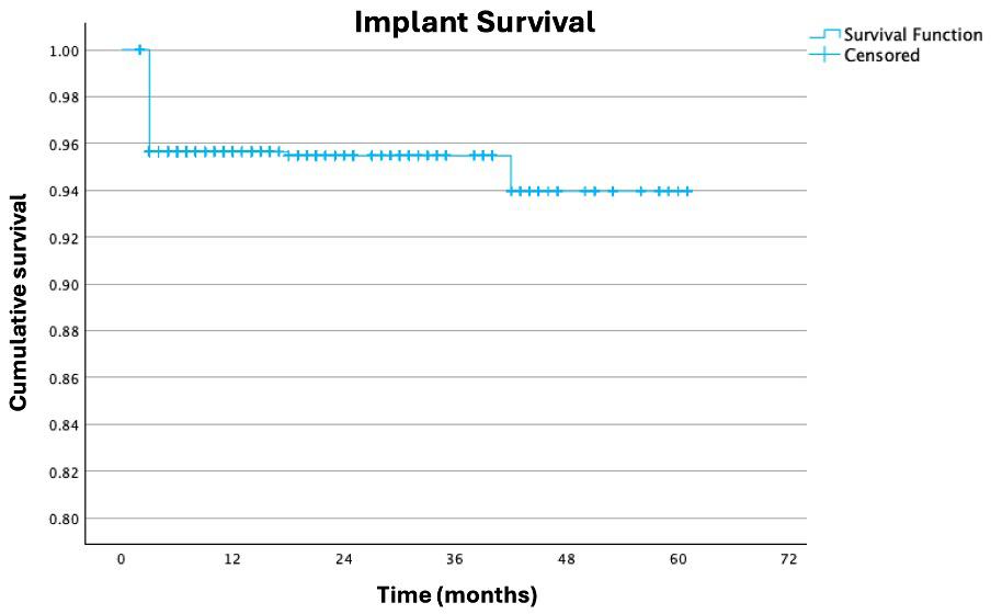
1. Kaplan–Meier curve of cumulative implant survival
Table 2. Multivariate Cox-regression analysis of variables impacting overall implant survival
wear and fracture, which was managed with recontouring and polishing.
Patient-reported outcome measures (PROMs)
PROMs were reported by 90 patients and 101 full arch rehabilitations at the 3–6-month review appointment using a 5-point Likert scale.
For the PROMs of comfort, aesthetics, function and overall happiness with treatment decision, 96%, 98%, 96% and 94% of patients reported a score of 5 respectively. A breakdown of all the PROMs is provided in Table 4
One patient gave a score of 4 for all four PROMs but did not report any complications.
For the PROM of comfort, the scores of 4 were given by one patient who had implant failure and required revision surgery, by one patient who had minor prosthesis chipping not necessitating a bridge remake and by two patients who reported no complications.
For the PROM of aesthetics, a score of 3 was given by a patient with gradual discoloration of their maxillary PMMA bridge, necessitating a bridge remake.
For the PROM of function, a score of 2 was given by a patient with no reported complications. The scores of 1 were given by a patient who
had both the maxillary and mandibular arches rehabilitated. They experienced an implant failure in the mandibular arch necessitating revision surgery but not requiring a bridge remake, and tooth wear and chipping in the maxillary arch not necessitating a bridge remake. This patient also gave scores of 2 for the PROM of overall happiness with treatment decision.
For the PROM of overall happiness with treatment decision, scores of 4 were given by one patient who had two maxillary implants fail but did not require a bridge remake, and three patients with no reported complications.
The cumulative 3-year implant survival rate of 95.5% is consistent with the immediate loading protocols published in the existing literature.18 Papaspyridakos and colleagues conducted a systematic review and meta-analysis in 2014 that demonstrated cumulative implant survival rates of 90.1%–100% for full arch immediate loading protocols.18 These results are predictable as the main difference with the AuDentes protocol is the immediate loading with a definitive prosthesis, which should not affect implant survival.
Ciabattoni and colleagues published a 3-year follow-up review of 32 patients who received full arch rehabilitation with an immediate
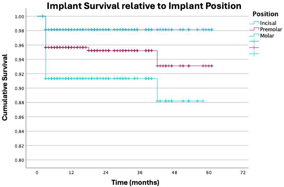
implant placement and loading protocol using a computer planned flapless guided protocol.13 They reported a cumulative implant survival rate of 97.54% and no prosthesis failures. The main differences between our protocols were the flapless approach taken in the Ciabattoni study and that patients who were identified as heavy smokers, defined as patients who smoked more than 10 cigarettes per day, were excluded from Ciabattoni’s study. Consistent with the literature, our study found that smoking was significantly associated with implant failure with 18 out of 48 implant failures (37.5%) occurring in smoking patients.17,28 The amount of smoking was not quantified in our study and should be recorded in future research. The increased risk of failure due to smoking should be discussed with patients in the informed consent process. Counselling advice and a referral to the general practitioner for assistance with smoking cessation should be recommended.
Consistent with the literature, our study found that male patients had a significantly increased risk of implant failure. Chrcanovic and colleagues published a systematic review and meta-analysis, which included 52 357 implants and that demonstrated increased implant failure rates in male patients.29
Our study demonstrated that implant failure was significantly associated with the implant position. Implants placed in the molar region were more likely to fail compared to implants placed in the premolar or anterior tooth regions. Similarly, implants placed in the premolar regions were more likely to fail compared to implants placed
in the anterior tooth region. There is no clear consensus on the survival of dental implants based on implant location.30 Comparisons between studies are difficult due to the heterogeneity of study methodologies. Del Fabbro and colleagues conducted a systematic review and meta-analysis evaluating the outcomes of full arch rehabilitations.31 They found slightly less favourable outcomes with maxillary implants versus mandibular implants after 10 years.31 We were unable to locate any other studies that evaluated the impact of implant position in the context of a full arch immediate implant placement and loading protocol. Our study indicates that a more anterior implant location is associated with increased implant survival. This makes sense from a bone density perspective and due to the relative reduction in occlusal forces present in the anterior regions. Furthermore, accessing the anterior dentition to maintain good oral hygiene is easier and could also contribute to increased implant survival rates over time. More research evaluating the effect of implant position on implant survival in the context of full arch rehabilitations with an immediate placement and loading protocol is required.
The prosthesis survival rate with the AuDentes protocol was 91.13%. This is slightly lower than the prosthesis survival rate of 93.75%–100% reported by Papaspyridakos and colleagues in their 2014 systematic review and meta-analysis.18 This could be attributed to different exclusion criteria or fabrication of the full arch prosthesis from different materials. In 2024, Crespi and colleagues published a review of 36 patients and 44 arches that were immediately loaded within 24 to 48 hours of the initial surgery with a definitive acrylic prosthesis.23 They reported a 100% prosthesis survival rate and 95.24% success rate with two of the 44 prostheses having minor prosthetic issues. Patients with a history of bruxism, who smoked more than 15 cigarettes per day, who had systemic chronic disease, and any sign of active infection or inflammation were excluded from their study. Bruxism and smokers were not excluded from our study. This could explain the disparity between prosthesis success and survival between these two studies. Despite the reduced survival rate in our study, PROMs were high, indicating high patient satisfaction at the 3–6-month review.
This study found a prosthesis success rate of 87.68% and the incidence of prosthesis complications was 12.32%. Korsch and colleagues published an article that compared the prosthetic success and survival of definitive versus temporary provisional restorations in patients treated with the all-on-four protocol.26 Their review included 126 patients with 136 arches, of which 92 patients and 94 arches were immediately loaded with a definitive restoration. In the definitive restoration group, an implant survival rate of 97.6% and a restoration survival rate of 92.6% were reported. Korsch and colleagues reported Necessitating a bridge remake (ie,
25 complications across the 94 arches, which equates to a prosthesis success rate of 73.40%.26 The prosthetic complications reported in their study included dehiscence, temporary hypoesthesia, peri-implant infection, sharp bone edge, temporary bleeding, implant loss, chipping or fracture of the composite teeth, fracture of the prosthetic screw, abutment loosening and loss of the screw channels resin cover.26 The large difference in prosthetic survival between our study (87.68%) and the Korsch study (73.40%) is likely due to a more thorough reporting of complications in the single-centred Korsch study versus the multicentred AuDentes study, which was conducted across more than 20 private clinics. Apart from implant loss, our study did not report any soft tissue complications. Given that 1069 individual implants were placed, it is likely that other soft tissue complications occurred. It is likely that these complications were not recorded or reported appropriately by the treating clinicians and therefore it is difficult to draw meaningful comparisons regarding complications between studies.
The results of our study demonstrate high patient satisfaction at the 3–6-month review with immediate implant placement and loading with a definitive prosthesis using the AuDentes protocol. The PROMs of comfort, aesthetics, function and overall happiness with treatment decision were high in this study with 96%, 98%, 96% and 94% of patients reporting a score of 5 respectively. These findings are consistent with prior studies, which evaluated patient satisfaction with patients rehabilitated using the all-on-four protocol.8,32 Direct interstudy comparison is difficult due to the different methodologies for recording and reporting PROMs. Multiple instruments for assessing patient perceptions exist, including visual analogue scales and questionnaires that assess oral health-related quality of life.4 This retrospective review used the 5-point Likert scale, which is a simple method for evaluating patient outcomes. Despite this, only 49.75 % of patients completed the PROM questionnaire. This is a limitation of our study and outlines the difficulty in data collection and assessing PROMs. Future prospective studies comparing the patient-reported and clinician-reported outcomes of different implant techniques should be conducted using a standardised patient perception scale.
Another limitation is that this study did not assess the impact of treatment on patient-related quality of life. This is because the PROMs were collected only once at the 3–6-month review using a 5-point Likert scale. Future research on the AuDentes protocol should also include baseline PROMs and consider collecting additional information on ability to masticate, phonetics, speech and selfesteem. These data would allow researchers to evaluate if the treatment improved the PROMs and would be a better marker of quality of life.
Duong and colleagues in 2022 discuss that there is poor agreement between the patients’ perception and the clinicians’ objective assessment of an aesthetic outcome.4 This study did not include the clinicians’ assessment of aesthetic outcomes, therefore we cannot compare the clinicians’ technical assessment versus the patients’ perception of aesthetic outcomes. Future research could include the clinicians’ perspective.
The Kaplan–Meier method of calculating cumulative survival relies on the assumption that censored data are represented in the remaining surviving cases. This becomes an inaccurate assumption as the sample size decreases and therefore the tail end of the curve should be disregarded if there is a high censorship rate. We attempted to mitigate this by only reporting 3-year survival. The effect of variables on survival requires events to occur for accurate estimation of effect. Our study has a relatively small event rate, and therefore, accuracy in detection of the effect of variables may be limited. Also, accuracy in the estimation of the magnitude effect of a variable is limited by a low event rate.
Insertion of an interim prosthesis enables peri-implant soft tissue conditioning to enhance the emergence profile and soft tissue appearance.25,33 Although there were no reported concerns with soft tissue aesthetics, specifically assessing soft tissue changes over time from a dental perspective and a patient-reported outcome perspective would be useful and should be explored further in a future prospective study.
This study has multiple limitations. The data was collected from multiple different dental clinics, and we were unable to obtain the radiographs to evaluate marginal bone loss over time. This study was a retrospective review, therefore we have no control group and are unable to directly compare our outcomes with other immediate placement and loading protocols.
The external validity of this study is good for clinicians using the AuDentes protocol for immediate full arch rehabilitation with immediate implant placement and loading with a PMMA prosthesis. The high PROMs are likely representative of patients who receive full arch rehabilitation with an immediate implant placement and loading protocol.
Within the limitations of this study, the short term implant and prosthetic outcomes of the AuDentes protocol are comparable with other immediate implant placement and loading protocols. The PROMs of the AuDentes protocol indicate high rates of patient satisfaction with treatment. Further research is required regarding longitudinal changes in PROMs, complications, soft tissue changes and marginal bone loss.
Bontempo D: Methodology, formal analysis, investigation, data curation, visualisation, writing – original draft, writing – review and editing, consultation.
Tan P: Conceptualisation, methodology, writing – review and editing, consultation.
Khadembaschi D: Formal analysis, investigation, data curation, writing – review and editing, consultation.
Gebauer D: Conceptualisation, methodology, formal analysis, investigation, data curation, writing – review and editing, consultation.
The authors thank the participants for their involvement in this study. No funding was required for this study.
The AuDentes protocol has been developed by Dr Philip Tan. Dr Tan’s role in this project was limited to the data collection phase and reviewing the final draft article. Dr Daniel Bontempo, Dr Darius Khadembaschi and Associate Professor Dieter Gebauer have no declarable conflicts of interest.
1 Fiske J, Davis D, Frances C, et al. The emotional effects of tooth loss in edentulous people. Br Dent J 1998; 184: 90-93.
2 Dierens M, Collaert B, Deschepper E, et al. Patientcentered outcome of immediately loaded implants in the rehabilitation of fully edentulous jaws. Clin Oral Implants Res 2009; 20: 1070-1077.
3 Joshipura KJ, Willett WC, Douglass CW. The impact of edentulousness on food and nutrient intake. J Am Dent Assoc 1996; 127: 459-467.
4 Duong H-Y, Roccuzzo A, Stähli A, et al. Oral healthrelated quality of life of patients rehabilitated with fixed and removable implant-supported dental prostheses. Periodontol 2000 2022; 88: 201-237.
5 Scala R, Cucchi A, Ghensi P, et al. Clinical evaluation of satisfaction in patients rehabilitated with an immediately loaded implant-supported prosthesis: a controlled prospective study. Int J Oral Maxillofac Implants 2012; 27: 911-919.
6 Yao CJ, Cao C, Bornstein MM, et al. Patient-reported outcome measures of edentulous patients restored with implant-supported removable and fixed prostheses: A systematic review. Clin Oral Implants Res 2018; 29 Suppl 16: 241-254.
7 Oh S-H, Kim Y, Park J-Y, et al. Comparison of fixed implantsupported prostheses, removable implant-supported prostheses, and complete dentures: patient satisfaction and oral health-related quality of life. Clin Oral Implants Res 2016; 27: e31-e37.
8 Gonçalves GSY, de Magalhães KMF, Rocha EP, et al. Oral health-related quality of life and satisfaction in edentulous patients rehabilitated with implant-supported full dentures all-on-four concept: a systematic review. Clin Oral Investig 2022; 26: 83-94.
9 Morton D, Gallucci G, Lin WS, et al. Group 2 ITI Consensus Report: Prosthodontics and implant dentistry. Clin Oral Implants Res 2018; 29: 215-223.
10 Patzelt SBM, Bahat O, Reynolds MA, et al. The all-on-four treatment concept: a systematic review. Clin Implant Dent Relat Res 2014; 16: 836-855.
11 Maló P, de Araújo Nobre M, Lopes A, et al. The All-on-4 treatment concept for the rehabilitation of the completely edentulous mandible: A longitudinal study with 10 to 18 years of follow-up. Clin Implant Dent Relat Res 2019; 21: 565-577.
12 Maló P, de Araújo Nobre M, Lopes A, et al. The All-on-4 concept for full-arch rehabilitation of the edentulous
The data underlying this article are available for sharing upon reasonable request to the corresponding author.
maxillae: A longitudinal study with 5-13 years of followup. Clin Implant Dent Relat Res 2019; 21: 538-549.
13 Ciabattoni G, Acocella A, Sacco R. Immediately restored full arch-fixed prosthesis on implants placed in both healed and fresh extraction sockets after computerplanned flapless guided surgery. A 3-year follow-up study. Clin Implant Dent Relat Res 2017; 19: 997-1008.
14 Del Fabbro M, Bellini CM, Romeo D, et al. Tilted implants for the rehabilitation of edentulous jaws: a systematic review. Clin Implant Dent Relat Res 2012; 14: 612-621.
15 La Monaca G, Pranno N, Annibali S, et al. Immediate flapless full-arch rehabilitation of edentulous jaws on 4 or 6 implants according to the prosthetic-driven planning and guided implant surgery: A retrospective study on clinical and radiographic outcomes up to 10 years of follow-up. Clin Implant Dent Relat Res 2022; 24: 831-844.
16 Niedermaier R, Stelzle F, Riemann M, et al. Implantsupported immediately loaded fixed full-arch dentures: evaluation of implant survival rates in a case cohort of up to 7 years. Clin Implant Dent Relat Res 2017; 19: 4-19.
17 Caramês JMM, Marques D, Caramês GB, et al. Implant survival in immediately loaded full-arch rehabilitations following an anatomical classification system-a retrospective study in 1200 edentulous jaws. J Clin Med 2021; 10: 5167.
18 Papaspyridakos P, Chen C-J, Chuang S-K, et al. Implant loading protocols for edentulous patients with fixed prostheses: a systematic review and meta-analysis. Int J Oral Maxillofac Implants 2014; 29: 256-270.
19 Papaspyridakos P, Bordin TB, Kim YJ, et al. Technical complications and prosthesis survival rates with implant-supported fixed complete dental prostheses: a retrospective study with 1- to 12-year follow-up. J Prosthodont 2020; 29: 3-11.
20 Marconcini S, Giammarinaro E, Covani U, et al. Immediate restoration of fixed full-arch prostheses placed on implants in both fresh and healed sockets using the flat one-bridge technique: a 7-year retrospective study. BMC Oral Health 2021; 21: 617.
21 Gallardo YNR, da Silva-Olivio IR, Gonzaga L, et al. A systematic review of clinical outcomes on patients rehabilitated with complete-arch fixed implant-supported prostheses according to the time of loading. J Prosthodont 2019; 28: 958-968.
22 Babbush CA, Kanawati A, Kotsakis GA, et al. Patientrelated and financial outcomes analysis of conventional full-arch rehabilitation versus the All-on-4 concept: a cohort study. Implant Dent 2014; 23: 218-224.
23 Crespi R, Vinci R, Capparé P, et al. A clinical study of edentulous patients rehabilitated according to the” all on four” immediate function protocol. Int J Oral Maxillofac Implants 2012; 27: 428-434.
24 Elsyad MA, Elgamal M, Mohammed Askar O, et al. Patient satisfaction and oral health-related quality of life (OHRQoL) of conventional denture, fixed prosthesis and milled bar overdenture for All-on-4 implant rehabilitation. A crossover study. Clin Oral Implants Res 2019; 30: 1107-1117.
25 Wittneben J-G, Wismeijer D, Brägger U, et al. Patientreported outcome measures focusing on aesthetics of implant- and tooth-supported fixed dental prostheses: A systematic review and meta-analysis. Clin Oral Implants Res 2018; 29: 224-240.
26 Korsch M, Walther W, Hannig M, et al. Evaluation of the surgical and prosthetic success of All-on-4 restorations: a retrospective cohort study of provisional vs. definitive immediate restorations. Int J Implant Dent 2021; 7: 48.
27 Feine J, Abou-Ayash S, Al Mardini M, et al. Group 3 ITI Consensus Report: Patient-reported outcome measures associated with implant dentistry. Clin Oral Implants Res 2018; 29 Suppl 16: 270-275.
28 Naseri R, Yaghini J, Feizi A. Levels of smoking and dental implants failure: a systematic review and meta‐analysis. J Clin Periodontol 2020; 47: 518-528.
29 Chrcanovic B, Albrektsson T, Wennerberg A. Dental implants inserted in male versus female patients: a systematic review and meta‐analysis. J Oral Rehabil 2015; 42: 709-722.
30 Do TA, Le HS, Shen Y-W, et al. Risk factors related to late failure of dental implant—A systematic review of recent studies. Int J Environ Res Public Health 2020; 17: 3931.
31 Del Fabbro M, Pozzi A, Romeo D, et al. Outcomes of fixed full-arch rehabilitations supported by tilted and axially placed implants: a systematic review and meta-analysis. Internat J Oral Maxillofac Implants 2022; 37: 1003-1025.
32 Babbush CA. Posttreatment quantification of patient experiences with full-arch implant treatment using a modification of the OHIP-14 questionnaire. J Oral Implantol 2012; 38: 251-260.
33 Furze D, Byrne A, Alam S, et al. Esthetic outcome of implant supported crowns with and without periimplant conditioning using provisional fixed prosthesis: a randomized controlled clinical trial. Clin Implant Dent Relat Res 2016; 18: 1153-1162.
Lewis PL (MBBS, PhD, FRACS(Orth), FAOrthA)1; Goss AN (DDSc, FRACDS (OMS), FICD)2,3; Esaian R (MPH)1; Smith PN (AM BMBS FRACS HonDUniv FAOrthA)1
The Australian Orthopaedic Association National Joint Replacement Registry (AOANJRR) collects and analyses information on patient characteristics, prosthesis types and surgical outcomes to identify factors associated with successful joint replacement. Initially focused on hip and knee replacements, data collection has expanded to include other joint procedures, such as shoulder, elbow, wrist, ankle and spinal disc replacements.
Before its establishment, there were limited data on prosthesis performance and revision rates. By monitoring the time between primary and revision surgery, the Registry provides valuable evidence regarding implant survival and surgical effectiveness,
The Australian Orthopaedic Association National Joint Replacement Registry (AOANJRR, hereafter, the Registry) was established to monitor joint replacement surgery in Australia and improve patient outcomes through systematic data collection and analysis. Since its inception, the Registry has evolved into a comprehensive national database capturing information on joint replacement procedures performed across the country. By analysing prosthesis performance, surgical techniques and patient characteristics, the AOANJRR provides valuable evidence to guide clinical practice and health care policy.
Operating under Commonwealth Government funding and legislative protection as a Federal Quality Assurance Activity, the Registry enables

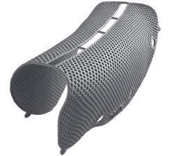

Spine
Wrist
Table 1. Number of procedures recorded in the Australian Orthopaedic Association National Joint Replacement Registry (AOANJRR) by type of joint replacement in March 2026
The foundations of the AOANJRR were laid in the late 1990s, born out of a critical information vacuum. At the time, orthopaedic surgeons lacked a centralised way to compare the performance of joint replacement prostheses across patient populations. Instead, they had to rely on a fragmented mix of personal experience, small-scale cohort studies, international meetings and industry-provided data. This lack of clear, objective evidence meant that while the Charnley total hip replacement was reported to account for 70% of prostheses used in Australia during the 1960s and 1970s, the accuracy of such claims—and the assessment of long term success of the implants— remained largely anecdotal.1
During the same period, novel prostheses were developed and introduced without adequate clinical evidence, tracking or surveillance. Introduced in the late 1970s, the Wagner hip resurfacing arthroplasty experienced early failures due to wear of the thin polyethylene acetabular cup and loss of bone under the metal femoral head from granuloma induced by polyethylene debris. Surgeons discovered the high failure rate from the article written by WC Head, who reported in 1981, 14 failures out of 41 Wagner resurfacing arthroplasties conducted.2 In the conclusion of the Head article, the failure rate for Wagner resurfacing was deemed unacceptably high; with multiple reasons given, including biomechanical design.
To bridge the knowledge gap, a registry pilot study led by Professor Don Howie was launched in Adelaide. This was modelled on the established hip and knee replacement registries from Sweden. The success of the pilot study secured federal government support for a national registry, leading to the appointment of Professor Steven Graves as the inaugural Registry Director, a role he held until 2022. Data collection officially commenced in 1999, and full national coverage was achieved for hip and knee replacements by mid2002. This milestone was transformative; national data allowed for meaningful benchmarking and the detection of rare mechanical failures that would otherwise go unnoticed in smaller datasets.
On 1 October each year, an annual report is published with an indepth analysis of hip and knee replacement procedures. Over the
decades, the Registry’s scope has expanded. In 2007, data collection was expanded to include other joint replacements such as shoulders, ankles, elbows, wrists and spinal discs. At first, the outcomes of shoulder replacement were reported separately, but since 2016, these have been included in the main annual report. In 2018, data collection was increased further to include Patient Reported Outcome Measures (PROMs), where patients complete electronic questionnaires before, and six months after, surgery. These surveys capture not only the perceived function of the replaced joint, but also measures of the patient’s general health.
From its inception, the Registry has maintained a practical focus. It collects a defined minimum dataset from hospitals, linking patient and procedure factors to a “hard” endpoint: revision surgery. By using revision as the definitive measure of failure, the AOANJRR has evolved into one of the world’s most robust clinical monitors, ensuring that the guesswork of the mid-20th century is replaced by rigorous, data-driven surgical standards.
Preceding the formation of AOANJRR, there was a lack of detailed information on the results of different procedures and types of joint replacements available, especially on factors known to influence the results. Surgeons have a wide selection of prostheses that they can utilise in the joint replacement procedure. Also, there are variations of techniques that can be used for correctly implanting the joint during surgery. The Registry can compare these variables and determine how they can influence the outcomes. The AOANJRR is also able to detail the results for different categories of implants, and determine if the patient’s age, sex, body mass index, general health and other reasons may affect the procedure. Utilising that access to data, surgeons can decide on the best type of prosthesis to use for any situation.
AOANJRR’s major contribution is patient safety through surveillance and feedback. By measuring revision rates and comparing performance across prosthesis types, fixation methods and techniques, the Registry helps clinicians and health services identify outlier implants or practices and shift towards better-performing options. Its scale strengthens this impact: recent profiling work reports that AOANJRR captures around 97.7% of primary and revision joint replacement procedures performed in Australia and has accumulated over 2 million procedures, enabling long term outcome reporting.
The Registry also underpins research and quality improvement across multiple joints (not only hips, knees and shoulders but including elbows, wrists, ankles and spinal discs), producing regular analyses that inform surgeons, hospitals, regulators and industry. The Registry turns what would otherwise be fragmented hospital-level data into a national learning system. In view of its completeness, scale and the depth of the analysis undertaken, the AOANJRR has become internationally recognised as a key source of information for regulators and researchers on the outcomes of joint replacement surgery.
Orthopaedic surgery manages most of the major joints in the human body, with the exception of the temporomandibular joint, which falls within the domain of oral and maxillofacial surgery. Accordingly, it is appropriate that temporomandibular joint replacement (TMJR) is monitored through the Australian and New Zealand Association of Oral and Maxillofacial Surgeons (ANZAOMS) TMJR Registry, following the principles established by the AOANJRR.
A pivotal benefit of the AOANJRR—and a cornerstone of its function as a Commonwealth Government Quality Assurance Activity (QAA)— is the robust legal protection known as qualified privilege. Established under Section 124X of the Commonwealth Health Insurance Act 1973, this legislative framework is specifically designed to foster a culture of transparent clinical audit by removing the threat of legal discovery. For the individual surgeon, the primary implication is confidentiality of performance data: information that identifies a specific practitioner, when collected solely for the purposes of the Registry’s quality assurance mandate, is legally protected from disclosure. This means that individual surgeon reports, which benchmark personal revision rates against national aggregate data, can be safely discussed in a closed audit forum to improve practice, and cannot be used as evidence in civil or criminal proceedings.1,3
This protection ensures that a surgeon’s proactive engagement with their own “outlier” data or a “Higher Than Anticipated Revision Rate” (HTARR) notification cannot be weaponised against them in a medical negligence suit. HTARR prostheses are prostheses that have statistically significant higher revision rates and are identified by the Registry following a standardised three-stage approach. The qualified privilege creates a “safe harbour” for clinical reflection, allowing surgeons to critically analyse their technique or implant selection without the data being discoverable. Furthermore, the Act imposes strict secrecy provisions on Registry personnel, making it a criminal offence to disclose identifiable information to unauthorised third parties, including medical boards or hospital administrations.
Ultimately, the benefit to the surgeon is the transition from a “blame culture” to a “safety culture.” By decoupling the act of rigorous selfaudit from the risk of professional liability, the QAA status empowers practitioners to utilise Registry data as a purely clinical tool for
1 Lewis PL, Gill DRJ, McAuliffe MJ, Stoney JD, Vertullo CJ, Wall CJ, et al. Lay Summary: Hip, Knee and Shoulder Replacement Supplementary Report. In: Australian Orthopaedic Association National Joint Replacement Registry 2025 Annual Report: Hip, knee and shoulder arthroplasty. Australian Orthopaedic Association. 2025. https://aoanjrr.sahmri.com/documents/d/guest/aoa-fullreport-2025
surgical refinement. It guarantees that the pursuit of prosthetic longevity and patient safety remains a protected, intellectual exercise, shielded by federal law from the volatility of the litigation landscape.
Running a national registry costs money—data collection, validation, analytics, reporting and governance. In Australia, Commonwealth support for the AOANJRR is tied to a cost-recovery model, where the costs of funding provided to administer the Registry are recovered through levies collected from joint replacement sponsors. Although funded by the Commonwealth Government, the Registry and its data are owned by the Australian Orthopaedic Association as an independent entity, so that there is no open access to the information that the Registry holds.
The key question is whether the Registry has been financially worthwhile, and the answer is an unequivocal yes. A 2025 economic evaluation concluded that the AOANJRR is highly cost-effective, estimating that about nine dollars were saved for every one dollar spent, primarily through reductions in the burden of revision surgery, and better-informed decision-making. The logic is straightforward: avoiding even a small number of revisions (which are costly, complex and harmful to patients) can offset registry operating costs many times over—while also improving quality of life for thousands of Australians undergoing joint replacement each year.4
Lewis P: Conceptualisation, article review, final editing.
Goss A: Article review, consultation, final editing.
Esaian R: Conceptualisation, initial drafting, article review, final editing.
Smith P: Conceptualisation, initial drafting, article review, final editing.
The authors declare that they have no conflicts of interest.
Registry data are available on request to the individual surgeon and will be provided as individual patient data. Otherwise, all data are restricted in accordance with the Commonwealth Government of Australia Quality Assurance status, the New Zealand Government Protected Quality Assurance Activity and the ANZAOMS TMJRR and AOANJRR Directives.
2 Head WC. Wagner surface replacement arthroplasty of the hip. Analysis of fourteen failures in forty-one hips. J Bone Joint Surg Am 1981; 63(3): 420-427.
3 Zhou Y, Wall CJ, Stevens J, et al. Data Resource Profile: The Australian Orthopaedic Association National Joint Replacement Registry (AOANJRR). Int J Epidemiol 2025; 54(4): dyaf078.
4 Okafor C, Nghiem S, Holder C, et al. Are joint replacement registries cost-effective? Economic evaluation of the Australian Orthopaedic Association National Joint Replacement Registry. Arch Orthop Trauma Surg 2025; 145(1): 408.
Goss A (DDSc, FRACDS (OMS), FICD)1,2; Burgess M (BDSc, MBBS, FRACDS (OMS), MBA)1,3; Moore L (BMedSc, B Dent, MBBS, FRACDS (OMS))1,4; Murdoch B (BDS, FDS, RCS (Eng, Edin), FFOMP (RCPA), NZSSM)1,5; Mellowes B (BMgt (RBA), MSocSc(IntlDev))1,6; Smith P (AM, BMBS, FRACS, FAOrthoA)7,8
The idea of establishing a bi-national Temporomandibular Joint Replacement Registry (TMJRR) was triggered in 2014 by the New Zealand Accident Compensation Corporation (ACC) requesting a meeting with the New Zealand Branch of the Australian and New Zealand Association of Oral and Maxillofacial Surgeons (ANZAOMS [NZ]) to investigate the indications and funding of temporomandibular joint replacements (TMJRs) in New Zealand. A subcommittee for the task was established with Dr John Edwards (oral and maxillofacial surgery [OMS], Auckland) as Chair, Dr Wayne Gillingham (OMS, Wellington) who was the ANZAOMS New Zealand Councillor and TMJR surgeon, Dr Bruce Murdoch (OMS, Palmerston North) who had placed many TMJR and Ms Rosemary Kennedy of the ACC. Professor Alastair Goss of Adelaide, Australia was appointed as the independent TMJR research consultant.
The subcommittee reviewed the available literature on TMJR, on current orthopaedic practice for monitoring joint replacement and the, at the time, newly established British TMJR Registry.1-7 The subcommittee produced pamphlets for the New Zealand Dental Association and the ACC with advice for New Zealand dentists regarding the general diagnosis of temporomandibular joint (TMJ) disorders and the specific indication for surgery on the TMJ.8 The subcommittee also conducted a retrospective study on TMJR placement in New Zealand between 2000 and
This article describes the regulatory and clinical need for an Australian and New Zealand clinical quality registry for temporomandibular reconstruction. Temporomandibular joint replacement (TMJR) prostheses have been developed internationally and in Oceania to restore pathologically destroyed temporomandibular joints with excellent success rates. Placement requires skilled surgery in the anatomically and functionally complex facial region.
The TMJR is then expected to continue to function for the rest of the patient’s life. This is expected by the authorities, the surgeon and the patient. Often the remaining life of the prosthesis and patient are longer than the remaining practising life of the surgeon. Complications and failure can occasionally occur and can be catastrophic.
The only means to safely monitor outcomes is by an independent national clinical quality registry. The success of this approach has been demonstrated by the success of the Australian Orthopaedic Association National Joint Replacement Registry (AOANJRR), which has documented over 2.4 million orthopaedic joints placed over the last 25 years.
Following the example of and in cooperation with the AOANJRR, the Australian and New Zealand Association of Oral and Maxillofacial Surgeons (ANZAOMS) has successfully piloted and established an Australian and New Zealand Temporomandibular Joint Replacement Registry (TMJRR).
1TMJRR Committee, Australian & New Zealand Association of Oral & Maxillofacial Surgery; 2Adelaide University, Adelaide, SA, Australia; 3Mater Adult Hospital, Brisbane, QLD, Australia; 4St Georges Hospital, Christchurch, New Zealand; 5Palmerston North, New Zealand; 6Australian & New Zealand Association of Oral & Maxillofacial Surgery; 7School of Medicine, Australian National University, Canberra, ACT, Australia; 8Australian Orthopaedic Association National Joint Replacement Registry.
Corresponding author: Alastair Goss ✉ alastair.goss@adelaide.edu.au | doi: 10.63717/2026.MS0051
Keywords: temporomandibular
2018 (Table 1). This study confirmed that TMJRs were successfully functioning.
The subcommittee decided that the establishment of a TMJRR was indicated. Informal approaches were made to the New Zealand Orthopaedic Association Joint Registry (NZOA Joint Registry) to see if a subset of TMJR could be included in their registry. At that time, the NZOA Joint Registry was experiencing some internal difficulties so declined to be involved. The response from the Australian Orthopaedic Association National Joint Replacement Registry (AOANJRR) was more positive, so the decision was made to have a single Australian and New Zealand TMJRR, based in Australia. At the time, those involved did not know how long and torturous the development timeline would be (Table 2).
This article discusses the establishment and development of the ANZAOMS TMJRR to date. It includes discussion of the regulatory environment, clinical quality registers, the pilot study, bi-national quality assurance, the initial results and the challenges ahead. Parallel to these developments, ANZAOMS has established a TMJRR Subcommittee and a subspecialisation group of temporomandibular surgeons.
The Therapeutic Goods Administration (TGA) of the Commonwealth of Australia and their New Zealand counterpart, Medsafe, are the responsible regulatory authorities for all foreign materials placed in humans.9 All suppliers of artificial joints must register their devices with the TGA and Medsafe. They must provide detailed information on the materials used, the performance of the devices and report any adverse outcomes.
An example of this reporting process was seen when Version 2 of the Maxoniq TMJR device had problems with fossa screws loosening. The ultra-high molecular weight polyethylene fossa could not become osseointegrated with the fossa bone, as well as the device not having sufficient length and number of fixation screws. This was reported to the TGA by the manufacturer and a notice was issued to all surgeons.10 The fossa was redesigned, and the current Version 4 of the implant has a titanium mesh backing so it can better integrate with the host bone. There is also increased screw fixation. The modification was designed and rigorously tested by the company in conjunction with the University of Melbourne, Victoria, Australia. The current version has TGA approval.
Failure to comply with TGA or Medsafe may result in severe penalties. For example, some dental practitioners who were importing unregistered dental implant devices have been caught and fined.11
In the TGA regulations, it states that any foreign materials implanted in humans must be monitored for the life of the implant or the patient.12 Similarly, surgeons are obligated to follow patients for the full post-operative period and longer on an elective basis for some chronic conditions. The national health registration bodies in Australia (Australian Health Practitioner Regulatory Authority) and New Zealand (Medical Council of New Zealand) state in their regulations
Joints
Type
106
TMJ Concepts
Outcome Functioning well 105
Failed: removed 1
Infected: removed and replaced 3
All were placed, or placement supervised, by Dr B Murdoch. Analysis by Prof AN Goss. The failed and removed TMJR was in a patient selected by another surgeon. The patient has a significant psychiatric condition and severe bruxism.
Table 1. Preliminary New Zealand results (2000–2018)
2014–2018 Investigation by ANZAOMS (NZ) and the New Zealand Accident Compensation Corporation (ACC) into temporomandibular joint replacements (TMJRs). TMJRR recommended.
2018 New Zealand Orthopaedic Association Joint Registry identified as appropriate, but participation declined.
2018–2023 Australian Orthopaedic Association National Joint Replacement Registry (AOANJRR) approached and receptive.
Pilot study by ANZAOMS and AOANJRR.
Completed governance, ethics, data form and analysis.
2022–2024 Commonwealth Government of Australia quality assurance status sought and obtained.
New Zealand Government quality assurance status sought and obtained.
2024–2025 ANZAOMS/AOANJRR negotiations for continuance.
2026–2031 Five-year contract for the ANZAOMS TMJRR backed by AOANJRR support and methodology.
Table 2. Timeline for the development of the Australian and New Zealand Association of Oral and Maxillofacial Surgeons (ANZAOMS) Australian and New Zealand Temporomandibular Joint Replacement Registry (TMJRR)
that practitioners “must participate in systems of quality assurance and surveillance monitoring of adverse events”.13,14
There is a problem with surgeons meeting their responsibilities for TMJR. The maximum practising life of an oral and maxillofacial surgeon from completion of training to retirement is about 30 years. The current maximum reported period for success of a TMJR is about 25 years. A TMJR placed in a young adult could be expected to be still functioning until the patient’s death in their eighties. That is sixty years, or twice the practising life of the surgeon. A solution to this problem is the placement of all TMJR patient results in a clinical quality register.
National clinical quality registers (CQRs) are organisations that monitor the quality of health care by collecting information from patients undergoing predefined medical procedures. There is an Australian Register of Clinical Registries, which includes over a hundred separate registries.15 The Australian Register of Clinical
Registries has a national organisation that meets regularly to refine the requirements involved in the operation of a successful registry. The organisation is based at the South Australian Health and Medical Research Institute (SAHMRI) in Adelaide.
Clinical registries cover a wide range of medical issues. An example is the intensive care registry, which includes all ventilator units in Australia. This knowledge was invaluable in the COVID-19 pandemic as patient demand could be balanced with the available facilities.16 There are multiple cancer registries, which are critical in defining the magnitude of the problem, outcomes of different treatment modalities and areas for further research. In dentistry, an Australian and New Zealand dental implant registry has been established.17
The most successful long term clinical registry in Australia is the AOANJRR, which records all hip, knee, shoulder or other orthopaedic joint replacements in Australia. It has been functioning for over 25 years with 100% surgeon involvement with over 2.4 million joint replacements recorded.18 The Commonwealth of Australia has declared the AOANJRR a Federal Quality Assurance Activity under part VC of the Commonwealth Health Insurance Act, 1973. This provides important protection for the patients and surgeons involved. The AOANJRR and quality assurance status are discussed in the article by Lewis and colleagues in this issue of The Australasian Journal of Oral and Maxillofacial Surgery 19
ANZAOMS approached the AOANJRR in 2018 with a view to establishing a TMJRR in association with the AOANJRR. In 2019, a formal four-year pilot study was established. The AOANJRR is based in Adelaide, South Australia at SAHMRI. Alastair Goss (University of Adelaide) was tasked with establishing the pilot study.
This developed quickly and within two years, a governance structure, ethics framework in both countries and a data form (Figure 1a and b) had been established. The data form was based on the AOANJRR knee replacement form. Although the same broad structure was followed, the form required some modification to include TMJRspecific information. A key difference was in age assessment. Any patient under 55 years of age is considered young for a knee replacement, whereas most TMJR patients are younger, with some as young as 16 years of age.19
In 2022, initial data was collected from a few surgeons and analysed. From these data, it became evident that a fully functioning TMJRR could be established. It was determined that the work up to 2022 would be maintained but not expanded to cover all Australian and New Zealand TMJR surgeons. The next key step was to obtain national quality assurance status in both countries. The extensive documentation was assembled and the applications made.20,21 At that time, the national health authorities were still grappling with the COVID-19 pandemic, so the application was accorded a low priority. The process took two years in Australia and two and a half in New Zealand, but finally national quality assurance was achieved.22,23 By that time, the initial 4-year pilot contract between ANZAOMS
and the AOANJRR had expired and there had been a complete change in the AOANJRR at an executive level, with the appointment of a new director, CEO and other officers. There were also serious funding difficulties between the AOANJRR, SAHMRI and the South Australian Government. Hence, negotiation between ANZAOMS and the AOANJRR took a further two years, mainly centring around the funding model. Throughout these contract negotiations, there remained goodwill towards the project and those working at the data face. In 2026, the new 5-year contract was finalised.24
There are several key features of the ANZAOMS TMJRR project. The results of the ANZAOMS TMJRR project are the intellectual property of ANZAOMS members. The registry is operated by the TMJRR Subcommittee of ANZAOMS, which reports to the ANZAOMS Directorate. The protocol and procedures of maintaining the registry follow those used by the AOANJRR, thus ensuring that a proven repeatable methodology is followed. This facilitates full understanding by other surgeons, manufacturers, theatre staff and hospitals. The data are well protected by national quality assurance in both Australia and New Zealand. The TMJRR meets all the regulatory requirements.
Involvement in any registry is voluntary. However, all orthopaedic surgeons in Australia participate in AOANJRR, and the equivalent in New Zealand. It would be confidently expected that oral and maxillofacial surgeons in Australian and New Zealand would similarly all participate in the ANZAOMS TMJRR. It is in their interest and medicolegal protection to do so. Similarly, patients are protected, with consent being on an “opt out” basis. The experience with the AOANJRR is that less than 0.01% of patients opt out.
Participation in the ANZAOMS TMJRR is free for all surgeons and patients as it is funded by the industry and ANZAOMS.
The initial results of the preliminary Australian data from 2000 to 2022 are presented in Tables 3a, 3b and 3c. The preliminary data are presented in three sections: demographics, indications and revisions.
The demographics follow the known distribution of end-stage TMJ pathology, namely a female predominance over a wide age range (Table 3a). Most commonly, a single joint was involved, and it could be replaced in a single-stage operation. The greatest number of operations were performed by a small group of surgeons with a larger number of surgeons operating on only a few patients. All the currently available TMJR prostheses available in Australia and New Zealand were recorded. All joints had a free periarticular fat graft.25
The second dataset presents the indication for TMJR (Table 3b). Most patients were in severe pain with limited jaw opening. The full range of diagnoses of TMJ pathology were included, with the most common being osteoarthritis. The challenge of indications for TMJR has recently been reviewed in the November 2025 issue of this journal.26
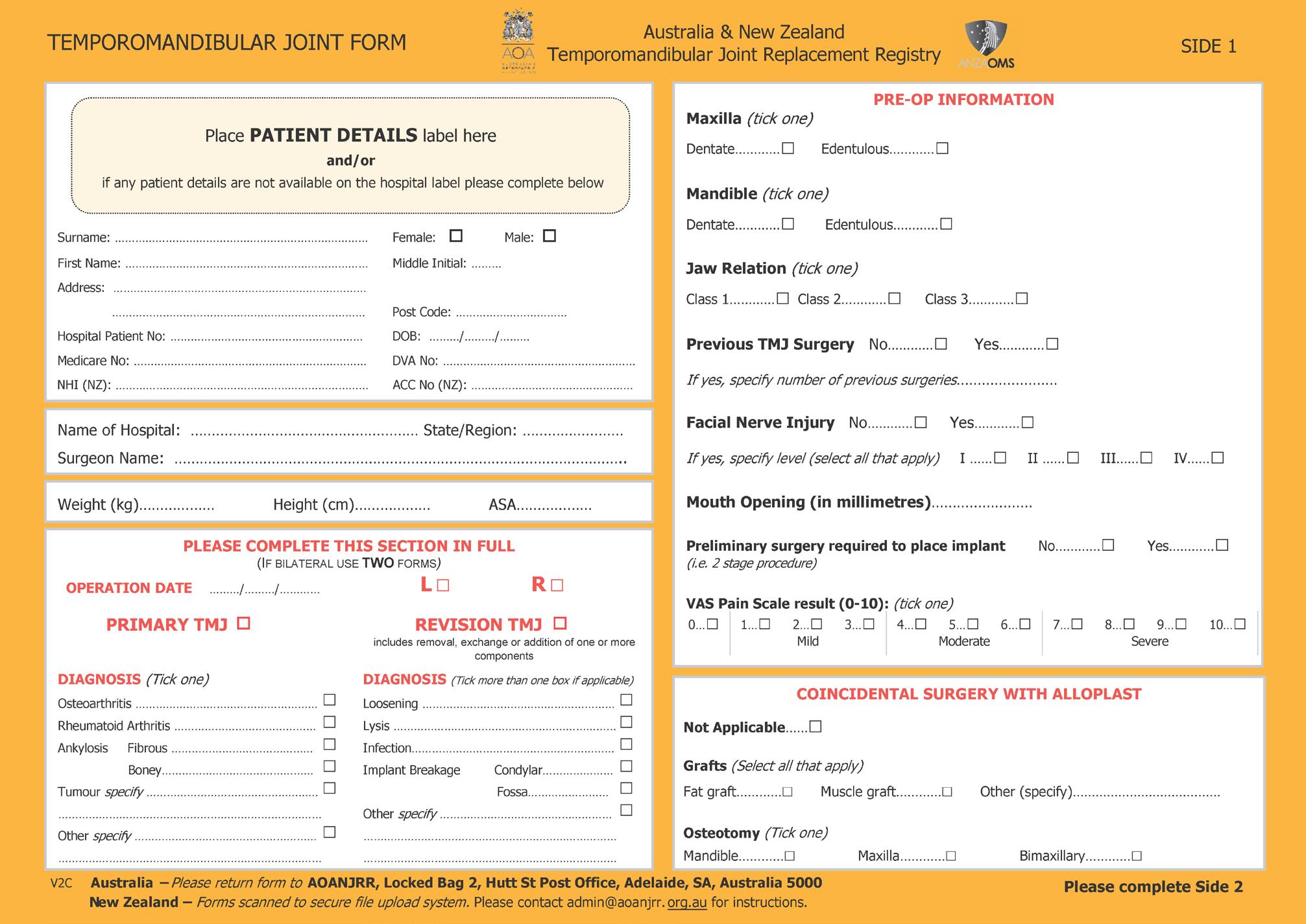
The third section of these results relates to revision (Table 3c). Only 14 of the 508 joints placed required reoperation. None failed totally and all 14 were successfully revised. This revision rate of 3% is acceptable and similar to other published studies.1,4,6,27,28 Joints that had previously undergone multiple operations or were grossly malformed and required a two-stage procedure were more likely to fail. The malposition of the TMJR was a technical problem noticed immediately so correctly reoperated. The regrafted case had a squeaking noise on joint movement so had a further placement of a fat graft, which resolved the issue.
The main difference is that most international studies1,4,6,27,28 involve one or two types of TMJR devices, whereas in Australia and New Zealand, there are 5 different types of devices, all of which have undergone revision over time.
The characteristics of the TMJR devices available are presented in Table 4. There are many common features shared by the different devices, all with fossa and condyle/ramus components ultimately stabilised by osseointegrated fixation screws.
The TMJ Concepts (now Stryker/TMJ Concepts, Ventura, CA, USA) device has the longest history and can be considered the gold standard device. It has substantial literature available on its performance, which is partly quoted in this article.1,29 The literature shows that a correctly placed Stryker/TMJ Concepts device can still be functioning 20+ years after placement. That study, however, did show a high unknown outcome rate of 25% to 34%.27,28 Twenty-five years may only be half the expected life of some patients. The aim of a high quality clinical register such as the ANZAOMS TMJRR is for a 100% known outcome over the life of the patient.
For many years, the TMJ Concepts device primarily relied on its American Food and Drug Administration (FDA) approval in the absence of Australian TGA or New Zealand Medsafe approval. When TMJ Concepts was taken over by Stryker (Kalamazoo, MI, USA) in 2021, the device underwent some minor improvements and obtained TGA approval. Unfortunately, for commercial reasons, Stryker is considering withdrawal from Oceania and negotiations are ongoing concerning future availability. At present, Stryker/TMJ Concepts has over 95% of the New Zealand market and about a third of the Australian market.
OrthoTiN is the second most common TMJR device in Australia. It was designed and is manufactured in America but does not have FDA approval and is not available for use in the USA. However, it does has Australian TGA approval with full materials and biomechanical testing evidence presented to the TGA.30 It has minimal peerreviewed evidence available on its performance and the only clinical articles available are two by the same authors on their technique of endoscopic insertion for the ramus component.31 They reported on the short term results of 104 TMJRs.32 There is no independent, medium term (5 to 10 year) data despite the device being available for that length of time. It has limited use in New Zealand.
The Maxoniq Arthrojaw, formerly known as OMX, TMJR has the longest TGA registration, being first registered in 2015. It is an Australian design developed by an Australian surgeon working with
(4 surgeons placed 78%)
Arthrojaw (formerly OMX)
Table 3a. Preliminary Australian results (2000–2025): demographics
* 0 = no pain; 10 = extreme pain.
Table 3b. Preliminary Australian results (2000–2025): indications
Table 3c. Preliminary Australian results (2000–2025): revisions
Make Fossa components
Stryker/TMJ Concepts
Titanium 6Al-4V alloy
Ultra-high molecular weight polyethylene (UHMWPE) fused to commercially pure titanium mesh backed fossa
Minimum four fixation screws
OrthoTiN
Titanium 6Al-4V alloy
UHMWPE insert with two locking tabs into the fossa
Minimum five screws
Condylecobalt chrome molybdenum
Ramus - titanium
Minimum eight screws
Maxoniq Arthrojaw (Version 4)
Titanium 6Al-4V alloy
UHMWPE fused to commercially pure titanium mesh fossa
Minimum four screws
Condyle - chrome cobalt
Ramus – titanium with chrome cobalt coating
Minimum six screws
Ramus - titanium G4
Tear drop shape
Minimum six screws
KLS Martin IPS TMJR Titanium 6Al-4V alloy
UHMWPE fused to fossa
Minimum four screws
Zimmer Biomet Stock
Titanium 6Al-4V alloy
UHMWPE fused to titanium mesh fossa
Minimum four screws
Available in three sizes (small, medium, large)
Condyle - chrome cobalt
Ramus – titanium with coronoid extension
Minimum eight screws
Titanium 6Al-4V alloy
Minimum five screws
Available in three sizes (small, medium, large)
CAD–CAM custom design
Computer controlled milled prosthesis
Designed for individual patient’s specific anatomy
American based company
Food and Drug Administration (FDA) approved
Device being withdrawn from Oceania by the manufacturer
CAD–CAM custom design
Computer controlled milled prosthesis
Designed for individual patient’s specific anatomy
American based company
Not FDA approved or available in the USA
CAD–CAM custom design
3D printed components
Designed for individual patient’s specific anatomy
Australian company based at the University of Melbourne
Available in Oceania and Southeast Asia
CAD–CAM custom design
3D printed components
Designed for individual patient’s specific anatomy
European International company
Recently introduced to the international market
Pilot in Oceania
Fossa and ramus template provided for size selection
American based company
FDA approved
Custom device not available in Oceania
Preauricular incision for fossa
Submandibular incision for ramus component
Allows concurrent osteotomy
Preauricular incision for fossa
Bearing fossa replaceable
Ramus component fitted endoscopically by submandibular incision
Allows concurrent osteotomy
Preauricular incision for fossa
Bearing fossa replaceable
Submandibular incision for ramus component
Allows concurrent osteotomy
Preauricular incision for fossa
Submandibular incision for ramus component
Allows concurrent osteotomy
Preauricular incision for fossa
Submandibular incision for ramus component
Select best fit by template
Remove bone to improve fit
Table 4. Types of temporomandibular joint replacement (TMJR) device available in Australia and New Zealand
the Biomechanical Department of the University of Melbourne.33,34 It has extensive peer-reviewed literature on its performance, which is partly presented in this article.35,36 As previously noted, it did have some limited early fossa failure, which was fully reported to the regulator and corrected. This caused the company some reputational damage. As well as Australia and New Zealand, it is available in Asia and South America.
The KLS Martin TMJR has only recently been developed and tested primarily in Europe.37 It has extensive literature on all aspects of its development, which is only partly presented in this article.38,39,40 Published survival reports are only at the 1-year stage.40 Some KLS Martin TMJR have been placed in Australia and New Zealand.
The Zimmer Biomet (Jacksonville, FL, US) stock device has been in use worldwide since its FDA approval in 2005.41 It has a proven medium to long term success rate.42,43 The disadvantage is that it comes in a limited number of sizes, which is not a problem when there is localised pathology to the condyle. However, when there is extensive anatomical destruction, such as post-traumatic bone ankylosis, the bone needs to be cut to fit the prosthesis rather than the prosthesis being made to fit the bone. The Zimmer Biomet custom TMJR is not FDA or TGA approved but is available in some other countries.
TMJR devices can be manufactured in a number of ways, with the titanium component being either variously milled or 3D printed. In his John Edwards Memorial Lecture, Louis Mercuri presented the
advantages and disadvantages of the different TMJR embodiment (materials, design and manufacturing) methodologies.44
At the time of writing, the TMJRR is facing several pressing tasks that require completion. Given the new contract and the evolution of the AOANJRR and the award of quality assurance status, the pilot documents written in the establishment phase will need to be updated and rewritten. That task is underway.
Currently less than half of all active TMJR surgeons in Australia and New Zealand are fully documented under the TMJRR. This includes their hospital and local ethics requirements. All their retrospective and current data need to be collected and analysed. This is also underway.
The AOANJRR has a well-established paper record, whereas the trend is towards electronic data records, with this being the preference of younger surgeons. The AOANJRR is moving carefully towards conversion to electronic records. With its vast trove of 25 years and 2.4 million records, it is moving slowly and cautiously, involving a strong information technology strategy towards developing its electronic data collection. When that occurs (probably first for the knee replacement registry), we will develop an electronic TMJR data form. However, it is important to do this cautiously over time. One of the reasons the British TMJR collapsed was that its
1 Mercuri LG, Wolford LM, Sanders B, et al. Custom CAD/ CAM total temporomandibular joint reconstruction system: preliminary multicenter report. J Oral Maxillofac Surg 1995; 53(2): 106-115; discussion 115-116.
2 Kent JN, Misiek DJ, Akin RK, et al. Temporomandibular joint condylar prosthesis: a ten-year report. J Oral Maxillofac Surg 1983; 41(4): 245-254.
3 van Loon JP, de Bont LG, Stegenga B, et al. Groningen temporomandibular joint prosthesis. Development and first clinical application. Int J Oral Maxillofac Surg 2002; 31(1): 44-52.
4 Murdoch B, Buchanan J, Cliff J. Temporomandibular joint replacement: a New Zealand perspective. Int J Oral Maxillofac Surg 2014; 43(5): 595-599.
5 New Zealand Orthopaedic Association. The New Zealand Joint Registry. Fourteenth Annual Report. 1999-2012. NZOA. 2013; 1-47. https://www.nzoa.org.nz/sites/default/ files/NJR%2014%20Year%20Report.pdf
6 Idle MR, Lowe D, Rogers SN, et al. UK temporomandibular joint replacement database: report on baseline data. Br J Oral Maxillofac Surg 2014; 52(3): 203-207.
7 Elledge R, Attard A, Green J, et al. UK temporomandibular joint replacement database: a report on one-year outcomes. Br J Oral Maxillofac Surg 2017; 55(9): 927-931.
8 Accident Compensation Corporation. Temporomandibular disorders – Guidelines for surgical treatment. ACC 7121. March 2015.
9 Australian Department of Health, Disability and Ageing. Therapeutic Goods (Medical Devices) Regulations 2002. Version 3.0. November 2021.https://www.legislation.gov. au/F2002B00237/latest/text
10 Therapeutic Goods Administration. Recall/hazard alert. June 2021.
simple electronic database was overwhelmed as the amount of data increased.
Once the TMJRR is fully established to capture all surgeons and all retrospective data, then research and further education can begin in earnest.45 Annual reporting will also be provided.
Goss A: Conceptualisation, registry development, initial drafting, article review, final editing.
Burgess M: Registry development, article review, final editing. Moore L: Registry development, article review, final editing. Murdoch B: Registry development, article review, final editing. Mellowes B: Registry development, article review, final editing. Smith P: Registry development, article review, final editing.
Author Alastair Goss is the Editor-in-Chief of The Australasian Journal of Oral and Maxillofacial Surgery and also the clinical advisor to the TMJRR. He declares this non-financial conflict. This article was independently reviewed. All other authors declare that they have no conflicts of interest.
Registry data are available on request to the individual surgeon and will be provided as individual patient data. Otherwise, all data are restricted in accordance with the Commonwealth Government of Australia Quality Assurance status, the New Zealand Government Protected Quality Assurance Activity and the ANZAOMS TMJRR and AOANJRR Directives.
11 Therapeutic Goods Administration. Canberra dental practice fined $266,400 for alleged unlawful supply of dental implants and bone grafts [media release]. 30 July 2021. https://www.tga.gov.au/news/media-releases/ canberra-dental-practice-fined-266400-allegedunlawful-supply-dental-implants-and-bone-grafts
12 Therapeutic Goods Administration. Understanding rules for boundary and combination products. 1 June 2004. https://www.tga.gov.au/resources/guidance/ understanding-rules-boundary-and-combinationproducts
13 Australian Health Practitioner Regulatory Authority. Good medical practice: a code of conduct for doctors in Australia. Ahpra, 2020. https://www. medicalboard.gov.au/Codes-Guidelines-Policies/ Code-of-conduct.aspx?_gl=1*19e93oe*_ ga*MTEyMTYwMTE4Ny4xNzc0NTkwNjMw*_ ga_F1G6LRCHZB*czE3NzQ1OTA2MjkkbzIwJGc xJHQxNzc0NTkwNjQyJGo0NyRsMCRoMA
14 Medical Council of New Zealand. Current standards. 2025. https://www.mcnz.org.nz/our-standards/currentstandards/
15 Australian Commission on Safety and Quality in Health Care. National Guidance for Clinical Quality Registries. https://www.safetyandquality.gov.au/our-work/ indicators-measurement-and-reporting/nationalguidance-clinical-quality-registries
16 Australian and New Zealand Intensive Care Society. Annual Report 2024. https://www.anzics.org/wp-content/ uploads/2025/01/ANZICS-Annual-Report-2024.pdf
17 Soukoulis S, Davies S, Goss A. The Australian and New Zealand Dental Implant Registry. Aust Dent J 2026 [in press].
18 Australian Orthopaedic Association. Australian Orthopaedic Association National Joint Replacement
Registry. Annual Report 2025: Hip, knee and shoulder arthroplasty. https://aoanjrr.sahmri.com/documents/d/ guest/aoa-full-report-2025
19 Lewis PL, Goss AN, Esaian R, Smith PN. The Australian Orthopaedic Association National Joint Replacement Registry: insights from 25 years. AJOMS 2026; 1: 40-42.
20 Australian Government Department of Health. Application for declaration. Registry of the Australian and New Zealand Temporomandibular Joint Replacement Registry. 25 August 2020.
21 New Zealand Government Department of Health. Application for declaration. Registry of the Australian and New Zealand Temporomandibular Joint Replacement Registry. 25 August 2020.
22 Kelly P. Chief Medical Officer, Department of Health & Aged Care Health Insurance (Quality Assurance Activity – Australian and New Zealand Temporomandibular Joint Replacement Registry). Declaration 2023. 24 July 2023.
23 Government of New Zealand. Correspondence from Reti, S (Hon Minister of Health). Health Practitioners (Protected Quality Assurance Activity – Australian and New Zealand Association of Oral and Maxillofacial Surgeons) Notice 2024. Section 54(3) of the Health Practitioners Competence Assurance Act 2003. 9 April 2024. https:// www.legislation.govt.nz/secondary-legislation/pcodrafted/2024/41/en/latest/#LMS938665
24 Funding Agreement between the Australian Orthopaedic Association and the Australian and New Zealand Association of Oral and Maxillofacial Surgeons. April 2024.
25 Liu A, Vu LC, Zhao DF, Dimitroulis G. The use of autologous fat grafting in temporomandibular total joint replacement surgery: a systematic review. AJOMS 2025; 2: 94-100.
26 Mian M, Woliansky M, Sklavos A, et al. What indication criteria are applied to patients receiving total temporomandibular joint replacement? A systematic review. AJOMS 2025; 2: 88-93.
27 Mercuri LG, Wolford LM, Sanders B, et al. Longterm follow-up of the CAD/CAM patient fitted total temporomandibular joint reconstruction system. J Oral Maxillofac Surg 2002; 60(12): 1440-1448.
28 Afzal Z, Umorin M, Mercuri LG, Warburton G. TMJ Concepts patient-fitted temporomandibular joint reconstruction prosthesis system: Results from a Food and Drug Administration postmarket surveillance prospective cohort study. J Oral Maxillofac Surg 2025; 83(1): 10-16.
29 Stryker. TMJ Concepts. Information for healthcare professionals. https: //www.stryker.com/pl/en/ craniomaxillofacial/products/tmj-concepts.html
30 OrthoTin. Orthotin TMJ Temporomandibular joint replacement system. OrthoTin LLC.
31 Kim SSY, Somers M. Total temporomandibular joint replacement without a submandibular incision. Journal of Oral Medicine and Oral Surgery 2020; 26(4): 43-47.
32 Somers M, Lim S, Kim SSY. Outcomes of temporomandibular joint replacement using an endaural incision and endoscopic assistance - A report of 104 cases. Br J Oral Maxillofac Surg 2025; 63(8): 583-588.
33 NovicoMedic. Maxoniq Arthrojaw Total Joint Replacement [website]. https://www.novicomedic.co/brands/maxoniqseries/arthrojaw
34 Woodford SC, Robinson DL, Abduo J, et al. Kinematics of the jaw following total temporomandibular joint replacement surgery. J Biomech 2023; 159: 111741.
35 Woodford SC, Robinson DL, Abduo J, et al. Muscle and joint mechanics during maximum force biting following total temporomandibular joint replacement surgery. Biomech Model Mechanobiol 2024; 23(3): 809-823.
36 Brierly G, Thomas A, Dimitroulis G. A five-year review of the OMX temporomandibular prosthetic total joint replacement system. Oral Maxillofac Surg 2023; 27(1): 131-139.
37 Materialise. Cranio-Maxillo Facial Solutions. TMJ Total Arthroplasty Surgery: Product highlights [website]. 2025. https://www.materialise.com/en/healthcare/hcps/cmf/tmjtotal-arthroplasty
38 Materialise. Study record: Long term safety and performance of Materialise patient specific TMJ systems [not yet recruiting]. Clinical Trials Gov ID NCT06951178. National Library of Medicine, National Centre for Biotechnology Information.
39 Andres A, Wickert K, Gneiting E, et al. Simulation of a custom-made temporomandibular joint-an academic view on an industrial workflow. Bioengineering (Basel) 2025; 12(5): 545.
40 de Sousa Gil AP, Velasques BD, Nelson Uzun N, et al. Total customized alloplastic reconstruction for treatment of severe temporomandibular joint pathologic conditions: a case series of combined intraoral and extraoral approach. J Craniofac Surg 2022; 33(3): e250-e253.
41 Zimmer Biomet. The Zimmer Biomet Total Mandibular Joint Replacement system [website]. https://www. zimmerbiomet.eu/en/products/total-joint-replacementsystem
42 Giannakopoulos HE, Sinn DP, Quinn PD. Biomet Microfixation Temporomandibular Joint Replacement System: a 3-year follow-up study of patients treated during 1995 to 2005. J Oral Maxillofac Surg 2012; 70(4): 787-794; discussion 795-796.
43 Abramowicz S, Barbick M, Rose SP, Dolwick MF. Adaptability of stock TMJ prosthesis to joints that were previously treated with custom joint prosthesis. Int J Oral Maxillofac Surg 2012; 41(4): 518-20.
44 Mercuri LG. Alloplastic temporomandibular joint replacement - past, present and future considerations. The 2022 ANZAOMS John Edwards Memorial Lecture. AJOMS 2024; 1: 85-92.
45 Bouloux GF, Chou J, DiFabio V, et al. Management of patients with orofacial pain and temporomandibular disorders. J Oral Maxillofac Surg 2025; 83(9): 1078.e11078.e83.
Lockyer JW (MBcHB, BDS)1; Kim SY (MBBS MMed BOH DSc Grad Dip Dent FRACDS (OMS))1
Temporomandibular joint dysfunction (TMD) affects a significant proportion of the population and has negative impacts on quality of life.1 Internal derangement, where the pathology and symptoms arise from within the joint capsule, often presents with restriction of motion, joint noises and pain.2 In late-stage disease, there are often structural and dynamic changes to the intra-articular disc, and the severity of symptoms generally correlates with progression of disease.3
Arthroscopy was first introduced by Onishi in 1975.4 In its simplest form, arthroscopy involves inserting an endoscope into the superior joint space, performance of diagnostic examination, lysis of adhesions and fluid lavage. This has been demonstrated to reduce pain and inflammation, with low morbidity related to its minimally invasive nature.5 As such, arthroscopy is the gold standard for the diagnosis of intra-articular pathology, while simultaneously providing symptomatic relief in around 80% of cases of internal derangement, usually measured by a reduction in pain and an improved mouth opening.6,7 Arthroscopy has demonstrated effectiveness in the long-term relief of internal derangement symptoms.8
Purpose: To evaluate early changes in pain, function, disability and mouth opening following arthroscopic lysis and lavage for internal derangement, using patient-reported outcome measures.
Methods: A retrospective study was conducted on 54 patients (65 joints) with Wilkes stage 3 to 5 internal derangement of the temporomandibular joint (TMJ) who underwent arthroscopic lysis, lavage and medial capsulotomy. All had failed non-surgical management. Patient-reported outcomes were assessed pre-treatment and at 6-weeks post-treatment using the Jaw Functional Limitation Scale (JFL), Characteristic Pain Intensity (CPI) scores and disability measures. Maximal interincisal opening (MIO) was measured by the clinician. Wilcoxon signed-rank tests were used for statistical analysis, with results stratified by Wilkes classification.
Results: Significant improvements were observed at 6-weeks posttreatment, as seen in mean CPI scores (63.3–43.2, P = 0.0001), JFL scores (3.9–2.7, P = 0.0002), disability scores (13.3–9.8, P = 0.0140) and MIO (24.2–35.1 mm, P = 0.0001). Improvements were most pronounced in patients with a Wilkes 4 TMJ. Younger patients were less likely to improve.
Conclusion: Arthroscopic lysis and lavage for internal derangement of the temporomandibular joint results in significant short-term improvements in pain, function, disability and mouth opening. Longer-term follow-up is needed to assess durability of these improvements.
1Prince of Wales Hospital, Sydney, NSW, Australia. Corresponding author: Jamie Lockyer ✉ jamie.lockyer@health.nsw.gov.au | doi: 10.63717/2026.MS0056
The ANZAOMS 2026 Annual Scientific Meeting will be held in the beautiful Otautahi/ Christchurch, New Zealand. The 2026 ASM promises to deliver an engaging and thought-provoking scientific program alongside a dynamic exhibition and social program.
Dr Deepak Kademani USA

Professor Andreas Neff Germany
6-8 AUGUST 2026 CHRISTCHURCH/OTAUTAHI NEW ZEALAND
Although most articles used pain and mouth opening as primary outcome measures following arthroscopy, quality of life is more complex and more important from the patient perspective.9 Indeed, the link between TMD and worse self-reported quality of life is well established, with a clear dose–response relationship. The Diagnostic Criteria for Temporomandibular Disorders (DC/TMD) tool attempts to incorporate this into the assessment, which comprises two axes: (i) physical assessment, and (ii) psychosocial disability and pain. The DC/ TMD incorporates the Graded Chronic Pain Scale (GCPS) and Jaw Function Limitation (JFL) Scale and has been validated in clinical and research settings.10 It is also important to obtain patient-reported measures, as there are demonstrable inconsistencies between surgeon’s and patient’s ratings of health status.11
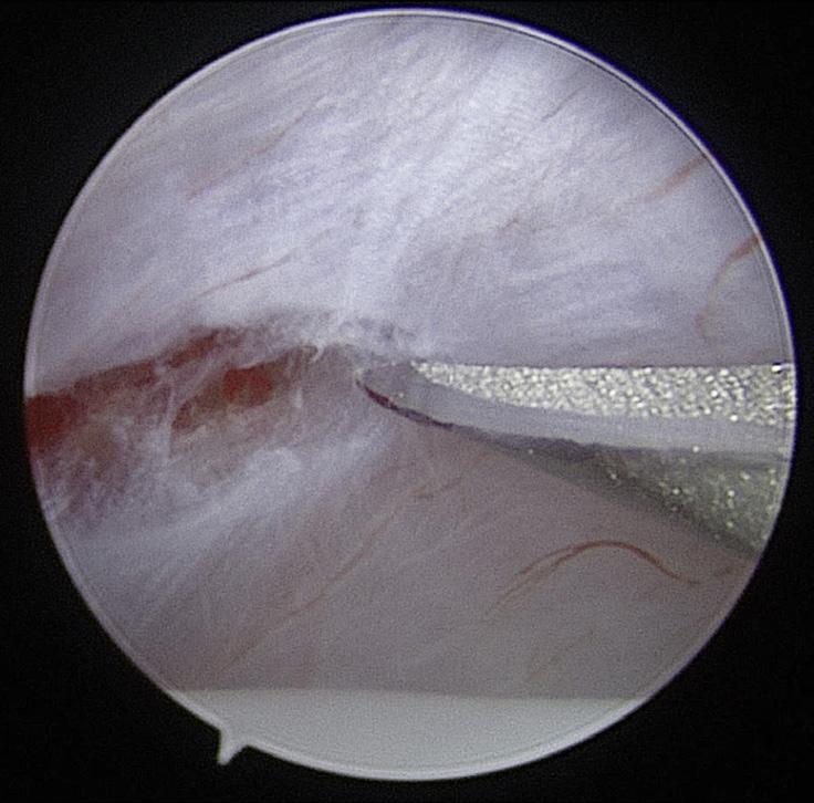
All patients presented with symptoms of internal derangement. A surgical referral was made if a patient failed management with non-surgical therapies (including medication, physiotherapy and bite appliance therapy, as appropriate) and opted to proceed to arthroscopy. All patients underwent a pre-operative magnetic resonance imaging and completed a preoperative questionnaire.
The following inclusion criteria were applied when selecting the study sample: (i) clinical evidence of internal derangement; (ii) failure of non-operative therapy; (iii) single operator; (iv) adequate pre-operative and post-operative documentation; (v) Wilkes classification 3, 4 or 5 on pre-operative evaluation (with the worst side recorded in bilateral cases).
This study aims to examine changes following arthroscopy in multiple domains: pain, function, disability and maximal interincisal opening using patient-reported outcomes.
The investigators performed a retrospective study of arthroscopic lysis and lavage over a two-year period from 1 January 2022 to 31 December 2023. The study compared mouth opening measurements, as well as pre-treatment patient questionnaires looking at function (derived from the JFL scale), pain (derived from the CPI scale), disability measures (derived from the OHR-QoL scale), with follow-up questionnaires repeated 6 weeks after treatment.
Patients underwent surgery in the private sector. All patients underwent arthroscopic lysis, lavage and medial capsulotomy under general anaesthesia (Figure 1). A 1.9-mm Dyonics 30° arthroscope (Smith & Nephew, England) was used with 150–300 mL Hartmann’s solution as lavage fluid. An intra-articular steroid (standard dose of 10 mg triamcinolone) was given at conclusion of the surgery. Patients completed a standardised regimen of home physiotherapy following the procedure.
All patients were followed up at 6-weeks post-treatment where the repeat questionnaire was completed.
Data variables
The maximal interincisal distance, in millimetres, was documented by a clinician. The questionnaire comprised three key components: pain, function and disability, and was derived from the OHR-QoL scale, as outlined below:
Table 1. Outline of components and scoring from the patient questionnaire
8
13
7
3
5 Side Left 24 Right 19
Table 2. Demographics of study participants
JFL: assesses functional ability related to orofacial pain in the previous month.12
CPI: derived from the first 4 items on the GCPS, converted to an overall score based on severity and frequency of symptoms.13
Disability: derived from the last three items on the GCPS to evaluate the impact of symptoms on patient lifestyle.
Mouth opening: interincisal opening distance as measured in millimetres.
The outline of components and scoring from the patient questionnaire are shown in Table 1
Statistical analysis
Abbreviations: CPI = characteristic pain intensity; JFL = jaw functional limitation-8 scale; Pre Tx = pre-treatment score; After Tx = after treatment score.
We compared pre-treatment and post-treatment scores for the following factors: CPI, JFL, disability and mouth opening. This comparison was done by conducting a Wilcoxon signed-rank test. Results were then stratified by Wilkes classification. P scores less than 0.05 were considered statistically significant.
A total of 54 patients underwent arthroscopy within the study period and met the inclusion criteria. Patient ages ranged from 16 to 75 years, with a mean age of 40.1 years. The male to female ratio was
Table 3. Comparison of pre-treatment and post-treatment outcome scores
about 1: 18. Nineteen patients had arthroscopy on the right side, 24 on the left, and 11 had bilateral treatment: a total of 65 joints. When stratified by Wilkes classification of the worst joint, there were 16 patients with Wilkes 3 temporomandibular joints (TMJs) (29.6%), 18 with Wilkes 4 TMJs (33.3%) and 20 with Wilkes 5 TMJs (37.0%) (Table 2).
Figures 2 to 8 demonstrate some key findings documented during arthroscopy. These findings have been used to stage the disease
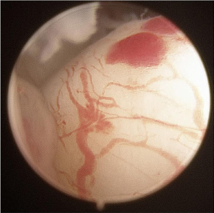
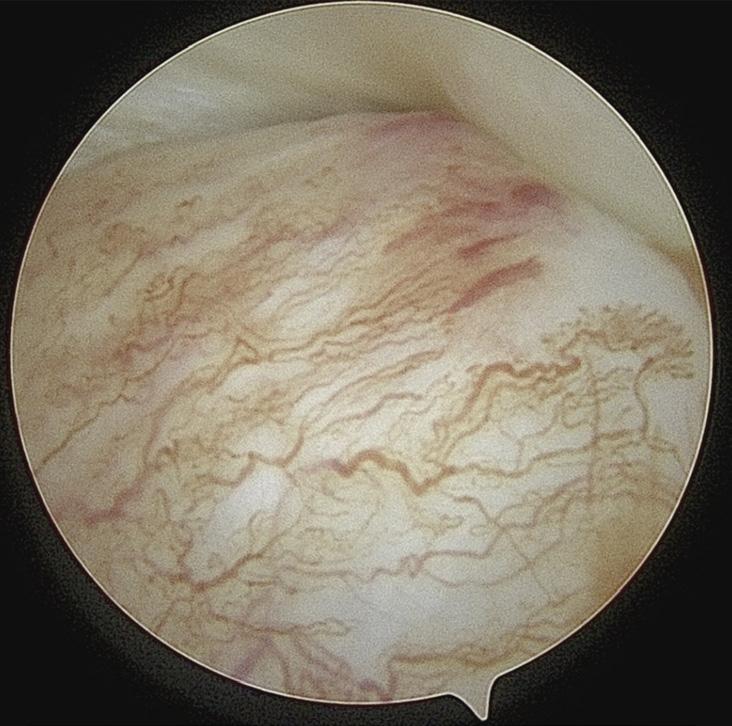
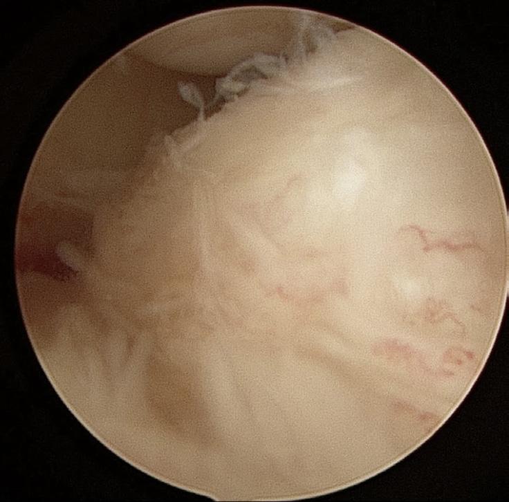
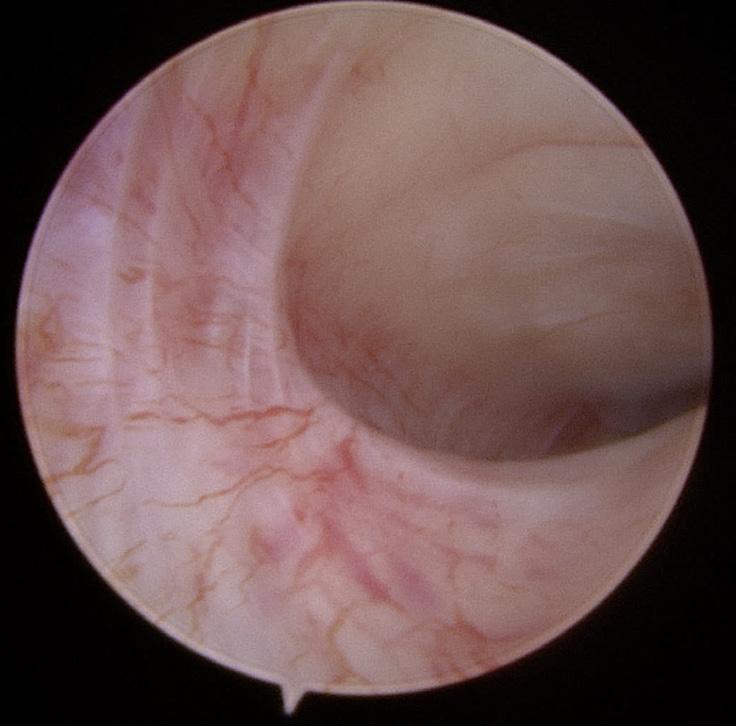
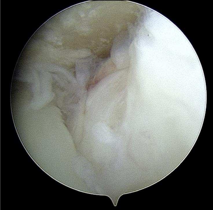
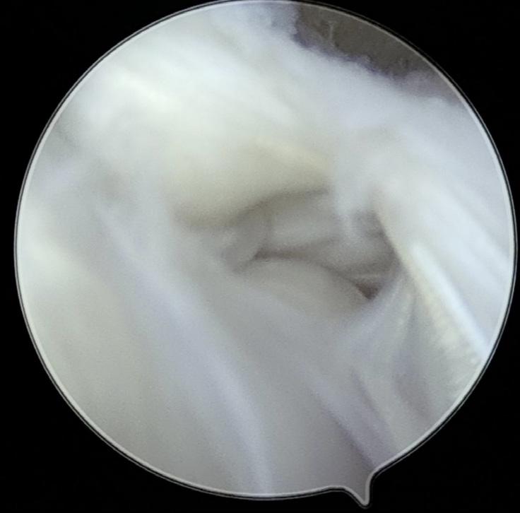
using the Wilkes classification. For example, findings associated with Wilkes 3 are demonstrated in Figures 2 and 3; Wilkes 4 in Figure 4, 5 and 6; and the most severe degenerative changes in Figure 7 and 8, consistent with a Wilkes 5 joint.
Following arthroscopy, the mean CPI scores reduced from 63.3 (standard deviation [SD], 18.2) to 43.2 (SD, 30.8). This change was statistically significant (P = 0.0001). When categorised by Wilkes classification: patients with Wilkes 3 TMJs had a reduction in the mean CPI score from 59.2 (SD, 24.6) to 32.7 (SD, 34.6). This was statistically significant (P = 0.0162). Patients with Wilkes 4 TMJs had a reduction in the mean CPI score from 63.0 (SD, 18.7) to 36.7 (SD, 26.4), which was statistically significant (P = 0.031). The change for patients with Wilkes 5 TMJs (67.0 to 54.8) was not statistically significant (P = 0.1500).
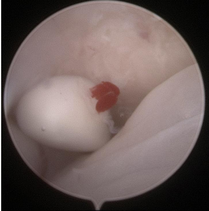
(P = 0.0140). When categorised by Wilkes classification: the mean reductions for patients with Wilkes 3 TMJs (9.1 to 8.8), and Wilkes 5 TMJs (16.2 to 14.3) were not statistically significant: with P values of 0.7160 and 0.4436, respectively. For patients with Wilkes 4 TMJs, there was a statistically significant reduction in disability score from 13.8 (SD, 11.7) to 5.6 (SD, 8.6), with a P value of 0.0008.
For disability scores, the mean score decreased from 13.3 (SD, 10.7) to 9.8 (SD, 11.4). This change was statistically significant
For JFL scores, the mean score changed from 3.9 (SD, 1.7) to 2.7 (SD, 2.3) following arthroscopy. The change was statistically significant (P = 0.0002). When categorised by Wilkes classification: the mean reductions for patients with Wilkes 3 TMJs (3.7 to 2.4), and Wilkes 5 TMJs (3.9 to 3.6) were not statistically significant: with P values of 0.0743 and 0.3406, respectively. For patients with Wilkes 4 TMJs, there was a statistically significant reduction in JFL score from 4.03 (SD, 1.8) to 1.8 (SD, 1.5), with a P value of 0.0007.
For MIO, the mean measurement changed from 24.2 mm (SD, 8.3 mm) to 35.1 mm (SD, 8.1 mm) following arthroscopy. The change was statistically significant (P = 0.0001). When categorised by Wilkes classification: the mean opening for patients with Wilkes 3 TMJs increased from 26.4 mm (SD, 7.0 mm) to 35.3 mm (SD, 7.9 mm) and was statistically significant (P = 0.0016). For patients with Wilkes 4 TMJs, the mean increase was from 29.4 mm (SD, 10.0 mm) to 36.7 mm (SD, 7.49 mm) and was statistically significant (P = 0.0073). Similarly, for patients with Wilkes 5 TMJs, the mean increase from 28.5 mm (SD, 7.7 mm) to 33.6 mm (SD, 8.8 mm) was also statistically significant (P = 0.0041) (Table 3).
Outcomes can be broadly divided into patients who improved and patients who did not. When an improvement was measured in at least three of the four measures (CPI, JFL, disability and mouth opening), the patient was thought to have improved. For this study, the overall rate of improvement was 64.8% (35/54). When categorised by Wilkes classification, improvements were seen in 11/16 of patients with Wilkes 3 TMJs (68.8%), 14/18 of patients with Wilkes 4 TMJs (77.8%), and 11/20 (55%) of patients with Wilkes 5 TMJs. Stratified by age, improvements were seen in 10/18 of the 16–30 year group (55.6%), 7/28 of the 31–59 year group (25%), and 7/8 (87.5%) of the 60 year and older group.
This study demonstrates statistically significant improvements across all measured domains (pain, function, disability and mouth opening) at 6-weeks following arthroscopy. When stratified by disease severity, these outcomes were most pronounced for patients with Wilkes 4 TMJs, which was also the group that had the most success following arthroscopy as described by Murakami and colleagues.14 However, Smolka and Iizuka reported lower rates of success with arthroscopy in patients with Wilkes 4 and 5 TMJs, and another study showed that these patients are more likely to proceed to open procedures, particularly total joint replacement.15,16
Most other literature measures success following arthroscopy as improvements in mouth opening (often measured as an interincisal distance >30 mm) and pain scores (often measured as an improvement in a visual analogue scale of 2 points or greater). Comparatively, outcomes in our study were determined by patientreported outcome measures derived from the DC/TMD tool, which comprised two axes: physical assessment, and psychosocial disability and pain. We think this is important as quality of life is more complex and more important to the patient.9 It is also important to seek patient-reported results, as research has demonstrated inconsistencies between surgeon and patient ratings of health status.11
Ulmner and colleagues reported that large scale studies investigating factors associated with successful outcomes following arthroscopy are lacking.17 They also identified that poorer outcomes are associated with higher levels of pre-operative global pain, muscle tenderness, psychiatric comorbidities, a younger age, a smaller
mouth opening and systemic rheumatic disease. This highlights the multifactorial nature of pain associated with TMD, particularly the role of extracapsular sources, that is, myofascial and centralised pain. Although our study demonstrated significant improvements in pain scores (CPI: 63.3 to 43.2), particularly for Wilkes 3 and 4 joints, we did not account for these comorbidities. However, the disability measure is more holistic and demonstrates overall improvements in quality of life following treatment.
Our study showed that 64.8% of patients improved following arthroscopy, defined as improvement in at least 3 of the 4 domains (CPI, JFL, disability and mouth opening). Although our rate of improvement is similar to Breik and colleagues, who reported 76% of patients to be improved and not require further surgery, this should be interpreted with caution as their criteria for success were different (post-operative visual analogue scale of ≤2, or ≥50% improvement).18 Breik and colleagues also found the highest rate of progression to further surgery in the 21–30-year-old group, with the lowest progression in the above 60-year-old group. This is similar to our findings, in that the youngest age group were least likely to have a successful outcome. However, unlike their study, we did not find that the most severe cases of internal derangement progressed to further surgery due to unsatisfactory outcomes. Tzanidakis and Sidebottom also showed poorer outcomes for patients with Wilkes 4 and above TMJs, as well as patients with higher pre-operative pain and lower mouth opening measurements.19 Comparatively, we found that 77.8% of patients with Wilkes 4 joints improved following treatment, demonstrating significant changes across all measured domains despite their relatively advanced disease.
Hashemi and colleagues showed a correlation between improvements in clinical measures and patient’s perceived outcomes at 12 months after surgery.20 The reduction in pain and improvement in mouth opening were associated with a high level of self-reported quality of life, which correlates with our findings of a reduction in disability measures following arthroscopy. Munoz-Guerra and colleagues agreed that arthroscopy provides effective relief in the short- to medium-term, However, on longer-term follow-up, Hashemi and colleagues demonstrated increasing symptoms beyond 12 months, which was beyond the follow-up period of our study (ie, six weeks).20,21
We acknowledge that a 6-week follow-up period can be considered inadequate, given the propensity for patients to improve or deteriorate over the months following treatment. For example, Sidebottom described that, following arthrocentesis, around 70% of patients will show improvement in the first six weeks, with a further 10% reporting further improvements over the next three to six months.22 While we would expect that patients in our series would have recovered from the complications of surgery (pain, swelling, etc) by six weeks, it is unlikely that all patients will have reached their potential outcome following intervention (further improvement or relapse). In future studies, it would be prudent to have a longer follow-up, that is, six months, or preferably multiple follow-up points during the postoperative period.
At six weeks following arthroscopy for internal derangement, there are statistically significant improvements in pain, function, disability and maximum interincisal opening across all Wilkes classifications. Improvements were more likely in patients with Wilkes 4 TMJs, and those over 60 years old.
Author contributions
Lockyer J: Ethics approval, data acquisition, analysis and interpretation, writing – original draft.
Kim S: Paper conception, data acquisition, analysis and interpretation, writing – review and edit.
1 Bitiniene D, Zamaliauskiene R, Kubilius R, et al. Quality of life in patients with temporomandibular disorders. A systematic review. Stomatologija 2018; 20: 3-9.
2 Maini K, Dua A. Temporomandibular syndrome [updated 2022 Apr 28]. In: StatPearls [Internet]. Treasure Island (FL): StatPearls Publishing. 2022.
3 Kaplan PA, Tu HK, Sleder PR, et al. Inferior joint space arthrography of normal temporomandibular joints: reassessment of diagnostic criteria. Radiology 1986; 159: 585-589.
4 Onishi M. Arthroscopy of the temporomandibular joint. J Stomatol Soc Jpn 1975; 42: 207-213.
5 Ulmner M, Weiner CK, Lund B. Predictive factors in temporomandibular joint arthroscopy: a prospective cohort short-term outcome study. Int J Oral Maxillofac Surg 2020; 49: 614-620.
6 Ahmed N, Sidebottom A, O’Connor M, Kerr HL. Prospective outcome assessment of the therapeutic benefits of arthroscopy and arthrocentesis of the temporomandibular joint. Br J Oral Maxillofac Surg 2012; 50: 745-748.
7 Smolka W, Yanai C, Smolka K, Iizuka T. Efficiency of arthroscopic lysis and lavage for internal derangement of the temporomandibular joint correlated with Wilkes classification. Oral Surg Oral Med Oral Pathol Oral Radiol Endod 2008; 106: 317-323.
8 Murakami K, Segami N, Okamoto M, et al. Outcome of arthroscopic surgery for internal derangement of the temporomandibular joint: long-term results covering 10 years. J Craniomaxillofac Surg 2000; 28: 264-271.
We are grateful to Sylvia Lim and David Power for their support in data collection and organisation. Thank you to Ricardo Maldonado for biostatistical support.
Conflicts of interest
All authors declare that there are no conflicts of interest associated with this research. No grants or financial support to declare.
Data sharing
The data underlying this article are available for sharing upon reasonable request to the corresponding author.
9 Anwar Z, McLeod N, Van den Bosch P, Cairns M. A review of the use of patient reported outcome measures (PROMS) in temporomandibular joint (TMJ) surgery. J Craniomaxillofac Surg 2024; 52: 181-187.
10 Schiffman E, Ohrbach R, Truelove E, et al. Diagnostic criteria for temporomandibular disorders (DC/TMD) for clinical and research applications: recommendations of the International RDC/TMD Consortium Network and Orofacial Pain Special Interest Group. J Oral Facial Pain Headache 2014; 28: 6-27.
11 Wartman SA, Morlock LL, Malitz FE, Palm E. Impact of divergent evaluations by physicians and patients of patients’ complaints. Public Health Rep 1983; 98: 141-145.
12 Ohrbach R, Larsson P, List T. The jaw functional limitation scale: development, reliability, and validity of 8-item and 20-item versions. J Orofac Pain 2008; 22: 219-230.
13 Von Korff M, Ormel J, Keefe FJ, Dworkin SF. Grading the severity of chronic pain. Pain 1992; 50: 133-149.
14 Murakami KI, Tsuboi Y, Bessho K, et al. Outcome of arthroscopic surgery to the temporomandibular joint correlates with stage of internal derangement: five-year follow-up study. Br J Oral Maxillofac Surg 1998; 36: 30-34.
15 Smolka W, Iizuka T. Arthroscopic lysis and lavage in different stages of internal derangement of the temporomandibular joint: correlation of preoperative staging to arthroscopic findings and treatment outcome. J Oral Maxillofac Surg 2005; 63: 471-478.
16 Lockyer JW, Lim CG. Arthroscopic outcomes in TMJ internal derangement and surgical progression in a single centre in New Zealand. Adv Oral Maxillofac Surg 2021; 2: 100039.
17 Soukoulis S, Davies S, Goss A. The Australian and New Zealand Dental Implant Registry: Regulatory Requirements and Registry Development. Aust Dent J 2026; 1-10. DOI: 10.1111/adj.70048.
18 Breik O, Devrukhkar V, Dimitroulis G. Temporomandibular joint (TMJ) arthroscopic lysis and lavage: outcomes and rate of progression to open surgery. J Craniomaxillofac Surg 2016; 44: 1988-1995.
19 Tzanidakis K, Sidebottom AJ. Outcomes of open temporomandibular joint surgery following failure to improve after arthroscopy: is there an algorithm for success? Br J Oral Maxillofac Surg 2013; 51: 818-821.
20 Hashemi M, Fransson P, Korduner M, Bengtsson M. Analysis of correlations between health-related quality of life and clinical measures after arthroscopic lysis and lavage of the temporomandibular joint. J Craniomaxillofac Surg 2025; 53: 528-532.
21 Muñoz-Guerra MF, Rodríguez-Campo FJ, EscorialHernández V, et al. Is there a relationship between age, personal factors or surgical findings, and outcome after temporomandibular joint arthroscopy? J Oral Maxillofac Surg 2021; 79: 1000-1008.
22 Sidebottom AJ. Current thinking in open temporomandibular joint surgery. Is this still indicated in the management of articular temporomandibular joint disorder? Br J Oral Maxillofac Surg 2024; 62: 324-328.


eoplewhohavemissingteethmaybenefitfromtechniquesthatallowartificialteeth
missingtoothtoreplacingmostteethintheupperandlowerjaws.Dentalimplants have
over
past30years.Millionsofpeoplearoundtheworldhavehadavarietyofdentalimplants. Asshowninthefigure(right),thebasicstructureconsistsoftheimplantandthedental
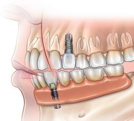
Patientinformationtoassistinformedconsent he temporomandibular

■protrudingrecedingchin jaw whenopenbite(thefrontteethdonottouch inabilitybitingtogether)tomakelipsmeetwithoutmouthchronicmouthbreathingwithdry andinflamedgums sleep apnoea (when sleeping,breathingproblemssuchassnoring).jaws,Yoursurgeonwillexamineyourfaceand andmaymakefacialmeasurements, ortakephotographsorusevideoimaging, YourrecommendX-raysorscansofyour surgeonwillassessyourbitefor toothevidenceoftoothwearandforirregular positionthatcauseproblemswith
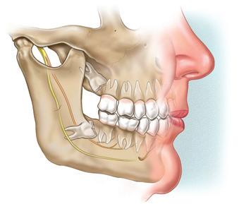
generalinformation.Itisnotasubstitute doesforadvicefromyoursurgeon.Thispamphlet
andtopic.Ifyouarenotsureaboutthebenefits
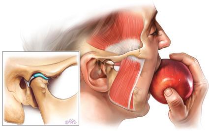
Patientinformationtoassistinformedconsent
Aidstoorthodonticssurgerytohelpanuneruptedtooth moveintoitsnormalpositionextractionofaneruptedandnormal tooththatiscrowdingotherteethor, inuncommoncases,oneormoreextra permanentteeth(supernumeraryteeth)
Patientinformationtoassistinformedconsent
andAn"openbite"leavesspacebetweentheupper lowerteethwhenthemouthisclosed.
isAprognathicjawoccurswhenthelowerjaw upperoverdevelopedandprotrudesbeyondthe jaw.Thisbitecanalsooccurwithan
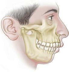
Patientinformationtoassistinformedconsent
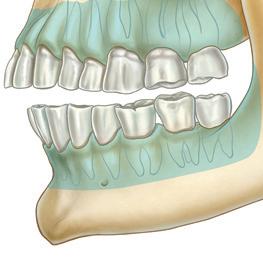
N
ecklumpsareacommoncondition. Most,butnotall, neck lumpsarebenign (non-cancerous). A persistent lump should be thoroughly investigatedandtreated,inaccordancewiththediagnosis. Malignant (cancerous) neck lumps haveusuallyspreadfromcancerslocated intheskinofthehead,neckororopharnyx(themiddlepartofthethroat,behind themouth).Anylumpthathasbeenpresentfor twoweeksormore,evenwithoutsymptoms,requires diagnosis.Notallnecklumpsrequiresurgery.Anecklumpmightdevelopfrom:
Aidstofittingofpartialorcomplete
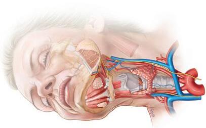
■ swollen lymph nodes (also called lymphglands),whicharethemostcommonnecklumps
■ a tumour, which is an abnormal growthoftissue.A tumourmightbe benignormalignant(cancerous).Benign lumpscangrowfromvarioustissues,such asnerves,fat,bloodvessels,muscles,or thesalivaryglands.Ifthelumpiscancerous,thenitislikelytohavespreadto necklymphnodesfromotherareas.The originalsiteofthecancermaybeinthe nearbyskinorinthemouthandthroat regions.(whetherbenignor malignant)increases withage.In children,necklumpsare uncommonandareusuallybenign.Lymphnodesandotheranatomy Theneckandtissuesabovethe collarbone containmanylymphnodes.Lymphnodes fightinfection.Theyaresmall,soft,nontenderlumps.Iftheygetbigger,then theirsizecanbefeltwhenexaminedby touch(palpation).Enlargedlymphnodes canbepainful,butthismaynotoccur withcancerouslymphnodes. Thelumpmaybe closeto blood vessels,nerves,lymphnodesandother tissues.Themostimportantneckstructuresinclude: ■ thecarotidarteryandjugularvein, whichcarrybloodtoandfromthebrain
To celebrate the latest edition of AJOMS, we are offering 10% off all ANZAOMS pamphlets.
Order online at www.mitec.com.au/ANZAOMS and enter code “AJOMS10” at the checkout.

The ANZAOMS pamphlets provide high-quality and peer-reviewed treatment information for the benefit of surgeons and their patients.
SURGEON Thispamphletprovidesgeneralinformation.Itisnota substituteforadvicefrom yoursurgeon.Termsareusedthatmay requirefurtherexplanationbyyoursurgeon. Thisinformationwillchangewithtime,due toclinicalresearchandnewtherapies.Read thispamphletcarefully,andsaveitforreference.Everyaspectofthesurgerycannotbe coveredinthispamphlet.Seekafullexplanationofyourcasefromyoursurgeon.Yoursurgeoncannotguaranteethatsurgerywillmeetallyour expectationsorthat surgeryhasnorisks.Ifuncertain,seekthe opinionofanotherspecialist.Usethispamphletonlyinconsultationwithyoursurgeon.
■ epidermoid,pilarorsebaceouscysts
■ abscesses
■ benignfattygrowthcalledalipoma
■ thyroidorparathyroidglands
■ abnormal blood vessels (haemangioma)
■ conditionsof the salivary glands, whicharetheparotidglands,submandibularglandsandsublingualglands.Specificcausesofalumpcaninclude:
■ inflamedtissue,duetovariouscauses (forexample,ablockedsalivaryduct)
■ infectionbybacteria,virusesorother infectiousorganisms
■ lymphomaorleukaemia
■ nervesthatcomefromthebrain:the accessory nerve, hypoglossal nerve, lingualnerve,facialnerve,vagusnerve, andtrigeminalnerve.Seepage4formore informationaboutpossible complicationscausedbyinjurytothesenerves.
Important:Fillinalldetailsonthestickerbelow.
DEARSURGEON: Afteryoudiscussthispamphletwithyourpatient,fillintheinformationonthissticker, peelitoff,andputitonthepatient'smedicalhistoryorcard.Thiswillremindyouandyourpatientthat thispamphlethasbeengiventothepatient.Somesurgeonsasktheirpatientstosignthesticker.
1number:Edition
ConsumerandCarerFeedback:Toprovidefeedbackaboutthispamphlet,sendcommentsto: feedback@mitec.com.auorbyposttoMi-tec,POBox24, Camberwell,VIC,3124,Australia.