Allergy Research Group ® Newsletter | Summer/Fall 2017
The Neuroelectric Origins of Asthma
Ba Hoang, MD, PhD, hopes to vanquish asthma with a novel treatment approach Page 2
Bronchial Epilepsy
A new model for asthma set out in three papers by Ba Hoang, MD, PhD Page 4
New Studies Confirm the Power of Three Chinese Herbs for Asthma
Traditional Chinese herbal formula effective in alleviating bronchoconstriction and inflammation Page 6
How One Nutritionist Uses a CortisolOptimizing Herbal Blend to Treat Fatigue
Laurie Hammer, NTP, recommends a blend of three Chinese herbs, along with Morinda citrifolia, for adrenal “fatigue”
Michael Ash, DO, ND, BSc, RNT, mIOD and Garth Nicolson PhD, MD(H) offer highlights from their recent publication Page 9
A
Allergy Research Group® 2300 North Loop Road Alameda, CA 94502

For more information, call 800.545.9960 or visit www.allergyresearchgroup.com 1
Phone: 800.545.9960 | 510.263.2000 Fax: 800.688.7426 | 510.263.2100 www.allergyresearchgroup.com
FOCUS
......................................................... Page 8
Membrane Lipid Replacement: A Rapidly Evolving Field
Sulfated Polysaccharides: Immune Boosters Extracted from Sea Life
novel galactofucan sulfate extract enhances immunity, inactivates viruses naturally and reduces inflammation .............................................. Page 10
Protect the Brain from Stress with a Unique Lipid Blend
Fatty acid blend soothes the stressed brain Page 14
Our summer/fall newsletter tackles that perennial autumn subject: asthma, which inevitably spikes during fall pollen season. In this issue, we present a novel hypothesis for a unique mechanism of action in the pathogenesis of asthma. Rather than tackling inflammation, Ba Hoang, MD, PhD, suggests that bronchopulmonary hyperexcitability may be at the root of many asthma symptoms. This approach may lead to new, effective, and less toxic treatment strategies and interventions.
We also discuss the strong science behind a versatile polysaccharide from the sea. Even though it has been available for years, it is only now gaining attention. I think you will enjoy reading about the far-reaching activity of fucoidan extract from seaweed.
We interview a nutritional therapist who offers up a novel use of a Chinese herbal blend to address adrenal dysfunction, and we look at the scientific research demonstrating the efficacy and safety of that innovative blend.
Lastly, a topic always at the forefront of my own clinical practice: the use of nutritional supplementation to enhance and optimize the function of the hypothalamic-pituitary-adrenal axis. I have used phosphatidylserine in my practice to help my patients with memory concerns, anxiety, insomnia, ADD/ADHD, and generalized stress, but have noticed that when combined with phosphatidic acid, the results are even more profound.
— Dr. Todd A. Born, ND, CNS Editor-in-Chief
The Neuroelectric Origins of Asthma
Ba Hoang, MD, PhD, Hopes to Vanquish
Asthma
with a Novel Treatment Approach
FOCUS: You have a unique approach to asthma. Rather than focus on inflammation, your model is similar to that for epilepsy, where seizures are the result of hyperexcitability in the tissue.
BA: Yes, during my practice in St. Petersburg in the 1990’s, I treated refractory asthma in children with natural, herbal anti-epileptic agents and dietary changes. When I was working in St. Petersburg, I found that the EEG of children with a history of life-threatening asthma attacks showed signs of hyperexcitatory brain activity, such as a slow dysrhythmia and occipital focal spikes or sharp waves. The paroxysmal, spasmodic character of asthma attacks may be similar to seizures.
FOCUS: How would you describe asthma?
BA: The primary clinical symptoms of asthma are attacks of shortness of breath, wheezing, and coughing resulting from excessive and inappropriate constriction of the airway smooth muscle.
I propose that asthma is caused by a toxic buildup of both exogenous and endogenous excitotoxins that primarily accumulate in smooth muscle tissue in our lungs. These excitotoxins can stem from infections, allergies, hyperventilation, pollutants, mental/physical stress, and exercise, as well as hormonal, nutritional, and environmental factors.
The genetic and environmental elements in the development of severe asthma are poorly understood. The pathology of severe asthma demonstrates a range of findings, including eosinophilic inflammation and other pathological changes that are likely due to airway remodeling.2 I do not think, however, that they are the cause of asthma.
FOCUS: How might excitotoxins trigger asthma?
BA: This new asthma model goes beyond descriptive studies of inflammatory immune mediators or abnormalities in smooth muscle function, and looks deeper at the dysfunction in the electrical potential of the cell membrane. The amino acids glutamate and aspartate are abundantly present in the central nervous system of mammals, and they are major excitatory
I propose that asthma is caused by a toxic buildup of both exogenous and endogenous excitotoxins that primarily accumulate in smooth muscle tissue in our lungs.
2 FOCUS | Summer/Fall 2017
From the Editor‘s Desk
neurotransmitters. Glutamate receptors have been found in the lungs and airways.3 Glutamate and the activation of glutamate receptors have led to increased airway secretions.4 So it is possible that excitotoxins might be an important mechanism of the airway inflammation and hyperreactivity found in asthma.5
This new asthma model goes beyond descriptive studies of inflammatory immune mediators or abnormalities in smooth muscle function, and looks deeper at the dysfunction in the electrical potential of the cell membrane.
There are no formal studies to investigate the role of the glutamate response modulator in asthma and other respiratory diseases, or to conclusively prove that asthma is a syndrome of inducible or genetically predisposed membrane hyperexcitability. But there is convincing evidence that this framework is valid. I call it a form of bronchial epilepsy.6 I have found that certain herbs that are capable of modulating the excitatory activity of smooth muscles, and increasing
the capacity of the patient’s serum to bind to allergic mediators, can reduce the severity and frequency of asthma attacks.
I have also found that these herbs can have remarkable efficacy in chronic obstructive pulmonary disease (COPD). I am in the process of translating into English a study conducted with my colleagues at the National Hospital of Traditional Medicine in Vietnam. We examined 60 patients diagnosed with C-level COPD according to the guidelines of the Global Initiative for Chronic Obstructive Lung Disease (GOLD). Patients were treated for one month with an herbal formula that contained the natural herbal compound Sophora flavescens, which reduces membrane hyperexcitability. The treatment group’s improvement was significantly better than that of the control group.
FOCUS: What inspired you to study herbs in an academic and scientific setting as you do today at the Keck School of Medicine?
BA: My family practiced medicine for three generations in Vietnam, where I grew up. My grandfather had an innate talent for healing and traditional medicine from a young age. He saved many lives, and when I was a child he conveyed his philosophy
that one can vastly reduce suffering by understanding the deeper cause of a disease and trying to reverse it with gentle, nontoxic molecules that support the body’s own healing ability. In this way, you can address functional declines in chronic and degenerative diseases. Fortunately, and happily, I can report to you now, after much practice and research, that my grandfather was correct.
References
1 Letter sent to Mary Tarnowka, Consul General-designate, Ho Chi Minh City, Vietnam. 2016 Aug 30.
2 Wenzel S. Mechanisms of severe asthma. Clin Exp Allergy. 2003;33:1622-8. PMID: 14656346
3 Said SI, Berisha HI, Pakbaz H. Excitotoxicity in the lung: N-methyl-D-aspartate-induced, nitric oxide-dependent, pulmonary edema is attenuated by vasoactive intestinal peptide and by inhibitors of poly(ADP-ribose) polymerase. Proc Natl Acad Sci USA. 1996;93:4688-92.
4 Haxhiu MA, Chavez JC, Pichiule P, et al. The excitatory amino acid glutamate mediates reflexly increased tracheal blood flow and airway submucosal gland secretion. Brain Res. 2000;883:7786. PMID: 11063990
5 Dickman KG, Youssef JG, Mathew SM, et al. Ionotropic glutamate receptors in lungs and airways: molecular basis for glutamate toxicity. Am J Respir Cell Mol Biol. 2004;30:139-44. PMID: 12855408
6 Hoang BX, Levine SA, Graeme Shaw D, et al. Bronchial epilepsy or broncho-pulmonary hyper-excitability as a model of asthma pathogenesis. Med Hypotheses. 2006;67(5):1042-51. PMID: 16797869
BIOGRAPHY Ba Hoang, MD, PhD, is a senior medical research associate at the Keck School of Medicine of the University of Southern California, where he works with a team of scientists at the Nimni-Cordoba Tissue Engineering and Drug Discovery Laboratory. Formerly a pediatric hematologist in St. Petersburg, Russia, he was awarded a fellowship by Swedish Pharmacy Research Foundation for his work on a method for detecting IgE-binding capacity in the serum of children with allergic disorders. In 1996, Dr. Ba was granted a United States visa for a person with extraordinary ability in science. His work “uses natural molecules to regulate cell energy metabolism … the major breakthrough discoveries have already shown positive effects in both preclinical and clinical applications,” according to USC’s Bo Han, PhD, and Marcel Nimni, PhD.1 Dr. Ba holds several patents, including one on a novel treatment approach to asthma. He comes from three generations of doctors, and learned about the medicinal power of herbs from his grandfather, a revered and widely respected folk healer. Dr. Ba is currently collaborating with Michael Roth, PhD, Director of the Pulmonary Cell Research group at Basel University in Switzerland. They are developing a naturally derived agent for asthma and COPD that restores normal function in smooth muscle tissue.
For more information, call 800.545.9960 or visit www.allergyresearchgroup.com
3
One can vastly reduce suffering by understanding the deeper cause of a disease.
Bronchial Epilepsy
A New Model for Asthma Set Out in Three Papers
by Ba Hoang, MD, PhD
Bronchial Epilepsy or Bronchopulmonary Hyper-excitability as a Model of Asthma Pathogenesis1
The first of three papers reframing asthma as a form of ”bronchial epilepsy” was published in 2006 by Ba Hoang, MD, PhD, and colleagues. It proposes that asthma may be, at root, a syndrome of inducible, genetically predisposed membrane hyperexcitability. Though inflammation and allergy are highly correlated with asthma, they are not always present. In active asthma, however, bronchoconstriction is always present.
The action of nerves, muscles, hormones, and neurotransmitters relies on cell excitability. Cells are excited by the reversal of resting membrane potential, and this reversal is mediated through what are known as gated ion channels. Most of the cell membrane is made of lipids, fatty molecules that act as a barrier to ions (electrically charged atoms) such as sodium, calcium, chloride, and potassium. Embedded in the cell membrane are special protein ”gatekeepers” known as ion channels that control the movement of ions. They open and close in response to a change in the electrical charge difference that is naturally generated across the cell membrane, or in response to molecular signals from inside or outside the cell. These ion channels are either excitatory or inhibitory. The major excitatory ion channels are glutamate- and acetylcholine-responsive. When these channels are activated, their electrical activity increases.2 Glutamate has an excitotoxic action, and its action in the lungs of rats mimics the asthmatic response. In contrast, gamma
aminobutyric acid (GABA) has an inhibitory, calming action, and GABA agonists inhibit bronchoconstriction, cough, and airway inflammation in animal studies.3
In addition, the authors suggest that sodium channels may be involved in membrane excitability (and asthma pathogenesis).4,5 Sodium influx can follow acetylcholine release, resulting in smooth muscle contraction. It is interesting to note that sodium channel blockers such as lidocaine (inhaled or intravenous) and phenytoin (an antiseizure medication) are effective in both epilepsy and asthma.6,7 Phenytoin blocks voltage-sensitive sodium channels. Studies have shown that lidocaine has potential as an asthma therapy even for patients with severe steroid-dependent asthma,8 and, like phenytoin, acts on voltage-sensitive sodium channels to block repetitive firing of neurons. Phenytoin is helpful for acute and paroxysmal asthma, and a majority of treated patients were found to have sustained benefit from the therapy after phenytoin was discontinued.9,10
The paper by Ba and his colleagues concludes that better asthma control and prevention might be attained if we explore the role of proven excitotoxins in food and the environment. In addition, since the ketogenic diet,11 glycine and taurine supplementation,12 and inhibition of glutamate have proven to be helpful therapies in epilepsy, the same might hold true of asthma.
TAKEAWAY
Asthma may be, at root, a syndrome of inducible, genetically predisposed membrane hyperexcitability.
2007 New Approach in Asthma Treatment
Using Excitatory Modulator13
In this 2007 study, Ba and colleagues test their hypothesis (that asthma is induced by excitotoxins) by utilizing an herb called Sophora flavescens on 14 individuals suffering from moderate to severe asthma. S. flavescens has long been used as an anti-asthmatic agent in traditional Chinese medicine. In addition to anti-inflammatory and antioxidant effects,14,15 the herb contains two matrine-type alkaloids— matrine and oxymatrine—which can act as modulators of membrane excitability.16
Six men and eight women, aged 22–70, were treated during the time period from February 1997 to December 2005. These patients had been diagnosed with asthma by their allergists and had been on medication for asthma for three to six years. Despite years of moderate to high doses of inhaled corticosteroids and beta2-agonists, they still suffered from episodes of dyspnea, expectoration, coughing, wheezing, or chest tightness more than two times a week and were waking up at night with asthma symptoms more than two times a week.
Patients were given capsules with a dose equal to 4 g of dried S. flavescens root with a progressively diminishing dose: thrice daily for three months, twice daily for six months, and once daily for 27 months thereafter. All patients had been followed for three years. The patients were evaluated every two weeks for the first month, once monthly for the next six months and every three months thereafter. A dried powder of a standard hot water
4 FOCUS | Summer/Fall 2017
2006
extract was used because it has been shown to contain a high content of matrine and oxymatrine (1.8%–3.2%). This corresponds to about 72–128 μg of matrine and oxymatrine, when taken three times daily. Results were based on the diaries of symptoms, PEF (peak expiratory flow, which is the maximum airflow during a forced expiration beginning with the lungs fully inflated), medication use, and quality of life.
Within the first two weeks, daytime asthma symptoms dropped by 65% and nighttime symptoms by 72%. Beta2-agonist medications were reduced by 67%. The dose of inhaled corticosteroids remained unchanged. The mean PEF rate improved by 9%.
By one month, daytime asthma symptoms were reduced by 78%, and nighttime symptoms by 75%. The beta2-agonist dose was reduced by 72% and the dose of corticosteroid inhaler reduced by 45%. The mean PEF rate improved by 12%.
At three months, the daytime asthma symptoms were reduced by 87%, and nighttime symptoms by 85%. The beta2-agonist use was reduced by 92% and the dose of corticosteroid inhaler was reduced by 84%. The mean PEF value increased by 15%.
At one year, daytime symptoms of asthma were reduced by 94%, and nighttime symptoms by 95%. Beta2agonist use was reduced by 95%; the dose of corticosteroid inhaler was reduced by 92%. The mean PEF had increased by 18%. Finally, at three years, daytime symptoms of asthma were reduced by 97%, and nighttime symptoms by 98%. The dose of beta2agonist was reduced by 97%, and no patients took inhaled corticosteroids. The mean PEF had increased by 21%.
At three years, nine of the 14 patients had achieved a symptom-free, medication-free, and asthma-free condition, in which they no longer developed asthma when exposed to the previous triggers of their asthma attacks. Ba and his colleagues conclude: “The excitatory modulator S. flavescens provided dramatic clinical
and functional results for patients with moderate and severe asthma. We acknowledge that our study is open and selective, which might have led to a higher rate of success. However, the short-term and long-term multifaceted benefits of this treatment are not due to just a placebo effect. S. flavescens root extract, with a high content of matrine-type alkaloid, may target the causes of asthma more specifically compared with the available standard pharmaceutical therapies.”
TAKEAWAY
be a mechanism by which asthma can be managed and prevented. Future studies will hopefully elucidate.
TAKEAWAY
References
1 Hoang BX, Levine SA, Graeme Shaw D, et al. Bronchial epilepsy or broncho-pulmonary hyper-excitability as a model of asthma pathogenesis. Med Hypotheses. 2006;67(5):1042-51. PMID: 16797869
2 Nakashima Y, Sugiyama S, Shindoh J, et al. Effects of sodium channel blockers on electrical field stimulation-induced guineapig tracheal smooth muscle contraction. Arch Int Pharmacodyn Ther. 1990;306:130-8. PMID: 1963767
3 Belvisi MG, Ichinose M, Barnes PJ. Modulation of nonadrenergic, non-cholinergic neural bronchoconstriction in guinea-pig airways via GABAB-receptors. Br J Pharmacol. 1989;97:1225-31. PMID: 2477104
4 Said SI, Berisha HI, Pakbaz H. Excitotoxicity in the lung: Nmethyl-D-aspartate-induced, nitric oxide-dependent, pulmonary edema is attenuated by vasoactive intestinal peptide and by inhibitors of poly(ADP-ribose) polymerase. Proc Natl Acad Sci USA. 1996;93:4688-92. PMID: 8643465
2010
Treating Asthma as a Neuroelectrical Disorder17
This 2010 paper reviews the proposed excitatory model of asthma. Lung tissue has a rich bed of nerve circuits, and the epithelial, submucosal, and smooth muscle cells of the lung carry both voltage-gated sodium channels and glutamate receptors. Sodium influx stimulates the release of acetylcholine, causing smooth muscle contraction.
There is a substantial body of evidence from basic research and clinical observations that indicate an important role of inducible and genetically predisposed airway membrane hyperexcitability in asthma pathogenesis and its relationship with emphysema and chronic lung disorders. The hyperexcitatory model of asthma pathogenesis may help to explain many unresolved issues of asthma’s pathology, epidemiology, clinical features, and therapeutic response. Ba and colleagues feel that modulation of membrane excitability may be found to
5 Chapman RW, Danko G, Rizzo C, et al. Prejunctional GABA-B inhibition of cholinergic, neurally-mediated airway contractions in guinea-pigs. Pulm Pharmacol. 1991;4:218-24. PMID: 1666856
6 De Giorgio CM, Altman K, Hamilton-Byrd E, et al. Lidocaine in refractory status epilepticus: confirmation of efficacy with continuous EEG monitoring. Epilepsia. 1992;33:913-6. PMID: 1396435
7 Hunt LW, Swedlund HA, Gleich GJ. Effect of nebulized lidocaine on severe glucocorticoid-dependent asthma. Mayo Clin Proc. 1996;71:361-8. PMID: 8637259
8 Hunt LW, Frigas E, Butterfield JH, et al. Treatment of asthma with nebulized lidocaine: a randomized, placebo controlled study. J Allergy Clin Immunol. 2004;113:853-9. PMID: 15131566.
9 Shulman MH. The use of dilantin sodium in bronchial asthma: a preliminary report. N Engl J Med. 1942;226:260-264.
10 Sayar B, Polvan O. Epilepsy and bronchial asthma. Lancet. 1968;1(7550):1038. PMID: 4171827
11 Panico LR, Demartini MG, Rios VG, et al. The ketogenic diet in infantile refractory epilepsy: electroclinical response, complications and secondary effects. Rev Neurol. 2000;31:212-20. PMID: 10996924
12 Schmieden V. Effects of taurine and glycine on epileptiform activity induced by removal of Mg2+ in combined rat entorhinal cortex-hippocampal slices. Epilepsia. 2003;44:1145-52. PMID: 12919385
13 Hoang BX, Shaw DG, Levine S, et al. New approach in asthma treatment using excitatory modulator. Phytother Res. 2007;21(6):554-7. PMID: 17295384
14 Jin JH, Kim JS, Kang SS, et al. Anti-inflammatory and anti-arthritic activity of total flavonoids of the roots of Sophora flavescens. J Ethnopharmacol. 2010;127(3):589-95. PMID: 20034551
15 Piao XL, Piao XS, Kim SW, et al. Identification and characterization of antioxidants from Sophora flavescens. Biol Pharm Bull. 2006;29(9):1911-5.
16 Liu M, Liu XY, Cheng JF. Advance in the pharmacological research on matrine. China J of Chinese materia medicine. 2003;28:801-4. PMID: 15015368
17 Hoang BX, Shaw DG, Pham P, et al. Treating asthma as a neuroelectrical disorder. Inflamm Allergy Drug Targets. 2010;9(2):130-4. PMID: 20359291
For more information, call 800.545.9960 or visit www.allergyresearchgroup.com
5
Ba reviews the evidence that modulation of membrane excitability might be used for the management and prevention of asthma.
Ba feels that Sophora flavescens may reduce cell membrane excitability and markedly improve asthma, so that by the three-year mark, nine of twelve patients in this study were symptomfree and medication-free.
New Studies Confirm the Power of Three Chinese Herbs for Asthma
Traditional Chinese Herbal Formula Effective in Alleviating Asthma
If you’ve ever tried to breathe through a straw, or felt as if an invisible gorilla had your chest in a vice, or simply had to focus all your will and attention on drawing each breath—if you’ve ever had a bad asthma attack, you know how frightening asthma can be.
One in every 13 Americans suffers from this chronic lung disease characterized by ongoing airway inflammation, episodes of bronchoconstriction, and trouble breathing.1 An estimated 39.5 million people (12.9%), including 10.5 million (14.0%) children in the United States have been diagnosed with asthma in their lifetimes.2 Asthma is the top reason for missed school days in children.3 In 2008, the group of children aged 5–17 years who each had one or more asthma attacks in the previous 12 months missed, in total, 10.5 million days of school. Adults who were employed and who each had one or more asthma attacks during the previous 12 months missed, in total, 14.2 million days of work due to asthma.4 Researchers estimate the annual cost of asthma in the United States is around $56 billion.5
The standard medications for asthma include inhaled corticosteroids and beta-2 agonists, leukotriene modifiers, oral corticosteroids, and theophylline.6 Steroids, however, can suppress endogenous cortisol production7 and immune function.8 Even inhaled steroids can suppress adrenal function, cause mucosal immunosuppression, and increase risk of respiratory infections.9 In spite of their risks, they are being overprescribed: “Inhalers have become almost a fashion accessory,” write two respiratory specialists in a 2015 editorial.10
Children under five, in particular, may be at risk of being overprescribed potent
oral steroids, when less potent inhaled steroids or beta agonist sprays would suffice, according to a 2017 study. The study analyzed the computerized databases of Texas Children’s Health Plan between January 2011 and January 2016.11 “Over the past 30 years, [oral corticosteroid] prescribing for children with asthma has gone from underuse to what now appears to be substantial overprescribing,” the authors write.
Even ”safer” medications such as betaagonists can lead to tolerance, and can actually increase airway inflammation.12 Long-acting beta-agonists may actually increase the risk for fatal and nonfatal asthma exacerbations.13
A Natural Alternative as Effective as Steroids— Without the Side Effects?
For the last 17 years, pediatric immunologist Xiu-Min Li, MD, and her team at the Icahn School of Medicine at Mount Sinai in New York have been proving the efficacy of a novel, nonsteroidal blend of traditional Chinese herbs for asthma.14 The formula is called ASHMI (anti-asthma
Cortisol was suppressed in the prednisone group, while in the ASHMI group, levels increased into the normal range. Prednisone decreased the secretion of interferon (IFN-γ), while ASHMI increased it, improving immune function.
herbal medicine intervention) and blends the three most effective herbs found in a traditional 14-herb formula15 with a long history of human use in China: lingzi (Ganoderma lucidum, commonly known as reishi), gancao (Glycyrrhiza uralensis, commonly known as Chinese licorice) and ku shen (Sophora flavescens, commonly known as shrubby sophora).16 ASHMI has broad therapeutic effects on the major pathogenic mechanisms of asthma, including inflammation and bronchoconstriction, and it also raises endogenous cortisol production instead of suppressing it as steroid medications do. In vitro studies conducted by the team have unraveled the ways in which the herbs actually function—modulating the activity of immune cells, inhibiting smooth muscle contraction, and increasing the production of cortisol. Human studies, in turn, have demonstrated the formula’s power and gentleness.
In our March 2008 edition of FOCUS (see: Herbal Adaptogens for Asthma and Adrenal Function), we reported on the team’s first human study, an FDA-approved Phase I and Phase II trial involving 13 researchers—11 of them physicians—from the Weifang Asthma Hospital, the Weifang School of Medicine, and the Mount Sinai School of Medicine. This study found ASHMI as effective as prednisone, without suppressing cortisol or immune function.14
During the four-week, double-blind, randomized placebo-control trial, 45 individuals with moderate to severe asthma received oral ASHMI capsules and placebo prednisone tablets (ASHMI group) and 46 matched individuals received oral prednisone tablets (20 mg) and placebo ASHMI capsules (prednisone group). The ASHMI
6 FOCUS | Summer/Fall 2017
group received four capsules three times a day, with each capsule containing 0.3 g of a dried aqueous extract of the three herbs. Placebo capsules were similar in appearance to prednisone. The prednisone group received oral tablets of 20 mg once a day in the morning.
Post-treatment lung function significantly improved in both groups, with the steroid group showing only a slightly greater benefit. Clinical symptom scores, use of beta agonist bronchodilators, and serum IgE levels were also reduced significantly in both groups. However, a key difference emerged: though both groups had low cortisol levels before the study, cortisol was even further suppressed in the prednisone group, while in the ASHMI group, levels increased into the normal range. And while prednisone decreased the secretion of interferon (IFN-γ), ASHMI increased it, improving immune function.
A second randomized, double-blind, placebo-controlled, dose-escalation study followed in 2009 to assess safety. 20 allergic individuals were included, in order to test three different doses of ASHMI: 600 mg, 1200 mg, or 1800 mg. For each test dose, four individuals were treated and two individuals were given the placebo.17 All three doses were well tolerated.18 Vital signs, electrocardiogram findings, and laboratory results obtained at preand post-treatment visits remained within normal range. No abnormal immunologic alterations were detected.
How ASHMI Works at the Cellular Level
Studies in mice reveal how ASHMI might work. First, it appears to prevent early reactivity to allergen exposure.19 It also reduces late airway reactions, and reduces airway inflammation and remodeling (the long-term thickening of the smooth muscles of the bronchi). Levels of collagen in lung fluid—which are associated with airway remodeling— are also reduced. In addition, ASHMI also helps inhibit constriction caused by the neurotransmitter acetylcholine, and increase levels of smooth-muscle
relaxants such as prostaglandins. It reduces lung inflammation and mucus in mice bred for late-onset asthma.20 A single dose of ASHMI dramatically reduces airway hyperresponsiveness in response to challenge with the neurotransmitter acetylcholine, which can contribute to asthma.21 Treatment of mice with ASHMI in drinking water for four weeks prevents the development of asthma, and this therapeutic effect remains at eight weeks post-therapy.22
for treatment and/or prevention for allergic asthma.”26 In sum, ASHMI improves lung function, reduces symptom scores in asthma, and leads to decreased beta2-agonist use—and does so without prednisone’s adverse effect on adrenal function, and with no overall immune suppression.
TAKEAWAY
Treatment of mice with ASHMI in drinking water for four weeks prevents the development of asthma, and this therapeutic effect remains at eight weeks post-therapy.
At the same time, in vitro studies reveal intriguing mechanisms underlying the formula’s effectiveness. In vitro studies using cells from human lungs found that one of the extracts, gancao (Chinese licorice), decreased levels of inflammatory chemicals such as interleukin (IL)-8.23 Other in vitro studies found that the formula decreased inflammatory molecules such as tumor necrosis factor (TNF) and nuclear factor kappa B (NF-κB).24 ASHMI also inhibited production of the inflammatory interleukins IL-4 and IL-5 as well as a molecule called eotaxin-1, which stimulates the migration of inflammatory white blood cells from blood into the lungs.25
A 2013 editorial in Clinical & Experimental Allergy, entitled Chinese Herbal Anti-asthma Tea to Go!, reviews the work by Li and her colleagues and concludes: “Experimental and clinical evidence suggests that ASHMI may be an effective future modality
References
1 Centers for Disease Control and Prevention (US). Asthma [Internet]. Atlanta (GA): U S Department of Health and Human Services; updated 2016 [cited 2017 Jul 22]. Available from: http://www.cdc.gov/asthma/default.htm
2 Centers for Disease Control and Prevention (US). Asthma facts [Internet]. Atlanta (GA): U S Department of Health and Human Services; 2013 [cited 2017 Jul 22]. Available from: http://www.cdc.gov/nchs/fastats/asthma.htm
3 Moorman JE, Akinbami LJ, Bailey CM, et al. National surveillance of asthma: United States, 2001-2010. Vital Health Stat 3. 2012;(35):1-58. PMID: 24252609
4 Akinbami LJ, Moorman JE, Liu X. Asthma prevalence, healthcare use, and mortality: United States, 2005-2009. Natl Health Stat Report. 2011;(32):1-14. PMID: 21355352
5 United States Environmental Protection Agency. Asthma facts [Internet]. Washington, DC: United States Environmental Protection Agency; 2013 [cited 2017 Jul 22]. Available from: https://www.epa.gov/sites/production/files/2017-08/ documents/2017_asthma_fact_sheet.pdf
6 Mayo Clinic staff. Asthma treatment and drugs [Internet]. Mayo Foundation for Medical Education and Research, Mayo Clinic; 2016 [cited 2017 Jul 22]. Available from: http://www.mayoclinic.org/diseases-conditions/asthma/basics/ treatment/con-20026992
7 The Asthma Center. Steroid dependence [Internet]. Philadelphia (PA): The Asthma Center Education and Research Fund [cited 2017 Jul 22]. Available from: http://www.theasthmacenter.org/index.php/disease_information/ asthma/medical_treatment_of_asthma/corticosteroids/ corticosteroid_risks/steroid_dependance
8 Fuenfer MM, Olson GE, Polk HC Jr. Effect of various corticosteroids upon the phagocytic bactericidal activity of neutrophils. Surgery. 1975;78(1):27-33. PMID: 1138397
9 Sabroe I, Postma D, Heijink I, et al. The yin and the yang of immunosuppression with inhaled corticosteroids. Thorax. 2013;68:1085-7. PMID: 23929790
10 Bush A, Fleming L. Is asthma overdiagnosed? Arch Dis Child. 2016;101(8):688-9. PMID: 27049005
11 Farber HJ, Silveira EA, Vicere DR, et al. Oral corticosteroid prescribing for children with asthma in a medicaid managed care program. Pediatrics. 2017;139(5). PMID: 28557753
12 Salpeter SR, Ormiston TM, Salpeter EE. Meta-analysis: respiratory tolerance to regular beta2-agonist use in patients with asthma. Ann Intern Med. 2004;140(10):802-13. PMID: 15148067
13 Salpeter SR, Buckley NS, Ormiston TM. Meta-analysis: effect of long-acting beta-agonists on severe asthma exacerbations and asthma-related deaths. Ann Intern Med. 2006;144(12):904-12. PMID: 16754916 References continued on p. 13
For more information, call 800.545.9960 or visit www.allergyresearchgroup.com
7
Experimental and clinical evidence suggests that ASHMI may be an effective future modality for treatment and/or prevention of allergic asthma.
How One Nutritionist Uses a Cortisol-Optimizing Herbal Blend to Treat Fatigue
By Laurie Hammer, NTP
CLINICAL PEARL
My practice largely centers around women aged 35–60, and for about 80% of them, I recommend an herbal formula that contains lingzi (Ganoderma lucidum, commonly known as reishi), gancao (Glycyrrhiza uralensis, commonly known as Chinese licorice), ku shen (Sophora flavescens, commonly known as shrubby sophora), and Morinda citrifolia (commonly known as noni fruit). I have found that this formula helps relieve adrenal “fatigue” (better understood as a dysfunctional hypothalamic-pituitaryadrenal axis) and naturally optimizes cortisol. I have also found it to enhance my clients’ mood.
Cortisol has many functions. It helps the body make sugar (glucose) and fat for energy (metabolism), but also makes fat and muscle cells resistant to insulin when elevated for too long in response to stress. Cortisol levels can be affected by many things, such as physical or emotional stress, strenuous activity, infection, injury, or when given synthetically.
My typical client is a woman in her mid-40s to early 50s, who presents with a lot of food cravings and persistent exhaustion. She might get out of bed in the morning with zero energy, and on a scale of one to ten (with ten being optimal), her energy will rarely rise over a four during the course of the day.
I had one such client in her mid-40s whose husband wouldn’t let her drive the car in the afternoon because she was so tired, she was falling asleep at the wheel! We initiated dietary changes, but she still had zero energy and fell asleep every afternoon.
I tested her adrenal function via saliva testing and it turned out she was hardly producing cortisol.
She began to take the herbal blend I recommend. She took four capsules in the morning when she woke, four at midday, and four in the midafternoon. Within two weeks, her energy had increased. She told me she didn’t ever remember being that energetic. Soon she was able to drive from Iowa to Indiana by herself.
I had another client with intense food cravings and a lot of fatigue. She was 57 years old. Her cortisol tested very low in the mornings and spiked in the early evening. For that reason, she was not sleeping well. There are many reasons this pattern can occur— from raising kids, psychological stress, or simply staying up too late every night. In her case, she had all four of her children very close together, and her diet was poor, focusing mainly on starches, breads, and pasta. She began to take four capsules of the herbal blend in the morning, four in the midmorning, and then two in the afternoon. She didn’t need as much in the afternoon, as her own cortisol would start rising. Within six weeks, she was sleeping through the night.
To test adrenal function, I have used both saliva testing (where the individual collects their saliva into a vial four times a day) and urine testing (where the patient urinates on tiny strips four or five times a day). I use the urine tests more commonly these days, because they provide indications of metabolic pathways and metabolites as well as cortisone and cortisol amounts.
I find that this herbal blend, along with dietary change and the addition of amino acids as building blocks, is a lovely combination that works very well in adrenal “fatigue” clients.
8 FOCUS | Summer/Fall 2017
BIOGRAPHY Laurie Hammer, NTP, received her degree in nutrition from the Nutritional Therapy Association in Tumwater, Washington, which is approved by the National Association of Nutrition Professionals (NANP). She practices at Walker Chiropractic & Wellness in Algona, Iowa.
Membrane Lipid Replacement: A Rapidly Evolving Field
By Michael Ash, DO, ND, BSc, RNT, mIOD, and Garth Nicolson, PhD, MD(H)
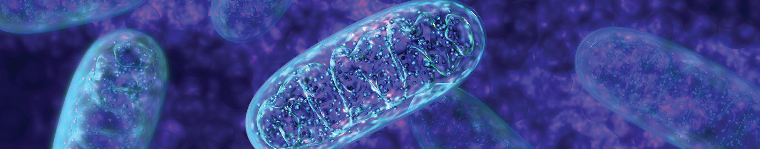
Healthy membranes are key to the function of both cells and their intracellular organelles, such as mitochondria. Healthy cellular membranes can fully maintain their chemical and electrical barrier functions, and also act as cellular signals to promote positive changes in cellular functions and health. Defects in cellular and intracellular membranes are characteristic of all chronic medical conditions, including cancer, and normal processes, such as aging.
In a special issue of the research journal Biochimica et Biophysica Acta— Biomembranes, entitled Membrane Lipid Therapy: Drugs Targeting Biomembranes, we published an invited review on the role of a specially prepared mixture of glycerolphospholipids on human health. This special mixture serves as a natural replacement of damaged membrane phospholipids.1 Our review, along with other recent articles,2,3 explores practical ways to replace damaged lipids in membranes.
This novel field, described as Membrane Lipid Replacement, is growing and evolving rapidly, providing treatments that are now in general use or that are being studied for their application in numerous conditions.1,2,3 Nowhere is this more apparent than in mitochondrial membranes, which provide most of our cellular energy needs (via the production of adenosine triphosphate, or ATP). This field has arisen from relevant discoveries on the behavior of membranes, and it paves the way to adopt new approaches in modern nutrition.1,2,3
The plasma or cell membrane was historically viewed by most scientists
as a fixed physical barrier where lipids arranged as a bilayer performed a mainly structural function, and proteins undertook the relevant catalytic and signaling tasks. This view of the membrane lipid bilayer sandwiched between protein layers prevailed until recent years. Studies carried out in the 1970s clearly indicated that membrane fluidity allowed lipids and proteins to move relatively freely along the plane of membranes, which could also regulate their activities.
Clinical trials have shown the benefits of Membrane Lipid Replacement in restoring mitochondrial function and reducing fatigue in aged subjects and chronically ill patients.
The landmark paper published by Singer and Nicolson in 1972 proposed the Fluid Mosaic Membrane model, which defined the membrane structure as a two-dimensional viscous solution of lipoproteins and glycolipids in thermodynamic equilibrium. These proteins are asymmetrically distributed across a dynamic lipid bilayer matrix.4
Over the years this model has been refined, but the basic concepts have remained, minimally changed in the past 45 years.5
We define “Membrane Lipid Replacement“ as the use of functional, oral supplements containing mixtures of cell membrane glycerolphospholipids, plus fructoo-ligosaccharides (for
protection against oxidative, bile acid, and enzymatic damage) and antioxidants. This unique blend can safely replace damaged, oxidized membrane phospholipids and restore membrane, organelle, cellular, and organ function.
When the glycerolphospholipids arrive at their membrane destinations, they can stimulate the natural removal and replacement of damaged membrane lipids. Clinical trials have shown the benefits of Membrane Lipid Replacement in restoring mitochondrial function and reducing fatigue in aged subjects and chronically ill patients.6 Recently, Membrane Lipid Replacement has been used to reduce pain and other symptoms, as well as removing hydrophobic chemical contaminants as part of ongoing clinical research, suggesting that there are additional new uses for this safe, natural medicine supplement.
References
1 Nicolson GL, Ash ME. Membrane Lipid Replacement for chronic illnesses, aging and cancer using oral glycerolphospholipid formulations with fructooligosaccharides to restore phospholipid function in cellular membranes, organelles, cells and tissues. Biochim Biophys Acta. 2017;1859:1704-24. PMID: 28432031
2 Nicolson GL. Membrane Lipid Replacement: clinical studies using a natural medicine approach to restoring membrane function and improving health. Intern J Clin Med. 2016;7:133-43. doi: 10.4236/ijcm.2016.72015.
3 Nicolson, GL, Rosenblatt S, Ferreira de Mattos G, et al. Clinical uses of Membrane Lipid Replacement supplements in restoring membrane function and reducing fatigue in chronic diseases and cancer. Discoveries. 2016;4(1):e54. doi: 10.15190/d.2016.1
4 Singer SJ, Nicolson GL. The fluid mosaic model of the structure of cell membranes. Science. 1972;175:720-31. PMID: 4333397
5 Nicolson GL. The Fluid-Mosaic Model of Membrane Structure: still relevant to understanding the structure, function and dynamics of biological membranes after more than 40 years. Biochim Biophys Acta. 2014;1838:1451-66. PMID: 24189436
6 Agadjanyan M, Vasilevko V, Ghochikyan A, et al. Nutritional supplement (NT Factor™) restores mitochondrial function and reduces moderately severe fatigue in aged subjects. Journal of Chronic Fatigue Syndrome. 2003;11(3):23-36.
For more information, call 800.545.9960 or visit www.allergyresearchgroup.com
9
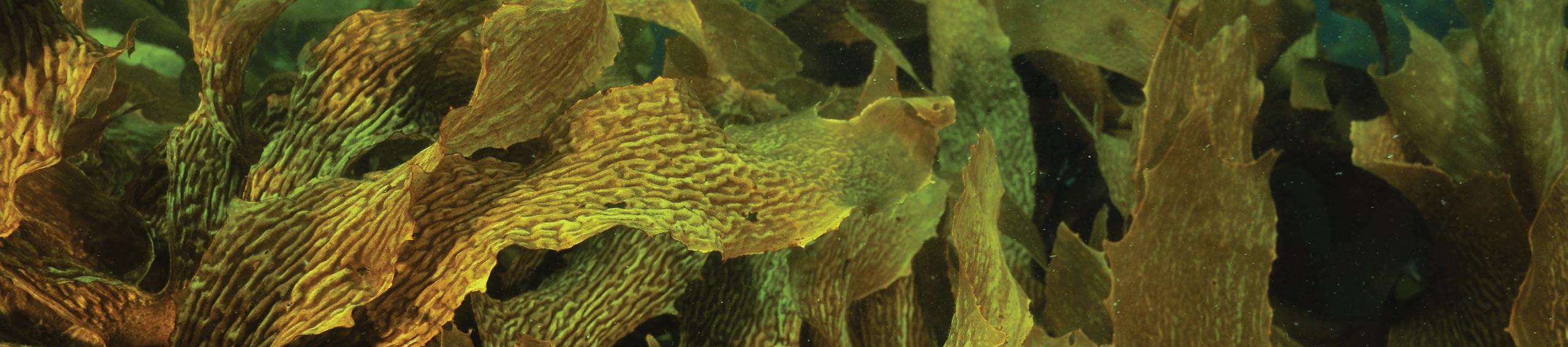
Sulfated Polysaccharides: Immune Boosters Extracted from Sea Life
Picture a frozen earth 635 million years ago, and envision the planet at last beginning to thaw from an extraordinary ice age that blanketed land and sea. As the permafrost begins to soften and melt, new life emerges from the ocean depths. It takes the form of sea plants, according to fossil finds discovered by scientists in 2011 in southern China.1
These sea plants were likely the earth’s first “complex” multicellular creatures—a grand harbinger of the flowering of plant and animal life to come. They are the ancestors of the familiar seaweeds of today, with their branches, splayed fans, and ribbony blossoms. And the solutions they have crafted to survive the salty, turbulent seas, at times swarming with microorganisms, offer surprising help for some of our own health challenges.2
It turns out that seaweeds contain large amounts of uniquely structured long-chain sugars, or polysaccharides, with many immune benefits.3 Polysaccharides are chains of single sugar molecules joined together, and include everything from the pectin in apples to glycogen, a form of sugar stored in your liver.4 Seaweed polysaccharides come in a variety of arrangements and molecules, varying by seaweed type, and each appears to have its own set of healing capacities.5
A significant body of seaweed research focuses on fucoidans, sulfated polysaccharides rich in a sugar called fucose. Fucoidans are commonly found in the cell walls of brown macroalgae isolated from Undaria pinnatifida, a large brown kelp native to Japan, and can also be found in other seaweeds
such as Fucus vesiculosus (also known as bladderwrack).6,7 Fucoidans are such an important component of seaweeds that they constitute up to 30% of their dry weight.8
Nearly 1500 peer-review studies have been published on fucoidans, and half of those in the last decade alone.9
Interest in fucoidans surged when scientists began to study the world’s longest-living people on the island of Okinawa, some of whom were over a century old.10,11 The Okinawan diet, which includes more than a dozen varieties of seaweed, is now considered one of the healthiest diets in the world.12
Dietary seaweed intake is correlated with lower all-cause mortality.13
Fucoidans have been called “a treasure trove of physiologically active polysaccharides.“
Fucoidans have been called “a treasure trove of physiologically active polysaccharides.”8 Their structures are surprisingly diverse, with variable sugar compositions that have different effects on our physiology.12 These include immune modulation,14 antibacterial and antiviral properties,15,16 antiinflammatory ability,17 modulation of gut flora,18 and antithrombotic activity.19,20
Fucoidans enhance immune function and stimulate both innate and adaptive immunity—in other words, they increase both our first line of defense, and our delayed, more specific memory-driven
immune response.6 T cells are impacted by fucoidan. T cells have numerous forms, but two significant ones are Th1 cells, which attack intracellular bugs such as bacteria and viruses, and Th2 cells, which fight helminths and other extracellular pathogens. Fucoidans modulate both Th1 and Th2 helper cells.21
Animal studies demonstrate that by stimulating Th1 cells, fucoidan can help eliminate infections such as Leishmania spp., which infects up to 12 million people in 98 different countries around the world.22 In turn, they also downregulate excessive Th2 responses, which can lead to allergies and inflammation. Oral consumption of fucoidans has been shown to relieve pulmonary inflammation in mice,23 while topical application relieves murine dermatitis as well.24
Fucoidans also increase natural killer cell activity, stimulate immune cells called macrophages, and help specialized immune training cells (dendritic cells) to mature.25,26 At doses of 300 mg a day for four weeks, a fucoidan extract stimulated the vaccine response to influenza in 70 volunteers over age 60.27 In this population, fucoidan was shown to increase antibody titers to three strains of the flu.
Fucoidans are antiviral, protecting against different types of viral infections.28 They directly inhibit herpes simplex viruses 1 and 2.29 Fucoidans also help protect against potentially deadly viruses, such as hepatitis C30 and a retrovirus known as HTLV-1.31 Fucoidans inhibit the adhesion of hantavirus to cells in vitro.32
10 FOCUS | Summer/Fall 2017
A Novel Galactofucan Sulfate Extract Enhances Immunity, Inactivates Viruses Naturally, and Reduces Inflammation
Some fucoidans mobilize stem cells and increase their number in the blood.33 Fucoidans might even exert anti-aging activity by increasing the expression of the sirtuins.34 Sirtuins are being studied for their anti-aging potential; it is thought that a boost in sirtuin activity may in part explain how calorie restriction extends life span.35 Finally, fucoidans are potent antiinflammatory molecules.36 Fucoidans may also protect the brain and nervous system from chronic inflammation, particularly via specialized cells called microglia. Thus, fucoidan may, some researchers believe, “offer substantial therapeutic potential for treatment of neurodegenerative diseases.”37
A Novel Aqueous Fucoidan Extract Balances the Immune System
“The role of marine compounds as therapeutic agents has immense potential, and it’s great to be at the cutting edge of this research,” says Stephen Myers, PhD, ND, BMed, of Southern Cross University in New South Wales, Australia. “Fucoidans have immense potential as immunemodulators and anti-inflammatory agents. The research on their benefits is only in its infancy, and we are going to see a wide range of uses for these compounds in future therapeutic applications.”38
New research is emerging on the immune and antiviral benefits of fucoidan isolated from either Undaria
pinnatifida seaweed (also known as wakame) or Fucus vesiculosus (also known as bladderwrack). The fucoidan extract is prepared using a novel aqueous extraction process that produces largely unaltered fucoidan fractions with consistent, measurable bioactivity. They yield specific and reproducible benefits in cellular, animal, and clinical studies.
A unique blend of fucoidan and algal polyphenols, along with the nutrients manganese, zinc, and vitamin B6, significantly enhanced immune function in a four-week pilot study of ten healthy adults.39 The study participants were randomly divided into two groups: five received 100 mg daily of the unique blend (containing 75 mg total fucoidan), and five received 1000 mg daily of the unique blend delivered as four 250 mg capsules (containing a total of 750 mg fucoidan). This lower and upper range was used to determine both safety and efficacy. The extract was well tolerated with no clinically relevant changes to measurements of liver or kidney function or to the formation of blood cells.
Changes in immune cells such as T lymphocytes, helper T cells, and cells of the innate immune system were measured at Day 1, Day 3, and Day 28. Numerous measures of immune function significantly improved, including the total number of T cells. There was a continual increase over the 28 days in the activity of innate immune cells called granulocytes and monocytes. These cells attack pathogens as soon as they are encountered.
The adaptive immune system responded as well—by Day 3, there were significant increases in killer T cells known as cytotoxic T cells, which recognize infected cells by the presence of antigens on their surface, and then kill them. The researchers conclude that “overall, these data suggest an immune-priming role of the nutrient seaweed complex.” The fucoidan extract was anti-inflammatory
as well. Cells in blood samples were “challenged“ with phytohemagglutinin (PHA)-M, a protein that stimulates T cells. After challenge, T cells produced less of an inflammatory molecule called interleukin (IL)-6 on Day 28 as compared to Day 1. Inappropriate levels of IL-6 have been linked to increased cardiovascular risk in the Women’s Health Study—a six-year
study with 619 participants.40 As the researchers note, “any agent capable of reducing IL-6 may confer substantive health benefits.” Finally, it may have the potential for lowering cholesterol, as there was a near-significant beneficial change in blood lipid levels by the end of the four-week study.41 It is notable that this small-scale study did not show a dose response effect in the efficacy outcomes between the two doses of the study preparation. Both doses were effective and well tolerated. However, the researchers point out that a larger study needs to be undertaken to determine dose responsiveness.
“This research set out to determine the potential activity of fucoidan and polyphenol rich seaweed extract on immune function,” explains the study’s lead researcher Stephen Myers. “Previous research had demonstrated that fucoidans have real potential as immune modifying agents, and our results have added to these findings.
For more information, call 800.545.9960 or visit www.allergyresearchgroup.com
11
“Fucoidans have immense potential as immune-modulators and anti-inflammatory agents. The research on their benefits is only in its infancy.“
— Stephen Myers, PhD, ND, BMed
A unique blend of fucoidan and algal polyphenols … significantly enhanced immune function in a four-week study of ten healthy adults.
You can never state with absolute certainty that we’d get the same result with a pure preparation of Fucus vesiculosus. However, I have little doubt the combination of fucoidan and polyphenols in the preparation tested were the principal active constituents.”38
At the higher dosage, the same combination reduced symptoms of osteoarthritis by over 50% in a 2010 pilot study of 12 individuals.39 The severity of the patients’ average comprehensive arthritis test (COAT) score was measured weekly. This test is comprised of four sub-scales: pain, stiffness, difficulty with physical activity, and overall symptom severity. The COAT score was reduced by 18% with the 100 mg treatment (75 mg fucoidan) and 52% with the 1000 mg dose (750 mg fucoidan) by the end of the study. This preparation also caused a significant increase in total antioxidant capacity, demonstrating potential for antioxidant activity.
The algal polyphenols themselves, separate from the fucoidan, are health-promoting, anti-inflammatory, and antioxidant.42 Phlorotannins, polyphenols present in the extract studied, are metabolized and absorbed in the colon and thus are bioavailable.43
Fucoidan may mobilize hematopoietic stem and progenitor cells (HSPC), which are critical for the maintenance and replenishment of blood cells in the bone marrow.44 In mice and nonhuman primates, intravenous (IV) fucoidan has a significant mobilizing effect on HPC.45 In humans, oral intake of fucoidan also mobilizes HPC.33
ulcer disease.46 H. pylori colonizes the stomach of half the world’s humans, and can cause chronic gastritis, peptic ulcer, gastric cancer, and gastric MALT lymphoma.47 Unfortunately, the efficacy of standard triple antibiotic combination therapy has declined in recent years to only 70%.48,49
Fucoidan may make a clinical difference in that antibiotic efficacy. Three fucoidan aqueous extracts were tested (from Fucus vesiculosus and Undaria pinnatifida), for their ability to inhibit H. pylori from adhering to the stomach lining. When human gastric epithelial cells were tested in vitro, it was found that fucoidans attach to H. pylori, potentially dislodging the bacteria from the gastric cell, or simply preventing it from adhering. In fact, analysis has shown that H. pylori carries several proteins on its outer membrane that bind to fucoidan.50
Fucoidan exhibited “excellent in vitro antiviral activity,” the researchers concluded, and was “significantly more active against clinical strains of HSV-2 than HSV-1.”29 This research has been confirmed in animal studies as well; in a 2008 study, oral administration of fucoidan protected mice from infection with HSV-1, as judged from the survival rate and lesion scores.29
Fucoidans offer a remarkable array of bioactivity, ranging for immune modulation to inhibiting infection by bacteria and viruses. This “food” from the sea is truly the ocean’s great medicinal gift.
References
1 Yuan X, Chen Z, Xiao S, et al. An early Ediacaran assemblage of macroscopic and morphologically differentiated eukaryotes. Nature. 2011;470(7334):390-3. PMID: 21331041
2 Percival E. The polysaccharides of green, red and brown seaweeds: their basic structure, biosynthesis and function. Br phycol J. 1979;103-17.
3 Da VD, Viswanathan P. Seaweed polysaccharides - New therapeutic insights against the inflammatory response in diabetic nephropathy Antiinflamm antiallergy agents med chem. 2017;15(3):178-90. PMID: 28215166
4 McGraw-Hill Concise Encyclopedia of Bioscience. Polysaccharide [Internet]. New York: The McGraw-Hill Companies; 2002 [cited 2017 Jul 28]. Available from: http://encyclopedia2.thefreedictionary.com/polysaccharide
5 Ruocco N, Costantini S, Guariniello S, et al. Polysaccharides from the marine environment with pharmacological, cosmeceutical and nutraceutical potential. Molecules. 2016;21(5). PMID: 27128892
6 Fitton J. Therapies from fucoidan; multifunctional marine polymers. Marine Drugs. 2011;9:1731-60. PMID: 22072995
7 Berteau O, Mulloy B. Sulfated fucans, fresh perspectives: structures, functions, and biological properties of sulfated fucans and an overview of enzymes active toward this class of polysaccharide. Glycobiology. 2003;13(6):29-40. PMID: 12626402
8 Kusaykin M, Bakunina I, Sova V, et al. Structure, biological activity, and enzymatic transformation of fucoidans from the brown seaweeds. Biotechnol J. 2008;3(7):904-15. PMID: 18543244
9 Fitton JH, Stringer DN, Karpiniec SS. Therapies from fucoidan: an update. Mar Drugs. 2015;13(9):5920-46. PMID: 4584361
10 Sho H. History and characteristics of Okinawan longevity food. Asia Pac J Clin Nutr. 2001;10(2):159-64. PMID: 11710358
Fucoidan Inhibits
the
Activity of Both Bacteria and Viruses
In 2005, the Nobel Prize in Physiology or Medicine was awarded to Barry Marshall and Robin Warren, Marshall’s long-time collaborator, for their discovery of the bacterium Helicobacter pylori and its role in gastritis and peptic
Just as fucoidan inhibits bacteria from attaching to cells, it can bind to receptors that some viruses use to enter cells, including herpes viruses such as those that cause oral and genital herpes. An extract of 75% pure fucoidan was tested in vitro against 32 clinical strains of herpes simplex virus (HSV), composed of 14 strains of HSV-1 and 18 strains of HSV-2. HSV-1 tends to cause oral herpes lesions, while HSV-2 causes genital herpes lesions.51 Many strains of herpes are now resistant to acyclovir, the most common antiviral drug used to combat infection.
11 Willcox BJ, Willcox DC, Todoriki H, et al. Caloric restriction, the traditional Okinawan diet, and healthy aging: the diet of the world’s longest-lived people and its potential impact on morbidity and life span. Ann N Y Acad Sci. 2007;1114:434-55. PMID: 17986602
12 Willcox D, Willcox BJ, Todoriki H, et al. The Okinawan diet: health implications of a low-calorie, nutrient-dense, antioxidantrich dietary pattern low in glycemic load. J Am Coll Nutr. 2009;28(suppl):500S-516S. PMID: 20234038
13 Iso H, Kubota Y. Nutrition and disease in the Japan Collaborative Cohort Study for Evaluation of Cancer (JACC). Asian Pac J Cancer Prev. 2007;8(suppl):35-80. PMID: 18260705
14 Zapopozhets TS, Besednova NN, Loenko IN. Antibacterial and immunomodulating activity of fucoidan. Antibiot Khimioter. 1995;40(2):9-13. PMID: 7605146
12 FOCUS | Summer/Fall 2017
Fucoidans attach to H. pylori bacteria, potentially dislodging the bacteria from the gastric cell.
15 Hirmo S, Utt M, Ringner M,Wadstrom T. Inhibition of heparan sulfate and other glycosaminoglycans binding to Helicobacter pylori by various polysulfated carbohydrates. FEMS Immunol Med Microbiol. 1995;10(3-4):301-6. PMID: 7539671
16 Adhikari U, Mateu CG, Chattopadhyay K, et al. Structure and antiviral activity of sulfated fucans from Stoechospermum marginatum. Phytochemistry. 2006;67(22):2474-82. PMID: 17067880
17 Cumashi A, Ushakova NA, Preobrazhenskaya ME, et al. A comparative study of the anti-inflammatory, anticoagulant, antiangiogenic, and anti-adhesive activities of nine different fucoidans from brown seaweeds. Glycobiology. 2007;17:541–52. PMID: 17296677
18 Shang Q, Shan X, Cai C, et al. Dietary fucoidan modulates the gut microbiota in mice by increasing the abundance of Lactobacillus and Ruminococcaceae. Food Funct. 2016;7(7):322432. PMID: 27334000
19 Millet J, Jouault SC, Mauray S, et al. Antithrombotic and anticoagulant activities of a low molecular weight fucoidan by the subcutaneous route. Thromb Haemost. 1999;81:391-5. PMID: 10102467
20 Atashrazm F, Lowenthal RM, Woods GM. Fucoidan and cancer: a multifunctional molecule with anti-tumor potential. Mar Drugs. 2015;13(4):2327-46. PMID: 25874926
21 Kar S, Sharma G, Das PK. Fucoidan cures infection with both antimony-susceptible and -resistant strains of leishmania donovani through Th1 response and macrophage-derived oxidants. J Antimicrob Chemother. 2011;66(3):618-25. PMID: 21393231
22 Barrett MP, Croft SL. Management of trypanosomiasis and leishmaniasis. Br Med Bull. 2012;104:175–96. PMID 23137768
23 Maruyama H, Tamauchi H, Hashimoto M, et al. Suppression of Th2 immune responses by Mekabu fucoidan from Undaria pinnatifida sporophylls. Int Arch Allergy Imm. 2005;137:4, 289-94. PMID: 15970637
24 Yang JH. Topical application of fucoidan improves atopic dermatitis symptoms in NC/Nga mice. Phytother Res. 2012;26(12):1898-903. PMID: 22431003
25 Shimizu J, Wada-Funada U, Mano H, et al. Proportion of murine cytotoxic T cells is increased by high molecular-weight fucoidan extracted from Okinawa mozuku (Cladosiphon okamuranus). J Health Sci. 2005;51(3):394-7. doi: 10.1248/ jhs.51.394
26 Hu Y, Cheng SC, Chan KT, et al. Fucoidan enhances dendritic cell-mediated T-cell cytotoxicity against NY-ESO-1 expressing human cancer cells. Biochem Biophys Res Commun. 2010;392(3):329-34. PMID: 20067767
27 Negishi H, Mori M, Mori H, et al. Supplementation of elderly Japanese men and women with fucoidan from seaweed increases immune responses to seasonal influenza vaccination. J Nutr. 2010;143(11):1794-8. PMID: 24005608
28 Hayashi K, Nakano T, Hashimoto M, et al. Defensive effects of a fucoidan from brown alga Undaria pinnatifida against herpes simplex virus infection. Int Immunopharmacol. 2008;8(1):109-16. PMID: 18068106
29 Thompson KD, Dragar C. Antiviral activity of Undaria pinnatifida against herpes simplex virus. Phytother Res. 2004;18(7):551-5. PMID: 15305315
30 Mori N, Nakasone K, Tomimori K, et al. Beneficial effects of fucoidan in patients with chronic hepatitis C virus infection. World J Gastroenterol. 2012;18(18):2225-30. PMID: 22611316
31 Araya N, Takahashi K, Sato T, et al. Fucoidan therapy decreases the proviral load in patients with human T-lymphotropic virus type-1-associated neurological disease. Antivir Ther. 2011;16(1):89-98. PMID: 21311112
32 Makarenkova ID, Kompanets GG, Zaporozhets TS. Inhibition of adhesion of the pathogenic microorganism to the eucaryotic cells. J Microbiol. 2006:121-5.
33 Irhimeh MR, Fitton JH, Lowenthal RM, et al. Fucoidan ingestion increases the expression of CXCR4 on human CD34+ cells. Exp Hematol. 2007;35(6):989-94. PMID: 17533053
34 Wang T, Zhu M, He Z. Low-molecular-weight fucoidan attenuates mitochondrial dysfunction and improves neurological outcome after traumatic brain injury in aged mice: involvement of Sirt3. Cell Mol Neurobiol. 2016;36(8):1257-68. PMID: 26743530
35 Guarente L. Sirtuins in aging and disease. Cold Spring Harb Symp Quant Biol. 2007;72:483-8. PMID: 18419308
36 Semenov AV, Mazurov AV, Preobrazhenskaia ME, et al. Sulfated polysaccharides as inhibitors of receptor activity of P-selectin and P-selectin-dependent inflammation. Quest Med Chem. 1998;44(2):135-43. PMID: 9634715
37 Park HY, Han MH, Park C, et al. Anti-inflammatory effects of fucoidan through inhibition of NF-κB, MAPK and Akt activation in lipopolysaccharide induced BV2 microglia cells. Food Chem Toxicol. 2011;49(8):1745-52. PMID: 21570441
38 Personal communication, 2017 Jul 28.
39 Myers SP, O’Connor J, Fitton JH, et al. A combined Phase I and II open-label study on the immunomodulatory effects of seaweed extract nutrient complex. Biologics. 2011;5:45-60. PMID: 21383915
40 Walston J, Xue Q, Semba RD, et al. Serum antioxidants, inflammation, and total mortality in older women. Am J Epidemiol. 2006;163(1):18-26. PMID: 16306311
41 Kumar SA, Magnusson M, Ward LC, et al. Seaweed supplements normalise metabolic, cardiovascular and liver responses in high-carbohydrate, high-fat fed rats. Mar Drugs. 2015;13(2):788-805. PMID: 25648511
42 Díaz-Rubio ME, Pérez-Jiménez J, Saura-Calixto F. Dietary fiber and antioxidant capacity in Fucus vesiculosus products. Int J Food Sci Nutr. 2009;60(suppl)2:23-34. PMID: 18951280
43 Corona G, Ji Y, Anegboonlap P, et al. Gastrointestinal modifications and bioavailability of brown seaweed phlorotannins and effects on inflammatory markers. Br J Nutr. 2016;115(7):124053. PMID: 26879487
44 Granick JL, Simon SI, Borjesson DL. Hematopoietic stem and progenitor cells as effectors in innate immunity. Bone Marrow Res. 2012;2012:165107. PMID: 22762001
45 Sweeney EA, Lortat-Jacob H, Priestley GV, et al. Sulfated polysaccharides increase plasma levels of SDF-1 in monkeys and mice: involvement in mobilization of stem/progenitor cells. Blood. 2002;99:44-51. PMID: 11756151
46 Weintraub P. The Dr. who drank infectious broth, gave himself an ulcer, and solved a medical mystery [Internet]. Waukesha (WI): Kalmbach Publishing Co., Discover Magazine; 2010 [cited 2017 Jul 28]. Available from: http://discovermagazine.com/2010/mar/07-drdrank-broth-gave-ulcer-solved-medical-mystery
47 Marshall BJ, Windsor HM. The relation of Helicobacter pylori to gastric adenocarcinoma and lymphoma: pathophysiology,epidemiology, screening, clinical presentation, treatment, and prevention. Med Clin North Am. 2005;89(2):313-44. PMID: 15656929
48 Mégraud F. H pylori antibiotic resistance: prevalence, importance, and advances in testing. Gut. 2004;53(9):1374-84. PMID: 15306603
49 Horiki N, Omata F, Uemura M, et al. Annual change of primary resistance to clarithromycin among helicobacter pylori isolates from 1996 through 2008 in Japan. Helicobacter. 2009;14(5):86-90. PMID: 19751432
50 Chua EG, Verbrugghe P, Perkins TT, et al. Fucoidans disrupt adherence of helicobacter pylori to AGS cells in vitro. Evid Based Complement Alternat Med. 2015;2015:120981. PMID: 26604968
51 WebMD. Herpes simplex: Herpes type 1 and 2 [Internet]. Atlanta (GA): WebMD LLC; [cited 2017 Jul 28]. Available from: http://www.webmd.com/genital-herpes/pain-managementherpes
Continued from p. 7, "New Studies Confirm the Power of Three Chinese Herbs for Asthma"
14 Wen MC, Wei CH, Hu ZQ, et al. Efficacy and tolerability of anti-asthma herbal medicine intervention in adult patients with moderate-severe allergic asthma. J Allergy Clin Immunol. 2005;116(3):517-24. PMID: 16159618
15 Li XM, Huang CK, Zhang TF, et al. The Chinese herbal medicine formula MSSM-002 suppresses allergic airway hyperreactivity and modulates TH1=TH2 responses in a murine model of allergic asthma. J Allergy Clin Immunol. 2000;106:660-8. PMID: 11031336
16 To learn more about Sophora flavescens, see both Bronchial Epilepsy (pp. 5–6) and The Neuroelectric Origins of Asthma (pp. 2–3) in this issue.
17 Note: Although the protocol specified enrollment of 18 individuals in the study (six per dose level), because of an error in randomizing, two additional individuals were recruited in order to complete the experiment while maintaining blinding.
18 Kelly-Pieper K, Patil SP, Busse PJ. Safety and tolerability of an antiasthma herbal Formula (ASHMI) in adult subjects with
asthma: a randomized, double-blinded, placebo-controlled, dose-escalation phase I study. Altern Complement Med. 2009;15(7):735-43. PMID: 19586409
19 Zhang T, Srivastava K, Wen MC, et al. Pharmacology and immunological actions of a herbal medicine ASHMI on allergic asthma. Phytother Res. 2010;24:1047-55. PMID: 19998324
20 Busse PJ, Schofield B, Birmingham N, et al. The traditional Chinese herbal formula ASHMI inhibits allergic lung inflammation in antigen-sensitized and antigen-challenged aged mice. Ann Allergy Asthma Immunol. 2010;104:236-46. PMID: 20377113
21 Srivastava K, Sampson HA, Emala CW, et al. The anti-asthma herbal medicine ASHMI acutely inhibits airway smooth muscle contraction via prostaglandin E2 activation of EP2/EP4 receptors.
J Physiol Lung Cell Mol Physiol. 2013;305(12):L1002-10. PMID: 24163140
22 Srivastava K, Sampson HA, Li X. The anti-asthma Chinese herbal formula ASHMI provides more persistent benefits than
dexamethasone in a murine asthma model. J All Clin Immun. 2011;2(127),p AB261
23 Matsui S, Matsumoto H, Sonoda Y, et al. Glycyrrhizin and related compounds down-regulate production of inflammatory chemokines IL-8 and eotaxin 1 in a human lung fibroblast cell line. Int Immunopharmacol. 2004;4:1633-44. PMID: 15454116
24 Li XM. Treatment of asthma and food allergy with herbal interventions from traditional Chinese medicine. Mount Sinai J Med. 2011;78:697-716. PMID: 21913200
25 Jayaprakasam B, Yang N, Wen MC, et al. Constituents of the anti-asthma herbal formula ASHMI(TM) synergistically inhibit IL-4 and IL-5 secretion by murine Th2 memory cells, and eotaxin by human lung fibroblasts in vitro. J Integr Med. 2013;11(3):195-205. PMID: 23743163
26 Wang YH, Hogan SP. Chinese herbal anti-asthma tea to go! Clin Exp Allergy. 2010;40(11):1590-2. PMID: 21039969
For more information, call 800.545.9960 or visit www.allergyresearchgroup.com
13
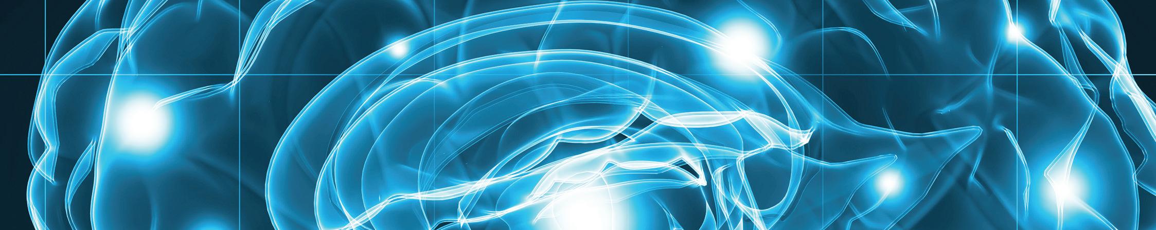
Protect the Brain from Stress with a Unique Lipid Blend
Fatty Acid Blend Soothes the Stressed Brain
Your brain is at least 60% pure fatty acids. These crucial molecules are necessary for your brain to perform its many functions.1 They include phospholipids, which compose the cell membrane of every neuron. Among those phospholipids, phosphatidylserine plays a special role. The sheath around your nerve cells, composed of myelin, is rich in phosphatidylserine.2
Phosphatidylserine supports memory in all its forms—both short- and longterm memory, as well as the ability to retrieve memories. Phosphatidylserine is critical to attention, concentration, problem solving, and communication. It especially supports rapid reactions and reflexes.3 It also helps protect the body and brain against stress.4 Oral phosphatidylserine is highly bioavailable and readily crosses the blood-brain barrier.5
A hallmark of the aging brain is the decreased ability to adapt to stress— including heat, cold, and chronic stress.6 Not only does age reduce stress resilience, chronic stress can accelerate the aging process, according to renowned neuroendocrinologist Robert Sapolsky, PhD, of Stanford University.7 The adrenal cortex secretes glucocorticoids in response to a variety of stressors.8 Glucocorticoids signal the brain, which then initiates a cascade of signals that may increase blood pressure and carbohydrate metabolism, as well as hormones, to improve energy and alertness.
Sapolsky has found that the adrenocortical axis, which is central to the stress response, can be impaired with aging. After experiencing stress,
the aged brain may continue to stimulate the secretion of stress hormones and cortisol. That continual glucocorticoid exposure can actually further impair the brain. Sapolsky calls this a “feed-forward cascade,” with potentially serious consequences. Excess secretion of cortisol is linked to depression, anxiety disorders, and metabolic syndrome (a precursor to diabetes and heart disease). Impaired cortisol secretion is linked to numerous chronic conditions, including fatigue, pain, irritability, fibromyalgia, arthritis, and chronic fatigue syndrome.9
One hallmark of aging in the human brain is decreased phosphatidylserine in the membranes of neurons.10,11,12 Given these links
A Blend of Phosphatidylserine and Phosphatic Acid Buffers Against Stress
Given the numerous studies on phosphatidylserine and stress, researchers have looked at the possible synergistic effect of a blend of phosphatidylserine with phosphatidic acid (PA), a precursor for other regulatory lipids.14 Phosphatidic acid also acts as signaling lipid, increasing in response to stress.15 Together, phosphatidic acid and phosphatidylserine can reduce cortisol levels and enhance stress resilience.16
A 2004 randomized, double-blind, placebo-controlled study found that a mixture of phosphatidylserine and phosphatic acid, a lipid messenger molecule, provides protection against stress.16 80 individuals (40 men and 40 women, aged 20–45) received the lipid blend (PAS) at either 400, 600, or 800 mg daily for three weeks. Each 100 mg PAS capsule contained 100 mg of phosphatidylserine and 125 mg of phosphatic acid, plus 270 mg of other phospholipids (phosphatidylcholine, phosphatidylinositol, phosphatidylethanolamine, and others).
between stress, aging, cortisol, and phosphatidylserine, researchers began to investigate the possibility that phosphatidylserine could act as a buffer against stress. Indeed, they soon found that it could decrease cortisol responses to acute physical and mental stress.13
All volunteers were given a test to trigger a moderate amount of anxiety. Called the Trier Social Stress Test, it entails a mock job interview, a five-minute speech, and a fiveminute math challenge, all performed in front of two observers. Though both groups experienced stress (increased heart rate and elevated scores on a questionnaire regarding anxiety and mood), those receiving 400 mg/day of PAS had significantly lower levels of
14 FOCUS | Summer/Fall 2017
One hallmark of aging in the human brain is decreased phosphatidylserine in the membranes of neurons.
stress hormones such as cortisol and adrenocorticotropic hormone (ACTH) in their saliva. This was the only effective dose—600 and 800 mg did not significantly impact stress hormones.
A second study in 2014 confirmed the efficacy of 400 mg of PAS on stress.17 75 healthy male volunteers were enrolled in this double-blind, placebocontrolled study and randomly assigned to placebo, 200 mg of PAS, or 400 mg of PAS for 42 days. Before the experiment began, they were given a test to measure chronic stress levels. Stress was induced via the Trier Social Stress Test. In those individuals who had tested high on chronic stress, a daily dose of 400 mg of PAS was effective in normalizing salivary and serum cortisol responses as well as adrenocorticotropic hormone (ACTH), which is secreted by the anterior pituitary gland in the brain and regulates levels of cortisol. PAS 200 and placebo were not effective. PAS supplementation had no significant effect on heart rate, pulse transit time, or psychological stress response.
It is notable that 400 mg of PAS was effective in both studies, whereas 200, 600, and 800 mg were not. This is, according to lead author Juliane Hellhammer, PhD, an “inverse U-shape reaction, a quite common finding. More is not always better in terms of efficacy.”18
In related research, PAS has been found to improve memory and mood in elderly individuals. One three-month, double-blind, placebocontrolled trial in functioning, nondepressive elderly people with memory problems, found that a blend of 100 mg phosphatidylserine and 80 mg phosphatidic acid three times daily improved mood and cognition.19
References
1 Chang CY, Ke DS, Chen JY. Essential fatty acids and human brain. Acta Neurol Taiwan. 2009;18(4):231-41. PMID: 20329590
2 Svennerholm L. Distribution and fatty acid composition of phosphoglycerides in normal human brain. J Lipid Res. 1968;9(5):570-9. PMID: 4302302
3 Glade MJ, Smith K. Phosphatidylserine and the human brain. Nutrition. 2015;31(6):781-6. PMID: 25933483
4 Benton D, Donohoe RT, Sillance B, et al. The influence of phosphatidylserine supplementation on mood and heart rate when faced with an acute stressor. Nutr Neurosci. 2001;4(3):169-78. PMID: 11842886
5 Rosadini G, Sannita WG, Nobili F, et al. Phosphatidylserine: Quantitative EEG effects in healthy volunteers. Neuropsychobiology. 1990-1991;24(1):42-8. PMID: 2132640
6 Sapolsky RM, Krey LC, McEwen BS. The neuroendocrinology of stress and aging: the glucocorticoid cascadehypothesis. Endocr Rev. 1986;7(3):284-301. PMID: 3527687
7 Sapolsky, RM. Stress, the Aging Brain, and the Mechanisms of Neuron Death. Cambridge, MA: Bradford Books-The MIT Press; 1992.
8 Munck A, Guyre P, Holbrook N. Physiological functions of glucocorticoids during stress and their relation to pharmacological actions. Endocr Rev. 1984 Winter;5(1):25-44. PMID: 6368214
A mixture of phosphatidylserine and phosphatic acid, a lipid messenger molecule, provides protection against stress. … Individuals receiving 400 mg/day of PAS (lipid blend of phosphatidylserine and phosphatic acid) had significantly lower levels of stress hormones in their saliva.
A second unpublished study, a twomonth randomized, double-blind, placebo-controlled trial, found that the same dose of PAS three times daily significantly improved memory and prevented “winter blues” in patients with Alzheimer’s disease (AD). This is not surprising, given the correlations between the aging brain and resilience to stress. As the researchers note, “The data encourage long-term studies in AD patients and other elderly with memory or cognition problems.”
PAS may have broader effects on other conditions impacted by stress, such as premenstrual syndrome. PMS patients have been shown to have lower cortisol concentrations premenstrually as compared to those without PMS.20 According to Dr. Hellhammer: “We just submitted a publication with very convincing data titled: A lecithin phosphatidylserine and phosphatidic acid complex (PAS) reduces symptoms of the premenstrual syndrome (PMS): results of a randomized, placebocontrolled, double-blind clinical trial.”18
Today, when novel concerns and ways of interacting are ratcheting up our stress levels, it’s good to know there are nutrients that can help shore up our resilience, both physical and mental.21
9 Nicolaides NC, Kyratzi E, Lamprokostopoulou A, et al. Stress, the stress system and the role of glucocorticoids. Neuroimmunomodulation. 2015;22(1-2):6-19. PMID: 25227402
10 Crook TH, Tinklenberg J, Yesavage J, et al. Effects of phosphatidylserine in age-associated memory impairment. Neurology. 1991;41(5):644-9. PMID: 2027477
11 Ball MJ. Neuronal loss, neurofibrillary tangles and granulovacuolar degeneration in the hippocampus with ageing and dementia. A quantitative study. Acta Neuropathol. 1977;37(2):111-8. PMID: 848276
12 Lippa AS, Critchett DJ, Ehlert F, et al. Age related alterations in neurotransmitter receptors: An electrophysiological and biochemical analysis. Neurobiol Aging. 1981;2:3-8. PMID: 6115325
13 Monteleone P, Beinat L, Tanzillo C, et al. Effects of phosphatidylserine on the neuroendocrine response to physical stress in humans. Neuroendocrinology. 1990;52:243-48. PMID: 2170852
14 Athenstaedt K, Daum G. Phosphatidic acid, a key intermediate in lipid metabolism. Eur J Biochem. 1999;266:1-16. PMID: 10542045
15 Sergeant S, Waite KA, Heravi J, et al. Phosphatidic acid regulates tyrosine phosphorylating activity in human neutrophils: enhancement of Fgr activity. J Biol Chem. 2000;276:4737-46. PMID: 11078731
16 Hellhammer J, Fries E, Buss C, et al. Effects of soy lecithin phosphatidic acid and phosphatidylserine complex (PAS) on the endocrine and psychological responses to mental stress. Stress. 2004;7(2):119-26. PMID: 15512856
17 Hellhammer J, Vogt D, Franz N, et al. A soy-based phosphatidylserine/ phosphatidic acid complex (PAS) normalizes the stress reactivity of hypothalamus-pituitary-adrenal-axis in chronically stressed male subjects: a randomized, placebocontrolled study. Lipids Health Dis. 2014;13:121. PMID: 25081826
18 Personal communication, 2017 Jul 21.
19 Moré MI, Freitas U, Rutenberg D. Positive effects of soy lecithin-derived phosphatidylserine plus phosphatidic acid on memory, cognition, daily functioning, and mood in elderly patients with Alzheimer’s disease and dementia. Adv Ther. 2014;31(12):1247-62. PMID: 25414047
20 Girdler SS, Pedersen CA, Straneva PA, et al. Dysregulation of cardiovascular and neuroendocrine responses to stress in premenstrual dysphoric disorder. Psychiatry Res. 1998;81(2):163-78. PMID: 9858034
21 American Psychological Association. Stress in America [Internet]. Washington, DC: American Psychological Association; 2017 [cited 2017 Aug 7]. Available from: http://www.apa.org/news/press/releases/stress/index.aspx
For more information, call 800.545.9960 or visit www.allergyresearchgroup.com
15
2300 North Loop Road Alameda, CA 94502
FOCUS on Allergy Research Group®
Editor-in-Chief: Todd A. Born, ND, CNS Editor at Large: Stephen A. Levine, PhD
Executive Editor: Jill Neimark
Medical Editor: Michael Ash, DO, ND, BSc, RNT
Assistant Editor: Carrie Decker, ND, MSME, MSBME
Graphic Designer: Matthew Ivan
FOCUS publishes emerging nutritional science and scientific theories that should not be construed to be conclusive scientific proof of any specific cause, effect, or relationship. The publication is for the educational use of healthcare practitioners and physicians. The articles in the publication are the independent scientific views and theories of the authors. FOCUS takes no position on the views and theories expressed but offers them for candid inquiry and debate. The articles are not intended for use in support of the sale of any commercial product and should not be construed as indicative of the use or efficacy of any commercial product. Emerging science and scientific theories do not constitute scientific proof of any specific cause, effect, or relationship. Copyright® 2017 Allergy Research Group®. Special permission is required to reproduce by any manner, in whole or in part, the materials contained herein.
16 FOCUS | Summer/Fall 2017
