1 oint
aestheticsjournal.com
@aestheticsgroup
Aesthetics Journal
Anatomical Basis of Festoons and Malar Mounds Mrs Sabrina Shah-Desai explains how to differentiate between festoons and malar mounds under the eye and discusses the most appropriate course of treatment Puffy, swollen eyelids are one of the common manifestations of under-eye ageing. Most patients with ‘eye bags’ present with skin laxity, a herniation of intraorbital eyelid fat, periorbital weakening of the soft tissues, bony resorption and a tear trough deformity.1 Although ‘baggy eyelids’ can be hereditary and are mainly a cosmetic concern, swollen eyelids can often be caused by seasonal allergies, blepharitis2 and, rarely, can be a cause of serious underlying conditions such as thyroid eye disease (over or under-active) or fluid retention due to compromised renal and liver function.1,3 Malar mounds and festoons Some people develop ‘saddle bags’; a mound with laxity of the skin and orbicularis muscle around the lower eyelid that starts accumulating fluid at the lid cheek junction. These ‘saddle bags’ can be split into two categories: a malar mound, which is a soft tissue elevation protruding from the lateral part of the malar eminence near the lower eyelid cheek junction (Figure 1), and a festoon, which is hanging tissues between two points (Figure 2). These two distinct entities are likely to fall within a complex continuum of anatomic findings, as when the skin loses elasticity the malar mound can become a festoon.1 Children can demonstrate the precursors of malar soft tissue convexities, which become defined with age, as creases delineate the upper and lower borders. Malar mounds can exhibit a familial inheritance pattern and occur at any age.4 Based on anatomic location, Furnas4 further distinguished between palpebral festoons (eye bags), which occur above the boundary of the inferior orbital rim, and malar festoons and mounds (saddle bags), which in terms of anatomic location, are found inferior to the bone. He divided these into pretarsal, preseptal, orbital, orbitomalar, and malar, although combinations of multiple sites may be concurrently present. He found that anatomically, festoons can affect any part of the upper or lower eyelid, but the hallmark is the sagging of the orbital segment of the orbicularis oculi muscle of the lower eyelid.1 Upper eyelid festoons can cause a droopy eyelid (ptosis) by a mechanical effect and can also sag beyond the eyelashes, thus obstructing the superior visual fields.5 The anatomic basis of these structures were poorly described until Pessa6 identified a specific connective tissue band in the orbitomalar
Aesthetics
region, which originates from the periosteum of the superior aspect of the infraorbital rim and inserts into cheek skin 2.5-3cm inferior to the lateral canthus (Figure 3). This fascial band of impermeable tissue delineates the inferomedial aspect of the malar mound and divides the suborbicularis oculi fat (SOOF) into a superior and inferior compartment. The lymphatics of the inferior compartment are continuous with the cheek drainage, whilst the superior compartment is compromised, and can be prone to collecting fluid in predisposed individuals.6 The SOOF has variable thickness, being most prominent in the central and lateral malar region. Medially, the SOOF engulfs the mimetic muscles and lies superficial to the periosteum.6 Pessa theorises that loss and/or ptosis of the SOOF and its adjacent deep fat compartments, in combination with the attenuation and relaxation of connective tissue, contributes to ‘malar bags’ associated with ageing.6 Aetiological factors 1. Genetically predisposed individuals with a familial history of festoons and malar mounds have compromised permeability characteristics of the malar septum. This predisposition to malar mounds may worsen by chronic lower eyelid oedema, which may progressively lead to the development of malar mounds and, over time, develop laxity of the attachments between the orbicularis and the deep fascia and cause festoons.1 2. Age is associated with a reducing orbicularis oculi muscle thickness and increasing orbital fat prolapse. Progressive sagging of the orbicularis oculi muscle can occur as it attenuates, until the orbital portion of the orbicularis oculi folds are suspended across the lid, akin to a curtain swag.4 Commonly, protruding intraorbital fat accompanies festoons and malar mounds; as the orbicularis oculi muscle becomes thinner, the orbital fat prolapse becomes more prominent. Occasionally, festoons are composed solely of muscle and skin. 3. Festoons and malar oedema can also occur as a complication from dermal filler injections in the infraorbital hollow and tear trough. This is related to various factors, including predisposed individuals, large
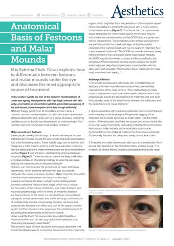
Figure 1: Patient presenting with mild malar mound and skin laxity at lid cheek junction
Figure 2: Patient with familial upper and lower lid festoons
Reproduced from Aesthetics | Volume 4/Issue 2 - January 2017