90
American Journal of Clinical Medicine® • Summer 2012 • Volume Nine Number Two
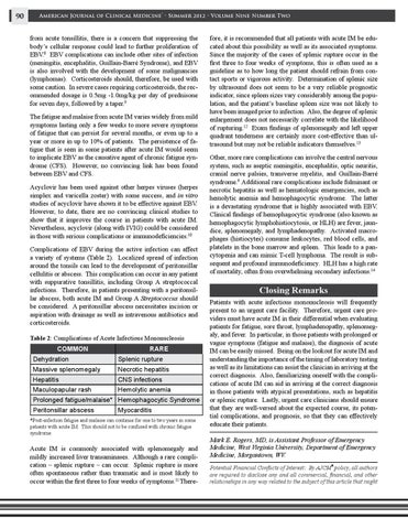
from acute tonsillitis, there is a concern that suppressing the body’s cellular response could lead to further proliferation of EBV.8 EBV complications can include other sites of infection (meningitis, encephalitis, Guillain-Barré Syndrome), and EBV is also involved with the development of some malignancies (lymphomas). Corticosteroids should, therefore, be used with some caution. In severe cases requiring corticosteroids, the recommended dosage is 0.5mg -1.0mg/kg per day of prednisone for seven days, followed by a taper.9 The fatigue and malaise from acute IM varies widely from mild symptoms lasting only a few weeks to more severe symptoms of fatigue that can persist for several months, or even up to a year or more in up to 10% of patients. The persistence of fatigue that is seen in some patients after acute IM would seem to implicate EBV as the causative agent of chronic fatigue syndrome (CFS). However, no convincing link has been found between EBV and CFS. Acyclovir has been used against other herpes viruses (herpes simplex and varicella zoster) with some success, and in vitro studies of acyclovir have shown it to be effective against EBV. However, to date, there are no convincing clinical studies to show that it improves the course in patients with acute IM. Nevertheless, acyclovir (along with IVIG) could be considered in those with serious complications or immunodeficiencies.10 Complications of EBV during the active infection can affect a variety of systems (Table 2). Localized spread of infection around the tonsils can lead to the development of peritonsillar cellulitis or abscess. This complication can occur in any patient with suppurative tonsillitis, including Group A streptococcal infections. Therefore, in patients presenting with a peritonsillar abscess, both acute IM and Group A Streptococcus should be considered. A peritonsillar abscess necessitates incision or aspiration with drainage as well as intravenous antibiotics and corticosteroids. Table 2: Complications of Acute Infectious Mononucleosis
Common
Rare
Dehydration
Splenic rupture
Massive splenomegaly
Necrotic hepatitis
Hepatitis
CNS infections
Maculopapular rash
Hemolytic anemia
Prolonged fatigue/malaise* Hemophagocytic Syndrome Peritonsillar abscess
Myocarditis
*Post-infection fatigue and malaise can continue for one to two years in some patients with acute IM. This should not to be confused with chronic fatigue syndrome.
Acute IM is commonly associated with splenomegaly and mildly increased liver transaminases. Although a rare complication – splenic rupture – can occur. Splenic rupture is more often spontaneous rather than traumatic and is most likely to occur within the first three to four weeks of symptoms.11 There-
fore, it is recommended that all patients with acute IM be educated about this possibility as well as its associated symptoms. Since the majority of the cases of splenic rupture occur in the first three to four weeks of symptoms, this is often used as a guideline as to how long the patient should refrain from contact sports or vigorous activity. Determination of splenic size by ultrasound does not seem to be a very reliable prognostic indicator, since spleen sizes vary considerably among the population, and the patient’s baseline spleen size was not likely to have been imaged prior to infection. Also, the degree of splenic enlargement does not necessarily correlate with the likelihood of rupturing.12 Exam findings of splenomegaly and left upper quadrant tenderness are certainly more cost-effective than ultrasound but may not be reliable indicators themselves.13 Other, more rare complications can involve the central nervous system, such as aseptic meningitis, encephalitis, optic neuritis, cranial nerve palsies, transverse myelitis, and Guillain-Barré syndrome.9 Additional rare complications include fulminant or necrotic hepatitis as well as hematologic emergencies, such as hemolytic anemia and hemophagocytic syndrome. The latter is a devastating syndrome that is highly associated with EBV. Clinical findings of hemophagocytic syndrome (also known as hemophagocytic lymphohistiocytosis, or HLH) are fever, jaundice, splenomegaly, and lymphadenopathy. Activated macrophages (histiocytes) consume leukocytes, red blood cells, and platelets in the bone marrow and spleen. This leads to a pancytopenia and can mimic T-cell lymphoma. The result is subsequent and profound immunodeficiency. HLH has a high rate of mortality, often from overwhelming secondary infections.14
Closing Remarks Patients with acute infectious mononucleosis will frequently present to an urgent care facility. Therefore, urgent care providers must have acute IM in their differential when evaluating patients for fatigue, sore throat, lymphadenopathy, splenomegaly, and fever. In particular, in those patients with prolonged or vague symptoms (fatigue and malaise), the diagnosis of acute IM can be easily missed. Being on the lookout for acute IM and understanding the importance of the timing of laboratory testing as well as its limitations can assist the clinician in arriving at the correct diagnosis. Also, familiarizing oneself with the complications of acute IM can aid in arriving at the correct diagnosis in those patients with atypical presentations, such as hepatitis or splenic rupture. Lastly, urgent care clinicians should ensure that they are well-versed about the expected course, its potential complications, and prognosis, so that they can effectively educate their patients. Mark E. Rogers, MD, is Assistant Professor of Emergency Medicine, West Virginia University, Department of Emergency Medicine, Morgantown, WV. Potential Financial Conflicts of Interest: By AJCM policy, all authors are required to disclose any and all commercial, financial, and other relationships in any way related to the subject of this article that might ®