III. Communicating with Message Maps
When discussing the topic of radiation, especially ionizing radiation, risk communicators face the daunting challenge of communicating about a topic that has virtually all the characteristics that create fear. Ionizing radiation is unfamiliar in people’s everyday lives and poorly understood outside of the science community. It cannot be seen, smelled, tasted, or felt, and people typically receive little if any education about sources of radiation in everyday life or how one measures the amount of radiation.
With such a topic, effective risk communication is critical. It can calm worried people, allowing them to hear much needed information. It can support logical decision making, such as allowing appropriate use of life saving medical imaging. Effective risk communication can save lives and promote physical and emotional well being. Poor risk communication will generate fear, amplify emotions, raise stress levels, undermine confidence in subject matter experts, and discourage people from receiving appropriate medical care. Poor communication can also send conflicting messages, which decrease trust in decisions and decision makers and lead to workflow challenges and increase demands and stress on medical staff.
I. Radiation “Hot Buttons”
Most people associate the term “radiation” with negative events and effects, such as nuclear weapons and failed nuclear power plants. Hollywood has long used radiation in science fiction stories to turn humans into superheroes, super villains, zombies, or other types of human mutants. Thus, simply thinking about radiation can trigger neurological and emotional hot buttons that greatly elevate fear.1 Neuroscience and behavioral science research indicate that these hot buttons function consciously and unconsciously within the brain. Several of the most important hot buttons are:
o Voluntariness. Risks from activities considered to be involuntary or imposed (e.g., mandatory vaccination) are judged to be greater, and are therefore less readily accepted, than risks from activities that are seen to be voluntary (e.g., smoking, sun bathing, or mountain climbing). Relevance to medical imaging radiation: being exposed to radiation from medical imaging can seem involuntary to patients seeking medical care.
o Controllability. Risks from activities viewed as under the control of others (e.g., flying on an airplane piloted by someone you do not know) are judged to be greater, and are less readily accepted, than those from activities that appear to be under the control of the individual (e.g., driving your own automobile or riding your own bicycle). Relevance to radiation from medical imaging: Individuals have only limited ability to control their need for medical imaging or how much radiation is delivered.
o Personal stake. Risks from activities viewed by people to place them (or their families) personally and directly at risk (e.g., living downstream from a dam) are judged to be greater than risks from activities that appear to pose no direct or personal threat (e.g., disposal of waste in remote areas).
 1 See e.g., Thornton et al. (2015) “Patient perspectives and preferences for Communication of medical imaging risks in the cancer care setting Radiology”; Hay et al. (2016)
“Prevalence and Correlates of Worry about the health harms of medical imaging radiation in the general population”. J Primary Care & Community Health 7(4).
1 See e.g., Thornton et al. (2015) “Patient perspectives and preferences for Communication of medical imaging risks in the cancer care setting Radiology”; Hay et al. (2016)
“Prevalence and Correlates of Worry about the health harms of medical imaging radiation in the general population”. J Primary Care & Community Health 7(4).
Edition: March 2022 4
Relevance to radiation from medical imaging: Individuals or family members are personally exposed to radiation from the medical imaging exam or procedure.2
o Familiarity. Risks from activities viewed as unfamiliar (e.g., eating exotic foods) are judged to be greater than risks from activities viewed as familiar (e.g., driving a car, riding a bike, or crossing the street). Relevance to radiation from medical imaging: medical imaging involves radiation producing equipment that most people have not seen or operated.
o Benefits. Risks from activities that seem to have unclear, questionable, or diffused personal or economic benefits (e.g., waste disposal facilities) are judged to be greater than risks from activities that have clear benefits (e.g., going to work to receive an income, driving an automobile to visit family or friends). Relevance to radiation from medical imaging: medical imaging carries fewer observable benefits to non experts since experts must interpret the results and make recommendations to patients on medical care. Although a negative test result may be very helpful to the clinician, it may be considered by patients to have no direct benefit, since the exam did not give a clear diagnosis.
o Catastrophic potential. Risks from activities viewed as having the potential to cause a significant number of deaths and injuries in a relatively short time or localized space (e.g., deaths and injuries resulting from major forest fires) are judged to be greater than risks from activities that cause deaths and injuries spread across time and space (e.g., automobile accidents). Relevance to radiation from medical imaging: radiation is often viewed as catastrophic since personal bodily harm, and particularly developing cancer, are considered catastrophic outcomes to an individual.
o Uncertainty. Risks from activities that are relatively unknown or that pose highly uncertain risks (e.g., risks from biotechnology and genetic engineering) are judged to be greater than risks from activities that appear to be relatively well understood (e.g., deaths related to automobile accidents, lung cancer from smoking). Relevance to radiation from medical imaging: the dose from medical imaging is highly variable, depending on exam indications and parameters, imaging equipment, practitioner preferences, and patient characteristics. The radiation dose to be delivered from a medical imaging exam is not precisely known prior to the exam and can only be estimated after the exam. Further, the risks associated with a given dose of radiation are uncertain, particularly at the low doses of radiation used in medical imaging, with substantial differences in expert opinion.
o Media attention. Risks from activities that receive considerable media coverage (e.g., passenger plane accidents) are judged to be greater than risks from activities that receive little (e.g., on the job accidents). Relevance to radiation from medical imaging: the public’s primary source of information about radiation is from the media, most often occurring after catastrophic accidents such as the those as the Chernobyl and Fukushima nuclear power plants
o Dread. Risks from activities that evoke fear, terror, or anxiety (e.g., exposure to cancer causing agents; AIDS) are judged to be greater than risks from activities that do not arouse such feelings or emotions (e.g., common colds, slipping on ice and breaking a bone). Relevance to radiation from medical imaging: the media, photographs of people suffering from radiation burns, and science
Edition: March 2022 5
2 https://www.psandman.com/articles/covello.htm
fiction can often portray radiation injury in a terrifying way (e.g., melting skin, glowing from radioactivity). High profile news stories documenting patient injury and death from overexposures to medical radiation show rare but tragic stories of pain and suffering that cannot be cured (e.g., overexposure of a patient’s brain stem to a radiation therapy beam due to medical error).
II. Best Practices in Risk/Benefit Communication for Medical Imaging Radiation3
Given these hot buttons and the fear, anger, and outrage they may engender, how can risk/benefit communicators best convey information about the radiation used in medical imaging? Listed below are several recommendations. Many of these recommendations may seem obvious. However, they are often not followed in practice, so it is important to explicitly discuss them
1. Listen to and understand your audience. In high stress, high concern situations, people are often more concerned about issues such as trust, empathy, control, competence, voluntariness, fairness, caring, and compassion than about mortality statistics and the details of quantitative risk/benefit assessments. If you do not listen to people, you cannot expect them to listen to you. Risk/benefit communicators are most effective when they actively listen to find out what people know and don’t know; what they fear and don’t fear; what may or may not outrage or concern them; what they are capable of doing and not capable of doing; what guidance they want and don’t want; and what broader cultural, economic, or political considerations are driving their perceptions and thinking. Risk/benefit communicators are most effective when they seek feedback.
2. Be honest and transparent. In communicating risk and benefit information, trust is your most precious asset. Trust is difficult to obtain and must be preserved. Once trust is lost, it is very difficult to fully regain. Risk/benefit communicators are most effective when they are open about what they know, what they don’t know, and what they are doing to learn more about what they do not know. Do not minimize or exaggerate the level of risk or benefit; speculate only with great caution. Lean toward sharing more information if the audience appears open to it, not less; discuss data uncertainties, strengths, and weaknesses, including those identified by credible sources; identify a worst case estimate as a worst case, citing ranges of risk estimates. It is essential that risk discussions are not held in a vacuum. The anticipated benefit is a critical element of risk discussions. It is also important to note that risks from exposure to ionizing radiation have been studied for over a century and a great deal of knowledge exists related to this topic. While the risks from the low doses of ionizing radiation used in medical imaging are not precisely known, upper boundaries of risk estimates have been well established.
3. Coordinate and collaborate with other credible sources. Allies and partners are critically important in helping communicate risk and benefit information. Coordinated messaging builds trust and effectiveness through consistency, where multiple experts share the same facts and information. Trust building is greatly enhanced when broad agreement exists among experts regarding both benefits and risks. Few things make risk communication more difficult than conflicts or public disagreements among credible sources. Communicators about the risks and benefits related to radiation from medical imaging are most effective when they take time to coordinate inter and intra organizational communication efforts; devote effort and resources to the hard work of building bridges and relationships with
3 https://riskcommunication.webnode.com/news/seven cardinal rules of risk communication/
Edition: March 2022 6
important allies and partners; use credible and authoritative intermediaries to communicate risk and benefit information; consult with allies and partners about who is best able to answer specific questions; and issue joint communications with trustworthy sources. Numerous studies indicate that the family physician or healthcare provider is considered by many to be the most trusted source of medical information, and medical imaging and radiation professionals would do well to coordinate and collaborate benefit and risk information about medical imaging with them.
4. Meet the needs of traditional and social media. Traditional print, broadcast, and social media outlets are prime sources of risk information. They play a critical role in choosing the topics that are set before the public and in driving public opinion. Communicators about the benefits and risks of the radiation from medical imaging are most effective when they are open and accessible, respect media deadlines, provide information tailored to the needs of each type of media, prepare in advance, provide background material on complex issues, follow up media stories with praise or constructive feedback, and are proactive.
5. Empathize and speak clearly. Neurological and behavioral research indicates that when people are stressed, they want to know that you care before they care what you know. They will have difficulty processing information until they believe that you have their best interest in mind. Stressed individuals will focus on what you say first and last, and they will often experience cognitive overload if you try to share more than three to five messages at a time. They will focus more on negative (risk) information than on positive (benefit) information. If you need positive information to be heard, it will be necessary to repeat it multiple times.
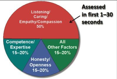
6. Understand how fear affects the brain. In a high stress situation, the brain typically processes fear faster than rational thought. The amygdala the brain’s “threat assessment” center takes control. The brain finds short cuts to process what could be life or death information as quickly as possible Hence, rational decision making takes a back seat to fear, and when facts and fears collide, fear often comes out the winner. Fear, such as that caused by a medical crisis and associated with radiation, makes it extremely difficult to process information. Neurological studies using the latest brain imaging techniques demonstrate that fear distorts risk perception and negatively impacts logical decision making, and that facts and reason are only part of how people make up their minds. Fear dramatically increases in situations where the people in charge are perceived as untrustworthy or uncaring. Lack of trust can dramatically increase fear. When risk communicators provide inaccurate, untimely, or inconsistent information, they are no longer trusted, and fear levels increase. As shown in Figure 1, caring, honesty, and competence are major determinants of trust. In high stress situations, whether the communicator can be trusted is often determined in less than 30 seconds.
Edition: March 2022 7
Figure 1: Trust determination in high stress situations
These six recommendations form the basic building blocks for effective communication about the risks and benefits associated with medical imaging. Each recommendation recognizes, in a different way, that effective communication is based on mutual trust, listening, caring, cooperation, and respect.
III. Building Trust and Overcoming Fear: The Eight Pillars of Risk Communication
So how can risk communicators use best communication practices to build trust and minimize fear related to the subject of interest?
Best practices in risk communication, documented in over 8,000 peer reviewed publications, are built on eight pillars.
1. The TBC (Trust/Benefits/Control) Pillar
2. The CCO (Compassion/Conviction/Optimism) Pillar
3. The PR (Primary/Recency) Pillar
4. The R3 (Rule of 3) Pillar
5. The 1N = 3P (One Negative Equals Three Positives) Pillar
6. The APP (Anticipate/Prepare/Practice) Pillar
7. The Overcoming Misinformation Pillar
8. The MM (Message Mapping) Pillar
Each pillar is described below.
1. The TBC (Trust/Benefit/Control) Pillar. Three of the most important perceptual factors driving fear are mistrust, unclear benefits, and lack of personal control. Translated into a best practice for risk communication, this means risk communicators should focus on dialogue and actions that build or repair trust, clearly identify the benefits of recommended guidance, and give people a sense of control.
Edition: March 2022 8
2. The CCO (Compassion/Conviction/Optimism) Pillar. People experiencing stress and fear typically want to know that you care before they care what you know. Translated into a risk communication best practice, the CCO pillar means risk communicators need to continually communicate messages that indicate listening, caring, empathy, and compassion.
3. The PR (Primacy/Recency) Pillar. People experiencing fear typically remember most the information they hear first and last. Translated into a best practice for crisis communication, the PR pillar means risk communicators should continually put their most important messages first and last.
4. The R3 (Rule of 3) Pillar. Fear can significantly reduce a person’s mental capacity to process information. In the psychological and neuroscience literature, this is called mental noise. Mental noise gives rise to the Rule of 3. Normally, people can remember seven discrete pieces of information, such as seven digit telephone numbers. During times of stress, this number is reduced to three. This is in part why emergency numbers worldwide have three digits, such as 9 1 1. Translated into a best practice for risk communication, the R3 pillar means risk communicators should organize their messages into groups or buckets of three.
5. The 1N = 3P (One Negative Equals Three Positives) Pillar. People experiencing fear typically put more weight on negative information than on positive information. Translated into a best practice for risk communication, this means a risk communicator should balance a negative message with at least three to four constructive or positive statements.
6. The APP (Anticipation/Preparation/Practice) Pillar. Risk communications are most successful when issues, scenarios, stakeholders, and stakeholder questions are anticipated; when messages and messengers are prepared; and when communications are practiced. Translated into a best practice for risk communication, this means risk communicators should, at a minimum, develop a preliminary list of likely stakeholder questions and concerns in advance. Generally, such a list would be developed through (1) an analysis of historical data and information; (2) a review of the communication literature on the subject of interest; (3) interviews with subject matter experts, and (4) survey data related to the subject of interest
IV. Message Maps: shorter and longer answers
This risk/benefit communication toolkit contains answers to important questions about the radiation associated with medical imaging presented in a format called a “message map.” A message map is a science based risk/benefit communication tool used to help organize complex information and make it easier for information to be shared with those who are not experts in the subject matter.
A message map distills information into a series of layered messages, from basic to more complex. Messages e.g., answers to questions are presented initially in a few bullets that convey key messages, ideally using the least number of words possible. The key messages are then followed by additional information.
A key assumption of message mapping is that people want clear answers to their questions about complex scientific issues as well as access to more detailed answers to those same questions. A second
Edition: March 2022 9
key assumption is that if stakeholders all those interested or impacted by the risk are well informed by the best and most up to date information about a complex scientific or technical issue, they are in a better position to engage, exchange information, and participate constructively in the decision making process.
Unfortunately, many scientists face challenges in sharing complex scientific information. These challenges are addressed in part by message maps. First, scientists must overcome a basic obstacle to effective communication: people facing a perceived threat and under stress typically have difficulty processing information hearing, understanding, and remembering. Second, scientists are used to communicating with their peers in a particular format, beginning with background information, moving to supporting details, and finally coming to their results and conclusions. For communicating with the public, however a more effective approach is to invert that pyramid and begin with the conclusions the bottom lines up front (BLUF). The top line of a message map the key messages are the conclusions. Third, scientists often fail to craft clear messages that can be easily understood and recalled by non experts. Message maps are designed to start with clear messages and build complexity through hierarchical layers. Fourth, because of details and lack of hierarchical structure, non experts often have difficulty sorting out what is important from what is less important. Fifth, scientists sometimes speak in a code known only to other scientists, using the technical jargon of their field instead of plain language. Many words that seem perfectly normal to scientists are incomprehensible jargon to a member of the public who is already stressed or fearful. Sixth, scientists often fail to put findings into the context of the larger body of knowledge of what is scientifically well understood. Seventh, scientists often lead with what they do not know instead of what they do know. Eighth, scientists often fail to anticipate common misunderstandings and misperceptions.
Message maps can be used to answer important questions in the form of a short answer consisting of ideally 3 (no more than 5) key messages expressed in 27 words. A longer answer consists of the shorter answer with supporting details. Best practices are to complete the answer to a question by repeating the shorter answer or key messages, which provides a soundbite that is easy to quote and remember.
The following message maps provide answers to commonly asked questions about the risks and benefits associated with medical imaging. The risk/benefit communicator should use the subset of message maps pertinent to a particular situation to provide the requested information without overwhelming the audience with information not pertinent to its concerns.
Edition: March 2022 10
101. Why is medical imaging important?
Key Messages/Shorter Answer (Soundbite):
1. Medical imaging has led to improvements in the diagnosis and treatment of disease and injury
2. Medical imaging, such as CT scanning, is fast, reliable, and can provide life saving information.
3. Medical imaging can reveal conditions that other diagnostic methods cannot
Longer Answer:
Medical imaging has led to improvements in the diagnosis and treatment of disease and injury.4, 5, 6, 7
• Medical imaging exams can include x rays (radiographs), fluoroscopy (x ray movies), computerized tomography (CT scanning), magnetic resonance imaging (MRI), ultrasound, and nuclear medicine and molecular imaging, such as positron emission tomography (PET scanning).
• Medical imaging can help determine the best treatment options for a patient.
• Medical imaging is needed to perform many advanced therapies, including opening blocked coronary arteries without open heart surgery and treating cancer with radiation therapy.
• Medical imaging is used to guide procedures that place catheters and stents into blood vessels, or to locate, biopsy, remove, or kill tumors.
Medical imaging, such as CT scanning, is fast, reliable, and can provide life-saving information.
• Medical imaging technology is used to:
o Noninvasively and painlessly diagnose disease and monitor therapy.
o Provide essential information for planning medical and surgical treatments
o Guide medical personnel when placing catheters, stents, or other devices to treat tumors, or remove blood clots or other blockages.
Medical imaging can reveal conditions that other diagnostic methods cannot. 8, 9
• Medical imaging can identify internal injuries, fractures, tumors, cancers, hemorrhage, blood clots, and changes in physiology and function.
• Medical imaging provides non invasive evidence based information that can assist your doctor in providing the best possible medical care.
4 https://www.fda.gov/radiation
5 https://www.health.harvard.edu/cancer/radiation


6 https://www.fda.gov/radiation
7 https://thorax.bmj.com/content/69/8/782.short
8 https://doi.org/10.1016/j.mayocp.2015.07.011
9 https://www.wsj.com/articles/in
Edition: March 2022 11
emitting products/medical imaging/medical x ray imaging#description
risk from medical imaging
emitting products/medical imaging/medical x ray imaging#description
image guided operating suites surgeons see real time mri ct scans 1424122291
102. What are the benefits of medical imaging?
Key Messages/Shorter Answer (Soundbite):
1. Medical imaging can determine whether an invasive procedure is necessary
2. Medical imaging can detect anatomic, physiologic, and molecular changes that cannot be found by other medical tests
3. Medical imaging allows more effective treatment and monitoring of treatment
Longer Answer:
Medical imaging can determine whether an invasive procedure is necessary. 10, 11
• Medical imaging can diagnose life threatening internal injuries that need immediate surgery
• Patients being assessed in the emergency department after a traumatic injury can be scanned quickly, letting surgeons know in a very short time whether and where to operate.
• Medical imaging has been shown to reduce unnecessary surgeries for many conditions, including appendicitis.
Medical imaging can detect anatomic, physiologic, and molecular changes that cannot be found by other medical tests. 12, 13
• Medical imaging can be used to detect conditions that cannot be found through blood or urine tests, measuring brain waves or cardiac rhythms, or physical examination.
• Medical imaging can quickly diagnose life threatening conditions, including tumors, aneurysms, congenital disorders, blocked blood vessels, internal bleeding, blood clots, spinal injuries, and bone fractures
Medical imaging allows more effective treatment and monitoring of treatment.
• Guiding treatment of common conditions
• Determining if surgery is necessary.
• Placing catheters and stents.
• Locating, destroying, or removing tumors.
• Reducing the length of hospitalization.
Edition: March 2022 12
10 https://www.imagewisely.org/Imaging Modalities/Computed Tomography/How to Understand and Communicate Radiation Risk 11 https://www.health.harvard.edu/cancer/radiation risk from medical imaging 12 https://doi.org/10.1016/j.mayocp.2015.07.011 13 https://www.wsj.com/articles/in image guided operating suites surgeons see real time mri ct scans 1424122291
Key Messages/Shorter Answer (Soundbite):
1. Scientists measure how often medical imaging provides the correct diagnosis.
2. Scientists measure the impact of medical imaging on patient care decisions.
3. Scientists measure improvements in health outcomes when medical imaging is used.
Longer Answer:
Scientists measure how often medical imaging provides the correct diagnosis. 14, 15
• Studies determine how often medical imaging correctly identifies injuries and disease and compares the results to the accuracy of other diagnostic tests
o In a study of almost 12,000 patients who received surgery to remove their appendix, about 10% of patients imaged using ultrasound received unnecessary surgery due to an incorrect diagnosis. With CT, the number of unnecessary surgeries fell to 2.5%. Unnecessary surgery occurred in almost 20% of patients who did not receive any imaging.
• Scientists evaluate evidence about the accuracy of medical imaging.
Scientists measure the impact of medical imaging on patient care decisions. 16, 17
• Scientists measure how medical imaging impacts patient diagnoses and management.
o Use of CT in the emergency department changed the diagnosis and patient management in 20 50% of evaluated patients
o In a study of patients with abdominal pain, weight loss, or blood in their urine, CT changed the doctor’s initial diagnosis in 50% of patients and changed clinical management in 35 54% of patients.
Scientists measure improvements in health outcomes when medical imaging is used. 18
• Scientists measure how much medical imaging improves health outcomes.
• Early detection of disease with medical imaging is especially important for conditions that do not display symptoms until the disease is in an advanced state, such as cancer.
o A 20% decrease in lung cancer deaths was observed in smokers and ex smokers who received low dose CT scans of their lungs compared to those who had a chest x ray
14
15
16 https://pubs.rsna.org/doi/full/10.1148/radiol.2015150473
17 https://pubs.rsna.org/doi/full/10.1148/radiol.2016152887
18 https://www.nejm.org/doi/full/10.1056/nejmoa1102873
103. How do scientists measure the benefits of medical imaging?
Edition: March 2022 13
https://www.americanjournalofsurgery.com/article/S0002 9610(18)30397 0/fulltext
https://jamanetwork.com/journals/jamasurgery/article abstract/2782155
:~:text=In the NLST, a 20.0,compared with the radiography group
Key Messages/Shorter Answer (Soundbite):
1. How we communicate benefits and risks of medical imaging can greatly influence a patient’s understanding and decision making processes.
2. Best practices use simple, clear, consistent messages, without jargon, technical terms, or acronyms.
3. Research is needed to better understand how emotions affect a patient’s medical decision making.
Longer Answer:
How we communicate benefits and risks of medical imaging can greatly influence the patient’s understanding and decision making processes. 19, 20, 21, 22
• Effective communication about medical imaging can address a patient’s concerns.
o Demonstrating caring and empathy is often the single most important element in helping alleviate a patient’s worries.
o Worried patients may have difficulty processing new information
o Worried patients may give greater weight to negative information and may be overly influenced by anecdotal reports communicated through social and mainstream media.
• Best practices include comparisons with naturally occurring sources of ionizing radiation.23
o Naturally occurring sources, such as cosmic radiation, deliver an average of 3 mSv per year, with a range from ~1 20 mSv/year, depending on where you live.
o One chest x ray (0.1 mSv) is equivalent to background exposure over 10 days.
o One mammogram (0.4 mSv) is equivalent to background exposure over 7 weeks.
o One abdominal CT scan (< 10 mSv) is equivalent to background exposure over 3.5 years. Best practices use simple, clear, consistent messages, without jargon, technical terms, and acronyms.
• Inconsistencies by experts undermines public trust in benefit and risk information
• Scientists measure radiation in different ways, and this can be confusing to patients. Research is needed to better understand how emotions affect a patient’s medical decision making.24
• Substances are viewed as being riskier when the science is perceived to be uncertain, the effect is delayed, the individual does not control their exposure levels, or the risk is sensationalized in the public media
• The human brain may use mental shortcuts that distort perceptions of radiation risk.
• Misinformation regarding risks from medical imaging can make providers hesitant to order medically appropriate imaging or patients hesitant to receive medically necessary imaging. https://www.imagewisely.org/Imaging https://www.health.harvard.edu/cancer/radiation https://www.cancer.gov/about https://www.imagegently.org/Portals/6/Parents/Image_Gently_8.5x11_Brochure.pdf https://pubs.rsna.org/doi/pdf/10.1148/radiol.2020192256 https://www.ajronline.org/doi/full/10.2214/AJR.10.5956
104. What are best practices in communicating the benefits and risks of medical imaging?
Edition: March 2022 14
19
Modalities/Computed Tomography/How to Understand and Communicate Radiation Risk 20
risk from medical imaging 21
cancer/causes prevention/risk/radiation 22
23
24
Key Messages/Shorter Answer (Soundbite):
1. Scientists classify radiation as either non ionizing or ionizing and medical imaging uses both types
2. Non ionizing radiation includes visible and certain types of ultra violet light as well as radio frequencies and microwaves.
3. Ionizing radiation, such as x rays, can have sufficient energy to cause changes in cells.
Longer Answer:
Scientists classify radiation as either non ionizing or ionizing and medical imaging uses both types.
• Non ionizing radiation has less energy than ionizing radiation.
• Health effects from both non ionizing and ionizing radiation are related to the amount and duration of your exposure.
25
Non ionizing radiation includes visible and certain types of ultra violet light as well as radio frequencies and microwaves.
• Exposure to large amounts of non ionizing radiation can damage tissue. Ionizing radiation, such as x rays, can have sufficient energy to cause changes in cells.
• Ionizing radiation can come from natural sources, such as radon and cosmic rays, or man made sources such as medical imaging equipment or air travel
• Medical imaging equipment such as x ray systems, CT scanner, or PET scanners use ionizing radiation to take pictures of the body and its functions
• Ionizing radiation can cause chemical changes in cells and damage DNA, which may increase the risk of developing health conditions such as cancer.
201. What type of radiation is used in medical imaging?
Edition: March 2022 15
25 https://www.who.int/medical_devices/global_forum/3rd_gfmd/Nonionizingradiationcosmetic.pdf?ua=1
Key Messages/Shorter Answer (Soundbite):
1. Some medical imaging procedures can cause skin damage
2. Although uncommon, fluoroscopy and MRI are most likely to damage skin.
3. Ongoing staff education and the use of safety protocols help minimize the risk of skin damage
Longer Answer:
Some medical imaging procedures can cause skin damage. 26, 27
• Skin can be damaged by prolonged exposures to x ray radiation or by heating caused by the radio waves used in MRI heating the skin.
• Skin reactions from medical imaging can be very mild, where the skin becomes red, or more moderate, where the skin becomes blistered and very sore.
Although uncommon, fluoroscopy and MRI can cause damage skin. 28
• Fluoroscopy creates a moving x ray image on a monitor and may require longer x ray exposures and higher doses to allow the physician to perform image guided procedures.
• MRI uses radio waves and strong magnetic fields to create images of the inside of the body.
• High intensity radio waves can cause tissue heating, which could lead to skin irritation.
• Burns can occur when electrical monitoring leads (metal wires) form a loop on the skin during an MRI scan
Ongoing staff education and the use of safety protocols help minimize the risk of skin damage. 29
• Optimizing the amount of radiation used helps prevent or minimize skin damage from fluoroscopy.
• Skin damage may be unavoidable during complex image guided interventional procedures, even when steps are taken to prevent and mitigate such events.
• Screening for implanted metal and careful placement of wires minimize the risk of skin damage during MRI.
• Patients with implanted medical devices should inform their healthcare providers before receiving an MRI study.
• Patients should contact their healthcare provider if they have skin changes after an imaging procedure. https://www.cdc.gov/nceh/features/medical
202. Can my skin be damaged by the radiation used in medical imaging?
Edition: March 2022 16
26
imaging procedures/index.html#:~:text=%20Medical%20imaging%20tests%20are%20non invasive%20procedures%20that,ionizing%20radiation%2C%20which%20can%20present%20risks%20to%20patients 27 https://www.ncbi.nlm.nih.gov/pmc/articles/PMC5489635/ 28 https://www.fda.gov/radiation emitting products/radiation emitting products and procedures/medical imaging 29 https://www.healthimaging.com/topics/practice management/battling burns mr
203. Can I get cancer from medical imaging?
Key Messages/Shorter Answer (Soundbite):
1. Theoretically, yes, but medical imaging exams use lower amounts of radiation than the amounts proven to increase cancer risk.
2. The risk to a patient depends on the type of exam, the amount of radiation used, and certain patient characteristics
3. If medical imaging is needed as part of your medical care, then the benefits are much larger than the potential risk from the radiation
Longer Answer:
Theoretically, yes, but medical imaging exams use lower amounts of radiation than the amounts proven to increase cancer risk. 30, 31, 32
• Ionizing radiation can damage cells.
• Cells with damaged DNA can either die, repair themselves, have no effect, or go on to cause a health effect such as cancer.
• Cancer risk is related to the amount (dose) of radiation received.
o Strong evidence exists that high doses of radiation can increase cancer risk
o Medical imaging uses low doses of radiation.
• Some epidemiological studies have associated increased cancer risk with low doses of radiation; however, such studies are associated with a great deal of uncertainty due to the difficulty of quantifying such a small risk.
The risk to a patient depends on the type of exam, the amount of radiation used, and certain patient characteristics. 33, 34, 35
• Medical imaging exams such as chest x rays and radiographs of the arms and legs deliver effective doses of <0.1 mSv and pose at most negligible risk.
• Most radiographic, fluoroscopic, nuclear medicine, and CT exams deliver effective doses of a few mSv to 20 mSv and pose at most very low risk.
If medical imaging is needed as part of your medical care, then the benefits are much larger than the potential risk from the radiation.36
• The benefits from medical imaging include earlier detection of disease, including cancer, which makes it easier for physicians to provide a cure.
• Medical imaging enables rapid diagnosis of life threatening injuries.
• Medical imaging reduces unnecessary procedures and surgeries.
30 https://www.cancer.org/treatment/understanding your diagnosis/tests/understanding radiation risk imaging tests.html https://www.cdc.gov/nceh/radiation/healthcare.html https://doi.org/10.1016/j.mayocp.2015.07.011 https://www.cdc.gov/nceh/radiation/health.html https://www.jacr.org/article/S1546 1440(12)00002 6/fulltext https://pubs.rsna.org/doi/full/10.1148/radiol.2020192256 https://www.cdc.gov/nceh/features/medical imaging procedures/index.html#:~:text=%20Medical%20imaging%20tests%20are%20non
Edition: March 2022 17
from
31
32
33
34
35
36
invasive%20procedures%20that,ionizing%20radiation%2C%20which%20can%20present%20risks%20to%20patients
Key Messages/Shorter Answer (Soundbite):
1. For most types of medical imaging, no radiation remains in your body.
2. After nuclear medicine imaging, radiation can stay in your body for a short time.
3. Breastfeeding mothers who receive nuclear medicine imaging may need to pause breastfeeding for a short time
Longer Answer:
1. For most types of medical imaging, no radiation remains in your body. 37
• The dose of radiation a person receives in a typical imaging exam is low.
• The exact amount of radiation exposure in an imaging procedure varies depending on the exam type and part of the body being imaged.
• After an X ray, fluoroscopy exam, or CT scan, no radiation remains in your body.
2. After nuclear medicine imaging, radiation can stay in your body for a short time. 38
• Nuclear medicine imaging, such as gamma camera, SPECT, or PET imaging, uses small amounts of radioactive material to detect and diagnose various health conditions.
• Nuclear medicine imaging can identify early signs of cancer, heart disease, and other disorders.
• Following a nuclear medicine imaging exam, detectable levels of radiation will disappear within a few days after the exam given the small amount of radioactive material used.
3. Breastfeeding mothers who receive nuclear medicine imaging may need to pause breastfeeding for a short time. 39
• For most nuclear medicine imaging exams, no interruption in breastfeeding is necessary.
• Some nuclear medicine imaging exams require a short pause in breastfeeding.
• If you are breastfeeding, consult your doctor or medical/health physicist prior to your exam
204. Am I radioactive after I have medical imaging?
Edition: March 2022 18
37 https://www.cdc.gov/nceh/features/medical imaging procedures/index.html#:~:text=%20Medical%20imaging%20tests%20are%20non invasive%20procedures%20that,ionizing%20radiation%2C%20which%20can%20present%20risks%20to%20patients 38 https://www.mountsinai.org/care/radiology/services/nuclear medicine/faqs 39 https://my.clevelandclinic.org/health/diagnostics/10123 pet scan
205. Will my vision be affected by medical imaging?
Key Messages/Shorter Answer (Soundbite):
1. The human eye is continuously exposed to natural sources of ionizing radiation.
2. Exposure to ionizing radiation can lead to cataract formation.
3. A medical imaging procedure is very unlikely to cause cataracts
Longer Answer:
1. The human eye is continuously exposed to natural sources of ionizing radiation. 40
• People are continuously exposed to low doses of ionizing radiation, including from cosmic and solar radiation
2. Exposure to ionizing radiation can lead to cataract formation. 41
• A cataract involves clouding of the eye lens.
• Ionizing radiation to the eyes has the potential to cause cataract formation.
• The leading cause of cataracts is the natural aging process and not radiation exposure.
o Frequent exposure to solar radiation can contribute to the formations of cataracts.
o Other risk factors for cataracts include alcohol and nicotine consumption, diabetes, and systemic use of corticosteroids.
3. A medical imaging procedure is very unlikely to cause cataracts.42
• The leading causes of vision loss in the United States are primarily age related, such as age related macular degeneration and cataract formation, diabetic retinopathy, and glaucoma
• Placing shielding over the eyes during medical imaging exams can interfere with the quality of the exam and is not recommended.
42
Edition: March 2022 19
40 http://www.iacrs rp.org/products/iacrs lens eye.pdf 41 https://www.rerf.or.jp/en/programs/roadmap_e/health_effects en/early en/cataract_en/#:~:text=Radiation%20cataract%20causes%20partial%20opacity,after%20exposure%20to%20lower%20doses
https://www.aapm.org/org/policies/details.asp?id=2547&type=
Key Messages/Shorter Answer (Soundbite):
1. Medical imaging tests provide pictures of tissues and organs inside the body.
2. A medical imaging test may reveal an incidental finding, which is a finding not related to the reason your doctor ordered the imaging test.
3. Your doctor will decide what to do about incidental findings based on what the images show and what medical guidelines recommend for such findings.
Longer Answer:
1. Medical imaging tests provide pictures of tissues and organs inside the body.
• Medical imaging tests are used to help diagnose medical problems.
• Medical imaging tests have revolutionized diagnosis and treatment, almost eliminating the need for painful, invasive, or potentially risky procedures, such as exploratory surgery.
2. A medical imaging test may reveal an incidental finding, which is a finding not related to the reason your doctor ordered the imaging test. 43, 44, 45
• Incidental findings can include cysts, benign growths, or other anatomic variations that have no health consequences.
• Sometimes an incidental finding could be important, such as if a CT scan of the chest looking for a blood clot shows a small growth in the lung or an aortic aneurism.
3. Your doctor will decide what to do about incidental findings based on what the images show and what medical guidelines recommend for such findings. 46, 47
• Most incidental findings are of little concern.
• Images of the incidental finding may be compared with any prior medical images that you might have to determine if the finding is new or changing.
• In some cases, your doctor may recommend additional imaging or testing to further evaluate incidental findings.
43 https://www.jacr.org/article/S1546 1440(10)00330 3/fulltext
44 https://www.ncbi.nlm.nih.gov/pmc/articles/PMC3473456/
45 https://www.cancer.org/cancer/lung cancer/detection diagnosis staging/how diagnosed.html
46 https://www.jacr.org/article/S1546 1440(12)00610 2/fulltext
47 https://pubmed.ncbi.nlm.nih.gov/21333775/
301. What happens if they find something in my medical images that was not expected?
Edition: March 2022 20
Key Messages/Shorter Answer (Soundbite):
1. When a pregnant woman experiences illness or injury, medical imaging can quickly provide an accurate diagnosis so that the patient can receive the best treatment.
2. Risks to the developing human fetus from medical imaging exams have not been observed.
3. The fetus is exposed to radiation only if the uterus is directly irradiated.
Longer Answer:
1. When a pregnant woman experiences illness or injury, medical imaging can quickly provide an accurate diagnosis so that the patient can receive the best treatment. 48, 49
• Pregnant women may express concern about exposing their unborn child to radiation.
• Increased risks to an unborn child from medical imaging have not been observed, even if the exam uses ionizing radiation.
• It is essential that pregnant women receive needed medical imaging exams to protect their own health and that of their baby
• Medical imaging necessary for the care of the mother, or the embryo or fetus, can be performed at any time during pregnancy
2. Risks to the developing human fetus from medical imaging exams have not been observed. 50, 51
• The amount of radiation a fetus receives from a medical imaging exam of the mother is frequently less than the fetus receives from natural sources of radiation during the pregnancy.
• For pregnant women, the low risk to the fetus needs to be balanced with the benefits of using the most appropriate imaging technique to care for the mother.
3. The fetus is exposed to radiation only if the uterus is directly irradiated. 52
• Medical imaging exams that do not include the pelvis expose the fetus to almost no radiation.
• Methods for reducing radiation dose to the fetus can be used for image guided interventional procedures.
302. What is the risk to my unborn baby if I receive a medical imaging exam?
Edition: March 2022 21
48 https://www.ajog.org/article/S0002 9378(08)00643 1/fulltext 49 https://ncrponline.org/publications/reports/ncrp report 174/ 50 https://www.acr.org/ /media/acr/files/practice parameters/pregnant pts.pdf 51 https://pubs.rsna.org/doi/10.1148/rg.274065149 52 https://pubs.rsna.org/doi/10.1148/rg.321115064
Key Messages/Shorter Answer (Soundbite):
1. Decisions about which exam to use are based on multiple factors, including which will provide the most accurate and relevant information for your medical situation.
2. Magnetic resonance imaging (MRI) and ultrasound imaging are preferred in some situations.
3. CT scans are preferred for many other situations, including trauma, bone, or lung imaging.
Longer Answer:
1. Decisions about which exam to use are based on multiple factors, including which exam will provide the most accurate and relevant information for your medical situation. 53
• The most important task of medical imaging is to provide answers that will help your doctor provide the best medical care for you.
• When deciding on the best exam for you, your healthcare provider should carefully consider professional guidelines that suggest the most appropriate tests for different situations.
• If you have questions about your exam, ask your healthcare or imaging provider.
2. Magnetic resonance imaging (MRI) and ultrasound imaging are preferred in some situations. 54, 55
• Magnetic resonance imaging (MRI) uses a powerful magnet and radio waves to create detailed cross sectional pictures of soft tissues, nerves, and blood vessels.
• MRI is highly effective in imaging soft tissues, such as the brain, but is not always available, is more expensive, may require sedation, and cannot be used for some patients with metallic implants.
• Ultrasound imaging uses high frequency sound waves and is often used for prenatal care.
• Ultrasound imaging is widely available and less expensive than CT and MRI but is operator dependent and doesn’t image large volumes as quickly or with the same level of detail.
3. CT scans are preferred for many other situations, including trauma, bone, or lung imaging. 56, 57
• A computed tomography scan (CT) is an imaging procedure that combines multiple X ray images to make detailed pictures of internal organs and structures.
o CT scans use images taken from different angles and a computer creates cross sectional images of the bones, blood vessels, and soft tissues inside your body.
o CT scans produce detailed images that identify tumors or damage to organs and may eliminate the need for higher risk and more invasive procedures like surgery.
o CT scans are very useful for diagnosing and staging cancer, monitoring whether treatment is working, and checking whether the cancer has come back.
• CT scans are good for imaging large areas quickly, such as the chest, abdomen, and pelvis
• Because CT scans are very fast, they can be helpful in young or ill patients who have difficulty holding still. https://doi.org/10.1016/j.mayocp.2015.07.011 https://www.hopkinsmedicine.org/health/treatment https://www.mskcc.org/news/ct
401. Are tests like MRIs or ultrasounds better than CT scans?
Edition: March 2022 22
53
54
tests and therapies/ct vs mri vs xray 55
vs mri what s difference and how do doctors choose which imaging method use 56 https://www.epa.gov/radiation/what ctcat scan and how it used 57 https://www.fda.gov/radiation emitting products/medical x ray imaging/computed tomography ct#3
402. Are MRIs or ultrasounds better for children?
Key Messages/Shorter Answer (Soundbite):
1. Children are generally considered to be more sensitive to ionizing radiation than adults.
2. The use of radiation in medical imaging should be optimized based on the patient size and medical need.
3. The choice of which exam to use is based on which will provide the most accurate and relevant information for your child’s medical situation.
Longer Answer:
1. Children are generally considered to be more sensitive to ionizing radiation than adults.58, 59
• Exposure to ionizing radiation is a topic of concern for pediatric patients because younger patients are generally considered to be more sensitive to radiation than adults.
• Compared to adults, children have a longer life expectancy and therefore a longer period of time in which negative health effects could occur.
• Because of their longer life expectancy, it is also of great importance to obtain accurate diagnostic information to effectively treat illness or injury in children.
2. The use of radiation in medical imaging should be optimized based on the patient size and medical need. 60, 61, 62, 63
• Often, less radiation is needed to get high quality medical images of smaller patients.
• Doctors should take care to “child size” the radiation dose.
• Even in children, the risk from a medically appropriate imaging exam is low when compared to the benefits of an accurate diagnosis and proper treatment.
3. The choice of which exam to use is based on which will provide the most accurate and relevant information for your child’s medical situation. 64
• Your healthcare provider will determine the most appropriate test based on your child’s condition and professional appropriateness criteria or other guidance documents.
• Avoiding a test that is deemed necessary due to the fear of radiation can lead to incorrect or delayed diagnosis and treatment.
• The risk of medical imaging in children is very small compared to the risk of not accurately diagnosing or treating their medical condition. https://www.unscear.org/docs/publications/2013/UNSCEAR_2013_Annex https://doi.org/10.1016/j.mayocp.2015.07.011 https://www.acr.org/Clinical Resources/ACR https://www.aapm.org/org/policies/details.asp?id=2548&type=PS https://www.fda.gov/consumers/consumer https://www.imagegently.org
Edition: March 2022 23
58
B.pdf 59
60
Appropriateness Criteria 61
62
updates/dose matters fdas guidance childrens x rays 63
64 https://www.acr.org/Clinical Resources/ACR Appropriateness Criteria
Key Messages/Shorter Answer (Soundbite):
1. A patient’s medical condition not regulations determines the amount of radiation that is needed.
2. State and federal agencies and accreditation and professional organizations play an important role in ensuring the quality and safety of medical imaging
3. Educational and professional certification requirements exist for medical imaging personnel, including medical physicists.
Longer Answer:
A patient’s medical condition determines the amount of radiation that is needed 65
• Regulations do not limit the total dose that a patient can receive from medical imaging; patients can receive the medical imaging care that is needed for their situation.
• Recommended values for image quality and dose metrics exist for imaging of standard test objects.
• Universal dose limits are not appropriate because the radiation dose must be adapted to the patient size and the medical need.
• Reducing the radiation dose too much can cause poor image quality and may lead to a missed or incorrect diagnosis.
State and federal agencies and accreditation and professional organizations play an important role in ensuring the quality and safety of medical imaging. 66, 67, 68, 69
• Individual states regulate the practice of medicine by licensing medical imaging providers, including radiologists, radiologic technologists, and medical physicists.
• Federal agencies such as the Food and Drug Administration and Nuclear Regulatory Commission regulate medical devices and radioactive materials
• Medical physicists perform state specific regulatory tests on imaging equipment to ensure proper and safe performance.
Educational and professional certification requirements exist for medical imaging personnel, including medical physicists. 70
• Board certification is a process used to ensure that medical imaging providers have the proper educational training and experience to practice in their field.
• There are standards for education and clinical experience, and competency is tested through written and oral examinations.
• Ongoing professional education is required to maintain certification.
65 https://www.epa.gov/sites/default/files/2015 05/documents/fgr14 2014.pdf https://www.epa.gov/radiation/who regulates radiation used medical https://www.fda.gov/radiation emitting products/radiation emitting https://www.cdc.gov/nceh/radiation/ionizing.htm https://www.nrc.gov/about nrc/radiation/protects you/nuclear medicine.html https://www.fda.gov/radiation
501. What protects patients from being exposed to too much radiation?
Edition: March 2022 24
66
procedures 67
products and procedures/medical imaging 68
69
70
emitting products/radiation safety/initiative reduce unnecessary radiation exposure medical imaging
502. What protects healthcare personnel from high radiation exposures?
Key Messages/Shorter Answer (Soundbite):
1. Federal and state agencies set maximum exposure levels for healthcare personnel and others working with or near radiation sources
2. Reasonable measures must be taken to minimize radiation doses that personnel receive during the performance of their job.
3. Radiation safety officers and imaging professionals oversee the use of radiation in healthcare organizations to ensure that regulations are followed and adequate safeguards are in place.
Longer Answer:
Federal and state agencies set maximum exposure levels for healthcare personnel and others working with or near radiation sources.71, 72
• National (e.g., U.S. Federal) and/or regional (e.g., U.S. state) agencies regulate dose levels, training requirements, and safety procedures for personnel working with radiation, with guidance from the International Commission on Radiological Protection, and in the U.S., the National Council on Radiation Protection and Measurements. 73, 74
Reasonable measures must be taken to minimize the amount of radiation received by healthcare personnel. 75, 76, 77
• Radiation experts at the U.S. National Council on Radiation Protection and Measurements (NCRP) provide detailed guidance regarding dose limits for healthcare personnel.
• The NCRP provides guidance regarding radiation exposures from x ray imaging, nuclear medicine and molecular imaging, radiation oncology, and dentistry.
• The NCRP recommends that radiation exposures to healthcare personnel be kept at levels that are as low as reasonably achievable (ALARA).
Radiation safety officers and imaging professionals oversee the use of radiation in healthcare organizations to ensure that regulations are followed and adequate safeguards are in place. 78
• Radiologists, medical physicists, or health physicists are trained in the safe use of radiation and typically serve as the radiation safety officer for healthcare organizations.
• Radiation safety officers direct radiation safety programs to monitor personnel doses.
• Radiation safety officers work with personnel with high exposure readings or who inform their employer that they are pregnant to limit future exposures. https://www.cms.gov/Medicare/Provider https://www.icrp.org/publication.asp?id=ICRP%20Publication%2075https://www.icrp.org/publication.asp?id=ICRP https://www.epa.gov/sites/default/files/2015 https://ncrponline.org/publications/reports/ncrp https://ncrponline.org/shop/reports/report
Edition: March 2022 25
71 https://www.fda.gov/radiation emitting products/radiation safety/initiative reduce unnecessary radiation exposure medical imaging 72
Enrollment and Certification/SurveyCertificationGenInfo/Accreditation of Advanced Diagnostic Imaging Suppliers 73
Publication 103 74 https://ncrponline.org/shop/reports/report no 180 management of exposure to ionizing radiation radiation protection guidance for the united states 2018 2018/ 75
05/documents/fgr14 2014.pdf 76
reports 107/ 77
no 180 management of exposure to ionizing radiation radiation protection guidance for the united states 2018 2018/ 78 https://www.birpublications.org/doi/full/10.1259/bjr.20200282
Key Messages/Shorter Answer (Soundbite):
1. Medical imaging is about performing the right test with the right dose at the right time.
2. Medical imaging has revolutionized medical diagnosis and treatment, eliminating the need for once common exploratory surgeries and many other invasive and potentially risky procedures.
3. Healthcare providers recommend medical imaging when the imaging is likely to benefit you by helping guide your medical care.
Longer Answer:
1. Medical imaging is about performing the right test with the right dose at the right time.79 , 80, 81
• Your doctor and the radiologist work together to make sure the test being considered is appropriate for your situation.
• Imaging exams are tailored to the medical situation and the information that your doctor needs.
• Medical imaging device operators and equipment features adjust the amount of radiation so that only the needed amount of radiation is used.
2. Medical imaging has revolutionized medical diagnosis and treatment, eliminating the need for exploratory surgeries and many other invasive and potentially risky procedures.82
• The many benefits from medical imaging include noninvasive and painless diagnosis, precise guidance of treatment, and monitoring of disease progression or remission.
• Medical imaging can provide life saving information and may eliminate the need for higher risk and more invasive procedures such as surgery.
• The benefits of medical imaging far outweigh the potential harm from the low doses of radiation used in to perform the exam.
• The risk of not getting a medically needed test is far greater than the radiation risk.
3. Healthcare providers recommend medical imaging when the imaging is likely to benefit you by helping guide your medical care.83
• Medical imaging can reveal health conditions undetected by other diagnostic tests.
• If your doctor recommends medical imaging, it is because they believe that the procedure will provide the information needed to diagnose your illness or injury and guide your treatment.
79 https://www.imagegently.org/ https://www.fda.gov/radiation emitting https://thorax.bmj.com/content/69/8/782.short https://www.fda.gov/radiation emitting products/medical https://www.wsj.com/articles/in image
601. How can we be sure that the benefit of medical imaging outweighs the risk?
Edition: March 2022 26
80
products/medical imaging/medical x ray imaging#description 81
82
imaging/medical x ray imaging#description 83
guided operating suites surgeons see real time mri ct scans 1424122291
Key Messages/Shorter Answer (Soundbite):
1. Healthcare providers are taught to recommend medical imaging only when medically justified.
2. Imaging providers are taught to use the amount of radiation needed to answer the clinical question
3. Patients have an important role in minimizing risk from medical imaging radiation.
Longer Answer:
1. Healthcare providers are taught to recommend medical imaging only when medically justified. 84
• When deciding on the best diagnostic test, the healthcare provider carefully determines the test that will deliver the most useful information for your specific condition.
• If a medical imaging technique that uses ionizing radiation is deemed medically appropriate, the goal is to optimize the use of radiation to answer the clinical question.
2. Imaging providers are taught to use the amount of radiation needed to answer the clinical question. 85
• The radiation dose depends on the specific procedure, the quality of the image needed to answer the medical question, and the size of the patient.
• Most imaging devices use automatic dose control technologies to help ensure the lowest dose possible without decreasing the effectiveness of the exam.
• Medical imaging techniques are now using much lower radiation doses than in the past.
o Pulsed fluoroscopy and “last image hold” features reduce the dose to patients from fluoroscopic procedures without reducing the quality of the exam.
o Over the past decade, the average doses from CT exams have fallen by at least 20%.
o Compared to the early days of CT, doses have fallen at least three fold
• National and international radiation regulations and guidelines help ensure that radiation doses used in medical imaging are appropriate
3. Patients have an important role in minimizing risk from medical imaging radiation. 86
• Ask your health care provider about how the medical imaging exam or procedure will help determine your treatment.
• Ask if there are other exams or procedures that might have lower risk while still providing an accurate diagnosis or best treatment guidance for your medical situation.
• If your healthcare provider explains there is no need for medical imaging, it is acceptable to ask for more details.
• If you switch health care providers, image files can be transferred to your new provider to avoid unnecessarily repeating an exam that you’ve already had
602. What is done to minimize my risk from medical imaging radiation?
Edition: March 2022 27
84 https://www.health.harvard.edu/cancer/radiation risk from medical imaging 85 https://www.who.int/publications/i/item/978924151034 86 https://www.hhs.gov/answers/public health and safety/how can i reduce radiation exposure from x rays/index.html
Key Messages/Shorter Answer (Soundbite):
1. Contrast dyes are used with medical imaging to help radiologists better view organs and tissues.
2. In rare cases, people have allergic reactions to contrast dyes.
3. Side effects experienced from contrast dyes are unrelated to medical imaging radiation.
Longer Answer:
1. Contrast dyes are used with medical imaging to help radiologists better view organs and tissues. 87
• Contrast dyes can be swallowed as a drink or injected into a vein, depending on the kind of exam or procedure.
• Contrast dyes are used to visualize organs and tissues more clearly, especially blood vessels and tumors.
• Contrast dyes can help the images distinguish between normal and abnormal conditions and allows for a more accurate diagnosis, which can lead to better medical care.
2. In rare cases, people have allergic reactions to contrast dyes. 88
• In rare cases, patients can have a life threatening allergic reaction to contrast dyes
• If there is a risk that you might have an allergic reaction to contrast dye, you may be given a small trial dose first.
• People who have had a prior reaction to contrast dye may need to take medicines in advance of their imaging test to help prevent or minimize another reaction.
• In rare cases, contrast dye can cause kidney problems in people with existing kidney disease, so your doctor may first do a blood test to check your kidney function.
3. Side effects experienced from contrast dye are unrelated to medical imaging radiation.
• Infrequently, patients may experience side effects from contrast dyes, including headache, nausea, vomiting, diarrhea, and nasal congestion.
o Patients should inform the imaging team about any medications that they are taking.
o Patients should specifically mention any known allergies to or problems with contrast dyes.
• Be sure to let your radiology technologist and your health care team know if you have concerns or notice any unusual symptoms after getting contrast dye
• Drink plenty of water after your procedure if you have a medical imaging exam that uses contrast dye.
• Your body naturally absorbs or expels the contrast dye after your test.
701. Are contrast dyes used in medical imaging safe and necessary?
Edition: March 2022 28
87 https://www.fda.gov/radiation emitting products/medical x ray imaging/computed tomography ct#3 88 https://www.acr.org/ /media/ACR/Files/Clinical Resources/Contrast_Media.pdf
Appendix B: Message Mapping
I. Overview
"Message maps" are risk communication tools used to organize complex information and make it easier to share. The development process distills information into easily understood messages written at approximately 6th to 8th grade reading levels.89
Messages are presented initially in no more than 3 5 short sentences that convey 3 5 key messages in as few words as possible. The approach is based on surveys showing that lead or front page media and broadcast stories usually convey only 3 key messages, usually in less than 9 seconds for broadcast media or 27 words for print.
Each key message has 3 5 supporting messages. These can be used to provide context for the issue being mapped.
Message Maps can be presented in either a bullet or box format as shown below.
II. SAMPLE MESSAGE MAP SMALLPOX (WITH KEYWORDS IN ITALICS)
Stakeholder: Public Question or Concern: How contagious is smallpox?
a. Bullet format Message Map
Shorter Answer:
• Smallpox spreads slowly compared to other diseases.
• The slow spread of smallpox allows time to find those infected.
• People infected with smallpox can be vaccinated to prevent illness.
Longer Answer:
• Smallpox spreads slowly compared to other diseases.
o People are only infectious when the rash appears.
o Smallpox typically requires hours of face to face contact.
o There are no smallpox carriers without symptoms.
• The slow spread of smallpox allows time to find those infected
o The time period before smallpox symptoms appear is 10 14 days
o Resources are available for finding people who may have become infected with smallpox.
o Finding people who have been exposed to smallpox and vaccinating them has proven successful.
• People infected with smallpox can be vaccinated to prevent illness.
o People who have never been vaccinated are the most important to vaccinate.
89
Edition: March 2022 31
https://www.epa.gov/sites/default/files/2015 08/documents/effective_risk_and_crisis_communication_during_water_security_emergencies.pdf
o Adults who were vaccinated for smallpox as children may still have some immunity.
o Adequate smallpox vaccine is on hand.
b. Box Format Message Map
Stakeholder: Public Question or Concern: How contagious is smallpox?
Key Message 1 Key Message 2 Key Message 3
Smallpox spreads slowly compared to other diseases.
The slow spread of smallpox allows time to find those infected.
People infected with smallpox can be vaccinated to prevent illness.
Supporting Information 1 1 Supporting Information 2 1 Supporting Information 3 1
People are only infectious when the rash appears.
The time period before smallpox symptoms appear is 10 14 days
People who have never been vaccinated are the most important to vaccinate.
Supporting Information 1 2 Supporting Information 2 2 Supporting Information 3 2
Smallpox typically requires hours of face to face contact.
Resources are available for finding people who may have become infected with smallpox.
Adults who were vaccinated as children may still have some immunity.
Supporting Information 1-3 Supporting Information 2-3 Supporting Information 3-3
There are no smallpox carriers without symptoms.
Finding people who have been exposed to smallpox and vaccinating them has proven successful.
III. Nine Principles of Message Mapping90
Adequate vaccine is on hand.
1) Limiting the number of key messages to a maximum of 3 5 using as few words as possible, ideally no more than 9 seconds or 27 words to express the necessary information.
2) Constructing messages that can be easily understood by an adult with a 6th to 8th grade education. This can be tested using the “readability” tool in word processing programs.
Edition: March 2022 32
90 https://journals.lww.com/health physics/Fulltext/2017/02000/Breaking_Bad_News_in_the_High_concern,_Low_Trust.1.aspx
3) Adhering to the “primacy/recency” or “first/last” principle, which states that the most important messages should occupy the first and last position in a list.
4) Citing sources that would be perceived as credible by the receiving audience.
5) Providing a preamble that indicates genuine empathy, listening, caring, and compassion crucial factors in establishing trust in high concern, high stress situations.
6) Developing graphics, visual aids, analogies, and narratives (such as personal stories), which can increase an individual’s ability to hear, understand, and recall a message by more than 50%.
7) Constructing messages in a way that recognizes the dominant role of negative thinking in high concern situations. Examples include avoiding unnecessary uses of absolutes, and of the words “no”, “not”, “never”, “nothing” and “none”; balancing or countering a negative key message with positive, constructive, or solution oriented key messages; and providing three or more positive points to counter a single negative point or bad news.
8) Presenting the message map using the repetitive structure found in the “Tell me, Tell me more, Tell me again model” (the “Triple T Model”): telling people the information in summary form (i.e., the three key messages; telling people more (i.e., the supporting information); and telling people again what was told in summary form (i.e., repeat the three key messages).
9) Developing key messages and supporting information that address important risk perception factors, such as trust, benefits, control, voluntariness, dread, fairness, reversibility, catastrophic potential, effects on children, morality, origin, and familiarity.
Edition: March 2022 33
Effective risk communication is based on several models that describe how risk information is processed, how risk perceptions are formed, and how risk decisions are made. Together, these models provide the intellectual and theoretical foundation for effective risk communication.
The risk perception model
One of the most important paradoxes identified in the risk perception literature is that the risks that harm people are often very different from the risks that concern, worry, and upset people. For example, there is virtually no correlation between the ranking of hazards according to statistics on expected annual mortality and the ranking of the same hazards by how upsetting they are to people. There are many risks that make people worried and upset but cause little harm. At the same time, there are many risks that harm many people but do not make people concerned, worried, or upset.
This paradox is explained in part by the factors that affect how risks are perceived. Two of the most important are perceived trust in the source of information, perceived benefits of the activity, and perceived adverse outcomes. These factors, together with actual risk numbers, determine a person’s emotional response to risk information. They affect levels of fear, worry, and anxiety.
The mental noise model
The mental noise model focuses on how people process information under stress. Mental noise is caused by stress and strong emotions. When people are stressed and upset, their ability to process information can become severely impaired. In high stress situations, people typically display a substantially reduced ability to process information. Exposure to risks associated with negative psychological attributes (for example, risks perceived to be involuntary, not under one’s control, low in benefits, unfair, or dreaded) creates large amounts of mental noise.
People under stress typically:
• have difficulty hearing, understanding, and remembering information;
• focus most on the first and last things they hear;
• focus more on the negative than the positive;
• process information at several levels below their educational level;
• can attend to no more than three to five messages at a time;
• focus intensely on issues of trust, benefit, and adverse outcomes.
• interpret non verbal cues negatively; and
• want to know that you care before they care what you know.
The negative dominance model
Edition: March 2022 34 Appendix C: Risk Communication Models 91
91 https://www.coursehero.com/file/27031263/Covello Risk Comm and Radpdf/
The negative dominance model describes the processing of negative and positive information in high concern and emotionally charged situations. In general, the relationship between negative and positive information is asymmetrical, with negative information receiving significantly greater weight.
The negative dominance model is consistent with the concept of “loss aversion,” a central theorem of modern psychology. According to the concept of “loss aversion,” people put greater value on losses (negative outcomes) than on gains (positive outcomes). When people face uncertainty, they do not typically evaluate the information carefully or compute the risks. Instead, they base their risk decisions and judgments on a brief list of emotions, instincts, and mental short cuts. As Joshua Lehrer points out, “These shortcuts aren’t a faster way of doing the math; they’re a way of skipping the math altogether.” People assign a much higher weight to the pain of loss than to the pleasure of gain. In human decision making, losses are feared more than gains. Negatives loom larger than positives.
One practical implication of the negative dominance model is that it typically takes several positive or solution oriented messages to counterbalance one negative message. On average, in high concern or emotionally charged situations, it takes three or more positive messages to counterbalance a single negative message. Another practical implication of negative dominance theory is that communications that contain negatives (e.g., words such as no, not, never, nothing, none, and words with strong negative connotations) tend to receive closer attention, are remembered longer, and have greater impact than messages with positive words. The use of unnecessary negatives in high concern or emotionally charged situations can have the unintended effect of drowning out positive or solution oriented information. Risk communications are typically more effective when they focus on what is being done rather than on what is not being done.
The trust determination model
A central theme in the risk communication literature is the importance of trust in effective risk communications. Trust is generally recognized as the single most important factor determining perceptions of risk. Only when trust has been established can other risk communication goals, such as consensus building, resilience, and dialogue, be achieved. Trust is typically built over long periods of time. Trust is easily lost, and once lost, it is difficult to regain.
Because of the importance of trust in effective risk communication, a significant part of the risk communication literature focuses on trust determination. Research indicates that the most important trust determination factors are: (1) listening, caring, empathy, and compassion; (2) competence, expertise, and knowledge; and (3) honesty, openness, and transparency. Other factors in trust determination include accountability, perseverance, dedication, commitment, responsiveness, objectivity, fairness, and consistency. Trust determinations are often made in less than 9 30 seconds. Initial trust impressions are often lasting impressions.
Trust is created in part by a proven track record. It can be substantially enhanced by endorsements from trustworthy sources. The most trustworthy individuals and organizations in many health, safety, and environmental risk controversies are (in no priority) informed citizen advisory panels, educators, firefighters, safety professionals, doctors, pharmacists, meteorologists, nurses, and faith leaders.
CHALLENGES TO EFFECTIVE RISK COMMUNICATION ABOUT MEDICAL IMAGING
Edition: March 2022 35
These four models form the backdrop for two of the most important challenges to effective risk communication about medical imaging: (1) selectivity and bias in media reporting about risks of medical imaging; and (2) psychological, sociological, and cultural factors that create public misperceptions and misunderstandings about the risks of medical imaging.
Selectivity and bias in media reporting about risks
The media play a critical role in the delivery of risk information. However, journalists are often highly selective in their reporting about risks. For example, they often focus their attention on:
• controversy;
• conflict;
• events with high personal drama;
• failures;
• negligence;
• scandals and wrongdoing;
• risks or threats to children; and
• stories about villains, victims, and heroes.
Much of the bias in media reporting stems from a host of professional and organizational factors, including deadlines, competition, technical expertise, and ratings.
Psychological, sociological, and cultural factors that create public misperceptions and misunderstandings about risks
People typically use only a small amount of available information to make risk decisions. Several factors contribute to this, including:
• Information availability. The availability of information about an event (i.e., information is accessible or easily remembered) often leads to overestimation of its frequency. Because of availability, people tend to assign greater probability to events of which they are frequently reminded (e.g., in the news media or in discussions with friends or colleagues) or to events easy to recall or imagine because of concrete examples or dramatic images;
• Conformity. This is the tendency on the part of people to behave in a particular way because everyone else is doing it, or because everyone else believes something;
• Overconfidence. Overconfidence in ability to avoid harm is most visible when high levels of perceived personal control lead to reduced feelings of susceptibility. A majority of people, for example, consider themselves less likely than average to get cancer, get fired from their job, or get mugged. Many people fail to use seat belts, for example, because of the unfounded belief that they are better or safer than the average driver. In a similar vein, many teenagers often engage in high risk behaviors (e.g., drinking and driving, smoking, unprotected sex) because of perceptions of invincibility;
• Confirmatory bias. Confirmatory bias is the tendency of people to: (1) seek out and accept information that is consistent with their beliefs or biases; (2) ignore information that is not consistent with their beliefs or biases; and (3) interpret information to support or confirm their beliefs or biases. Once a belief about a risk is formed, new evidence is generally made to fit,
Edition: March 2022 36
contrary information is filtered out, ambiguous data is interpreted as confirmation, and consistent information is seen as “proof.” Strongly held beliefs about risks, once formed, change very slowly. They can be extraordinarily persistent in the face of contrary evidence; and
• Risk aversion. Risk aversion often translates into a marked preference and demand by the pubic for statements of fact over statements of probability the language of risk assessment. People often want absolute answers, such as whether food is “safe” or “unsafe.” For example, people often demand to know exactly what will happen, not what might happen.
A broad range of strategies can be used to help overcome distortions in risk information caused by psychological, sociological, and cultural factors. Several of the most important strategies derive from the risk perception model. For example, because risk perception factors such as perceived trust are as relevant as measures of hazard probability and magnitude in judging the acceptability of a risk, efforts to increase trust and understanding are as significant as efforts to reduce the risk itself.
Table Desirable characteristics of a risk communicator
• Listen to, acknowledge, and respect fears and anxieties.
• Focus on what is known and not known
• Tell people what follow up actions will be taken if a question cannot be answered immediately or tell people where to get additional information.
• Offer authentic statements and actions that communicate compassion, conviction, and optimism.
• Be honest, candid, transparent, ethical, frank, and open.
• Remember that first impressions are lasting impressions they matter.
• Avoid humor because it can be interpreted as uncaring or trivializing the issue.
• Be extremely careful in saying anything that could be interpreted as an unqualified absolute (“never” or “always”) it only takes one exception to disprove an absolute.
• Balance bad news with three or more positive, constructive, or solution oriented messages.
• Avoid mixed or inconsistent verbal and non verbal messages.
• Be readily available.
• Develop and offer three concise key messages in response to each major concern.
• Continually look for opportunities to repeat the prepared key messages.
• Use clear non technical language free of jargon and acronyms.
• Make extensive but appropriate use of visual material, personal and human interest stories, quotes, analogies, and anecdotes.
• Monitor what is being said on the internet as much as other media.
• Avoid guessing check and double check the accuracy of facts.
• Provide information on a continuous and frequent basis.
• Be able to cite other credible sources of information.
• Acknowledge uncertainty.
• Make extensive use of support from credible third parties.
When responding to any high stress or emotionally charged question:
• Use the message map template to develop answers to anticipated questions;
• Be brief and concise in your first response: no more than 27 words, 9 seconds, and 3 messages;
Edition: March 2022 37
• Use the CCO TEMPLATE when asked a question with high emotion. Steps: Compassion, Conviction, Optimism;
• Develop only a limited number of key messages (ideally three key messages or one key message with three parts) that address the concerns of key stakeholders;
• Develop messages that are clearly understandable by the target audience, typically at or below their average reading grade level [see, for example, CDC’s Plain English Thesaurus for Health Communications at www.nphic.org/files/editor/file/thesaurus_1007.pdf; see also NCRP Composite Glossary (2008). Available online at www.ncrponline.org/PDFs/NCRP Composite Glossary.pdf];
• Adhere to the “primacy/recency” or “first/last” principle by putting the most important messages in the first and last position in lists;
• Cite credible third parties that support or can corroborate key messages;
• Provide information that indicates genuine empathy, listening, caring, and compassion;
• Use graphics, visual aids, analogies, and narratives (such as personal stories); and
• Balance negative information with positive, constructive, or solution oriented messages.
In addressing risks, experts often find it tempting to use risk comparisons to put risks in perspective, especially for invisible or unfamiliar risks. Risk comparisons are valuable because they are an effective way to address public questions and concerns about risks during and after an emergency. In responding to questions from the public, experts are often irritated or frustrated by the public’s ignorance, distorted mental models, and misperceptions about radiation. However, while helpful in providing perspective, risk comparisons can be dangerous to credibility. Many variables affect their effectiveness, including context and the trustworthiness of the source of the comparison.
The most effective comparisons appear to be:
• comparisons of the same risk at two different times;
• comparisons with a regulatory standard (such as a public health or food safety standard);
• comparisons of the risk of doing something vs. not doing something;
• comparisons of alternative solutions to the same problem; and
• comparisons with the same risk as experienced in other places.
All these types of comparisons have some claim to relevance and legitimacy. The most difficult comparisons to communicate effectively are those that disregard the risk perception factors people consider important in evaluating risks. The most important of these include trustworthiness, fairness, benefits, alternatives, control, dread, catastrophic potential, and familiarity. Because of these factors, some risk comparisons (for example, comparing radiation to radiation from a banana and a Brazil nut) may appear condescending or as an attempt to distract the audience from the risk in question. A more effective comparison would be to compare the amount to radiation to a professional or regulatory standard or to a familiar source of radiation (such as cosmic rays).
Edition: March 2022 38


