

Benign Breast Disease
An Educational Guide for Patients
Introduction
Women commonly experience changes in their breasts which are non-cancerous or benign (beenine) Normal breast changes occur during puberty, the menstrual cycle, pregnancy, and menopause For patients, it may be difficult to tell the difference between benign and cancerous conditions because the symptoms can be similar In a benign breast condition, there can be the presence of a lump or thickened area, which may or may not be tender It is important to inform your physician of the breast mass or lump, and any symptoms such as pain, skin or nipple changes, and fever Statistically, eighty percent of all breast masses are non-cancerous, but even benign breast masses should be evaluated by a physician. Yearly mammograms are suggested for women starting at the age of forty, or earlier if there is a first degree relative (i.e. mother, sister) with a history of breast cancer. It is recommended that mammograms should be started 10 years younger than the youngest family member diagnosed with breast cancer. For the purposes of this document, the terms women and men refer to cisgender females and cisgender males, respectively. Cancer risk for individuals with other gender identities may differ and can be influenced by factors such as the type and extent of gender affirming care received.
The Breast
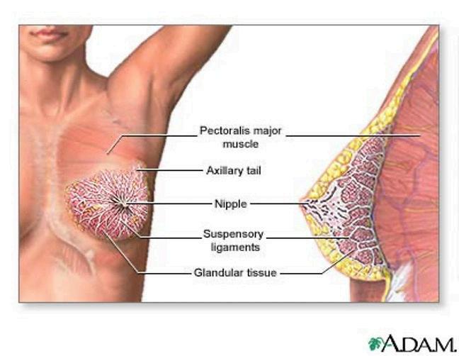
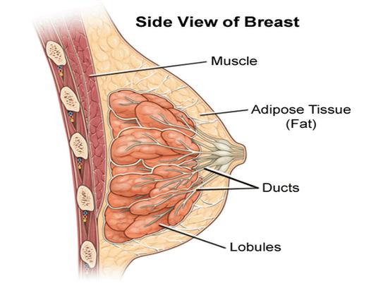
The breast of a woman is made up of the sacs that make the breast milk (the lobules), the tubes that carry the milk to the nipple (the ducts), and the surrounding fatty tissue The areola is the darker colored skin surrounding the nipple Nearly all breast cancers begin in either the lobules or the ducts
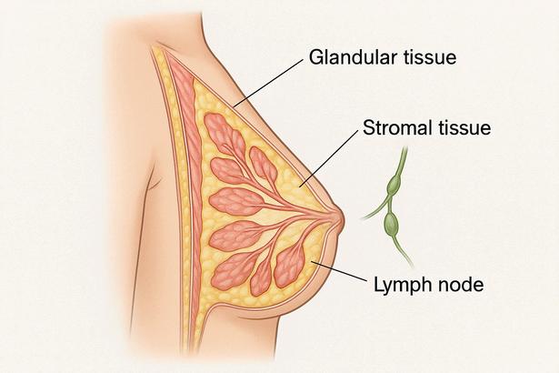
Blood vessels and lymph (pronounced limf) channels are found in each breast in the fatty tissue Lymph nodes are small lima bean shaped organs that are part of our body's immune system They are found from our head to our toes Part of their job is to filter germs and abnormal cells that are released into the lymph channels There are groups of lymph nodes in our axilla (armpit), above our collarbone, and in our chest which drain the fluid coming from the breast
Normal Breast Changes
Many women experience normal breast changes throughout the course of their life As mentioned above, these changes can occur during puberty, the menstrual cycle, pregnancy, and menopause In addition, weight gain and loss could contribute to breast changes
Puberty: As early as age 8, girls may develop tender lumps underneath one or both nipples called breast buds These signify the beginning of puberty During puberty the breasts may grow at different rates causing a discrepancy in size Many adult women have one slightly larger breast which is a normal variation
Menstrual Cycle: Hormonal changes occur during a period that can cause the breasts to swell, feel tender, sore, or lumpy. Birth control pills convince the body that it is pregnant. This is called pseudopregnancy. This may cause breasts to become firmer, tender, and slightly larger.
Pregnancy: Breast changes can occur during a woman's entire pregnancy and after childbirth. During pregnancy, the breast lobules become enlarged in preparation for the breasts to produce milk, which causes them to get bigger. The breasts may be tender, and the areola may be darker at this time In the third trimester, breasts will start leaking a yellow fluid called colostrum, the precursor to breast milk After delivery or when the individual stops breast feeding, pregnancy-related breast changes will slowly go away and the ducts and lobules of the breast tissue will go back to a prepregnancy state once milk production has ended
Menopause: With age, breasts may be less firm, lumpy, sore, or swollen The period of perimenopause is associated with much less predictable hormone cycles and therefore can be associated with increased symptoms or tenderness and benign lesions like cysts (see description below) These changes will be less evident if you are using hormone replacement therapy (HRT), as HRT can maintain the breast in a pre-menopausal state, including breast cysts, which would typically go away without HRT Women should experience less benign lumps during menopause; therefore, any new lump should be investigated by a physician
Self-Breast Exam
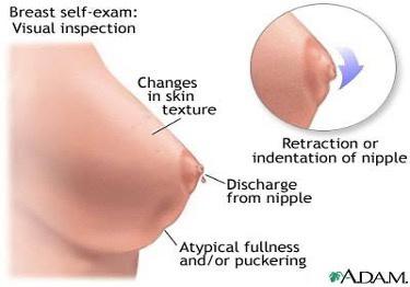
It is important to perform a regular breast self-exam to detect any unusual changes in the appearance or feel of the breasts. Optimally, a breast self-exam should be done once a month. Breasts should be checked approximately 7-10 days from the beginning of the menstrual cycle, or roughly the same time each month if periods are no longer occuring
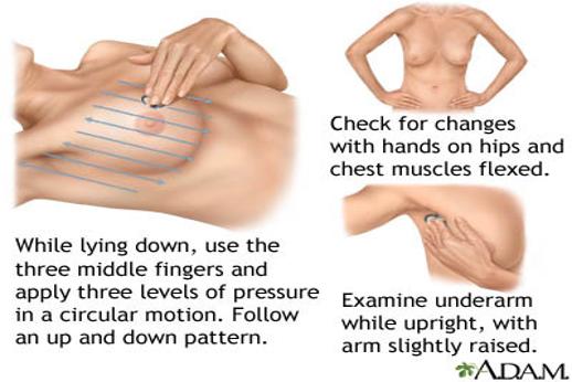
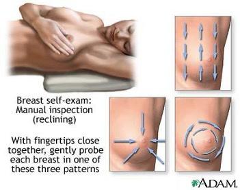
When Performing a Self-Breast Exam
1 Face the mirror with hands by the side of the body and then above the head Notice flattening, bulging, or puckering of the breasts and any changes in skin color
2.Next, lie down and raise one arm over the head. Use the other hand to examine one breast. Then switch hands for the other breast.
Use the pads of the index, middle, and ring fingers in an up-and-down or circular motion with light, medium, and firm pressure pushing the breast tissue against the ribcage. Check the nipples for any changes or discharge by squeezing them gently.
Common Benign Conditions
Fibrocystic Changes
These changes occur in the glandular (lobules and ducts) and stromal tissues of the breast (the "supporting framework" including fatty and connective tissues that gives the breast its shape) and can cause the breasts to feel lumpier, thicker, and in some cases swollen, sore, or tender Fibrocystic changes affect half of all women at some point Women between twenty and fifty years most often experience fibrocystic changes If a biopsy is done for an indeterminant finding, there are many pathologic terms that may be used, ALL of which are totally benign, and do not indicate an increase in the risk of developing breast cancer. These terms include:
Usual ductal hyperplasia
Flat ductal atypia (FEA)
Pseudoangiomatous stromal hyperplasia (PASH)
Low risk ductal intraepithelial hyperplasia
Fibroadenomatoid changes
Lymphocytic lobulitis
Stromal calcifications, or calcifications in benign ducts
Papillomas
Stromal Fibrosis
Cysts
These are fluid-filled sacs that feel like movable lumps within the breasts Cysts are usually bigger and more painful before the onset of the menstrual cycle and resolve somewhat after the period ends Women with breast cysts are not known to have a greater risk of developing cancer Cysts do not require any procedure once accurately diagnosed unless they become painful and then they can be drained as an office procedure. If bloody fluid is found in a cyst, then the cyst should be removed for evaluation.
Fibroadenomas
They are the most common benign tumor of the breast Fibroadenomas can be small or large and can move freely within the breast tissue They are commonly found in women in their teens, twenties and thirties, but can be seen at all ages Women who form a single fibroadenoma are more likely to produce additional fibroadenomas during their lifetime These tumors are estrogen-stimulated and can grow larger over time in cases such as pregnancy Fibroadenomas can be associated with discomfort, especially if they are very large and press on other parts of the breast If the fibroadenoma does not appear to be growing over time or causing discomfort, then it does not need to be removed.
Phyllodes Tumors
Phyllodes tumors are rare breast tumors that develop in the connective (stromal) tissue rather than the ducts or lobules. They are typically found as a firm, fast-growing lump and can be benign, borderline, or malignant, though most are not cancerous. Treatment usually involves surgical removal with a margin of normal tissue to reduce the risk of recurrence. Regular follow-up is important, as these tumors can sometimes come back after treatment.
Intraductal Papillomas
These are benign growths in the milk ducts of the breast which can sometimes have atypical (precancerous) cells Typically, they cause clear or bloody nipple discharge These may require excision for full diagnosis or to rule out
Mammographic Calcifications
These are calcium deposits in the breasts that are not caused by calcium in food or supplements, but by normal aging, inflammation, and trauma Calcifications can be characterized as macrocalcifications and microcalcifications. Macrocalcifications are large calcium deposits related to non-cancerous conditions and do not need further investigation. Microcalcifications are small specks of calcium that can appear alone or in clusters. Certain shapes or patterns of microcalcifications may be a cause of concern and require further imaging or biopsy. Calcifications cannot be felt and will be picked up by a routine mammogram.
Fat Necrosis
This is a benign lump that forms in the breast due to damaged or degenerated fatty tissue, usually because of trauma, previous surgery, and previous radiation therapy to the chest Women with very large breasts more commonly experience fat necrosis
Other Benign Conditions
Lipoma
This is a benign lump or mass that is made of normal fat cells A lipoma can be large in size, and they can be found anywhere on the body It is soft in texture and can be visible under the skin Often, there may be multiple lipomas present. Family history appears to play a role in the development of a lipoma.
Galactocele
This is a milk-filled cyst that is usually caused by a blocked milk duct in women who are pregnant or lactating It is always benign
Gynecomastia
This is the term used to describe breast enlargement in a male. Nearly all boys develop this at some point during puberty, but it is usually temporary. Adult men can develop breast masses or breast enlargement, and it is often associated with the use of certain medications. This is not a risk factor for the future development of breast cancer, but any new breast mass in an adult male should be evaluated as men can also develop breast cancer.
Breast Pain
Most women experience breast pain or breast tenderness at some point in their lives It is unclear why this occurs; however, it is rarely a sign of breast cancer Statistically only 1 2% to 6 7% of patients having breast pain are diagnosed with breast cancer Women with breast pain may also present with redness, skin thickening, and a warm sensation to the skin over the breast These symptoms should always be brought to a physician’s attention to be evaluated since they may represent an infection (mastitis) requiring antibiotics, or a rare form of breast cancer (inflammatory breast cancer) Possible causes of breast pain could be due to hormonal changes, trauma, caffeine intake, infection or cysts. If breast pain is persistent and annoying, then it is recommended to limit caffeine intake, take ibuprofen (Advil), wear a supportive garment, and apply warm compresses or ice packs to relieve it.
Nipple Discharge
Clear or milky nipple discharge from the nipple is common among women and usually noncancerous. Bloody nipple discharge, or spontaneous, persistent, non-bloody discharge other than milk is a cause for concern and should be checked. Ninety-five percent of the time it is benign and related to an intraductal papilloma or inflammation of the milk duct, but a surgical excision may be necessary for diagnosis A radiologic procedure called a ductogram may also be recommended
Visual Nipple Changes
All women should be aware of the normal appearance and texture of their breasts It is important to look for visual changes in the breasts, and especially the nipple A visible rash or scaly change in the skin of the nipple may present Paget’s disease, which is a rare type of ductal breast cancer It is important to notify a physician immediately if nipple changes arise Nipple changes will be evaluated first by imaging and sometimes require a biopsy depending on the findings Also, if new nipple retraction occurs then a physician should be contacted to evaluate this symptom.
Breast Imaging
Most breast cancers found in the United States today are picked up through routine screening x-rays of the breast The term screening refers to any test or procedure done on a patient without symptoms, to find a cancer before it can cause symptoms There are several different types of radiological studies available to evaluate the breast for the possibility that a cancer is present
Mammograms
A mammogram is an x-ray of the breast. A screening mammogram is a routine study, done every 1-2 years after the age of 40, for woman who are an average risk for development of breast cancer. Those who are deemed to be at a higher risk would start at an earlier age. This annual test should be done on younger women if they have a family history of early breast cancer. The breast tissue is compressed (squeezed) from two different directions and pictures are taken that will be evaluated by the radiologist. The breast is checked for changes from last year, new calcium deposits that are grouped in one area of the breast, or new masses or densities A letter will be sent directly to you from the radiologist notifying you about the results of your screening mammogram
A diagnostic mammogram is used when there is a breast complaint or symptom, or when an abnormality has been discovered on a routine screening mammogram Once someone has been diagnosed and treated for breast cancer, it is recommended they complete a diagnostic mammogram for the first year after diagnosis A diagnostic mammogram is a series of pictures used to evaluate the area of concern This may involve pictures with special paddles or magnification to better examine the tissue. It may also require an additional step of performing an ultrasound for a complete evaluation. The radiologist will speak to you directly about the findings and recommendations. A biopsy may be required if there is something still of concern.
Tomosynthesis (3-D Mammography)
The Center for Breast Cancers at Smilow Cancer Hospital and Yale Cancer Center performs 3-D mammography, also known as digital breast tomosynthesis. This technology allows the radiologist to view the breast in thin “slices” rather than as a whole. This improves detection of lesions and reduces false positives due to overlapping normal tissues We use the new 3-D technology in addition to our standard 2-D imaging This combination will provide the radiologist with an opportunity to compare the familiar standard images with your prior mammograms The 3-D imaging is performed simultaneously so the length of your exam does NOT change There is a small increase in radiation dose However, even with this increase, the total radiation does remains below the federal standards for mammography
Ultrasound
A breast ultrasound is a test that evaluates the breast tissue using sound waves. This is most helpful in determining whether a palpable mass is solid (in which case it could be benign or malignant) or a fluid filled sac (a benign cyst) As mentioned above, ultrasound can be used to look at questionable areas seen on the mammogram to better determine what they might represent
An ultrasound is a painless test where you will be comfortably lying on your back, and the technician will rub a handheld probe covered in gel over your breast Biopsies frequently can be done under the guidance of breast ultrasound
Screening breast ultrasound can be a useful backup tool in evaluating dense breast tissue Dense breast tissue can result in poorer detection of cancers by mammography. In addition, it is now believed that dense breast tissue slightly increases a woman's risk for developing breast cancer. For these reasons additional imaging studies, such as ultrasound or MRI, may be indicated depending on your particular risk factors. In this case, all areas of both breasts will be evaluated with the sound wave probe. It is important to remember that there are pros and cons of this additional screening test. While ultrasound may detect cancers not found on mammography, it is possible that additional findings may require additional imaging follow-up or biopsy.
Magnetic Resonance Imaging (MRI)
A breast MRI is a very sensitive examination which uses a powerful magnetic field and a computer to take detailed pictures of the breast tissue A dye called gadolinium is injected into your vein as part of this scan This test can be used to help detect abnormalities that are not easily seen with either mammography or ultrasound You will lie on your stomach on a table which slides, feet first, inside the magnet During imaging it can be noisy so headphones will be placed over your ears MRI is performed for several reasons It is often used in patients who present with a lymph node that has cancer, but the mammogram and ultrasound are negative If there is a question of cancer invading the chest wall or an implant rupture, MRI may also be useful Many surgeons will use MRI to better evaluate the extent of malignancy to help with surgical planning, or to assess the response of a cancer after preoperative chemotherapy. Breast MRI can also be used for screening in high-risk individuals.
Appropriate patients for high-risk screening by breast MRI include:
A known pathogenic variant (mutation) in a gene associated with a lifetime risk of breast cancer of 20% or more in a person or a first or second degree relative Strong family history (lifetime risk of >20% by Tyrer-Cuzick or similar risk assessment model) Prior chest wall radiation given between the ages of 10-30 years
Like ultrasound, there are pros and cons to screening breast MRI and false positives requiring additional imaging or biopsy are common
Methods of Breast Biopsy
If any part of your evaluation shows an indeterminate or suspicious abnormality in your breast, a biopsy may be done A small piece of tissue is removed and sent to pathology where it is studied under a microscope There are four main types of breast biopsies
Core Needle Biopsy
This type of biopsy provides a sample of tissue for the pathologist to examine and is done under a local, or numbing, anesthetic. It is often done with the help of an imaging study like ultrasound, mammogram (called stereotactic needle biopsy), or MRI. A small metal clip is usually left to mark the area where the biopsy was taken for future reference. This marker clip will remain in the breast unless the pathology results show something which needs to be removed. This procedure takes about 30 to 60 minutes. It is recommended that patients avoid strenuous upper body activity, such as weight lifting for 1-2 days following the procedure. In addition, it is recommended that patients avoid submerging the breast under water, such as swimming or taking a bath, for 1-2 days postprocedure, though showering is permissible
Excisional Biopsy
This type of biopsy is performed by the surgeon if the abnormality cannot be biopsied with ultrasound, mammogram, or MRI This is much less common than it used to be, and only a small percentage of lesions are biopsied this way After using local numbing medicine, usually with a medication that makes you sleepy, the surgeon makes an incision on the breast to remove the abnormal tissue. If the abnormality cannot be felt, it may be necessary for a guide wire to be placed into the breast to mark the location (called a needle localized excision). This wire is placed by the radiologist and/or surgeon either 1-2 weeks prior to surgery or sometimes immediately before the procedure.
Skin Punch Biopsy
This type of biopsy is performed in the surgeon’s office using local anesthesia to numb the skin before a small piece of skin and tissue is removed. This technique is used only for findings which are seen in the skin of the breast
Learning that you have a breast abnormality and need a biopsy can be stressful and anxiety producing Many women find that anticipating the biopsy procedure and then waiting for the results can be very stressful With that in mind, we want you to know that there is a social worker in the breast center who is available to talk with you, help you cope with the “limbo of not knowing,” and discuss anything else that might be on your mind
Pathological Findings that Cause an Increased Risk for Cancer
It is important to understand that even though there is an overall increased risk for cancer with these listed pathological findings, women with these conditions are more likely to never get breast cancer than to get it Surgical biopsies are sometimes performed on these lesions to prove that they are not associated with cancer
Lobular Intraepithelial Neoplasia (LIN & LCIS) and Atypical Hyperplasia (ADH)
These are two different pathologically-seen lesions which describe abnormal growth of cells lining the lobules or the milk ducts and reflect a higher risk for development of breast cancer. Some of these findings are surgically removed, some are treated with medication, and some are monitored with imaging.
Complex Sclerosing Lesions or Radial Scars
This is a benign breast lesion that distorts the normal breast tissue and will be monitored for changes by your doctor
Cancer 101
The body is made up of hundreds of millions of living cells of many different types. Normal cells grow, divide and die in a very orderly process. This helps replace normal cells and helps to keep us all healthy.
Sometimes cells begin to divide even when new cells are not needed, and these extra cells can form a mass of tissue called a tumor. Not all tumors are cancerous. Benign tumors can grow large and press on other organs but have no ability to invade other tissues or spread to other parts of the body. Benign breast tumors are never a threat to your life.
Malignant tumors are cancer, and their cells are not normal Instead of dying, cancer cells keep on growing and forming new cancer cells Cancer cells can invade, or grow into, other nearby tissues They can also break away from their original site and enter the bloodstream or lymphatic channel This is how a metastasis (muh-tas-tuh-sis) begins These breakaway cells can then form new tumors in other vital organs, and it is this process that can make cancer life-threatening
Breast cancer is a malignant tumor that starts from cells that line the milk ducts or in the lobules that make up the breast. If cancer has spread outside the breast, it is commonly seen in the lymph nodes under the armpit (axillary nodes). Cancer cells may also spread to other parts of the body, like the lungs, liver, bones or brain. If cancer spreads to other parts of the body, the new spots are still named after the primary tumor. For example, if breast cancer spreads to the bones, a biopsy of this area would show breast cancer cells. We would call this metastatic breast cancer, not bone cancer.
Causes of Breast Cancer
DNA is the chemical in each of our cells that helps make up our genes, and genes are the blueprint for how our cells are supposed to work. Sometimes a change occurs in DNA that can make a normal breast cell become cancer Most of these changes occur spontaneously, and the causes are not known There are DNA changes which will run through families called mutations, some of which will increase the risk for developing breast cancer
There are a number of genes known to be associated with an increased risk to develop breast cancer When working properly, these genes keep cancer from developing by regulating the way cells grow and divide When a mutation is present from birth the person has less of this regulating message and are at increased risk to develop certain cancers The most well known genes associated with hereditary breast cancer are BRCA1 and BRCA2. People born with a mutation in one of these genes are at increased risk to develop breast, ovarian, pancreatic, melanoma, and prostate cancer.
All patients who are women or assigned female at birth are at risk for breast cancer. Men or patients assigned male at birth can also get breast cancer, but this is rare, accounting for only one percent of all breast cancers. Gender-diverse or transgender patients, especially those taking supplemental hormones, may be at increased risk of breast cancer development regardless of their sex at birth. We recommend discussing specific risks with your doctor
The two most important risk factors for developing breast cancer are being a female and getting older Here is a chart which shows the chances of developing breast cancer by age:
By age 25: one in 19,608
By age 35: one in 622
By age 45: one in 93
By age 55: one in 33
By age 65: one in 17
By age 75: one in 11
By age 85: one in 9
By age 95: one in 8
Finding Cancer
There are two primary ways that breast cancers are discovered They can either be felt by the patient on a breast self-examination or by her physician on a clinical breast examination, or they are found on an imaging test of the breast We will discuss both of these two possibilities
Palpable Breast Cancers
These cancers present as a mass, or lump, in the breast. This can be a new lump or a lump that has grown or changed. There are other changes in the breast that can be the first indication of breast cancer. All these symptoms, as well as any new or changing breast mass, should certainly be brought to your health care provider's attention. These include:
A change in the size or shape of the breast
A retraction or dimpling of the skin anywhere in the breast
Red, flaky or swollen skin anywhere on the breast
New localized breast pain
A nipple that is tender or suddenly turns inward (gets retracted)
Bloody nipple discharge, or spontaneous persistent non-bloody discharge other than milk
Image-Detected Cancers
Many cancers are found on mammograms, ultrasounds or MRIs – before they are ever felt For more information about each of these tests, see the imaging section of this guide If you have an abnormal mammogram, you may be called back for additional views and/or other studies. If something abnormal is still seen, a biopsy may be needed to find out what this is.
Risk Factors
Risk Factors You Cannot Change
Sex
Breast cancer is 100 times more common in women than men
Male-specific Risk Factors
Increasing age, genetic mutations (particularly BRCA2), family history of breast cancer, liver disease, and radiation exposure to the chest
Age
About two out of three women with invasive breast cancer are 55 years old or older
Family History
Breast cancer risk is higher in women who have close blood relatives on either their mother’s or father’s side of the family. This risk increases with the number of individuals affected.
Genetic Risk Factors
Approximately 5-10% of breast cancers are caused by inherited mutations in genes like BRCA1 and BRCA2.
Personal History of Breast Cancer
A woman with a breast cancer in one breast has a greater chance of getting a new breast cancer in either breast
Race
White women are more likely to develop breast cancer than African American women, but African American women are more likely to die of their cancer due to a high prevalence of triple negative breast cancer
Asian Hispanic and Native American women have a lower risk of developing or dying from breast cancer
Early Breast Radiation
Women who have had radiation treatments to the chest area during puberty or young adulthood have a greatly increased risk of developing breast cancer
Atypical Breast Lesions
Women who have had previous biopsies showing atypical proliferative lesions have an increased risk for developing breast cancer. There is a long list of these lesions including LCIS, atypical ductal hyperplasia, and papillomatosis.
Menstrual History
Women who begin to have their periods at a younger age, or who have menopause at an older age are at a slightly increased risk for developing breast cancer.
Risk Factors Related to Lifestyle Decisions
Pregnancy History
Women who have not had children or who have their children after the age of 30 have an increased risk of breast cancer
Hormone Replacement Therapy
Postmenopausal combination hormone replacement with both estrogen and progesterone increases the risk of developing breast cancer Estrogen alone does not appear to increase the risk
Alcohol
Women who have one drink a day have a small increased risk of developing breast cancer
Heavy alcohol users have much more risk
Obesity
Women who are overweight or obese, especially after menopause, have a higher risk of breast cancer.
Sedentary Lifestyle
Recent studies clearly show that exercise decreases the risk of breast cancer.
Conclusion
It is important to remember that it is normal to experience changes in the breasts throughout life Most breast masses and lumps are benign
If there are concerns about any changes in your breasts, we encourage you to consult your physician for an evaluation.
A new lump or mass, especially if it is firm, painless, and does not move easily
Swelling or thickening of breast tissue
Skin changes, such as dimpling, redness, puckering, or scaling
Nipple changes, including new inversion (turning inward), thickening, or skin changes
Nipple discharge, particularly if it is bloody or occurs without squeezing
Persistent breast pain that is new, localized, and not associated with injury
Enlarged lymph nodes, especially in the armpit or near the collarbone
A change in breast size or shape that is new and unexplained