3 minute read
“Dormant” Cone Photoreceptors Continue to Function in Degenerating Retina, Indicating Potential Treatment Target


Amultidisciplinary team of UCLA Stein Eye Institute researchers has found that what have long been thought to be dormant cone photoreceptors in the degenerating retina actually continue to function, producing responses to light and driving retinal activity for vision. The group’s findings, published in the journal Current Biology, suggest that therapeutic interventions to protect these cells, or enhance their sensitivity, have the potential to preserve nearly normal daytime vision.
The vast majority of inherited retinal degenerative diseases are triggered by mutations leading to the death of rod photoreceptors—the cells that encode dim-light vision. The cone photoreceptors—those that are active in daylight— are typically kept alive for some time, undergoing morphological changes in response to the rod degenerations in which they retract the outer segments required for producing light responses, as well as the pedicle that communicates with the downstream cells in the retina.
It was believed that this process rendered the cone photoreceptors dormant. “The presumption with dormancy is that the cones are just sitting there, not doing anything, in retinal degenerative diseases,” says Alapakkam P. Sampath, PhD, Grace and Walter Lantz Endowed Chair in Ophthalmology at the UCLA Stein Eye Institute and the study’s senior author. Previous research suggested that these dormant cells were not functional, and attempts to record from them revealed no light-driven activity, Dr. Sampath explains.
But in an examination of the membrane properties of cones in mice following the degeneration of rods, Dr. Sampath and his colleagues—including labs led by Drs. Greg D. Field, Gordon L. Fain , and David S. Williams —found otherwise. For the study, they used the patch clamp recording method, a laboratory technique for measuring currents in living cells while controlling the cells’ membrane potential, or membrane voltage. These single-cell recordings can establish key features of the cell’s activity—including the presence of specific membrane currents, whether the cell has light responses, and whether they might connect to downstream neurons in the retina. In addition, the investigators used multi-electrode array recordings that establish the activity of all retinal ganglion cells, and that can show the ganglion cell’s ability to respond to visual stimuli that vary in spatial location over time.
These recordings revealed not only that the remaining cones in a retina where the rods had mostly degenerated were still functional, but that, based on downstream signals recorded from the retina, visual processing was not as compromised as might be expected. “This study shows, for the first time, that these cones have residual light responses, generated by somewhat normal mechanisms,” Dr. Sampath says. “However, the response is much less sensitive than normal because the specialized part of the cone that senses light and increases the efficiency of light capture in phototransduction is missing.”
Importantly, though, these light responses appear to be driving responses in the middle of the retina cells, likely protecting vision to some extent. “We were surprised to find that the drop-off in sensitivity for the ganglion cells that project to the brain was much less,” Dr. Sampath says. “It seems that adaptational mechanisms in the inner retina might be trying to minimize the sensitivity difference to preserve robust signaling in the ganglion cells. This is consistent with what we know about the brain: Homeostatic mechanisms that respond to injury and disease typically cover up the deficiency.”
The findings on the retinal ganglion cells, contributed by the laboratory led by Dr. Field, have significant clinical implications, Dr. Sampath says. “The fact that the spatial and temporal properties of the retinal ganglion cell responses are largely unaffected suggests that if you can increase the light sensitivity a little bit, you may be able to restore almost normal daytime vision, at least as far as cones go,” he explains.
The previous research that was unable to record light responses from cone photoreceptors after the death of rods did find, encouragingly, that placing lightgated ion channels in the residual cones drove visual responses, Dr. Sampath notes. Currently, optogenetics clinical trials are underway in Europe that place light-gated ion channels into the retinas where the rods and cones have died in an effort to restore vision.
Dr. Sampath says the important findings resulting from his group’s collaboration illustrate a great strength of the UCLA Stein Eye Institute. “Sweeping studies like this can be done entirely in-house because of the expertise we have built in retinal cell and circuit-level biology,” he explains.
The next step for the Stein Eye researchers is to establish the extent to which the neuroprotection or enhancement of the dormant cones will allow the rescue of vision in various forms of blindness. “Protecting these cells is step number one from the perspective of maintaining vision,” Dr. Sampath says. “Step two, it appears from our research, is to look to increase the light sensitivity of these residual cones.”
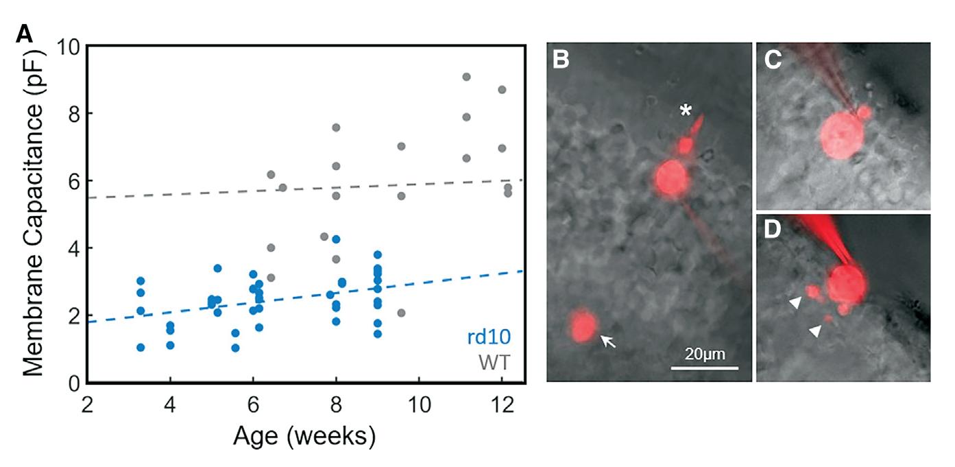
Membrane capacitance and morphology of degenerating cones
(A) Membrane capacitance was measured for WT cones (gray, n = 22) and rd10 cones (blue, n = 42) and plotted as a function of animal age. Dash lines represent fits with linear regression: WT slope = 0.05, 95% CI = [-0.06, 0.17] for data from 6 to 12 weeks, rd10 slope = 0.14, 95% CI = [0.03, 0.26] for data from 3.5 to 9.5 weeks. (B–D) Images of cones filled with Alexa-750 from a WT mouse (B), and rd10 mice ages 4 weeks old (C), and 9 weeks old (D). Note large outer segment (asterisk) and cone pedicle (arrow) of the WT cone and processes of 9-week rd10 cone (arrowheads). Scale bars, 20 m m.
Dr. Sampath says the important findings resulting from his group’s collaboration illustrate a great strength of the UCLA Stein Eye Institute.


