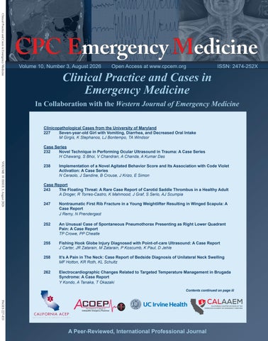
Clinical Practice and Cases in Emergency Medicine
l a c i M eedincrease
Open Access at www.cpcem.org
Clinical Practice and Cases in Emergency Medicine
Clinicopathological Cases from the University of Maryland 227 Seven-year-old Girl with Vomiting, Diarrhea, and Decreased Oral Intake M Girgis, K Stephanos, LJ Bontempo, TA Windsor Case Series Novel Technique in Performing Ocular Ultrasound in Trauma: A Case Series 232 H Chawang, S Bhoi, V Chandran, A Chanda, A Kumar Das VOLUME 10 ISSUE 3, August 2026
EMERGENCY MEDICINE IS THE ONLY SPECIALTY WITH ITS OWN BUDGET ALLOCATION.
238
Implementation of a Novel Agitated Behavior Score and Its Association with Code Violet Activation: A Case Series N Ceraolo, J Sandine, B Crouse, J Krizo, E Simon
Case Report The Floating Threat: A Rare Case Report of Carotid Saddle Thrombus in a Healthy Adult 243 A Droger, R Torres-Castro, K Mahmood, J Graf, S Serio, AJ Scumpia 247
Nontraumatic First Rib Fracture in a Young Weightlifter Resulting in Winged Scapula: A Case Report J Remy, N Prendergast
252
An Unusual Case of Spontaneous Pneumothorax Presenting as Right Lower Quadrant Pain: A Case Report TP Crowe, PP Cheatle
255
Fishing Hook Globe Injury Diagnosed with Point-of-care Ultrasound: A Case Report J Carter, JR Zatarain, M Zatarain, P Koscumb, K Paul, D Jehle
258
It’s A Pain in The Neck: Case Report of Bedside Diagnosis of Unilateral Neck Swelling MF Hotton, KR Roth, KL Schultz
262
Electrocardiographic Changes Related to Targeted Temperature Management in Brugada Syndrome: A Case Report Y Kondo, A Tanaka, T Okazaki Contents continued on page iii
PAGES 227-433
@CALIFORNIAACEP
www.californiaacep.org
ISSN: 2474-252X
In Collaboration with the Western Journal of Emergency Medicine
rat e
CALIFORNIA ACEP SECURED $200M ANNUALLY TO INCREASE MEDI-CAL REIMBURSEMENT FOR EMERGENCY PHYSICIANS!
Volume 10, Number 3, August 2026
A Peer-Reviewed, International Professional Journal
Penn State Health Emergency Medicine
JOIN OUR TEAM
EMERGENCY MEDICINE OPPORTUNITIES AVAILABLE
About Us: Penn State Health is a multi-hospital health system serving patients and communities across central Pennsylvania. We are the only medical facility in Pennsylvania to be accredited as a Level I pediatric trauma center and Level I adult trauma center. The system includes Penn State Health Milton S. Hershey Medical Center, Penn State Health Children’s Hospital and Penn State Cancer Institute based in Hershey, Pa.; Penn State Health Hampden Medical Center in Enola, Pa.; Penn State Health Holy Spirit Medical Center in Camp Hill, Pa.; Penn State Health Lancaster Medical Center in Lancaster, Pa.; Penn State Health St. Joseph Medical Center in Reading, Pa.; Pennsylvania Psychiatric Institute, a specialty provider of inpatient and outpatient behavioral health services, in Harrisburg, Pa.; and 2,450+ physicians and direct care providers at 225 outpatient practices. Additionally, the system jointly operates various healthcare providers, including Penn State Health Rehabilitation Hospital, Hershey Outpatient Surgery Center and Hershey Endoscopy Center. We foster a collaborative environment rich with diversity, share a passion for patient care, and have a space for those who share our spark of innovative research interests. Our health system is expanding and we have opportunities in both academic hospital as well community hospital settings.
Benefit highlights include: • Competitive salary with sign-on bonus • Comprehensive benefits and retirement package • Relocation assistance & CME allowance • Attractive neighborhoods in scenic central Pennsylvania
FOR MORE INFORMATION PLEASE CONTACT: Amber Winters, MBA Penn State Health Physician Recruiter awinters@pennstatehealth.psu.edu
Penn State Health is fundamentally committed to the diversity of our faculty and staff. We believe diversity is unapologetically expressing itself through every person’s perspectives and lived experiences. We are an equal opportunity and affirmative action employer. All qualified applicants will receive consideration for employment without regard to age, color, disability, gender identity or expression, marital status, national or ethnic origin, political affiliation, race, religion, sex (including pregnancy), sexual orientation, veteran status, and family medical or genetic information.
CALAAEM 2026 WESTERN REGION SAVE THE DATE
CONFERENCE
OCTOBER 17 | 8AM - 5PM UCSD SIXTH COLLEGE SAN DIEGO, CA
Plan to join California’s emergency medicine community for a day of advocacy, education, networking, and career growth! FREE for Cal/AAEM Members | $35 for AAEM members and non-members Please note: You must be a CAL/AAEM member for free registration.
More event details and registration information to be announced!
Clinical Practice and Cases in Emergency Medicine Indexed in PubMed and full text in PubMed Central
Rick A. McPheeters, DO, Editor-in-Chief Kern Medical/UCLA- Bakersfield, California Patrick Meloy, MD, Deputy Editor Emory University Hospital Midtown, Atlanta, Georgia Mark I. Langdorf, MD, MHPE, Senior Associate Editor University of California, Irvine School of Medicine- Irvine, California Shahram Loftipour, MD, MPH, Senior Associate Editor University of California, Irvine School of Medicine- Irvine, California Shadi Lahham, MD, MS, Associate Editor Kaiser Permanente- Orange County, California John Ashurst, DO, Decision Editor/ ACOEP Guest Editor Kingman Regional Health Network, Arizona Anna McFarlin, MD, Decision Editor Louisiana State University Health Science Center- New Orleans, Louisiana
Christopher Sampson, MD, Decision Editor University of Missouri- Columbia, Missouri Joel Moll, MD, Decision Editor Virginia Commonwealth University School of Medicine- Richmond, Virginia Alexander Scumpia, DO, MHA, MSc, Decision Editor HCA FL Aventura Hospital Florida Steven Walsh, MD, Decision Editor Einstein Medical Center Philadelphia-Philadelphia, Pennsylvania Melanie Heniff, MD, JD, Decision Editor University of Indiana School of Medicine- Indianapolis, Indiana Austin Smith, MD, Decision Editor Vanderbilt University Medical Center-Nashville, Tennessee Rachel A. Lindor, MD, JD, Decision Editor Mayo Clinic College of Medicine and Science, Minnesota
Lev Libet, MD, Decision Editor Kern Medical/UCLA- Bakersfield, California J. David Gatz, MD, Decision Editor University of Maryland School of Medicine, Maryland Ezhilkugan Ganessane, MD, Decision Editor All India Institute of Medical Sciences (AIIMS) Madurai, India Ryan Ley, MD, MBA, MS, Decision Editor Arrowhead Regional Medical Center, California
Amin A. Kazzi, MD, MAAEM The American University of Beirut, Beirut, Lebanon Anwar Al-Awadhi, MD Mubarak Al-Kabeer Hospital, Jabriya, Kuwait Arif A. Cevik, MD United Arab Emirates University College of Medicine and Health Sciences, Al Ain, United Arab Emirates Abhinandan A.Desai, MD University of Bombay Grant Medical College, Bombay, India Bandr Mzahim, MD King Fahad Medical City, Riyadh, Saudi Arabia Barry E. Brenner, MD, MPH Case Western Reserve University Brent King, MD, MMM University of Texas, Houston Daniel J. Dire, MD University of Texas Health Sciences Center San Antonio David F.M. Brown, MD Massachusetts General Hospital/Harvard Medical School
Jacqueline K. Le, MD, Decision Editor Desert Regional Medical Center Arihant Jain, MD, Decision Editor All India Institute of Medical Sciences (AIIMS), New Delhi Grace Hickam, MD, MEd, Decision Editor Virginia Commonwealth University, Virginia
Editorial Board Khrongwong Musikatavorn, MD King Chulalongkorn Memorial Hospital, Chulalongkorn University, Bangkok, Thailand
Edward Michelson, MD Texas Tech University Edward Panacek, MD, MPH University of South Alabama
Leslie Zun, MD, MBA Chicago Medical School
Erik D. Barton, MD, MBA Icahn School of Medicine, Mount Sinai, New York
Linda S. Murphy, MLIS University of California, Irvine School of Medicine Librarian
Francesco Dellacorte, MD Azienda Ospedaliera Universitaria “Maggiore della Carità,” Novara, Italy
Nadeem Qureshi, MD St. Louis University, USA Emirates Society of Emergency Medicine, United Arab Emirates
Francis Counselman, MD Eastern Virginia Medical School Gayle Galleta, MD Sørlandet Sykehus HF, Akershus Universitetssykehus, Lorenskog, Norway
Niels K. Rathlev, MD Tufts University School of Medicine
Hjalti Björnsson, MD Icelandic Society of Emergency Medicine
Pablo Aguilera Fuenzalida, MD Pontificia Universidad Catolica de Chile, Región Metropolitana, Chile
Jacob (Kobi) Peleg, PhD, MPH Tel-Aviv University, Tel-Aviv, Israel
Peter A. Bell, DO, MBA Baptist Health Science University
Jonathan Olshaker, MD Boston University
Peter Sokolove, MD University of California, San Francisco
Katsuhiro Kanemaru, MD University of Miyazaki Hospital, Miyazaki, Japan
Robert M. Rodriguez, MD University of California, San Francisco
Advisory Board
Amal Khalil, MBA UC Irvine Health School of Medicine
Elena Lopez-Gusman, JD California ACEP American College of Emergency Physicians DeAnna McNett, CAE American College of Osteopathic Emergency Physicians John B. Christensen, MD California Chapter Division of AAEM Randy Young, MD California ACEP American College of Emergency Physicians Mark I. Langdorf, MD, MHPE UC Irvine Health School of Medicine
Robert Suter, DO, MHA UT Southwestern Medical Center Robert W. Derlet, MD University of California, Davis Rosidah Ibrahim, MD Hospital Serdang, Selangor, Malaysia Samuel J. Stratton, MD, MPH Orange County, CA, EMS Agency Scott Rudkin, MD, MBA University of California, Irvine Scott Zeller, MD University of California, Riverside Steven Gabaeff, MD Clinical Forensic Medicine Steven H. Lim, MD Changi General Hospital, Simei, Singapore Terry Mulligan, DO, MPH, FIFEM ACEP Ambassador to the Netherlands Society of Emergency Physicians Vijay Gautam, MBBS University of London, London, England Wirachin Hoonpongsimanont, MD, MSBATS Siriraj Hospital, Mahidol University, Bangkok, Thailand
Editorial Staff
Jorge Fernandez, MD California ACEP American College of Emergency Physicians University of California, San Diego Peter A. Bell, DO, MBA American College of Osteopathic Emergency Physicians Baptist Health Science University Robert Suter, DO, MHA American College of Osteopathic Emergency Physicians UT Southwestern Medical Center
Luke Garcia, BS Executive Editorial Director
Cassandra Saucedo, MS Executive Publishing Director
Acacia Jang, BS WestJEM Editorial Director
Isabella Choi, BS WestJEM Publishing Director
Isabella Cao, BS CPC-EM Editorial Director
Annie Zhang, BS WestJEM Associate Publishing Director
Stephanie Burmeister, MLIS WestJEM Staff Liaison
Omid Haghkhah, BS CPC-EM Publishing Director June Casey, BA Copy Editor
Shahram Lotfipour, MD, MPH UC Irvine Health School of Medicine Brian Potts, MD, MBA California Chapter Division of AAEM Alta Bates Summit-Berkeley Campus
Official Journal of the California Chapter of the American College of Emergency Physicians, the America College of Osteopathic Emergency Physicians, and the California Chapter of the American Academy of Emergency Medicine
Available in MEDLINE, PubMed, PubMed Central, Google Scholar, eScholarship, DOAJ, and OASPA. Editorial and Publishing Office: WestJEM/Depatment of Emergency Medicine, UC Irvine Health, 3800 W. Chapman Ave. Suite 3200, Orange, CA 92868, USA Office: 1-714-456-6389; Email: Editor@westjem.org
Volume 10, No. 3: August 2026
i
Clinical Practice and Cases in Emergency Medicine
Clinical Practice and Cases in Emergency Medicine Indexed in PubMed and full text in PubMed Central
This open access publication would not be possible without the generous and continual financial support of our society sponsors, department and chapter subscribers.
Professional Society Sponsors American College of Osteopathic Emergency Physicians California ACEP
California Chapter Division of American Academy of Emergency Medicine
Academic Department of Emergency Medicine Subscribers Albany Medical College Albany, NY
INTEGRIS Health Oklahoma City, OK
Robert Wood Johnson University Hospital New Brunswick, NJ
University of Florida, Jacksonville Jacksonville, FL
American University of Beirut Beirut, Lebanon
Kaweah Delta Health Care District Visalia, CA
Rush University Medical Center Chicago, IL
University of Illinois at Chicago Chicago, IL
Arrowhead Regional Medical Center Colton, CA
Kennedy University Hospitals Turnersville, NJ
Southern Illinois University Carbondale, IL
Augusta University Augusta GA
Kern Medical Bakersfield, CA
St. Luke’s University Health Network Bethlehem, PA
University of Illinois College of Medicine Peoria, IL
Baystate Medical Center Springfield, MA
Lakeland HealthCare St. Joseph, MI
Beaumont Hospital Royal Oak, MI
Lehigh Valley Hospital and Health Network Allentown, PA
Stanford/Kaiser Emergency Medicine Residency Program Stanford, CA
Beth Israel Deaconess Medical Center Boston, MA
Loma Linda University Medical Center Loma Linda, CA
Boston Medical Center Boston, MA
Louisiana State University Health Sciences Center New Orleans, LA
Brigham and Women’s Hospital Boston, MA
Madigan Army Medical Center Tacoma, WA
Brown University Providence, RI
Maimonides Medical Center Brooklyn, NY
Carl R. Darnall Army Medical Center Fort Hood, TX
Maricopa Medical Center Phoenix, AZ
Conemaugh Memorial Medical Center Johnstown, PA
Massachusetts General Hospital Boston, MA
Desert Regional Medical Center Palm Springs, CA
Mayo Clinic College of Medicine Rochester, MN
Doctors Hospital/Ohio Health Columbus, OH
Mt. Sinai Medical Center Miami Beach, FL
Eastern Virginia Medical School Norfolk, VA
North Shore University Hospital Manhasset, NY
Einstein Healthcare Network Philadelphia, PA
Northwestern Medical Group Chicago, IL
Emory University Atlanta, GA
Ohio State University Medical Center Columbus, OH
Genesys Regional Medical Center Grand Blanc, Michigan
Ohio Valley Medical Center Wheeling, WV
Hartford Hospital Hartford, CT
Oregon Health and Science University Portland, OR
Hennepin County Medical Center Minneapolis, MN
Penn State Milton S. Hershey Medical Center Hershey, PA
Henry Ford Hospital Detroit, MI
Presence Resurrection Medical Center Chicago, IL
University of Iowa Iowa City, IA University of Louisville Louisville, KY
Staten Island University Hospital Staten Island, NY
University of Maryland Baltimore, MD
SUNY Upstate Medical University Syracuse, NY
University of Michigan Ann Arbor, MI
Temple University Philadelphia, PA
University of Missouri, Columbia Columbia, MO
Texas Tech University Health Sciences Center El Paso, TX
University of Nebraska Medical Center Omaha, NE
University of Alabama, Birmingham Birmingham, AL
University of South Alabama Mobile, AL
University of Arkansas for Medical Sciences Little Rock, AR University of California, Davis Medical Center Sacramento, CA University of California Irvine Orange, CA University of California, Los Angeles Los Angeles, CA University of California, San Diego La Jolla, CA University of California, San Francisco San Francisco, CA UCSF Fresno Center Fresno, CA
University of Southern California/Keck School of Medicine Los Angeles, CA University of Tennessee, Memphis Memphis, TN University of Texas, Houston Houston, TX University of Texas Health San Antonio, TX University of Warwick Library Coventry, United Kingdom University of Washington Seattle, WA
University of Chicago, Chicago, IL
University of Wisconsin Hospitals and Clinics Madison, WI
University of Colorado, Denver Denver, CO
Wake Forest University Winston-Salem, NC
University of Florida Gainesville, FL
Wright State University Dayton, OH
State Chapter Subscribers Arizona Chapter Division of the American Academy of Emergency Medicine California Chapter Division of the American Academy of Emergency Medicine Florida Chapter Division of the American Academy of Emergency Medicine
Great Lakes Chapter Division of the American Academy of Emergency Medicine Tennessee Chapter Division of the
International Society Partners Emergency Medicine Association of Turkey Lebanese Academy of Emergency Medicine Mediterranean Academy of Emergency
Medicine Norwegian Society for Emergency Medicine
American Academy of Emergency Medicine Uniformed Services Chapter Division of the American Academy of Emergency Medicine Virginia Chapter Division of the American Academy of Emergency Medicine
Sociedad Argentina de Emergencias Sociedad Chileno Medicina Urgencia Thai Association for Emergency Medicine
To become a WestJEM departmental sponsor, waive article processing fee, receive print and copies for all faculty and electronic for faculty/residents, and free CME and faculty/fellow position advertisement space, please go to http://westjem.com/subscribe or contact: Stephanie Burmeister WestJEM Staff Liaison Phone: 1-800-884-2236 Email: sales@westjem.org
Clinical Practice and Cases in Emergency Medicine
ii
Volume 10, No. 3: August 2026
Clinical Practice and Cases in Emergency Medicine Indexed in PubMed and full text in PubMed Central JOURNAL FOCUS Clinical Practice and Cases in Emergency Medicine (CPC-EM) is a MEDLINE-indexed internationally recognized journal affiliated with the Western Journal of Emergency Medicine (WestJEM). It offers the latest in patient care case reports, images in the field of emergency medicine and state of the art clinicopathological and medicolegal cases. CPC-EM is fully open-access, peer reviewed, well indexed and available anywhere with an internet connection. CPC-EM encourages submissions from junior authors, established faculty, and residents of established and developing emergency medicine programs throughout the world.
Table of Contents continued 266
Sorely Mistaken—Soft Palatal Myxedema in Decompensated Hypothyroidism Presenting as a Sore Throat: Case Report
G Miller, B Myers 269
A Split from Traditional Orbital Compartment Syndrome Intervention: Case Report of Vision-saving Vertical Lid Split Procedure H Chason, B Zimmerman, A Jenzer, D Fay, J Elpers
273
Unusual Etiology and Presentation for Hyperkalemia—Dialysis Access Recirculation: A Case Report
277
Clinical Application of Intravenous Lipid Emulsion Therapy in Cocaine-associated Cardiac Arrest: A Case Report
JL Kim, JM Glazer, AE Achufusi, RE Tsuchida
R Offman, SK Baribeau
280
284
288
291
295
301
Incidental Wolff-Parkinson-White Syndrome Discovered Following Dicyclomine Use: A Case Report
J Wahhab, N Loveridge, M Siaj
Penetrating Neck Injury in a Child Presenting as a Simple Laceration: A Case Report
B Davitt, E Quinn
Retrograde Intubation in a Severe Fixed Spinal Deformity: A Case Report
S Joshi, S Nayak, S Chakraborty, H Kaur, N Siddiqua, S Bhoi
Cerebral Kounis Syndrome—A Rare Case Report of Cerebral Vasospasm Following Anaphylaxis
MH Ismail, S Muniandy, MS Azmi
Differentiating Weakness—an Atypical Presentation of Acute Neuromuscular Paralysis: A Case Report
J Golder, L Tjiattas-Saleski
Stretching the Limits: A Rare Case Report of Perimesencephalic Subarachnoid Hemorrhage During Yoga
A Vierra, M Sinha, L Jamal, N Khan, T Liebert, A Bokhari
306
Keeping An Eye Out for Stroke—Herpes Zoster Ophthalmicus Leading to Acute Ischemic Stroke S Burke, M Armbruster, J Le
Policies for peer review, author instructions, conflicts of interest and human and animal subjects protections can be found online at www.cpcem.org. Volume 10, No. 3: August 2026
iii
Clinical Practice and Cases in Emergency Medicine
Clinical Practice and Cases in Emergency Medicine Indexed in PubMed and full text in PubMed Central JOURNAL FOCUS Clinical Practice and Cases in Emergency Medicine (CPC-EM) is a MEDLINE-indexed internationally recognized journal affiliated with the Western Journal of Emergency Medicine (WestJEM). It offers the latest in patient care case reports, images in the field of emergency medicine and state of the art clinicopathological and medicolegal cases. CPC-EM is fully open-access, peer reviewed, well indexed and available anywhere with an internet connection. CPC-EM encourages submissions from junior authors, established faculty, and residents of established and developing emergency medicine programs throughout the world.
Table of Contents continued 310
Subungual Myiasis Presenting to the Emergency Department: A Case Report TD Marquis, AY Sheng
313
Emergency Department Presentation of Duloxetine-induced Acute Extrapyramidal Symptoms: A Case Report MW Shulby, CM Powell
316
Severe Tetanus Following a Rooster-Peck Injury Requiring Nasotracheal Intubation: A Case Report R White, H Singh, K Craver, R Cranston, K Hodge, E Vandervort
320
Yellow Oleander (Thevetia peruviana) Toxicity from a Misrepresented Dietary Supplement: A Case Report SE Long, A Grim, SK Bhandari, HQ Zaidi
324
Magnetic Mishaps—Small Bowel Obstruction Caused by Ingested Magnets Complicated by Appendicitis: A Case Report S Ball, M Wierzbicki
328
Dynamic Supraglottic Airway Collapse Diagnosed Using Airway Point-of-care Ultrasound: A Case Report A Jain, AM Muthanikkatt, SM Ayyan
333
Entrectinib-related Myocarditis Causing a Triangular QRS-ST-T Waveform Electrocardiographic Pattern: A Case Report W Dean, J Dean II, H Lichaa, C Wilbert
336
Blunt Thoracic Aortic Injury Presenting as Hemodynamically Stable: A Case Report J Knudsen, S Lucas, J Mangano
341
Adult Intussusception and Ischemic Bowel Potentially Associated with CurQD Supplementation: A Case Report of A Diagnosis Driven by Point-of-care Ultrasound A Parambath, B Patel, TJ Batchelor, NG Ashenburg, TL Ahern
345
Unmasking the Silent Liver-Lung Connection: A Pediatric Hepatopulmonary Syndrome Case Report SA Haupt, D Lalos, C Muntean, D Vaysman
Policies for peer review, author instructions, conflicts of interest and human and animal subjects protections can be found online at www.cpcem.org. Clinical Practice and Cases in Emergency Medicine
iv
Volume 10, No. 3: August 2026
Clinical Practice and Cases in Emergency Medicine Indexed in PubMed and full text in PubMed Central JOURNAL FOCUS Clinical Practice and Cases in Emergency Medicine (CPC-EM) is a MEDLINE-indexed internationally recognized journal affiliated with the Western Journal of Emergency Medicine (WestJEM). It offers the latest in patient care case reports, images in the field of emergency medicine and state of the art clinicopathological and medicolegal cases. CPC-EM is fully open-access, peer reviewed, well indexed and available anywhere with an internet connection. CPC-EM encourages submissions from junior authors, established faculty, and residents of established and developing emergency medicine programs throughout the world.
Table of Contents continued 350
Tension Hydrocele—How Point-of-care Ultrasound Helps in the Emergency Department: A Case Report S Potalivo, S Fornbacher, J Abadilla, R Goubert, ET Hirashima
354
Ultrasound-guided Hernia Reduction—Preventing Surgery for an Incarcerated Ventral Hernia: A Case Report A Lochner, E Stovicek, N Kman, S Petelinsek, J Cotton
358
VenoArterial Extracorporeal Membrane Oxygenation in Cardiac Arrest Suspected due to Massive Pulmonary Embolism: A Case Report T Chopra, L Dahlke, N Salinas, M Shabbir, T Gunn, S Torbati
363
Use of Balloon Tamponade Device for Aortoesophageal Fistula: A Case Report
367
Case Report: Lidocaine Toxicity Presenting with Focal Neurologic Findings T Yeung, I Estrada, TH Phan, EJ Samones, S Kalam
370
Persistent and Progressive Exfoliative Dermatitis: A Case Report
374
Use of Corrected QT Cutoffs Derived from Biological Variation to Predict Adverse Events Due to Antipsychotic Drugs: Case Report AHB Wu, M Alamillo, K Kendrick
380
A Diagnostic Pitfall in the Emergency Department —Aortic Dissection Masquerading as Acute Paraplegia: A Case Report
E Burton, T Wray
J Boukouris, S Elsherif
S Nagarajan, E Ganessane, Kowsthubha BG, A Somasundaram, N Balamurugan
385
Incidental Diagnosis of ST-Elevation Myocardial Infarction on Computed Tomography in a Burn Patient: A Case Report MJ Araujo, E Sandhu, A Romero, J Randhawa, R O’Donnell
388
Purulent Pericarditis Identified with Point-of-Care Echocardiography: A Case Report D Baquet, A Blevins, D Casey, C Glass, J Seaback, J Schoeneck
Policies for peer review, author instructions, conflicts of interest and human and animal subjects protections can be found online at www.cpcem.org. Clinical Practice and Cases in Emergency Medicine
v
Volume 10, No. 3: August 2026
Clinical Practice and Cases in Emergency Medicine Indexed in PubMed and full text in PubMed Central JOURNAL FOCUS Clinical Practice and Cases in Emergency Medicine (CPC-EM) is a MEDLINE-indexed internationally recognized journal affiliated with the Western Journal of Emergency Medicine (WestJEM). It offers the latest in patient care case reports, images in the field of emergency medicine and state of the art clinicopathological and medicolegal cases. CPC-EM is fully open-access, peer reviewed, well indexed and available anywhere with an internet connection. CPC-EM encourages submissions from junior authors, established faculty, and residents of established and developing emergency medicine programs throughout the world.
Table of Contents continued 392
Berberine Poisoning with Polymorphic Ventricular Tachycardia: A Case Report
395
Misplaced Central Venous Catheter Leading to a Chemothorax: Case Report HE Miller, S Lentz
399
Immunoglobulin A Vasculitis-Associated Ileoileal Intussusception in an Adult Male: Case Report
403
Altered Mental Status in Emergency Department Patient with Cerebral Septic Emboli from Infective Endocarditis: Case Report AG Theophanous, RG Theophanous
408
Transaminitis from Duloxetine: Case Report
MP Mesmin, MS Tweet, SH Kim, TJ Fulks
P Sawhney, P Frost, P Stueve, E Boccio
G Galletta, S Mehta, D Jancura, D Ryan, I Li
Images in Emergency Medicine 411 Stridor and Dysphagia Unmasking an Aberrant Right Subclavian Artery in a Toddler M Yasuda, T Ito 414
Yellow Granular Material in Hair: A Bedside Clue to Overdose from Cold Medication Containing Acetaminophen S Sakamoto
417
Elderly Male with a Painful Red Eye D Dahdal, A Nama, W Deng, B Hahn
419
Popliteal Artery Aneurysm Thrombosis Diagnosed with Point-of-Care Ultrasound A Gonedes, B Kohen, A Diallo, M McKenna, R Farrow II, E Boccio
421
Emergency Department Transvenous Pacemaker Placement Complicated by Tricuspid Mass K Molyneux, N Krejchi, M Fulton, M Youssef
424
Ruptured Abdominal Aortic Aneurysm Identified on Point-of-Care Ultrasound MH Sherman
Policies for peer review, author instructions, conflicts of interest and human and animal subjects protections can be found online at www.cpcem.org. Clinical Practice and Cases in Emergency Medicine
vi
Volume 10, No. 3: August 2026
Clinical Practice and Cases in Emergency Medicine Indexed in PubMed and full text in PubMed Central JOURNAL FOCUS Clinical Practice and Cases in Emergency Medicine (CPC-EM) is a MEDLINE-indexed internationally recognized journal affiliated with the Western Journal of Emergency Medicine (WestJEM). It offers the latest in patient care case reports, images in the field of emergency medicine and state of the art clinicopathological and medicolegal cases. CPC-EM is fully open-access, peer reviewed, well indexed and available anywhere with an internet connection. CPC-EM encourages submissions from junior authors, established faculty, and residents of established and developing emergency medicine programs throughout the world.
Table of Contents continued 426
Retained Stingray Barb in the Sole of the Foot S Sakamoto
429
Cutaneous Anthrax SC Christos, A Mazouni
Letter to the Editor 432 Recurrence of Guillain-Barré Syndrome Is not Uncommon, and a Relapse May Require More Aggressive Treatment than the Original Condition J Finsterer
Policies for peer review, author instructions, conflicts of interest and human and animal subjects protections can be found online at www.cpcem.org. Clinical Practice and Cases in Emergency Medicine
vii
Volume 10, No. 3: August 2026
Emergency Physician
Whanganui Hospital | Clinical Leadership Opportunity
Whanganui Hospital Emergency Department is seeking an experienced emergency physician to join a department with active clinical leadership, quality improvement, and teaching functions. The department is pursuing FACEM training accreditation and building an Emergency Medicine Training Network with regional partners. This is an opportunity to help shape an emerging academic programme in a high-autonomy environment, without the administrative burden of large tertiary institutions. ABEM certification recognised. Relocation support available. New Zealand’s publicly funded system offers a quality of professional life increasingly rare in North American practice — true consultancy, collegial culture, and sustainable hours.
How to Apply Please send your application to Honey Pillai, Senior Recruitment & Operations - SMOs at Honey.Pillai@wdhb.org.nz Alternatively, you can apply via the company online portal here.
Clinicopathological Cases from the University of Maryland
Seven-year-old Girl with Vomiting, Diarrhea, and Decreased Oral Intake Mary Girgis, MD* Kathleen Stephanos, MD† Laura J. Bontempo, MD, MEd† T. Andrew Windsor, MD†
* University of Maryland Medical Center, Department of Emergency Medicine and Pediatrics, Baltimore, Maryland † University of Maryland School of Medicine, Department of Emergency Medicine, Baltimore, Maryland
Section Editor: Joel Moll, MD Submission history: Submitted April 26, 2026; Revision received July 25 2026; Accepted July 28, 2026 Electronically published August 5, 2026 Full text available through open access at http://escholarship.org/uc/uciem_cpcem DOI: 10.5811/cpcem.64687
A seven-year-old girl presented to the pediatric emergency department with three days of nonbloody, nonbilious vomiting, nonbloody diarrhea, and decreased oral intake. She also had weight loss and fatigue over the preceding month. The examination showed persistent tachycardia despite antipyretics and fluids. This presentation addresses the many causes of persistent tachycardia in a child with a diagnostic test sent from the emergency department revealing the ultimate diagnosis. [Clin Pract Cases Emerg Med. 2025;10(3):227–231.] Keywords: thyrotoxicosis; Graves disease; CPC.
CASE PRESENTATION (DR. MARY GIRGIS) A seven-year-old girl presented with her mother to the pediatric emergency department (ED) for a chief complaint of vomiting and diarrhea for the prior three days. Given the patient’s age, her mother helped provide the history. The mother noted that the patient had been having nonbloody, nonbilious vomiting for the prior three days, coupled with diarrhea without blood or mucous. The patient had been complaining of diffuse abdominal pain during this time. Her mother also noted that she had not been eating very well for the prior few days but did confirm that she had normal urine output. The patient had been feeling fatigued and had been having occasional headaches as well. Her mother noted that she thought her daughter had been losing weight over the preceding month. The patient stated that she felt “like my heart is beating out of my chest.” There were no episodes of fever and no upper respiratory symptoms such as cough, rhinorrhea, or sore throat. The patient had no medical or surgical history, no known allergies, and took no daily medications. She was homeschooled and the family had not traveled recently. The patient’s two sisters were also sick at home with similar symptoms of vomiting, diarrhea, and abdominal pain. Her parents and siblings were generally healthy. The only
Volume 10, No. 3: August 2026
known significant family history was a paternal aunt with a history of thyroid disease. The patient was up to date on her vaccinations. The patient was appropriately alert and oriented for her age. The vital signs on arrival were as follows: temperature, 36.9° Celsius; heart rate, 140 beats per minute (bpm); blood pressure, 106/63 mm Hg; respiratory rate, 22 breaths per minute; and oxygen saturation, 98% on room air. The patient’s body mass index (BMI) was 13.8 kg/m2 (less than the fifth percentile). She appeared thin and pale but was interactive with the examination. Her head was normocephalic and atraumatic. Otoscopic examination revealed unremarkable external auditory canals and tympanic membranes bilaterally. The nasal mucosa was unremarkable, without congestion or rhinorrhea. Mucous membranes were moist, and the oropharynx was clear without exudate or posterior oropharyngeal erythema. There was no ocular discharge, and conjunctivae were normal bilaterally. The patient had nontender bilateral anterior cervical lymphadenopathy. Her heart had a regular tachycardic rhythm. Pulses were normal, and heart sounds were without any murmurs, rubs, or gallops. She had a normal respiratory effort and clear lung sounds without any wheezing, stridor or decreased breath sounds. Her abdomen was soft and flat
227
Clinical Practice and Cases in Emergency Medicine
Seven-year-old Girl with Vomiting, Diarrhea, and Decreased Oral Intake
Image 1. Electrocardiogram of a seven-year-old girl with vomiting, diarrhea, and decreased oral intake.
without any distension. She was mildly tender diffusely but without guarding or rebound, and no masses or hernias were noted. There was mild hepatomegaly without splenomegaly. Her bowel sounds were normal. Her musculoskeletal, skin, and neurological exams were all normal, and genitourinary exam was deferred. The patient’s initial laboratory studies are shown in the table. An electrocardiogram (ECG) (Image 1) and chest radiograph (Image 2) were also obtained. On reassessment, the patient was noted to have continued tachycardia with a heart rate of 150 bpm and was febrile to 38.1° Celsius. She was given a 20 mL/kg intravenous fluid bolus and oral antipyretics. She was reassessed approximately one hour later; despite volume expansion, her heart rate was unchanged, but her fever had improved. Two hours later, a new pediatric ED team took over her care. At that point, additional testing was ordered, and a diagnosis was made. CASE DISCUSSION (DR. KATHLEEN STEPHANOS) When I first began to review this case, I was struck by how vague the initial symptoms were in this child. Vomiting was one of the complaints, but this is so prevalent in young children that it was even removed from the algorithm for pediatric head trauma.1 The combination of fatigue, headache, and gastrointestinal symptoms may point many clinicians toward a diagnosis of viral illness. This is one of the most common diagnoses in the pediatric ED; however, these symptoms can also be the presenting complaint for more serious pathology, as is likely the case in this patient.2 The frequency of these symptoms can result in confirmation bias, recency bias, and anchoring bias. The fact that the patient’s siblings had similar symptoms further pushes the clinician toward premature closure. However, anchoring and bias must be avoided to ensure that a correct diagnosis is made. To recap, this is a case of a homeschooled 7-year-old who presented to an ED with vomiting, diarrhea, fatigue, headaches, palpitations, and weight loss. Her examination
Clinical Practice and Cases in Emergency Medicine
Girgis et al.
Image 2. Chest radiograph of a seven-year-old girl with vomiting, diarrhea. and decreased oral intake.
was notable for tachycardia and diffuse abdominal discomfort but was otherwise reassuring. She was later noted to have a fever. Her chest radiograph showed clear lung fields with no pulmonary edema and a normal cardiac silhouette. Her ECG was notable for an ectopic atrial tachycardia, as identified by the fast rate with an upright P wave in lead aVR. The laboratory tests, which I will discuss in more detail, showed mild leukopenia, anemia, hyponatremia, mild transaminitis, and an elevated N-terminal pro-brain natriuretic peptide (NTproBNP). From the patient’s history I learned several things about the child that stand out and begin to help me formulate a differential beyond the simple “viral illness.” When hearing that the patient attended home school, it is worth noting that home-schooled children have higher rates of nonvaccination or undervaccination; thus, this may increase the risk of vaccine-preventable diseases, particularly if vaccination status is unknown or unreported.3 While important to consider, ultimately this child was (or at least was reported to be) fully vaccinated, and none of the patient’s symptoms fit well with any of the common illnesses that would be covered by the typical vaccine schedule. I next began to approach the topic of the child’s weight loss. In children, weight loss is almost always a red flag for serious pathology, often due to indolent processes, and warrants immediate investigation.4 Because children are expected to grow continuously, true weight loss is particularly concerning. When evaluating a child with weight loss or poor weight gain, it is essential to confirm this using a growth chart. Typically, failure to thrive first appears as poor weight gain, reflected by declining weight percentiles, before actual weight loss occurs. Later findings include decreased height growth velocity followed by small-for-age head circumference. Notably patterns that show short stature or small head circumference before inappropriate weight gain should prompt other investigation into nonnutritional causes, further supporting the importance of growth chart use. In
228
Volume 10, No. 3: August 2026
Seven-year-old Girl with Vomiting, Diarrhea, and Decreased Oral Intake
Girgis et al.
Table. Laboratory values of a seven-year-old girl who presented with vomiting, diarrhea. and decreased oral intake. Test
Patient value Reference Range
Complete Blood Count White Blood Cell
3.8 K/mcL
4.5 - 11 K/mcL
Hemoglobin
9.5 g/dL
11.9 - 15.7 g/dL
Hematocrit
29.8%
35.0 - 45.0%
Platelets
228 K/mcL
153 – 367 K/mcL
Sodium
130 mmol/L
136 - 145 mmol/L
Potassium
4.1 mmol/L
3.5 - 5.1 mmol/L
Chloride
99 mmol/L
98 – 107 mmol/L
Bicarbonate
25 mmol/L
21 -30 mmol/L
Anion Gap
7
5-16
Blood urea nitrogen
10 mg/dL
7 – 17 mg/dL
Creatinine
0.23 mg/dL
0.52 - 1.04 mg/dL
Glucose
88 mg/dL
70-100 mg/dL
Complete Metabolic Panel
Calcium
9.3 mg/dL
8.8-10.8 mg/dL
Total Protein
6.2 g/dL
6.2-8.1 g/dL
Albumin
3.6 g/dL
3.2 - 4.6 g/dL
Total bilirubin
0.6 mg/dL
0.3 - 1.2 mg/dL
Direct bilirubin
0.2 mg/dL
≤0.4 mg/dL
Aspartate aminotransferase 71 units/L
14 - 36 units/L
Alanine aminotransferase
46 units/L
0 - 34 units/L
Alkaline phosphatase
193 units/L
38 - 126 units/L
Additional Labs Troponin I
<0.02 ng/mL ≤0.06 ng/mL
N-terminal pro-brain natriuretic peptide
484.0 pg/mL
20.0 - 217.0 pg/ mL
K, thousands; mcL, microliter; g, grams; dL, deciliter; mmol, millimole; L, liter; mg, milligram; mcg, microgram; mL, milliliter; ng, nanogram; pg, picogram.
this patient, I was not given these other variables. Inadequate intake can cause weight loss in cases of socioeconomic instability or disordered eating. Neither of these are clearly indicated by the history of this patient. Caloric losses could be from inflammatory bowel disease, celiac disease, pancreatic insufficiency, or protein-losing enteropathies. The history of diarrhea may support this; however, it appears that the patient’s weight loss preceded any gastrointestinal symptoms. Increased metabolic demand can also cause weight loss in the cases of congenital heart disease, chronic kidney disease, severe anemia, malignancies, hyperthyroidism, or diabetes. With that differential in mind, I began to focus on the abdominal pain, vomiting, and diarrhea with vague tenderness on exam. This did initially expand my differential slightly to include a focal intraabdominal process such as cholecystitis or pancreatitis, vasculitis, and myocarditis; however, none of these fit the patient’s clinical picture. Pediatric patients
Volume 10, No. 3: August 2026
without a history of hereditary hemoglobinopathy typically do not have cholecystitis, while children without known cystic fibrosis rarely have pancreatic issues unless they are anatomic in nature or idiopathic. While we do not know whether this child receives regular medical care, it would be unusual for a child to reach the age of seven years without identifying these diagnoses. Importantly, the examination did not demonstrate a focal area of tenderness to support specific organ involvement of her symptoms. Vasculitis, on the other hand, may present with vague gastrointestinal symptoms, which may be present before rash development. However, this only occurs in 1040% of patients, and the rash is typically the hallmark of diagnosis.5 I reviewed the ECG, which showed an ectopic atrial tachycardia. This is atypical for children and usually indicates a structural cardiac abnormality (such as an atrial mass), myocarditis, or heart failure. Rarely, it can also occur in children with excessive cardiac output due to excessive metabolic demand. Overall, this is an unusual rhythm and made me more concerned that something more is going on than a simple virus. Notably, the ECG itself does not have signs of hypertrophy. The downward T wave, predominant S wave, and lack of dominant R in V1 indicate no evidence of right ventricular hypertrophy, while the R wave in V6 not reaching the baseline of V5 roughly estimates no evidence of left ventricular hypertrophy.6 The laboratory values allowed us to remove some of the diagnoses from the differential for this patient. The blood glucose was not elevated, removing diabetes as a possibility. The patient did have anemia, although not to the level that would explain her tachycardia or require emergent intervention. Because there is no history to suggest acute blood loss, the anemia could be secondary to an underlying condition and is unlikely to be the primary cause of all the patient’s presenting symptoms. The mild elevation of liver enzymes and mild hyponatremia may be associated with many diagnoses and are more suggestive of nonspecific inflammation than a primary diagnosis. The NT-proBNP level is elevated. This is generally elevated due to a cardiac cause of symptoms, such as heart failure or myocarditis, both of which could occur because of a recent viral infection, as described in this patient. Infants typically will present with vague symptoms for either of these diagnoses, with tachypnea or sweating during feeds, or poor feeding in general, while older children may have more typical symptoms as seen in adults with poor exercise tolerance and possible fluid overload.7 Elevated NT-proBNP may also be due to high output states, and this is a common cause of elevation in children. Importantly, in this case the chest radiograph did not demonstrate cardiac enlargement or fluid overload. While myocarditis remains a concern, typically the presentation for myocarditis is not this indolent. Putting all the information together, I kept coming back to
229
Clinical Practice and Cases in Emergency Medicine
Seven-year-old Girl with Vomiting, Diarrhea, and Decreased Oral Intake the initial weight loss coupled with her persistent tachycardia. As the case went on, the patient continued to be tachycardic. She did have a fever, but even with defervescence, the tachycardia persisted. Her weight loss resulting in a BMI below the fifth percentile means that the process she is dealing with likely was present for a long time. The weight loss was possibly worsened by the diarrhea and possible viral infection that she and her sibling shared, rather than being caused by the viral illness itself. Given the reassuring laboratory values and no evidence of supraventricular tachycardia on her electrocardiogram, this eliminates many common causes of pediatric tachycardia. It seems, from the ECG and elevated NT-proBNP with a normal chest radiograph that while there is a cardiac impact of the underlying disease, it does not appear to be the primary cause. Notably the patient’s blood pressure remains normal despite fluid boluses, again pointing away from a cardiogenic shock picture. Coupling the findings of severe weight loss with persistent tachycardia and ultimately a fever, I am suspecting a potential underlying hypermetabolic disorder, particularly hyperthyroidism. Most cases of hyperthyroidism in children are due to the autoimmune disease known as Graves.8 Other causes of hypermetabolism in children include inborn errors of metabolism, severe combined immunodeficiency, oncologic processes, or burns. Inborn errors of metabolism and immunodeficiency do not usually present in an otherwise healthy 7-year-old with no prior history. An oncologic process could be considered but she has no other symptoms of night sweats, lymphadenopathy, or splenomegaly to suggest this etiology. Although she did have mild hematopoietic suppression, it was more consistent with her recent viral illness than the profound suppression seen in pediatric oncologic disorders. Clearly history and physical examination eliminate burns. Ultimately, the additional labs I want to obtain include a thyroid stimulating hormone (TSH) level in conjunction with a thyroxine (T4) level to assess for hyperthyroidism. CASE OUTCOME (DR. MARY GIRGIS) The diagnostic test was a thyroid panel which showed a TSH less than 0.01 mIU/L (reference range, 0.5-4.50 mIU/L) and an elevated free T4 level of 6.2 ng/dL (0.6-2.5 ng/dL), demonstrating that the patient was suffering from thyrotoxicosis. The patient was started on propranolol (8 mg) three times a day and methimazole (20 mg daily) and was admitted to the pediatric floor. While admitted, the patient was positive for thyroid-stimulating immunoglobulin and TSH receptor antibodies (TRAb) which confirmed that she had Graves disease. RESIDENT DISCUSSION (DR. MARY GIRGIS) Thyroid disease occurs on a spectrum. The starting point is hyperthyroidism, a biochemical definition in which
Clinical Practice and Cases in Emergency Medicine
Girgis et al.
the thyroid produces excess triiodothyronine (T3) and T4, causing a negative feedback loop to the pituitary gland, which results in a low TSH. Thyrotoxicosis occurs when a patient has clinical symptoms because of these abnormal values. Thyrotoxicosis is relatively common in adults, but it is rare in preadolescent children. Graves disease accounts for approximately 95% of cases of hyperthyroidism in children, with an incidence of only about 1 to 3 per 100,000 children per year before 15 years of age.8 The term “thyroid storm” describes the development of end-organ dysfunction secondary to thyrotoxicosis.9 This condition is an endocrine emergency. The incidence of thyroid storm in patients younger than 15 years is estimated at 0.9 per 100,000 population per year, according to a United Kingdom study.10 For all ages in the United States the incidence of thyroid storm ranges between 0.2 and 0.76 per 100,000 persons per year, with an incidence of 4.8–5.6 per 100,000 hospitalized patients.9 The mortality of thyroid storm was historically quite high, but newer data suggest that it is approximately 1.2%–3.6% in the United States.9 Usually, thyroid storm can be linked to a precipitating event, such as infection, surgery, or medication noncompliance.11 It can also be from an autoimmune process such as Graves disease, as in this case. Graves disease occurs when TRAb bind to the thyrotropin receptor, causing overproduction of thyroid hormone and thyroid growth. This can continue unchecked, which results in many downstream effects. These effects present as hyperthyroidism, which can range from mild to severe and can lead to thyrotoxicosis if untreated.12 The clinical presentation of thyrotoxicosis is varied; thyrotoxicosis can be mild to severe, and it can affect most organ systems. For example, patients can have cardiovascular effects that range from tachycardia and atrial fibrillation to decompensated heart failure. Patients may have neurological symptoms such as agitation, anxiety, delirium, psychosis, and even coma. Patients can present with gastrointestinal symptoms such as nausea, vomiting, or diarrhea. In severe cases, patients may have hepatic failure. Patients may also have systemic signs such as hyperpyrexia and sweating. On exam, a patient may have tremors, and they may be warm and sweaty. They may have a lid lag, exophthalmos, and a goiter. Other laboratory abnormalities that can be seen include hypercalcemia, hyperglycemia, elevated liver enzymes, leukocytosis, or leukopenia.11 The treatment of thyrotoxicosis and thyroid storm involves a combination of medications that both inhibit the synthesis of thyroid hormone and block the conversion of T4 into T3. Patients should first be treated with a beta blocker for symptomatic management of sympathetic hyperactivity. Several beta blocker options exist for clinical management. Propranolol is frequently used because it partially blocks conversion of throxine (T4) to tridothyrodine (T3); however,
230
Volume 10, No. 3: August 2026
Girgis et al.
Seven-year-old Girl with Vomiting, Diarrhea, and Decreased Oral Intake
atenolol and metoprolol are more cardio selective. Notably, atenolol should be avoided in pregnant patients.13 The dose for propranolol is 10-40 mg three to four times a day in adults and one to four mg/kg/day in divided doses for pediatric patients, titrating based on the patient’s heart rate and blood pressure.14 After controlling the sympathetic hyperactivity, patients should then be given an antithyroid drug such as methimazole or propylthuracil (PTU). These block the synthesis of thyroid hormone.12, 13 Because methimazole, is teratogenic, PTU is preferred in pregnancy. Patients may also receive iodine, which inhibits the release of stored thyroid hormone. This must be given after the sympathetic pathway has been blocked by administration of methimazole or PTU. Steroid administration is another option because it decreases peripheral conversion of T4 to T3. It can be helpful if there are concerns for concomitant adrenal insufficiency.13, 14 Thyrotoxic patients presenting to the ED should be admitted and monitored for improvement of symptoms. Patients in thyroid storm may need to be admitted to an intensive care unit. They should be followed outpatient by an endocrinologist because medication titration is often necessary.
Conflicts of Interest: By the CPC-EM article submission agreement, all authors are required to disclose all affiliations, funding sources and financial or management relationships that could be perceived as potential sources of bias. The authors disclosed none.
FINAL DIAGNOSIS Thyrotoxicosis in a pediatric patient secondary to Graves disease
5. Roache-Robinson P, Killeen RB, Hotwagner DT. IgA vasculitis
KEY TEACHING POINTS • Weight loss in children is abnormal and often portends serious illness. • Thyrotoxicosis and thyroid storm can affect any organ system and can lead to end-organ dysfunction. • Consider thyroid storm in patients who have refractory hyperpyrexia and/or tachycardia that is unexplained by other processes. • Remember that methimazole and atenolol are generally avoided in pregnancy.
6. Evans W, Acherman R, Mayman G, et al. Simplified pediatric
Copyright: © 2026 Girgis et al. This is an open access article distributed in accordance with the terms of the Creative Commons Attribution (CC BY 4.0) License. See: http://creativecommons.org/ licenses/by/4.0/ REFERENCES 1. Borland ML, Dalziel SR, Phillips N, et al. Vomiting with head trauma and risk of traumatic brain injury. Pediatrics. 2018;141(4):e20173123. 2. McDermott KW, Stocks C, Freeman WJ. Overview of pediatric emergency department visits, 2015. In: Healthcare Cost and Utilization Project (HCUP) Statistical Briefs [Internet];2018. Agency for Healthcare Research and Quality (US). 3. Mohanty S, Joyce CM, Delamater PL, et al. Homeschooling parents in California: attitudes, beliefs and behaviors associated with child’s vaccination status. Vaccine. 2020;38(8):1899-1905. 4. Levinson B. Evaluation of weight loss in children beyond 6 months. AAP Pediatric Care Online. 2025. Available at: https://doi. org/10.1542/aap.ppcqr.396125. Accessed April 24, 2026. (Henoch-Schönlein purpura). In: StatPearls. StatPearls Publishing; 2024. Available at: https://www.ncbi.nlm.nih.gov/books/NBK537252/. Accessed September 17, 2025. electrocardiogram interpretation. Clin Pediatr. 2010;49(4):363-72. 7. Jayaprasad N. Heart failure in children. Heart Views. 2016;17(3):92-99. 8. Hanley P, Lord K, Bauer AJ. Thyroid disorders in children and adolescents: a review. JAMA Pediatr. 2016;170(10):1008-1019. 9. Farooqi S, Raj S, Koyfman A, Long B. High risk and low prevalence diseases: thyroid storm. Am J Emerg Med. 2023;69:127-135. 10. Williamson S, Greene SA. Incidence of thyrotoxicosis in childhood: a national population-based study in the UK and Ireland. Clin Endocrinol (Oxf). 2010;72(3):358-363. 11. Sarlis NJ, Gourgiotis L. Thyroid emergencies. Rev Endocr Metab Disord. 2003;4(2):129-136.
The authors attest that their institution requires neither Institutional Review Board approval, nor patient consent for publication of this clinicopathologic case. Documentation on file.
12. American Thyroid Association. Graves’ disease. American Thyroid Association. 2023. Avaiable at: https://www.thyroid.org/gravesdisease/. Accessed April 24, 2026. 13. Ross DS, Burch HB, Cooper DS, et al. 2016 American Thyroid Association guidelines for diagnosis and management of hyperthyroidism and other causes of thyrotoxicosis. Thyroid.
Address for Correspondence: T. Andrew Windsor, MD, University of Maryland, Department of Emergency Medicine, 110 S. Paca Street, 6th Floor, Suite 200, Baltimore, MD 21201. Email: awindsor@som.umaryland.edu.
Volume 10, No. 3: August 2026
2016;26(10):1343-1421. 14. Shenoi RP, Timm N. Committee on Drugs; Committee on Pediatric Emergency Medicine. Drugs used to treat pediatric Eemergencies. Pediatrics. 2020;145(1):e20193450.
231
Clinical Practice and Cases in Emergency Medicine
Case Series
Novel Technique in Performing Ocular Ultrasound in Trauma: A Case Series Hannah Chawang, MD Sanjeev Bhoi, MD Vineeth Chandran MD Anmol Chanda, MD Anand Kumar Das, MD
JPN Apex Trauma Centre, Department of Emergency Medicine, All India Institute of Medical Sciences, New Delhi, India
Section Editor: Shadi Lahham, MD Submission history: Submitted June 18, 2025; Revision received October 17, 2025; Accepted December 31, 2025 Electronically published June 25, 2026 Full text available through open access at http://escholarship.org/uc/uciem_cpcem DOI: 10.5811/cpcem.48653
Introduction: Ocular ultrasound is highly effective for diagnosing traumatic eye injuries such as retinal detachment, vitreous hemorrhage, foreign bodies, and retrobulbar hematomas. However, it is contraindicated in cases of suspected globe rupture, as applying external pressure to the eye could cause further damage. When globe injury is clinically suspected, computed tomography is typically used to confirm the diagnosis. However, delays can occur if other life-threatening injuries require immediate attention or there are long wait times. We propose a novel method for performing ocular ultrasound in such cases—the modified water bath technique—which allows for the diagnosis of globe rupture upon the patient’s arrival in the emergency department. Case Series: We present five cases where the modified water bath technique was used. A glove partially filled with saline is prepared and gently placed on the affected eye while the patient lies supine. A small amount of gel is applied to a high-frequency linear probe, which is then positioned transversely on the glove. An ocular scan is then conducted in dual mode to compare both eyes. Conclusion: This method produces clear, well-defined images and offers the advantage of being performed bedside immediately. This eliminates diagnostic delays and allows for prompt initiation of treatment. Additionally, the high-quality images provided by this technique can be used to diagnose other eye emergencies through ultrasound. [Clin Pract Cases Emerg Med. 2026;10(3):232–237.] Keywords: modified water bath technique; ocular ultrasound, trauma; facial injuries; case series
INTRODUCTION Nearly 60 million cases of ocular trauma were reported worldwide in 2019, resulting in over 400,000 years lived with disability, according to the Global Burden of Disease Study, Eye injuries are a major cause of visual impairment, with 1.6 million cases of blindness globally attributed to ocular trauma.1 Unintentional injuries such as intraocular foreign bodies, mechanical forces, and falls were the leading causes, followed by self-harm, interpersonal violence, and transportrelated injuries.2 Vision-threatening injuries, such as open globe injuries, vitreous hemorrhage, retinal detachment, and
Clinical Practice and Cases in Emergency Medicine
retrobulbar hematoma, require prompt recognition and early ophthalmologic intervention to optimize visual outcomes. About 27% of serious ocular injuries in the United States result in legal blindness (visual acuity worse than 20/200 where normal is considered 20/20).3 Ocular ultrasound is a vital diagnostic tool in the emergency department (ED), particularly for assessing traumatic eye injuries. Common pathologies identified via this modality include retinal detachment, vitreous haemorrhage, intraocular foreign bodies, and retrobulbar hematomas. It has been useful when physical examination has proven to be
232
Volume 10, No. 3: August 2026
Chawang et al.
Novel Technique in Performing Ocular Ultrasound in Trauma: A Case Series
inadequate, especially when the visual axis is obstructed due to foreign body or blood. Even with extensive facial swelling, lid manipulation will be painful and may even cause more harm if there is globe rupture. Ultrasound allows for visualisation of the retina without dilating the pupil.4 Despite its utility, ocular ultrasound also presents distinct challenges. In cases of suspected globe rupture, without proper precautions even minimal pressure on the eyelid can exacerbate the injury, increasing the risk of vitreous extrusion. Additionally, the procedure can be intensely painful and uncomfortable for the patient, further complicating its execution. Another common issue is the difficulty in visualising near-field structures, which are often positioned too close to the transducer, resulting in suboptimal imaging. In such scenarios, the diagnostic gold standard has been computed tomography (CT) for globe rupture, presence of foreign body, retrobulbar hematoma, etc.5 However, reliance on CT can lead to delays, particularly when resources are limited or other critical injuries take precedence. To address these challenges, we propose the modified water bath technique for ocular ultrasound. This technique is an adaptation of the water bath technique used for evaluating injuries to extremities, such as fractures in the hand or foot. By using water as a medium to optimize image clarity in those cases, this method significantly improves the visualisation and interpretation of fractures.6 This novel approach to ocular ultrasound offers a noninvasive, bedside diagnostic option that produces high-quality images without compromising patient safety or comfort. In this paper we detail the methodology and discuss the clinical advantages of this innovative technique.
CPC-EM Capsule What do we already know about this clinical entity? Ocular ultrasound is a valuable diagnostic tool for eye injuries, but direct probe contact may cause discomfort and risk further increase in intraocular pressure, especially in patients with traumatic eye injuries. What makes this presentation of disease reportable? This report describes a modified water bath technique which is a novel technique in performing ocular ultrasound that enables a safer, more comfortable method with better quality images when compared to the conventional method. What is the major learning point? The modified water bath technique permits high-quality ocular ultrasound images while minimising patient discomfort and avoiding an increase in globe pressure. How might this improve emergency medicine practice? This technique expands safe ocular ultrasound use in emergency settings, particularly for trauma or painful eye conditions, while providing superior image quality.
CASE SERIES We report five cases in which the modified water bath technique was used to perform ocular ultrasound in the ED of a trauma centre. Every patient was initially managed by performing the primary and secondary survey. Once adequate resuscitation had been confirmed and the patient was hemodynamically stable, ultrasound was done in those patients with suspected ocular injuries to help guide further treatment. Modified Water Bath Technique 1. Preparation: • Partially fill a sterile surgical glove with saline solution and secure it to prevent leakage. • Gently place the glove over the closed eyelid of the affected eye while the patient remains in a supine position, as shown in Image 1. 2. Ultrasound Setup: • Prepare to position a high-frequency 6-13 megahertz linear transducer by gently placing saline-soaked
Volume 10, No. 3: August 2026
•
•
cotton on the surface, ensuring optimal conductivity and reducing artifacts during imaging. Select ocular preset, and keep the thermal index < 1 and spatial-peak temporal-average < 50 milliwatts per square centimeter, per the U.S. Food and Drug administration guidelines for ophthalmic ultrasound manufacturers.7 Position the transducer transversely on the salinefilled glove and lay it on one of the gloved fingers, avoiding direct contact with the eyelid or globe.
3. Imaging Protocol: • We used a dual-mode imaging protocol, enabling simultaneous comparison of the affected eye with the unaffected eye. • Images were captured systematically, covering all relevant ocular structures to ensure a comprehensive
233
Clinical Practice and Cases in Emergency Medicine
Novel Technique in Performing Ocular Ultrasound in Trauma: A Case Series
Chawang et al.
quality image, plays a critical role. Finally, technical issues, which include difficulty visualising near-field structures as well as peripheral structures, may lead to difficulty in acquiring and optimizing the image. Ocular ultrasound was performed by an emergency medicine senior resident. Patient comfort and procedural feasibility were evaluated through patient verbal feedback and clinician observations.
Image 1. Modified water bath technique in a case series describing its use to treat ocular trauma in the emergency department.
Image 2. Ocular ultrasound performed using (A) modified water bath* technique and (B) conventional method. *Saline solution was used as the medium.
evaluation. Image 2 shows the dual-mode imaging of ocular ultrasound performed on a patient using both the modified water bath technique and the conventional method. Ocular ultrasound has been pivotal in diagnosing vision-threatening injuries. However, it has several limitations in trauma cases. Firstly, uncooperative patients often complicate probe placement as the procedure causes discomfort. Secondly, in patients with suspected globe rupture, a generous amount of gel must be applied over the eyelid to minimize the risk of vitreous extrusion caused by excessive pressure. Therefore, the skill of the operator in applying minimal pressure, whilst also acquiring good-
Clinical Practice and Cases in Emergency Medicine
DISCUSSION The National Trauma Data Bank (2008–2014) showed that 58.2% of patients with major trauma and ocular injuries had traumatic brain injuries and were often associated with facial fractures (61.3%).8 This leads to challenges while examining patients initially. Extensive periorbital swelling makes lid manipulation difficult, and a patient with altered mentation will not be as compliant with an eye examination. It can also be difficult to examine the eye when foreign bodies or blood obscures the visual axis.4 Additionally, life-threatening conditions take priority, which may delay the identification of ocular trauma. Addressing these limitations, we propose the use of a modified water bath technique for identifying ocular trauma, which is a modification of the water bath technique already in use to evaluate extremity injuries. Shrimal and the study group of our institute studied this technique by immersing the affected limb in a basin filled with lukewarm water. The ultrasound transducer was positioned near the water’s surface without making direct contact with the skin. The quality of images significantly improved when water was used as a medium compared to the conventional gel. This further improved the visualisation and interpretation of fracture and showed a sensitivity and specificity of 97% and 94%, respectively, for identifying fractures of the. hand and foot.6 Using the same concept, we developed the modified water bath technique for ocular ultrasound. Table 1 summarises the five cases with patient details, the mechanism of injury, final diagnosis, ultrasound findings using the modified water bath technique and the findings on CT. The modified water bath technique consistently produced clear, well-defined ocular images. These high-quality images were instrumental in identifying globe rupture, foreign body, and vitreous haemorrhage with diagnostic precision. The anterior chamber, iris, lens, and vitreous chamber were all visualised well. The near-field structures as well as peripheral ocular structures were clearly visualised. The conventional method resulted in lower image quality as the patient was uncomfortable due to pain, which prevented full placement of the ultrasound probe on the eyelid. After viewing the images in ultrasound using the modified water bath technique, we correlated the images with head CT. Computed tomography is considered the gold standard in trauma because it is useful for both diagnosing facial fractures and
234
Volume 10, No. 3: August 2026
Novel Technique in Performing Ocular Ultrasound in Trauma: A Case Series
Chawang et al.
Table 1. Patient characteristics and findings in a case series describing use of a modified water bath technique for ocular trauma. Mechanism Age(yrs)/sex Of injury
Diagnosis
Ocular ultrasound finding
CT finding
Case 1 40 yr/male
Road traffic incident
Left globe injury with multiple facial fractures
Complete prolapse of the intraocular Left globe rupture contents with extrusion of the vitreous and uveal tissue as evident from the echogenic material without any globe contour (Image 3)
Case 2 32 yr/male
Assault
Right globe injury
The irregular contour of the right globe which appears flattened due to the loss of intraocular pressure can be seen (Image 4)
Case 3 35 yr/male
FireworksLeft globe rupture Ruptured globe and highly echogenic related injury with intraocular structures with posterior acoustic foreign body shadowing indicating intraocular foreign bodies. (Image 5)
Left globe rupture with intraocular foreign body
Case 4 12 yr/male
Hit by bat to the face
Left globe injury
Decrease of globe size with vitreous hemorrhage settled as a membrane at the bottom (Image 6)
Left globe injury
Case 5 42 yr/female
Assault
Right globe injury
Globe injury with choroidal detachment seen (Image 7)
Right globe injury— mildly deformed globe with choroidal detachment. Mild proptosis with thinned and stretched optic nerve. Retrobular hematoma noted along medial rectus.
Right eye globe shows loss of contour, with non- visualisation of lens likely globe rupture
CT, computed tomography; yr, year.
Image 3. Case 1. Modified water bath technique used in a patient globe rupture: A) normal right eye; B) globe rupture of left eye; and C) computed tomography suggestive of left globe rupture.
Volume 10, No. 3: August 2026
Image 4. Case 2. Ocular ultrasound modified water bath technique performed: A) right globe injury with loss of globe volume and decrease in size; B) normal left eye; and C) computed tomography findings suggestive of right globe injury.
235
Clinical Practice and Cases in Emergency Medicine
Novel Technique in Performing Ocular Ultrasound in Trauma: A Case Series
Image 5. Case 3. Ocular ultrasound modified water bath technique performed: A) right normal eye; B) left globe rupture with intraocular foreign body seen as echogenic structures with posterior acoustic shadowing (marked with asterisks); and C) computed tomography suggestive of left globe rupture.
Image 6. Case 4. A patient with suspected vitreous haemorrhage and globe rupture: A) using modified water bath technique (red arrow); B) using conventional method on the same eye using copious amount of gel (yellow arrow); and C) computed tomography suggestive of left globe injury.
Clinical Practice and Cases in Emergency Medicine
Chawang et al.
Image 7. Case 5. A patient with right eye choroidal detachment: A) conventional method of ocular ultrasound (red arrow); B) modified water bath technique (yellow arrow); and C) computed tomography image of the globe injury.
evaluating orbital soft tissue injuries and foreign body. It has a higher specificity (90-100%) for identifying open globe injuries where most specific CT indicators are alterations in the globe and presence of vitreous haemorrhage (specificity 98%).9 Computed tomography can detect intraocular foreign body as small as 0.06 mm³ with a sensitivity exceeding 65% and specificity of 90%.10,11 However, detection rates are based on the material of the foreign body. Also, subtle soft tissue injuries can be missed, which is why CT is never used as the sole imaging modality. Ultrasound should complement examination. as soft tissue injuries such as vitreous haemorrhage, retinal detachment, choroid detachment, lens dislocation, etc, are better visualised with a sensitivity of approximately 93% and a specificity of 98%.12 Ocular ultrasound compares favourably with CT in all measures of accuracy.13 Since the image quality using the modified water bath technique supersedes the quality of conventional ocular ultrasound imaging, we infer that the correlation between images obtained using the water bath technique and images seen in the CT were better as well, as seen in all five cases. This technique is also very straightforward, requiring minimal additional equipment beyond standard ultrasound tools. The simplicity of the method makes it highly adaptable for emergency settings, where time and resources are often limited. Feedback from patients indicated a high level of comfort during the procedure. The use of a saline-filled glove, rather than direct application of the probe, reduced the sensation
236
Volume 10, No. 3: August 2026
Chawang et al.
Novel Technique in Performing Ocular Ultrasound in Trauma: A Case Series
Table 2. Comparison between modified water bath technique and conventional ocular ultrasound in a case series describing their use in diagnosing ocular trauma. Modified water bath technique
Conventional ocular ultrasound
Better quality images; hence better correlation with computed tomography
Poorer quality images
Materials easy to procure
Copious use of gel required
Less pressure transmitted to the eye when placed over the glove; Pressure of the probe directly over the eye posed a risk for exachence, safer erbating the ocular injury. Increased patient comfort
Patients had a more uncomfortable experience
of pressure on the eye, alleviating patient anxiety and minimizing the risk of exacerbating the injury. Table 2 summarises the benefits of this novel technique. While the results of this small case series are promising, larger scale studies are needed to validate the technique’s efficacy across a broader patient population. Future research could also explore adaptations of this method for other ocular conditions and further refine the imaging protocol.
2. Li C, Fu Y, Liu S, et al. The global incidence and disability of eye injury: an analysis from the Global Burden of Disease Study 2019. EClinicalMedicine. 2023;62:102134 3. Gardin MA, Wolf BJ, Kahn PA. Nationwide Emergency Medical Services Guidelines for Care of Ocular Emergencies. Ophthalmology. 2022;129(11):1332-1333. 4. Ma OJ, Mateer JR. Ocular. In: Ma OJ, Mateer JR, eds. Ma and Mateer’s Emergency Ultrasound. 4th ed. New York, NY: McGraw Hill; Access Emergency Medicine. Accessed November 29, 2024.
CONCLUSION The modified water bath technique provides clear, high-quality ocular images, enhancing diagnostic accuracy while minimizing patient discomfort. Its simplicity and minimal equipment requirements make it well-suited for emergency settings. However, larger studies are needed to validate its efficacy and explore broader applications.
Available at: https://accessemergencymedicine.mhmedical.com/ content.aspx?bookid=2966§ionid=249987824 5. Cellina M, Cè M, Marziali S, et al. Computed tomography in traumatic orbital emergencies: a pictorial essay—imaging findings, tips, and report flowchart. Insights Imaging. 2022;13(1):1-17. 6. Shrimal P, Bhoi S, Sinha TP, et al. Sensitivity and specificity of waterbath ultrasound technique in comparison to the conventional methods in diagnosing extremity fractures. Am J Emerg Med.
The authors attest that their institution does not require Institutional Review Board approval. Patient consent has been obtained and filed for the publication of this case report.
2022;53:118-121. 7. Nabili M, Geist C, Zderic V. Thermal safety of ultrasound-enhanced ocular drug delivery: a modeling study. Med Phys. 2015;42(10):5604. 8. Kodali S, He CH, Patel S, et al. Characteristics of ocular injuries
Address for Correspondence: Sanjeev Bhoi, MD, JPN Apex Trauma Centre, Department of Emergency Medicine, Room 12, Dept of Emergency medicine, Jai Prakash Narayan Apex Trauma Centre, All India Institute of medical sciences, New Delhi, India 110029 Email: sanjeevbhoi@gmail.com.
associated with mortality in patients admitted with major trauma. BMC Ophthalmol. 2024;24(1):1-13. 9. Zhou Y, Disclafani M, Jeang L, et al. Open globe injuries: review of evaluation, management, and surgical pearls. Clin Ophthalmol. 2022;16:2545.
Conflicts of Interest: By the CPC-EM article submission agreement, all authors are required to disclose all affiliations, funding sources and financial or management relationships that could be perceived as potential sources of bias. The authors disclosed none.
10. Management of Intraorbital Foreign Bodies. American Academy of Ophthalmology. Accessed December 7, 2024. Available at: https:// www.aao.org/eyenet/article/management-of-intraorbital-foreign-bodies.
Copyright: © 2026 Chawang et al. This is an open access article distributed in accordance with the terms of the Creative Commons Attribution (CC BY 4.0) License. See: http://creativecommons.org/ licenses/by/4.0/
11. Al-Ani A, Bondok M, Madjedi K, et al. Clinical outcomes and characterization of intraocular foreign body injuries from a Canadian centre: a 20-year retrospective study and literature review. Canadian Journal of Ophthalmology. 2025;60(1):e83-e91. 12. Haddad RI, Shwayat WA, Al Abed SA, et al. The role of ocular ultrasound in evaluating patients with ocular trauma. Eur J Pharm
REFERENCES 1. Bashir MT, Bouamra O, Kirwan JF, et al. Ocular injuries among
Med Res. 2019;6(3):573-575. 13. Ojaghihaghighi S, Lombardi KM, Davis S, et al. Diagnosis of
patients with major trauma in England and Wales from 2004 to 2021.
traumatic eye injuries with point-of-care ocular ultrasonography in the
Eye. 2024;38(14):2761-2767.
emergency department. Ann Emerg Med. 2019;74(3):365-371.
Volume 10, No. 3: August 2026
237
Clinical Practice and Cases in Emergency Medicine
Case Series
Implementation of a Novel Agitated Behavior Score and Its Association with Code Violet Activation: A Case Series Negin Ceraolo, MD, MS*† Julianna Sandine, BS† Bethany Crouse, PharmD*†‡ Jessica Krizo, PhD†§ Erin Simon, DO*†
*Cleveland Clinic Akron General, Department of Emergency Medicine, Akron, Ohio † Northeast Ohio Medical University, Rootstown, Ohio ‡ Cleveland Clinic Akron General, Department of Pharmacy, Akron, Ohio § Cleveland Clinic Akron General, Department of Health Sciences, Akron, Ohio
Section Editor: Joel Moll, MD Submission history: Submitted February 5, 2026; Revision received April 11, 2026; Accepted April 12, 2026 Electronically published August 3, 2026 Full text available through open access at http://escholarship.org/uc/uciem_cpcem DOI: 10.5811/cpcem.62328
Introduction: Agitation in the emergency department (ED) poses a significant safety concern for staff and patients. Multidisciplinary responses, such as Code Violet activations, are common but resource-intensive. Early identification and intervention may reduce escalation, but ED-specific data are limited. The objective of the study was to evaluate the implementation of a novel agitated behavior score (ABS) and assess whether higher ABS scores are associated with increased risk of Code Violet activation. Case Series: This prospective observational study included adult patients admitted to a behavioral health unit in a community teaching hospital ED. Trained staff administered a novel 25-point ABS incorporating altered mentation, verbal agitation, and motor agitation. The primary outcome was Code Violet activation; secondary outcomes included pharmacologic interventions and substance use. Among 83 patients, 27 (33%) experienced ≥ 1 Code Violet activation. And those patients with Code Violet activation had higher initial ABS scores (mean 10.9 [6.35] versus 4.6 [4.43]; P <.001). Overall rates of psychotropic medication use were similar between groups; however, time to first medication was longer in the Code Violet group (4.0 versus 2.7 hours). Patients with Code Violet activations more frequently received parenteral medications, whereas those without the code activation more commonly received oral agents. Conclusion: The agitated behavior score was associated with Code Violet activation and may help identify patients at greater risk of behavioral escalation. While it measures current agitation rather than predicting future agitation, its structured format may facilitate early recognition and management of agitation. [Clin Pract Cases Emerg Med. 2025;10(3):238–242.] Keywords: agitation; assessment tool; pharmacological sedation; physical restraint; case report.
INTRODUCTION Agitation is a common and potentially dangerous behavioral presentation in the emergency department (ED), affecting approximately 2–3% of visits.1,2,9 When unrecognized or inadequately managed, agitation can escalate to violence, delay medical care, and increase reliance on sedative medications or physical restraints, interventions that pose significant safety risks
Clinical Practice and Cases in Emergency Medicine
to both patients and staff.6-8 Initial management should prioritize nonpharmacologic strategies, particularly verbal de-escalation.3-5 When these measures fail, ED teams may activate a Code Violet, a multidisciplinary emergency response for violent or combative patients. Such activations frequently necessitate pharmacologic or physical restraint, leading to increased patient morbidity and resource use.11-15
238
Volume 10, No. 3: August 2026
Ceraolo et al.
Implementation of a Novel Agitated Behavior Score and Its Association with Code Violet Activation
Despite the critical importance of early agitation recognition, few tools exist to assess agitation, specifically in the ED setting. Commonly used scales, including the Richmond Agitation and Sedation Scale (RASS), Agitated Behavior Scale (ABS), Positive and Negative Syndrome Scale, Excited Component (PANSS-EC), and Behavioral Activity Rating Scale (BARS), focus on current agitation severity, and were developed for psychiatric or intensive care environments.10 In this pilot study we evaluated a novel agitated behavior score (ABS) designed for use in the ED behavioral health unit. We compared ABS scores among patients who experienced Code Violet activations with those who did not, aiming to determine whether the ABS could aid in early risk stratification and escalation prevention. This prospective observational study was conducted in the ED of an urban, Level I trauma center, community teaching hospital. The study focused on patients admitted to the ED’s behavioral health unit, which specializes in the evaluation and management of psychiatric and behavioral presentations. Eligible patients were adults (≥ 18 years of age) triaged to the ED behavioral health unit between May–November 2024. A convenience sample of patients in this unit were screened and scored using the ABS upon arrival. No exclusion criteria were applied during this pilot phase. Agitated Behavior Score (ABS) The novel ABS is a 25-point scale composed of three domains (Image):
CPC-EM Capsule What do we already know about this clinical entity? Agitation in the emergency department (ED) is common and dangerous; existing scales assess severity but are not designed for ED-specific risk stratification. What makes this presentation of disease reportable? This study introduces a novel ED-specific agitated behavior score and demonstrates its association with Code Violet activation risk. What is the major learning point? Higher agitation scores correlate with escalation risk, and delayed pharmacologic intervention may contribute to Code Violet activation. How might this improve emergency medicine practice? Structured agitation scoring may enable earlier recognition, timely intervention, and reduce escalation, restraints, and resource-intensive responses.
Image. Agitated behavior score rubric designed for use in an emergency department behavioral health unit. ET, endotracheal tube; IV, intravenous; pt(s), point(s).
Volume 10, No. 3: August 2026
239
Clinical Practice and Cases in Emergency Medicine
Implementation of a Novel Agitated Behavior Score and Its Association with Code Violet Activation
Ceraolo et al.
Figure 1. Distribution of psychiatric diagnoses among study participants.
• Altered mentation (6 points): impulsivity, attention, hallucinations, mood lability • Verbal agitation (7 points): verbal threats, loudness, cooperation, outbursts • Motor agitation (12 points): wandering, physical resistance, pulling at equipment, self-harm behaviors Each domain was scored through direct observation and interaction by trained clinical staff. In addition, staff assigned a subjective Code Violet risk assessment (low, moderate, high) estimating the likelihood of agitation escalation within the next two hours. Data was abstracted from the electronic health record (EHR) (Epic Systems Corporation, Verona, WI) by a trained data analyst who was blinded to the study hypothesis. Demographic data, psychiatric history (Figure 1), substance use (Figure 2), ED course, medication administration
Figure 2. Substance use identified among study participants based on toxicology screening. Frequencies represent the number of patients with positive results for each substance. Patients may have tested positive for multiple substances; therefore, totals exceed 100%.
Clinical Practice and Cases in Emergency Medicine
Figure 3. Time to first pharmacologic intervention in patients with and without Code Violet activation. Patients who experienced Code Violet activation had a longer mean time to initial medication administration compared to those without Code Violet activation (4.0 versus 2.7 hours). avg, average; CV, Code Violet; h, hours.
(including time, route, and type), Code Violet activation, and lab results (eg, urine drug screens, serum alcohol levels) were abstracted retrospectively from the EHR. Psychiatric conditions were identified based on documentation in the EHR. Categories are not mutually exclusive, and some patients had multiple coexisting diagnoses. The primary outcome was Code Violet activation during the ED encounter (binary: yes/no). Secondary outcomes included time to medication administration, documentation of substance use, psychiatric diagnosis, and subjective Code Violet risk assessment. We compared mean ABS scores between patients with and without Code Violet activations using a two-tailed t-test. Descriptive statistics were used for demographic and clinical variables. A P-value <. .05 was considered statistically significant. CASE SERIES The study included 83 patients, 18–87 years of age. The cohort included 57 males (69%) and 26 females (31%); 45 patients (54.2%) had positive toxicology screens; 12 (14.5%) presented with acute alcohol intoxication. A total of 27 patients (33%) experienced at least one Code Violet activation, and among these 11 patients had multiple activations. Patients who had Code Violet activations showed a higher mean ABS score of 10.9 (6.35), compared to a mean ABS score of 4.6 (4.43) in patients without Code Violet activations. This difference between the two groups was statistically significant (t = 6.75; P = 1.73 × 10⁻⁹). A total of 172 ABS assessments were completed.
240
Volume 10, No. 3: August 2026
Ceraolo et al.
Implementation of a Novel Agitated Behavior Score and Its Association with Code Violet Activation
Patients with higher ABS were more likely to be subjectively labeled “high risk” for Code Violet. Among non-Code Violet patients, five were still assessed as “high risk.” A total of 47 patients (56.6%) received psychotropic medications, with no significant difference in administration rates between patients with and without Code Violet activations. The mean time to first medication was longer in the Code Violet group, at 4.0 hours compared to 2.7 hours (see Figure 3). In the Code Violet group, commonly administered medications included parenteral droperidol (56%), haloperidol (17%), midazolam (19%), and lorazepam (11%). In contrast, the non-Code Violet group more frequently received oral medications, particularly olanzapine (30%) and lorazepam. Of the 13 patients who were initially given oral medications, only three (23.1%) went on to experience a Code Violet activation. A total of 45 patients (54.2%) tested positive for drugs, including 16 who experienced Code Violet activations. Twelve patients presented with alcohol intoxication, of whom six (50%) had Code Violet activations that occurred on average within 38 minutes of arrival to the ED. The range of detected illicit substances included amphetamines in 20 patients (44.4%), barbiturates in one patient (2.2%), benzodiazepines in 15 patients (33.3%), cocaine in eight patients (17.8%), opiates in three patients (6.7%), cannabinoids in 28 patients (62.2%), and phencyclidine in one patient (2.2%) (see Figure 2). DISCUSSION In this pilot study, higher ABS values were significantly associated with Code Violet activations, indicating that patients with greater behavioral dysregulation at triage were more likely to require multidisciplinary intervention. Subjective Code Violet risk ratings closely aligned with actual Code Violet occurrences, supporting internal consistency and construct validity. Earlier administration of oral medications appeared to mitigate escalation, whereas patients requiring parenteral agents were more likely to experience agitation severe enough to trigger a Code Violet (Figure 3). An important finding in this study was the longer time to pharmacologic intervention in patients who ultimately required Code Violet activation. This delay is notable given that these patients demonstrated higher levels of agitation on initial assessment. Several factors may explain this pattern. Diagnostic uncertainty, particularly in patients without clear intoxication or psychiatric history, may have contributed to hesitation in early medication administration. Additionally, initial agitation in the Code Violet group may have been intermittently manageable with verbal de-escalation, delaying escalation to pharmacologic treatment. Operational barriers, including staff availability, safety considerations, and time required to mobilize resources, may have further contributed to delays. Variability in clinician comfort with early sedation in undifferentiated agitation may also play a role. These findings highlight an opportunity for earlier, standardized intervention strategies in high-risk patients identified by Volume 10, No. 3: August 2026
elevated ABS scores. These findings reinforce the importance of structured assessment in managing agitation, a behavioral presentation affecting approximately 2–3% of ED visits.1,2 Unrecognized or inadequately managed agitation can escalate to violence, delay care, and increase reliance on physical restraint or sedative medications, each posing risks to both patients and staff.6-8 Several existing agitation scales, such as the RASS, ABS, PANSS-EC, and BARS, were developed for psychiatric or intensive care settings and primarily measure current agitation severity rather than risk of escalation.10 The novel ABS was designed specifically for use in the ED behavioral health environment, emphasizing rapid administration and broad usability among nursing and ancillary staff. While the agitated behavior score quantifies present behavioral intensity rather than predicting future agitation in a time-dependent manner, its structured approach facilitates consistent assessment, enhances communication between physicians, and promotes early recognition of risk. Early identification allows for proactive, less invasive interventions such as verbal de-escalation or timely oral pharmacologic treatment.3,4 Incorporating a standardized agitation assessment tool into the ED workflow may improve patient and staff safety. Objective risk scoring can guide targeted allocation of resources such as security support or medication preparation and may reduce reliance on emergent parenteral administration or physical restraint. Structured documentation of agitation levels also supports departmental quality improvement initiatives and provides measurable benchmarks for future intervention studies. LIMITATIONS This study has several limitations. The small sample size and single-center convenience sampling design may limit generalizability. Subjective elements within the Code Violet risk assessment could have introduced observer bias. Additionally, this pilot did not include comparison with established agitation scales (eg, RASS, BARS) or clinician gestalt, limiting external validation. Data abstraction from the EHR may also have been subject to documentation variability. Future studies should validate the ABS in larger, multicenter cohorts and assess interrater reliability across different clinician roles. Comparative analyses with existing agitation scales and clinician assessments would provide additional validity data. Longitudinal studies using serial ABS measurements could help determine whether the tool predicts agitation escalation in real time. Furthermore, protocolized interventions triggered by elevated ABS scores such as early oral pharmacologic treatment or increased monitoring should be evaluated for their effect on safety outcomes and resource use. CONCLUSION The agitated behavior score was strongly associated with
241
Clinical Practice and Cases in Emergency Medicine
Implementation of a Novel Agitated Behavior Score and Its Association with Code Violet Activation Code Violet activation in an ED behavioral health population. Its structured, rapid format may facilitate early identification of patients at higher risk of behavioral escalation, enabling timely and less invasive management strategies. Although the ABS measures current rather than predictive agitation, it shows promise as a standardized, scalable approach to agitation risk stratification and escalation prevention in emergency care settings.
Ceraolo et al.
4. Holloman GH Jr and Zeller SL. Overview of Project BETA: best practices in evaluation and treatment of agitation. West J Emerg Med. 2012;13(1):1-2. 5. Roppolo LP, Morris DW, Khan F, et al. Improving the management of acutely agitated patients in the emergency department through implementation of Project BETA (Best Practices in the Evaluation and Treatment of Agitation). J Am Coll Emerg Physicians Open. 2020;1(5):898-907. 6. Cohen S, Meyer A, Ifrach N, et al. Physical restraint and associated agitation. Nurs Crit Care. 2024;29(5):1132-1141.
The Institutional Review Board approval has been documented and filed for publication of this case series. The authors attest that their institution does not require patient consent for publication of this case series.
7. Knox DK and Holloman GH Jr. Use and avoidance of seclusion and restraint: consensus statement of the American Association for Emergency Psychiatry Project Beta Seclusion and Restraint Workgroup. West J Emerg Med. 2012;13(1):35-40. 8. Yap CYL, Taylor DM, Kong DCM, et al. Risk factors for sedationrelated events during acute agitation management in the emergency
Address for Correspondence: Negin Khosravi, MD, MS, Cleveland Clinic Akron General, Department of Emergency Medicine, 1 Akron General Avenue, Akron, OH 44307. Email: orfanin2@ccf.org. Conflicts of Interest: By the CPC-EM article submission agreement, all authors are required to disclose all affiliations, funding sources and financial or management relationships that could be perceived as potential sources of bias. The authors disclosed none.
department. Acad Emerg Med. 2019;26(10):1135-1143. 9. American College of Emergency Physicians. Emergency department violence poll results. 2022. Available at: https://www. emergencyphysicians.org/siteassets/emphysicians/all-pdfs/acepemergency-department-violence-report-2022-abridged.pdf. Accessed January 3, 2026. 10. Thiessen MEW, Godwin SA, Hatten BW, et al. Clinical policy: critical issues in the evaluation and management of adult out-of-hospital or
Copyright: © 2026 Ceraolo et al. This is an open access article distributed in accordance with the terms of the Creative Commons Attribution (CC BY 4.0) License. See: http://creativecommons.org/ licenses/by/4.0/
emergency department patients presenting with severe agitation. Ann Emerg Med. 2024;83(1):e1-e30. 11. Miner JR, Klein LR, Cole JB, et al. The characteristics and prevalence of agitation in an urban county emergency department. Ann Emerg Med. 2018;72(4):361-370. 12. Gault TI, Gray SM, Vilke GM, et al. Are oral medications effective in
REFERENCES
the management of acute agitation? J Emerg Med. 2012;43(5):854859.
1. Sharma NP, Huecker MR. (2025). Agitation. In: StatPearls [Internet] (p.5). Treasure Island, Florida: StatPearls Publishing. Available at:
13. Mullinax S, Shokraneh F, Wilson MP, et al. Oral medication for agitation of psychiatric origin: a scoping review of randomized
https://www.ncbi.nlm.nih.gov/books/NBK493153/. Accessed January
controlled trials. J Emerg Med. 2017;53(4):524-529.
1, 2025. 2. Miner JR, Klein LR, Cole JB, et al. The characteristics and
14. Knott JC, Taylor DM, Castle DJ. Randomized clinical trial comparing
prevalence of agitation in an urban county emergency department.
intravenous midazolam and droperidol for sedation of the acutely
Ann Emerg Med. 2018;72(4):361-370.
agitated patient in the emergency department. Ann Emerg Med. 2006;47(1):61-67.
3. Richmond JS, Berlin JS, Fishkind AB, et al. Verbal de-escalation of the agitated patient: consensus statement of the American
15. Kynoch K, Wu CJ, Chang AM. Interventions for preventing and
Association for Emergency Psychiatry Project Beta De-Escalation
managing aggressive patients admitted to an acute hospital setting: a
Workgroup. West J Emerg Med. 2012;13(1):17-25.
systematic review. Worldviews Evid Based Nurs. 2011;8(2):76-86.
Clinical Practice and Cases in Emergency Medicine
242
Volume 10, No. 3: August 2026
Case Report
The Floating Threat: A Rare Case Report of Carotid Saddle Thrombus in a Healthy Adult Ariel Droger, DO Rolando Torres-Castro, MD Kashan Mahmood, DO Jason Graf, MD Sean Serio, MD Alexander John Scumpia, DO
HCA FL Aventura Hospital, Department of Emergency Medicine, Aventura, Florida
Section Editor: Joel Moll, MD Submission history: Submitted July 22, 2025; Revision received September 19, 2025; Accepted November 13, 2025 Electronically published May 23, 2026 Full text available through open access at http://escholarship.org/uc/uciem_cpcem DOI: 10.5811/cpcem.48938
Introduction: Carotid free-floating thrombus is a rare and potentially devastating cause of ischemic stroke. Diagnosis remains challenging due to the dynamic nature of the lesion. Case Report: We report the case of a 46-year-old female presenting with neck pressure and gait instability, who was found to have a free-floating thrombus at the brachiocephalic-carotid junction. Despite early anticoagulation, she developed biparietal ischemic strokes. Conclusion: This case highlights the challenges in management of carotid free-floating thrombus including appropriate anticoagulation, contraindications to thrombolysis, and the need for multidisciplinary involvement. Emergency physicians must maintain high suspicion for vascular pathology in atypical neurologic presentations and recognize that even optimal medical therapy does not eliminate stroke risk. [Clin Pract Cases Emerg Med. 2026;10(3):243–246.] Keywords: free-floating thrombus; saddle embolus; stroke; carotid artery; anticoagulation.
INTRODUCTION Carotid free-floating thrombus is a rare clinical finding characterized by a thrombus attached to the arterial wall without causing complete occlusion.1 While typically more prevalent in males and younger populations compared to other carotid pathologies,4 their incidence is likely under-recognized, with improved detection attributed to the growing use of noninvasive vascular imaging.15 Incidence of carotid free-floating thrombus with carotid disease, strokes, or transient ischemic attack (TIA) is approximately 0.6-2.5%. This figure reflects the rarity and varying incidence of this condition based on the population studied and diagnostic methods used. True incidence in the general population is likely lower as most data are derived from selected cohorts of stroke and carotid intervention patients. Carotid free-floating thrombus remains an uncommon but clinically significant cause of stroke.3,17 The diagnosis remains challenging due to the dynamic nature of the lesion. Volume 10, No. 3: August 2026
Despite increasing numbers of carotid free-floating thrombus being identified, standardized treatment guidelines are lacking; current management strategies are based predominantly on case reports and small case series.7,10,12,14,19 Treatments range from anticoagulation therapy alone to surgical options such as thromboendarterectomy or carotid artery stenting.2 The majority of symptomatic cases are attributed to atherosclerotic plaque rupture; however, other etiologies include hypercoagulable states, endothelial injury, trauma, substance use, dissection, cardioembolism, malignancy, or idiopathic causes.2,4,11 While digital subtraction angiography has been proposed as the gold standard for detection, non-invasive imaging modalities like computed tomography angiography (CTA) and magnetic resonance angiography (MRA) are increasingly being used.3 Patients commonly present with TIAs or strokelike symptoms, often due to microembolization rather than complete thrombus migration.4 The internal carotid artery is
243
Clinical Practice and Cases in Emergency Medicine
The Floating Threat: A Rare Case of Carotid Saddle Thrombus in a Healthy Adult the most frequently involved vessel, although carotid freefloating thrombus has been reported in other supra-aortic vessels including the common carotid, brachiocephalic, and subclavian arteries.4,5,8
CPC-EM Capsule
CASE REPORT A 46-year-old female with a history of daily tobacco use presented with right-sided head and neck pressure, gait instability, and a history of recent dental work complicated by toothache and steroid injections. Physical examination revealed a rightward gait deviation and positive Romberg sign, with otherwise normal speech, strength, and sensation. Vital signs were stable. Computed tomography angiography imaging revealed a 1-cm free-floating saddle thrombus at the junction of the left common carotid artery and brachiocephalic trunk (Image). Laboratory testing showed an isolated leukocytosis with a white blood cell count of 18.5 ×10^9/liter (L) (reference range: 4.5-11.0 x 10^9/L). Consultations with vascular surgery, neurointerventional radiology, and neurology resulted in a consensus for anticoagulation with heparin bolus and infusion, intensive care unit admission, permissive hypertension, and hourly neurologic checks. Several hours after initiation of heparin, the patient developed acute left hemiplegia and hemineglect, rightward gaze deviation, dysarthria, and sensory deficits with a cumulative National Institutes of Health Stroke Scale of 13. This stroke occurred with heparin at supratherapeutic levels, 109-150 seconds, as partial thromboplastin time goal was 80-100 seconds per neurointerventional radiology. Repeat CTA showed hypoperfusion of the watershed zones between the right anterior and posterior cerebral circulation without evidence of residual thrombus. Magnetic resonance imaging confirmed biparietal ischemic strokes without hemorrhagic transformation. Thrombolysis with tissue plasminogen activator (tPA) was contraindicated due to therapeutic anticoagulation with heparin. Transthoracic and transesophageal echocardiography excluded cardioembolic sources; and no patent foramen ovale, left atrial appendage thrombus, infective endocarditis, or valvular vegetations were identified. Cerebral angiography revealed
Image. Computed tomography angiogram of the head and neck, (A) axial view and (B) coronal view, demonstrating a free-floating thrombus (arrows) at the junction of the left common carotid artery and brachiocephalic trunk.
Clinical Practice and Cases in Emergency Medicine
Droger et al.
What do we already know about this clinical entity? Carotid free-floating thrombus is a rare but highrisk cause of ischemic stroke with no standardized treatment guidelines. What makes this presentation of disease reportable? This patient developed bilateral ischemic strokes despite supratherapeutic heparin for a rare carotid saddle thrombus. What is the major learning point? Stroke risk remains high in carotid free-floating thrombus despite early anticoagulation and multidisciplinary care. How might this improve emergency medicine practice? Emergency physicians should suspect vascular pathology in atypical neurologic symptoms and obtain early vascular imaging.
acute ischemic changes without mycotic aneurysms. Further workup revealed positive lupus anticoagulant antibody (Ab) but negative antinuclear antibodies, double stranded DNA Ab, beta-2 glycoprotein immunoglobulin G Ab, anticardiolipin Ab, and normal homocysteine levels. Of note, the patient was on heparin at the time, which could potentially cause a false positive lupus anticoagulant Ab. Lumbar puncture showed elevated myelin basic protein at 11.3 nanograms per milliliter (ng/mL) and was largely unremarkable with no oligoclonal bands, negative for Venereal Disease Research Laboratory, Borrelia burgdorferi and Cryptococcus. The patient improved gradually with heparin anticoagulation and intensive rehabilitation. Seven days after the acute ischemic event, the patient was walking with the help of family at bedside with significantly improved left lower extremity weakness and with near full recovery of ability to perform activities of daily living. She was discharged on apixaban for six months with plans for outpatient hypercoagulability evaluation. DISCUSSION This case illustrates a rare instance of carotid free-floating thrombus leading to bilateral ischemic strokes despite appropriate anticoagulation. Most cases are associated with carotid atherosclerotic disease; however, absence of flowlimiting stenosis in this patient necessitated evaluation for
244
Volume 10, No. 3: August 2026
Droger et al.
The Floating Threat: A Rare Case of Carotid Saddle Thrombus in a Healthy Adult
alternative causes.2,4,11 Management of carotid free-floating thrombus remains controversial. Successful treatment has been achieved with both anticoagulation and surgical interventions such as thromboendarterectomy or stenting.4,5,9,17 No clear consensus exists regarding superiority, although some studies report lower recurrence rates with surgery in selected cases.17 In emergency settings, rapid imaging (CTA or MRA) is crucial for patients presenting with atypical neurologic symptoms and neck pain. Duplex ultrasound may miss proximal or saddle lesions.3 The decision to withhold tPA was appropriate in this case as American Heart Association guidelines contraindicate thrombolysis in patients on therapeutic heparin with an elevated activated partial thromboplastin time, due to increased hemorrhagic risk. Emergency physicians must involve vascular, neurology, and neurointerventional teams early, even in cases without carotid stenosis, given the embolic risk of carotid free-floating thrombus. This case reinforces that despite appropriate medical therapy, stroke risk remains high in carotid free-floating thrombus patients, particularly in the first week after diagnosis.11,19 Intensive care unit monitoring with permissive hypertension and neurologic surveillance is recommended. Ultimately, management should be multidisciplinary and individualized based on thrombus characteristics, imaging findings, and patient comorbidities. CONCLUSION We present a rare case of a free-floating carotid thrombus in a 46-year-old female who developed biparietal strokes despite proper anticoagulation, with gradual return to near-full recovery. This case illustrates the importance of early imaging, multidisciplinary management, and recognition of contraindications to thrombolysis in the emergency department.
REFERENCES 1. Cancer-Perez S, Alfayate-García J, Vicente-Jiménez S, et al. Symptomatic common carotid free-floating thrombus in a COVID-19 patient: case report and literature review. Ann Vasc Surg. 2021;73:122-128. 2. Shiozaki E, Morofuji Y, Kawahara I, et al. Free-floating thrombus in the carotid artery without atherosclerosis dissolved by antithrombotic therapy. Neurol India. 2021;69(5):1269-1270. 3. Ferrero E, Ferri M, Viazzo A, et al. Free-floating thrombus in the internal carotid artery: diagnosis and treatment of 16 cases in a single center. Ann Vasc Surg. 2011;25(6):805-812. 4. Bhatti AF, Leon LR Jr, Labropoulos N, et al. Free-floating thrombus of the carotid artery: literature review and case reports. J Vasc Surg. 2007;45(1):199-205. 5. Oki N, Inoue Y, Kotani S. Free-floating thrombus of the aorta: 3 case reports. Surg Case Rep. 2021;7(1):141. 6. Yang P, Li Y, Huang Y, et al. A giant floating thrombus in the ascending aorta: a case report. BMC Surg. 2020;20(1):321. 7. Christou N, Gourgiotis I, Dakis K, et al. Embolic strokes in a patient with a large floating thrombus in the ascending aorta. Hippokratia. 2021;25(4):172-174. 8. Torrealba JI, Valdés FJ, Garrido L, et al. Acute symptomatic free-floating thrombus in the innominate artery: a case series. Vasc Endovascular Surg. 2022:15385744221098812. 9. Chang H, Rockman CB, Narula N, et al. Presentation, diagnosis and management of innominate artery thromboembolism. J Endovasc Ther. 2022;29(3):451-456. 10. Jayyusi F, AlBarakat MM, Al-Rousan HH, et al. The efficacy of medical interventions for free-floating thrombus in cerebrovascular events: a systematic review. Brain Sci. 2024;14(8):801. 11. Dowlatshahi D, Lum C, Menon BK, et al. Aetiology of extracranial carotid free-floating thrombus in a prospective multicentre cohort. Stroke Vasc Neurol. 2023;8(3):194-196.
Patient consent has been obtained and filed for the publication of this case report.
12. Bouchal S, Essayeh G, Naouli H, et al. Recurrent floating common carotid artery thrombus related to COVID-19: a case report. J Med Vasc. 2023;48(1):31-35. 13. Vassileva E, Daskalov M, Stamenova P. Free-floating thrombus in
Address for Correspondence: Ariel Droger, DO, HCA FL Aventura Hospital, Emergency Department, 20900 Biscayne Blvd, Aventura, FL, 33180. Email: arieldroger@gmail.com.
stroke patients with nonstenotic internal carotid artery: an ultrasonographic study. J Clin Ultrasound. 2015;43(1):34-38. 14. Gülcü A, Gezer NS, Men S, et al. Management of free-floating
Conflicts of Interest: By the CPC-EM article submission agreement, all authors are required to disclose all affiliations, funding sources and financial or management relationships that could be perceived as potential sources of bias. The authors disclosed none.
thrombus within the arcus aorta and supra-aortic arteries. Clin Neurol Neurosurg. 2014;125:198-206. 15. Lane TR, Shalhoub J, Perera R, et al. Diagnosis and surgical management of free-floating thrombus within the carotid artery. Vasc
Copyright: © 2026 Droger et al. This is an open access article distributed in accordance with the terms of the Creative Commons Attribution (CC BY 4.0) License. See: http://creativecommons.org/ licenses/by/4.0/
Volume 10, No. 3: August 2026
Endovascular Surg. 2010;44(7):586-593. 16. Müller MD, Raptis N, Mordasini P, et al. Natural history of carotid
245
artery free-floating thrombus: a single center, consecutive cohort analysis. Front Neurol. 2022;13:993559.
Clinical Practice and Cases in Emergency Medicine
The Floating Threat: A Rare Case of Carotid Saddle Thrombus in a Healthy Adult 17. El Harake S, Doche E, Bertolino J, et al. Symptomatic carotid
2024-. Available at: https://www.ncbi.nlm.nih.gov/books/NBK499911/.
free-floating thrombus: management of 50 cases in a referral neurovascular center. J Clin Med. 2023;12(23):7238.
Accessed September 5, 2025. 19. Fridman S, Lownie SP, Mandzia J. Diagnosis and management of
18. Kelley JD, Kerndt CC, Ashurst JV. Anatomy, thorax, aortic arch. In:
carotid free-floating thrombus: a systematic literature review. Int J
StatPearls [Internet]. Treasure Island, FL: StatPearls Publishing;
Clinical Practice and Cases in Emergency Medicine
Droger et al.
Stroke. 2019;14(3):247-256.
246
Volume 10, No. 3: August 2026
Case Report
Nontraumatic First Rib Fracture in a Young Weightlifter Resulting in Winged Scapula: A Case Report Jessica Remy, DO Nicole Prendergast, MD
Atrium Health Wake Forest Baptist, Department of Emergency Medicine, WinstonSalem, North Carolina
Section Editor: Anna McFarlin, MD Submission history: Submitted July 7, 2025; Revision received October 15, 2025; Accepted December 6, 2025 Electronically published April 29, 2026 Full text available through open access at http://escholarship.org/uc/uciem_cpcem DOI: 10.5811/cpcem.49035
Introduction: Shoulder pain is a common emergency department (ED) presentation. Scapular winging is a rare condition often associated with long thoracic nerve injury.9 This case report describes an even rarer case of dorsal scapular nerve injury caused by a nontraumatic first rib fracture in a young weightlifter, an injury mechanism not previously reported in the literature. Case Report: A 17-year-old male presented to the ED with left shoulder pain following weightlifting. Physical examination demonstrated scapular winging, and a clinical diagnosis of dorsal scapular neuropraxia was made. Imaging revealed a nontraumatic first rib fracture. The patient was treated conservatively with nonsteroidal anti-inflammatory drugs and rest, resulting in complete resolution of symptoms within two weeks at clinic follow-up. Conclusion: To our knowledge, this is the first case in the emergency medicine literature of a nontraumatic rib fracture with associated dorsal scapular nerve injury and scapular winging. This case highlights the importance of thorough visual inspection and movement assessment by emergency physicians in patients presenting with shoulder pain, as key findings such as scapular asymmetry and neurologic injuries may otherwise be missed. Recognizing rare injuries like nontraumatic first rib fractures with associated neurologic deficits is critical for timely diagnosis and management, which can lead to excellent outcomes. [Clin Pract Cases Emerg Med. 2026;10(3):247–251.] Keywords: shoulder pain; scapular winging; dorsal scapular nerve; first rib fracture; case report.
INTRODUCTION Shoulder pain is a common presenting complaint in the emergency department (ED).1 While scapular winging is a rare diagnosis, it is an important clinical finding that can signify underlying neurologic or structural injury. Scapular winging most frequently results from injury to the long thoracic nerve, affecting the serratus anterior muscle. However, less common etiologies include injury to the spinal accessory or dorsal scapular nerves, which are rarely reported in the literature.2-4 The first rib, located in the cephalic thorax, is infrequently injured. Traumatic fractures typically result from significant blunt force, whereas nontraumatic first rib fractures are exceedingly rare and most associated with repetitive stress or muscle force imbalance. Adolescent athletes are particularly vulnerable due to the discrepancy between rapid muscle Volume 10, No. 3: August 2026
development and delayed bony maturation.5,6 We report a unique case of a 17-year-old male presenting to the ED with left shoulder pain and scapular winging secondary to dorsal scapular nerve injury associated with a nontraumatic first rib fracture. To our knowledge, this is the first reported instance of this combination of injuries. This case emphasizes the importance of visual inspection and movement assessment in patients presenting with shoulder complaints and highlights the potential for conservative management to achieve complete recovery in such cases. CASE REPORT A 17-year-old male with no significant past medical history presented to the ED with five days of left shoulder pain. The pain began after he performed deadlifts in
247
Clinical Practice and Cases in Emergency Medicine
Nontraumatic First Rib Fracture in a Young Weightlifter Resulting in Winged Scapula weightlifting class at school the day prior to symptom onset. He described pain in his posterior shoulder with no known trauma or injury. The patient’s mother observed an abnormal appearance of his left upper back, prompting the visit. On examination, visual inspection demonstrated protraction of the inferior aspect of the left scapula with resulting asymmetry consistent with scapular winging (Image 1). The patient was noted to have edema and tenderness on palpation to the posterior, inferior portion of the shoulder joint. Neurovascular examination showed intact sensation and pulses bilaterally, with 5/5 strength in all planes of motion. Special testing, including the empty-can test, was negative. There were no signs of shoulder droop or scapular elevation. Initial radiographs of the left scapula were obtained, which incidentally revealed a first rib fracture (Image 2). This was confirmed on subsequent chest and cervical spine radiographs. The shoulder was located without additional bony or soft tissue abnormalities. A clinical diagnosis of scapular winging resulting from neuropraxia was made in the ED. This was attributed to stretching of the nerve caused by opposing muscle forces acting on the first rib during weightlifting, resulting in fracture of the rib. The patient was treated conservatively with a 14-day course of diclofenac 75 mg twice daily. At follow-up in the sports medicine clinic eight days after initial ED presentation, the patient demonstrated complete resolution of symptoms, including the scapular winging. He was advised to avoid lifting greater than 10 pounds and refrain from participating in contact sports for four weeks. Further followup was deemed unnecessary unless symptoms recurred. DISCUSSION Scapular winging resulting from nerve injury is an uncommon finding in the ED. Most cases are attributed to long thoracic nerve damage affecting the serratus anterior muscle. The dorsal scapular nerve innervates the rhomboid muscles, which insert on the inferomedial scapula and are responsible for retracting and elevating its medial border.
Image 1. A) Visual inspection with upper extremities protracted; B) white line delineating approximate medial scapular border, demonstrating winging of the scapula in a teenage male whose pain began after performing deadlifts.
Clinical Practice and Cases in Emergency Medicine
Remy et al.
CPC-EM Capsule What do we already know about this clinical entity? Adolescent athletes are at high risk for overuse, stress and non-contact injuries due to bone growth and its impact on surrounding muscles, ligaments and tendons. What makes this presentation of disease reportable? This rare case of a nontraumatic rib fracture caused dorsal scapular nerve injury and scapular winging, with potential for long-term deficits if it had been missed. What is the major learning point? Exposure and careful visual inspection are essential in the evaluation of musculoskeletal complaints as asymmetry can reveal subtle neurologic injury. How might this improve emergency medicine practice? Understanding adolescent musculoskeletal injuries underscores the value of visual inspection in the physical exam.
Injury to the dorsal scapular nerve is a rare cause of scapular winging and typically arises from nerve entrapment in the middle scalene muscle or excessive traction during repetitive activities.4,7,8 This case is unique in that the dorsal scapular neuropathy, a rare entity itself, was associated with a similarly uncommon nontraumatic first rib fracture. The first rib, due to its anatomic positioning behind the clavicle and shoulder muscles in the thorax, is seldom injured without significant trauma. Nontraumatic fractures are rare and primarily occur in adolescent athletes due to the disproportionate strength of developing musculature compared to immature bone.5 There are multiple muscular attachments to the first rib: the scalene; intercostal; and serratus anterior muscles. The scalene muscles exert a superior force on the rib while the intercostal and serratus anterior muscles attach on the inferior surface, pulling the rib downward with their contraction. The first rib’s subclavian groove, the narrowest portion, has been identified in literature as the most prone to fracture. The mechanism of injury in this case likely involved opposing forces of the scalene and serratus anterior muscles, described above,
248
Volume 10, No. 3: August 2026
Remy et al.
Nontraumatic First Rib Fracture in a Young Weightlifter Resulting in Winged Scapula
Image 2. Scapula radiograph demonstrating incidental finding of first rib fracture marked by arrow, in the case of a teenage male injured while weightlifting.
exerted on the first rib during a deadlift maneuver. This hypothesis is supported by previous case reports that have described similar mechanisms of nontraumatic first rib fractures in adolescent athletes, such as weightlifters and cheerleaders, due to sudden neck muscle contraction.5,6 However, neurologic injuries associated with first rib fractures are exceptionally uncommon, and dorsal scapular nerve involvement is particularly rare.4 Although, cases of first rib fracture and dorsal scapular nerve injuries have been previously described independently, none to our knowledge have reported a fracture with the concurrent presence of dorsal scapular neuropathy leading to scapular winging. A previous case report in a boxer described dorsal scapular neuropathy secondary to muscle imbalance between the serratus anterior and rhomboid muscles created by microtrauma resulting in stretching and nerve impairment.7 While injury to the scalene muscles can result in dorsal scapular nerve entrapment, we believe the fracture mechanism in this case reflects the reverse process—forceful contraction of the scalenes generating the first rib fracture. We propose two prospective mechanisms for injury of the dorsal scapular nerve: traction-related stretch injury; and entrapment due to muscle hypertrophy or inflammation. Our patient exhibited significant scalene contraction during the
Volume 10, No. 3: August 2026
lift, generating enough force to fracture the rib, potentially producing traction on the dorsal scapular nerve as it traverses the middle scalene. Additionally, local swelling or hypertrophy of the middle scalene after fracture may have led to dorsal scapular nerve entrapment (Table).9 Our patient’s scapular winging resolved with conservative management, including nonsteroidal anti-inflammatory drug therapy and rest, like the previous case described, which is consistent with a diagnosis of neuropraxia.7 In this case, we were able to make a clinical diagnosis of scapular winging due to dorsal scapular nerve pathology. Based on our patient’s reassuring exam without evidence of neurovascular compromise, no advanced imaging was indicated in the ED. Rapid clinical recovery negated the need to pursue magnetic resonance imaging (MRI) in this patient. Persistent weakness or scapular dyskinesis at follow-up would warrant MRI and/or electromyography to evaluate for nerve compression or anatomic variants. In select cases, point-ofcare ultrasound may assist with diagnosis—detecting rib fractures, assessing scalene hypertrophy, or visualizing dorsal scapular nerve entrapment. This innovative use of dynamic ultrasound has been described in adolescent athletes.10 Our patient had no history of direct trauma; therefore, computed tomography was not felt to be appropriate in this pediatric patient to minimize exposure to radiation. This presentation underscores the need for careful inspection and movement assessment in the evaluation of ED patients with shoulder pain to identify rare, subtle findings like scapular asymmetry, which is likely under-reported and may indicate underlying neurologic or structural injuries.11 Failure to diagnose scapular winging or its etiology can predispose patients to further injury or muscle atrophy. The stability of the scapulothoracic joint depends on the coordinated function of the supporting musculature, and undiagnosed deficits can lead to secondary injuries of the glenohumeral joint.12 This case highlights the critical role of emergency physicians in identifying subtle physical exam findings and considering rare diagnoses in the evaluation of shoulder pain. CONCLUSION Nontraumatic first rib fractures with associated neurologic injury resulting in scapular winging is a rare diagnosis. Adolescent athletes may be more predisposed to nontraumatic first rib fracture, which should be considered in any athlete presenting with shoulder pain. This case underscores the importance of the physical exam, including visual inspection at rest and range of motion assessment of the shoulder girdle and scapulothoracic joint as it can be essential for accurate diagnosis of scapular winging, a subtle indicator of underlying neurologic or structural injury. Emergency physicians must consider this fracture diagnosis in the appropriate adolescent athlete population. Scapular winging should also be considered in most others with nontraumatic shoulder pain to
249
Clinical Practice and Cases in Emergency Medicine
Nontraumatic First Rib Fracture in a Young Weightlifter Resulting in Winged Scapula
Remy et al.
Table. Scapular winging nerve, muscle groups involved, scapular appearance, and differential in the case of a teenage athlete injured while weightlifting. Nerve involved
Muscle group involved
Scapular movement observed Differential
Lateral scapular winging
Long thoracic nerve
Serratus anterior
Superior portion of the scapula moves laterally while tip of the scapula moves medially.
Primary neurogenic: • Spinal accessory nerve palsy (most often iatrogenic after neck dissections/lymph node biopsy; also traction, blunt/penetrating trauma) • Dorsal scapular nerve palsy with rhomboid weakness (entrapment at middle scalene, Cervical nerve 5 radiculopathy/ traction injury Systemic/neuromuscular: • Facioscapulohumeral dystrophy (relative weakness of trapezius/rhomboids/levator versus deltoid/rotator cuff).
Medial scapular winging
Spinal accessory Rhomboids, nerve (cranial trapezius nerve XI), dorsal scapular nerve
Entire scapula moves laterally
Primary Neurogenic • Long thoracic nerve palsy (traction/overuse, compressive, iatrogenic; eg, chest tube, thoracic/axillary surgery) • Serratus anterior myopathy • Parsonage-Turner/brachial neuritis • Cervical nerve VII radiculopathy • Brachial plexopathy. Systemic/neuromuscular: • Facioscapulohumeral dystrophy; limb-girdle muscular dystrophies • Inflammatory/viral neuropathies (eg, Guillain-Barré syndrome, Lyme disease, systemic lupus erythematosus associations). Structural and trauma: • Serratus anterior avulsion • Inferior pole scapular fracture
ensure timely identification and treatment. Early recognition and conservative management, as demonstrated in this case, can lead to excellent prognostic outcomes, preventing longterm complications such as muscle atrophy or secondary injuries to the shoulder joint.
REFERENCES 1. Spangler M and Weinstock MB. Shoulder pain in the urgent care. J Urgent Care Med. Published online June 1, 2015. Available at: https://www.jucm.com/shoulder-pain-in-the-urgent-care/. Accessed on January 17, 2025. 2. Didesch JT and Tang P. Anatomy, etiology, and management of scapular winging. J Hand Surg Am. 2019;44(4):321-330.
Patient consent has been obtained and filed for the publication of this case report.
3. Gooding BW, Geoghegan JM, Wallace WA, et al. Scapular winging. Shoulder Elbow. 2014;6(1):4-11. 4. Meininger AK, Figuerres BF, Goldberg BA. Scapular winging: an update. J Am Acad Orthop. 2011;19(8):453-462.
Address for Correspondence: Jessica Remy, DO, Atrium Health Wake Forest Baptist, Department of Emergency Medicine, 1 Medical Center Blvd. Winston-Salem, NC 27157. Email: Jessica. Remy@advocatehealth.org.
5. Weis JL. Non-traumatic first rib fractures secondary to opposing
Conflicts of Interest: By the CPC-EM article submission agreement, all authors are required to disclose all affiliations, funding sources and financial or management relationships that could be perceived as potential sources of bias. The authors disclosed none.
pseudoarthrosis in a weight lifter. Med J Armed Forces India.
muscle contractions: a case series. Int J Sports and Exerc Med. 2019;5(1):1-4. 6. Satija L, Debnath J, George R, et al. Bilateral first rib fractures with 2011;67(2):184-186. 7. Farrell T, Kennedy M, O’Brien C. A case of dorsal scapular neuropathy in a young amateur boxer. J Rehabil Med Clin Commun.
Copyright: © 2026 Remy et al. This is an open access article distributed in accordance with the terms of the Creative Commons Attribution (CC BY 4.0) License. See: http://creativecommons.org/ licenses/by/4.0/
Clinical Practice and Cases in Emergency Medicine
250
2018;1(1):1000002. 8. Azman F, Yildiz FG, Temucin CM. Winged scapula: clinical and electrophysiological features and common causes based on 20 years of experience in a referral center in Turkey. J Clin Neurophysiol.
Volume 10, No. 3: August 2026
Remy et al.
Nontraumatic First Rib Fracture in a Young Weightlifter Resulting in Winged Scapula
2021;40(4):286-292.
swimmers: a case report. J Sports Sci. 2015;34(13):1266-1270.
9. Sheng DL, Burnham K, Boutin RD, et al. Ultrasound identifies first rib
16. Morante de Los Reyes A, Bacle G, Chaves C, et al. Scapular winging
stress fractures: a case series in National Collegiate Athletic
due to rhomboid muscle paralysis: alinical assessment of 4 cases
Association Division I athletes. J Athl Train. 2023;58(7-8):664-668.
and anatomic study of the dorsal scapular nerve. J Shoulder Elbow
10. Yoon Y, Lam KH, Park C, et al. Innovative dynamic ultrasound
Surg. 2022;31(12):2595-2601.
diagnosis of first rib stress fracture in an adolescent athlete—a case
17. Park SB. Winging of the scapula. In: StatPearls [Internet]. June 2,
report. Diagnostics. 2025;15(19):2437.
2025. Available at: https://www.ncbi.nlm.nih.gov/books/NBK541005/.
11. Akgun K, Aktas I, Terzi Y. Winged scapula caused by a dorsal
Accessed November 10, 2025.
scapular nerve lesion: a case report. Arch Phys Med Reh.
18. Piacentini L, Jouve C, Lacroix VJ, et al. First rib fracture presenting
2008;89(10):2017-2020.
as interscapular pain in a competitive adolescent tennis player. Curr
12. Paine R and Voight ML. The role of the scapula. Int J Sports Phys
Sports Med Rep. 2022;21(10):355-357.
Ther. 2013;8(5):617-629.
19. Safran MR. Nerve injury about the shoulder in athletes, part 2. Am J
13. Achenbach L, Le Hanneur M, Camenzind RS, et al. Systematic
Sports Med. 2004;32(4):1063-1076.
bifocal decompression for isolated long thoracic nerve paresis: a
20. Sakellaridis T, Stamatelopoulos A, Andrianopoulos E, et al. Isolated
case series of 12 patients. Interdiscip Neurosurg.
first rib fracture in athletes. Br J Sports Med. 2004;38(3):1063–1076..
2022;27(101338):101384.
21. Sclafani MP, Amin NH, Delehanty E, et al. Rehabilitation following an
14. Funakoshi T, Furushima K, Kusano H, et al. First-rib stress fracture in overhead throwing athletes. JB JS. 2019;101(10):896-903.
report and literature review. Int J Sports Phys Ther. 2014;9(7):
15. Low S, Kern M, Atanda A. First-rib stress fracture in two adolescent
Volume 10, No. 3: August 2026
acute traumatic first rib fracture in a collegiate football player: a case 1021-1029.
251
Clinical Practice and Cases in Emergency Medicine
Case Report
An Unusual Case of Spontaneous Pneumothorax Presenting as Right Lower Quadrant Pain: A Case Report Timothy P. Crowe, DO Patrick P. Cheatle, MD
St. Luke’s Hospital, Department of Emergency Medicine, Bethlehem, Pennsylvania
Section Editor: Christopher Sampson, MD Submission history: Submitted October 2, 2025; Revision received December 15, 2025; Accepted January 9, 2026 Electronically published April 29, 2026 Full text available through open access at http://escholarship.org/uc/uciem_cpcem DOI: 10.5811/cpcem.53014
Introduction: Primary spontaneous pneumothorax generally presents with symptoms of chest pain and shortness of breath. Progression to a tension pneumothorax results in a medical emergency. Rare presentations with abdominal pain are possible and must be considered to expedite appropriate treatment of pneumothorax. Case Report: We report a case of a 21-year-old male with primary spontaneous pneumothorax who initially presented to the emergency department with right lower quadrant abdominal pain. History and physical exam were suggestive of acute appendicitis. A large right pneumothorax was incidentally found on computed tomography. Conclusion: This case highlights unusual presentations of pneumothorax. Emergency physicians should consider atypical presentations of chest pathology such as pneumothorax in patients presenting with symptoms consistent with an acute abdomen. [Clin Pract Cases Emerg Med. 2026;10(3):252–254.] Keywords: pneumothorax; appendicitis; thoracic surgery; emergency medicine; case report.
INTRODUCTION Primary spontaneous pneumothorax is a potentially life-threatening condition that must be recognized expeditiously. It typically presents with symptoms of pleuritic chest pain and/or shortness of breath; however, in rare cases patients present with symptoms suggestive of an acute abdomen. We report an unusual case of primary spontaneous pneumothorax in a 21-year-old male who presented to the emergency department (ED) with symptoms mimicking acute appendicitis. CASE REPORT A 21-year-old male presented to the ED for evaluation of right lower quadrant abdominal pain since shortly after waking the morning of presentation. The pain was sudden in onset, “sharp,” constant, and worse with attempted oral intake. He was walking when the pain started. He spoke with his mother, a healthcare professional, who advised him to go to the ED for probable appendicitis. He had no significant Clinical Practice and Cases in Emergency Medicine
medical, surgical, or social history. Review of systems was positive for abdominal pain and mild anorexia but was otherwise negative. Vital signs were unremarkable except for mild tachycardia at 110 beats per minute. Temperature was 98.1 °F, respiratory rate was 18, blood pressure was 143/80 millimeters of mercury, and oxygen saturation was 97% on room air. The patient was a thin but otherwise normal-appearing male. Breath sounds were auscultated bilaterally. Abdominal examination was significant for right lower quadrant tenderness but no rebound or guarding. Physical exam was otherwise unremarkable. Bloodwork included a complete blood count, complete metabolic panel, and lipase. No significant abnormalities were found. Electrocardiogram showed normal sinus rhythm with nonspecific T-wave changes. The patient declined pain medications. He continued to endorse right lower quadrant abdominal pain during his ED stay, and tachycardia resolved spontaneously. Computed tomography abdomen and pelvis
252
Volume 10, No. 3: August 2026
Crowe et al.
An Unusual Case of Spontaneous Pneumothorax Presenting as Right Lower Quadrant Pain
showed no acute abdominopelvic pathology. Incidentally, there was a large right pneumothorax with significant atelectasis of the right lung (Image 1). A chest radiograph (CXR) was then taken (Image 2). Thoracic surgery was consulted per patient/family request. A percutaneous 12 French chest tube was placed at bedside without incident or complication. Several days later the patient was taken to the operating room for right videoassisted thoracoscopic surgery pleurodesis and right apical blebectomy. On hospital day seven, the chest tube was removed, and he was discharged to home. Post-removal CXR showed no pneumothorax. The patient was stable at two-week follow-up visit.
CPC-EM Capsule What do we already know about this clinical entity? Pneumothorax is a potentially life-threatening condition that presents typically with pleuritic chest pain or dyspnea. It classically presents in tall, thin males. What makes this presentation of disease reportable? This report describes a rare presentation of pneumothorax as right lower quadrant abdominal pain.
DISCUSSION A primary spontaneous pneumothorax can occur with the rupture of subpleural air sacs called blebs. This leads to an air leak into the pleural space and eventual lung collapse. Risk factors include being a tall, thin male or having a smoking history or history of lung disease.1 Pneumothorax can become life-threatening if it progresses to a tension pneumothorax. Therefore, prompt diagnosis is necessary. Primary spontaneous pneumothorax was not initially high on the differential in this case. The patient did not complain of shortness of breath or chest pain. Additionally, breath sounds were auscultated bilaterally; however, these sounds could have been transmitted from the collapsed or contralateral lung. While primary spontaneous pneumothorax generally presents with chest pain or shortness of breath, rare cases have
Image 1. Computed tomography demonstrating incidental rightsided pneumothorax in a thin young man.
Volume 10, No. 3: August 2026
What is the major learning point? Pneumothorax can atypically present as abdominal pain. How might this improve emergency medicine practice? Physicians should be aware of non-classical presentations of pneumothorax.
Image 2. Chest radiograph demonstrating large right-sided pneumothorax.
253
Clinical Practice and Cases in Emergency Medicine
An Unusual Case of Spontaneous Pneumothorax Presenting as Right Lower Quadrant Pain
Crowe et al.
been documented where the patient presents with symptoms of acute abdominal pain. Traditionally, hemopneumothorax2 and primary spontaneous pneumothorax3 can present as right upper quadrant pain suggesting gallbladder disease. There are also several noted cases of it presenting as epigastric pain mimicking pancreatitis.4,5,6 In this case, the patient demonstrated lower abdominal pain mimicking acute appendicitis. Several possible mechanisms of referred pain have been previously discussed.5 These include diaphragm depression due to tension pneumothorax, pleural effusion causing upper quadrant pain, or the collapsed lung pulling upward on the pulmonary ligament, which interferes with the diaphragm. Diaphragmatic irritation of the phrenic nerve, (C3-C5) can refer pain to the shoulder (Kehr sign) and sometimes produce upper abdominal pain; however, right lower quadrant pain would not be expected. In this patient, the possible etiology of pain could have been secondary to inflammation of the peripheral portions of the diaphragm and their innervation. The anterior branches of the thoracic intercostal nerves (T7-T12) can refer pain to the lower abdominal dermatomes. The thoracoabdominal nerves originate from the ventral rami of the T7 to T12 spinal nerves.7,8 Although mechanisms of abdominal pain from primary spontaneous pneumothorax are limited, diaphragmatic irritation could explain the presentation in this patient.
Address for Correspondence: Timothy Crowe, DO, St. Luke’s Hospital, Department of Emergency Medicine, St. Luke’s Hospital, 801 Ostrum Street, Bethlehem, PA 18015. Email: timothy.crowe@ sluhn.org
CONCLUSION Primary spontaneous pneumothorax can present with symptoms suggesting an acute abdomen, specifically acute appendicitis. Emergency clinicians should consider primary spontaneous pneumothorax on the list of differential diagnoses in a patient presenting with lower abdominal pain.
5. Mizumoto J. Pneumothorax presenting as epigastric pain. J Gen Fam
Conflicts of Interest: By the CPC-EM article submission agreement, all authors are required to disclose all affiliations, funding sources and financial or management relationships that could be perceived as potential sources of bias. The authors disclosed none. Copyright: © 2026 Crowe et al. This is an open access article distributed in accordance with the terms of the Creative Commons Attribution (CC BY 4.0) License. See: http://creativecommons.org/ licenses/by/4.0/
REFERENCES 1. Huan NC, Sidhu C, Thomas R. Pneumothorax: classification and etiology. Clin Chest Med. 2021;42(4):711-727. 2. Chen TH, Tseng YH, Tseng CM, et al. Spontaneous hemopneumothorax simulating acute abdominal affections. Pediatr Pulmonol. 2014;49(1):E1-4. 3. Lien WC, Yuan A, Tsai KC, et al. Primary spontaneous pneumothorax with clinical manifestation mimicking acute cholecystitis. J Emerg Med. 2004;26(3):354-356. 4. Al-Hurani MF and Kocher GJ. A 24-year-old man with spontaneous right tension pneumothorax presenting with abdominal pain with an initial symptomatic diagnosis of acute pancreatitis. Am J Case Rep. 2023;24:e939036. Med. 2021;22(5):291-92. 6. Ogawa R, Yamamoto Y, Haraguchi N, et al. Spontaneous pneumothorax presenting as epigastric pain. Am J Emerg Med. 2005;23(4):572–74. 7. Kudzinskas A and Cunha B. Anatomy, anterolateral abdominal wall nerves. In: StatPearls. StatPearls Publishing, Updated 2025 January. Available at: https://www.ncbi.nlm.nih.gov/books/NBK556034/.
The authors attest that their institution requires neither Institutional Review Board approval, nor patient consent for publication of this case report. Documentation on file.
Accessed on September 2, 2025. 8. Kanakarajan S, High K, Nagaraja R. Chronic abdominal wall pain and ultrasound-guided abdominal cutaneous nerve infiltration: a case series. Pain Med. 2011;12(3):382-6.
Clinical Practice and Cases in Emergency Medicine
254
Volume 10, No. 3: August 2026
Case Report
Fishing Hook Globe Injury Diagnosed with Point-of-care Ultrasound: A Case Report Jeremy Carter, DO John Robert Zatarain, MD Mia Zatarain Paul Koscumb, MD Krishna Paul, BS Dietrich Jehle, MD
University of Texas Medical Branch, Department of Emergency Medicine, Galveston Texas
Section Editor: Rick A. McPheeters, DO Submission history: Submitted November 18, 2025; Revision received December 22, 2025; Accepted January 9, 2026 Electronically published July 10, 2026 Full text available through open access at http://escholarship.org/uc/uciem_cpcem DOI: 10.5811/cpcem.53929
Introduction: Globe injuries constitute true ophthalmologic emergencies and require prompt surgical intervention. When direct physical examination is limited, due to trauma or swelling, point-of-care ultrasound (POCUS) can serve as a valuable adjunct in evaluating globe integrity. Case Report: We report the case of a 31-year-old male who presented to the emergency department with a barbed fishing hook embedded in his right eyelid. The patient reported pain localized to the eyelid but denied any visual disturbances or direct eye involvement. On examination, a large fishing hook embedded in the right upper eyelid was visualized. His eye was swollen, and he was unable to fully open it, limiting direct assessment of the globe. Given the limited exam, POCUS of the right eye revealed a foreign body traversing the anterior chamber toward the lens, raising concern for globe injury. Ophthalmology was consulted, and computed tomography of the orbits was obtained. The following morning, the patient underwent surgical removal of the barbed fishing hook and repair of the globe. Conclusion: Point-of-care ultrasonography is a valuable diagnostic adjunct in the evaluation of traumatic eye injuries and useful when physical examination is limited. In this case, POCUS was used to differentiate an intraocular foreign body and globe injury from eyelid foreign body. While POCUS is not routinely recommended in cases of suspected globe rupture, when physical exam is unclear it may be a useful adjunct. [Clin Pract Cases Emerg Med. 2026;10(3):255–257.] Keywords: fishing hook injury; traumatic eye injury; point-of-care ultrasound; POCUS; case report.
INTRODUCTION Globe injuries constitute true ophthalmologic emergencies and require prompt surgical intervention. When direct physical examination is limited due to trauma, swelling, or patient discomfort, point-of-care ultrasound (POCUS) can serve as a valuable adjunct in evaluating intraocular foreign bodies and globe integrity. This is especially important in resource-limited settings where advanced imaging modalities such as computed tomography (CT) or ophthalmology consultation may not be readily
Volume 10, No. 3: August 2026
available. However, care must be taken to avoid exerting pressure on the globe during ultrasound, as this could worsen the injury. In this case, bedside ultrasound facilitated rapid identification of an intraocular foreign body and probable globe injury, expediting specialty consultation and definitive care. CASE REPORT A 31-year-old male presented to the ED after sustaining a fishhook injury to his right upper eyelid. The injury
255
Clinical Practice and Cases in Emergency Medicine
Fishing Hook Globe Injury Diagnosed with POCUS
Carter et al. CPC-EM Capsule What do we already know about this clinical entity? Globe Injuries and retained ocular foreign bodies are a surgical emergency. Point-of-care ultrasound (POCUS) can be used as an adjunct to rapidly diagnose the globe injury and foreign body at bedside, when there is clinical ambiguity. What makes this presentation of disease reportable? In this case, POCUS was used to differentiate an intraocular foreign body and globe injury from eyelid foreign body. What is the major learning point? While POCUS is not routinely recommended in cases of suspected globe rupture, when physical exam is unclear it may be a useful adjunct.
Image 1. Photo of a fishhook embedded in the right eyelid with concern for globe injury.
How might this improve emergency medicine practice? POCUS is routinely used for the emergency diagnoses of multiple patjologies such as retinal detachments,vitreous hemmorhage and lens dislocations. When physical exam is unclear, it can also be used to diagnose foreign bodies and globe injury. Although care must be taken to limit pressure on the eye, which could worsen the injury.
occurred while he was fishing. After his hook became lodged in debris, he pulled forcefully on the line, causing the hook to dislodge and embed in his eyelid. The patient reported pain localized to the eyelid but denied any visual disturbances or direct eye involvement. On examination, a large fishhook embedded in the right upper eyelid was visualized (Image 1), Given the limited exam, POCUS of the right eye was performed and revealed a foreign body traversing the anterior chamber toward the lens, raising concern for globe injury (Video). Ophthalmology was consulted, and a CT of the orbits was obtained to further evaluate the extent of the injury (Image 2). The following morning, the patient underwent surgical removal of the barbed fishing hook and repair of the globe. Three days later, he required a lensectomy due to rupture of the lens capsule. DISCUSSION Open globe injuries with retained intraocular foreign bodies are considered ophthalmologic emergencies. According to the National Library of Medicine, ocular trauma is the leading cause of monocular vision loss in the United States, with open globe injuries being a significant contributor to ocular morbidity and blindness. Recent epidemiological data estimate the incidence of open globe injuries in the U.S. at 4.49 per 100,000 population, with the associated economic burden—primarily from ED visits and hospital admissions—approaching $793 million.1 Fishing hooks are a well-documented cause of ocular trauma, particularly in recreational fishing accidents. The mechanism of injury often involves high-velocity retraction of the hook, which can lead to penetration of the eyelid, orbit, or even the globe.2-6
Clinical Practice and Cases in Emergency Medicine
Image 2. Computed tomography of the orbits, axial image of foreign body in anterior chamber of the right eye.
Point-of-care ultrasonography is a valuable diagnostic adjunct in the evaluation of traumatic eye injuries. It offers
256
Volume 10, No. 3: August 2026
Fishing Hook Globe Injury Diagnosed with POCUS
Carter et al. high sensitivity and specificity for identifying conditions such as lens dislocation, vitreous hemorrhage, globe rupture, and retrobulbar hematoma. While orbital CT and formal ophthalmologic evaluation remain the gold standard, POCUS is particularly useful when physical examination is limited by swelling, pain, or restricted eyelid movement.7-10 However, caution must be exercised when using ultrasound in suspected globe injuries. Applying pressure to a compromised globe may lead to extrusion of intraocular contents and worsen the injury. Ultrasound should be avoided in the presence of clear clinical signs of globe rupture, including visible penetrating injury, vision loss, hyphema, or significant periorbital trauma.11,12 In this case, POCUS was used to differentiate an intraocular foreign body and globe injury from eyelid foreign body. While POCUS is not routinely recommended in cases of suspected globe rupture, when physical exam is unclear it may be a useful adjunct. The patient believed the injury was limited to the eyelid, and the physical exam allowed for the possibility that the globe was uninvolved. The ultrasound exam was performed quickly (< one minute), using a generous amount of gel and applying minimal pressure to avoid further injury. Point-of-care ultrasound aided in prompt diagnosis and escalation of care.
Address for Correspondence: Jeremy Carter, DO, University of Texas Medical Branch, Department of Emergency Medicine, 301 University Blvd., Galveston, TX 77555-1173. Email: jemcarte@utmb.edu. Conflicts of Interest: By the CPC-EM article submission agreement, all authors are required to disclose all affiliations, funding sources and financial or management relationships that could be perceived as potential sources of bias. The authors disclosed none. Copyright: © 2026 Carter et al. This is an open access article distributed in accordance with the terms of the Creative Commons Attribution (CC BY 4.0) License. See: http://creativecommons.org/ licenses/by/4.0/ REFERENCES 1. Vingopoulos F, Wang Y, Grob S, et al. Open-globe injury with intraocular foreign body. J Vitreoretin Dis. 2020;5(4):288-294. 2. Nakatsuka AS, Khanamiri HN, Merkley KH. Fishhook injury of the anterior chamber angle of the eye. Hawaii J Med Public Health. 2019;78(6):200-201. 3. Ang CS. Ocular fishhook injuries. Med J Malaysia. 2001;56(2):252-254. 4. Sonu John E, Krishan A, Thomas G. Fish hook injury. Kerala State Ophthalmic Soc J. 2007;19(4):446-447. 5. Bartholomew RS and Macdonald M. Fish hook injuries of the eye. Br J Ophthalmol. 1980;64(7):531-533. 6. Knox FA, Chan WC, McAvoy CE, et al. Penetrating ocular injuries from
CONCLUSION Point-of-care ultrasonography is a valuable diagnostic adjunct in the evaluation of traumatic eye injuries and useful when physical examination is limited. In this case, POCUS was used to differentiate an intraocular foreign body and globe injury from eyelid foreign body. While POCUS is not routinely recommended in cases of suspected globe rupture, when physical exam is unclear it may be a useful adjunct.
fish-hooks. Int Ophthalmol. 2004;25(5-6):291-294. 7. Ojaghihaghighi S, Lombardi KM, Davis S, et al. Diagnosis of traumatic eye injuries with point-of-care ocular ultrasonography in the emergency department. Ann Emerg Med. 2019;74(3):365-371. 8. Blaivas M, Theodoro D, Sierzenski PR. A study of bedside ocular ultrasonography in the emergency department. Acad Emerg Med. 2002;9(8):791-799. 9. Propst SL, Kirschner JM, Strachan CC, et al. Ocular point-of-care ultrasonography to diagnose posterior chamber abnormalities: a
Video. Point-of-care ultrasound video of the right eye with foreign body traversing the anterior chamber of the eye with concern for globe injury. The blue arrow shows the fishhook entering the anterior chamber and terminating just above the lens. The red arrow shows the posterior chamber.
systematic review and meta-analysis. JAMA Netw Open. 2020;3(2):e1921460. 10. Schmitz G, Long B, April MD. Are emergency practitioners able to diagnose posterior chamber abnormalities with point-of-care ocular ultrasonography? Ann Emerg Med. 2020;76(6):767-769. 11. Frasure SE, Saul T, Lewiss RE. Bedside ultrasound diagnosis of vitreous hemorrhage and traumatic lens dislocation. Am J Emerg Med. 2013;31(6):1002.e1-1002.e2
The authors attest that their institution does not require Institutional Review Board approval. Patient consent has been obtained and filed for the publication of this case report.
Volume 10, No. 3: August 2026
12. Garcia GA, Kolseth C, Bodnar ZM. Ultrasonographic diagnosis of traumatic eye and orbital injuries: critical evaluation of patient selection criteria. Ann Emerg Med. 2020;75(1):121-122.
257
Clinical Practice and Cases in Emergency Medicine
Case Report
It’s A Pain in The Neck: Case Report of Bedside Diagnosis of Unilateral Neck Swelling Maxx F. Hotton, MD* Kevin R. Roth, DO† Kristine L. Schultz, MD†
*Lehigh Valley Health Network, Department of Emergency and Hospital Medicine, Allentown, Pennsylvania † Lehigh Valley Health Network / USF Morsani College of Medicine, Department of Emergency and Hospital Medicine, Division of Point of Care Ultrasound, Allentown, Pennsylvania
Section Editor: Shadi Lahham, MD Submission history: Submitted October 15, 2025; Revision received December 22, 2025; Accepted January 16, 2026 Electronically published April 29, 2026 Full text available through open access at http://escholarship.org/uc/uciem_cpcem DOI: 10.5811/cpcem.53021
Introduction: Lemierre syndrome is a rare but potentially severe thrombophlebitis of the internal jugular vein. It most often presents after oropharyngeal infection, likely stemming from anaerobic bacteria, commonly Fusobacterium necrophorum. The potential severity of this condition underscores the importance of early and accurate diagnosis. The gold standard diagnosis relies on computed tomography and blood cultures; however, point-of-care ultrasound offers a rapid and costeffective tool. Case Report: A 58-year-old woman with chronic obstructive pulmonary disease, migraines, and recent dental extractions presented with two days of worsening right-sided neck pain and swelling. She denied fever, chills, or recent upper respiratory symptoms. Examination revealed a tender anterior neck mass without airway compromise. Point-of-care ultrasound demonstrated a 1.22 x 1.80 centimeters hyperechoic intraluminal mass within the right internal jugular vein with surrounding cobblestone edema; the external jugular vein and carotid artery were normal. Computed tomography imaging confirmed the diagnosis. Laboratory studies were unremarkable. Blood cultures were obtained, and empiric intravenous beta lactamase-resistant antibiotics was initiated. Anticoagulation was considered but not started. Conclusion: A delayed diagnosis of Lemierre syndrome is common, as its early presentations are commonly nonspecific. The classic triad is a recent oropharyngeal infection, internal jugular vein thrombosis, and septic emboli. The importance of early diagnosis and treatment cannot be overstated, as it improves outcomes while reducing costs and radiation exposure for the patient. Point-of-care ultrasound is a valuable first-line imaging modality for Lemierre syndrome. Its use in patients with symptoms such as unexplained neck swelling and tenderness can facilitate timely diagnosis and treatment, thereby averting serious adverse outcomes. [Clin Pract Cases Emerg Med. 2026;10(3):258–261.] Keywords: Lemierre syndrome; internal jugular thrombus; point-of-care ultrasound; case report.
INTRODUCTION Lemierre syndrome is a thromboembolic and septic complication most commonly caused by Fusobacterium necrophorum following oropharyngeal infections.1,2 The
Clinical Practice and Cases in Emergency Medicine
classic triad includes recent oropharyngeal infection, internal jugular vein thrombosis, and metastatic septic emboli.1-3 While traditionally diagnosed through computed tomography (CT) imaging and blood cultures,3,4 the importance of point-of-care
258
Volume 10, No. 3: August 2026
Hotton et al.
It’s A Pain in The Neck: Case Report of Bedside Diagnosis of Unilateral Neck Swelling
ultrasound (POCUS) has increased in emergency medicine due to its rapid, cost-effective bedside utility.5-7 Delayed diagnosis of Lemierre syndrome is associated with poor outcomes such as septic shock, intensive care unit admission and, in one study, a 30-day mortality rate of 2%.8 Additionally, analysis of 712 cases found that new thromboembolic complications, peripheral septic lesions, and even death were more prevalent in patients with delayed diagnosis.9 The treatment standard is currently intravenous antibiotics with anaerobic coverage, usually β-lactam and metronidazole.3 The recommended duration of treatment is 3-6 weeks, including the transition to oral therapy.3 The role of anticoagulation remains controversial, and its use is deferred to physician evaluation in each case, with particular attention to those with extensive thrombosis or persistent septic emboli.5 Surgical drainage for abscess is occasionally done and is reserved for atypical cases.10 Considering the risks and nonspecific presentation, this case illustrates POCUS as a frontline diagnostic tool in Lemierre syndrome. CASE REPORT A 58-year-old woman with a history of chronic obstructive pulmonary disease, migraines, and recent dental extractions presented to the emergency department with two days of progressively worsening right-sided neck pain and swelling. She denied systemic symptoms such as fever, chills, or recent upper respiratory infections. However, she did note an unexplained lung opacity seen on a recent shoulder radiograph, which was currently undergoing monitoring by her primary care physician. Physical examination revealed a tender, swollen anterior neck mass with mild pharyngeal erythema but no airway compromise or neurologic deficits. A POCUS examination of the neck revealed a 1.22 × 1.80 cm hyperechoic mass in the right internal jugular vein with surrounding cobblestone edema (Images 1 and 2). These findings were consistent with Lemierre syndrome, a type of septic thrombophlebitis specific to the internal jugular vein (Video). The external jugular vein and carotid artery appeared normal. The patient was admitted to the hospital and further diagnostic testing confirmed her initial diagnosis. Laboratory results, including complete blood count, complete metabolic panel, and liver function tests, were within normal limits. Blood cultures were drawn to assess anaerobic pathogens, although the patient was given empiric intravenous beta lactamase-resistant antibiotics prior to blood culture results, with coverage tailored toward anaerobes. The blood cultures returned later as negative, displaying no growth. She was discharged on hospital day four and has had no further complications to date.
Volume 10, No. 3: August 2026
CPC-EM Capsule What do we already know about this clinical entity? Lemeirre syndrome is a rare but severe thrombophlebitis of the internal jugular vein, most often presenting after oropharyngeal infection. What makes this presentation of disease reportable? This case report highlights point-of-care ultrasound (POCUS) findings in still image and video format, demonstrating results in real time. What is the major learning point? POCUS is a versatile imaging modality that can expedite diagnosis in patients with suspected Lemierre syndrome. How might this improve emergency medicine practice? Expedited diagnosis of Lemierre syndrome using POCUS could significantly decrease the chance of poor outcomes such as septic shock and Intensive Care Unit admission.
DISCUSSION Lemierre syndrome presents a diagnostic challenge in the ED due to its rarity and nonspecific symptoms in the early stages.2,3,11 Although CT remains the gold standard for diagnosis, POCUS offers real-time vascular assessment and may provide earlier recognition, as demonstrated in this case.4,12,13 Point-of-care ultrasound shows high specificity and sensitivity to detect internal jugular vein thrombosis when performed by trained emergency physicians.5,6,14 It offers a low-cost, noninvasive, and rapid alternative when CT is not immediately available or when radiation exposure is a concern.5,6,12 Additionally, POCUS can help monitor the resultant treatment by evaluating the progression of the internal jugular vein thrombus.11 In this case, early bedside identification allowed for prompt initiation of antibiotics, preventing further complications such as sepsis, persistent emboli, and metastatic abscesses.11,15
259
Clinical Practice and Cases in Emergency Medicine
It’s A Pain in The Neck: Case Report of Bedside Diagnosis of Unilateral Neck Swelling
Hotton et al.
CONCLUSION Lemierre syndrome is a rare, life-threatening condition for which early diagnosis is central to improving patient outcomes. Early symptoms are nonspecific and can be easily overlooked. This case underscores the importance of point-of-care ultrasound in rapid diagnosis in the emergency department.
Image 2. Close-up ultrasound image highlighting the internal jugular vein thrombus (arrow) in a patient diagnosed with Lemierre syndrome. Video. Point-of-care ultrasound neck examination. The anatomical structures of the internal jugular vein (white dotted circle) and carotid artery (red asterisk) are prominent. The thrombus within the internal jugular vein (red shadow) and cobblestone edema (yellow arrows), indicative of Lemierre syndrome, can also be seen. The authors attest that their institution requires neither Institutional Review Board approval, nor patient consent for publication of this case report. Documentation on file.
Address for Correspondence: Maxx F. Hotton, MD, Lehigh Valley Health Network, Department of Emergency and Hospital Medicine, LVHN-M-South 5th Floor, 2545 Schoenersville Road, Bethlehem, PA 18017. Email: Maxx.Hotton@Jefferson.edu. Conflicts of Interest: By the CPC-EM article submission agreement, all authors are required to disclose all affiliations, funding sources and financial or management relationships that could be perceived as potential sources of bias. The authors disclosed none. Image 1. Ultrasound measurement of the internal jugular vein demonstrating a 1.22 × 1.80 centimeters intraluminal hyperechoic thrombus in a 58-year-old woman who presented to the emergency department with two days of progressively worsening rightsided neck pain and swelling.
Clinical Practice and Cases in Emergency Medicine
Copyright: © 2026 Hotton et al. This is an open access article distributed in accordance with the terms of the Creative Commons Attribution (CC BY 4.0) License. See: http://creativecommons.org/ licenses/by/4.0/
260
Volume 10, No. 3: August 2026
Hotton et al.
It’s A Pain in The Neck: Case Report of Bedside Diagnosis of Unilateral Neck Swelling
REFERENCES nationwide retrospective study. Clin Microbiol Infect.
1. Allen BW, Anjum F, Bentley TP. Lemierre syndrome. In: StatPearls
2020;26(8):1089.e7-e12.
[Internet], Treasure Island, FL: StatPearls Publishing. Available from
9. Valerio L, Zane F, Sacco C, et al. Patients with Lemierre syndrome
https://www.ncbi.nlm.nih.gov/books/NBK499846/. Accessed August
have a high risk of new thromboembolic complications, clinical
21, 2025.
sequelae and death: an analysis of 712 cases. J Intern Med.
2. Eilbert W and Singla N. Lemierre’s syndrome. Int J Emerg Med.
2021;289(3):325-339.
2013;6(1):40. 3. Carius BM, Koyfman A, Long B. High risk and low prevalence
10. Righini CA, Karkas A, Tourniaire R, et al. Lemierre syndrome: study of 11 cases and literature review. Head Neck. 2014;36(7):1044-1051.
diseases: Lemierre’s syndrome. Am J Emerg Med. 2022;61:98-104. 4. Gaillard F, Bell D, Di Muzio B, et al. Lemierre syndrome. Available
11. Gudinchet F, Maeder P, Neveceral P, Schnyder P. Lemierre’s
from: https://radiopaedia.org/articles/lemierre-syndrome?lang=us.
syndrome in children: high-resolution CT and color Doppler sonography patterns. Chest. 1997;112(1):271-273.
Accessed August 21, 2025. 5. Lee WS, Jean SS, Chen FL, et al. Lemierre’s syndrome: a forgotten and
12. Craven P, End B, Griffin P. Emergency department point-of-care ultrasound identification of suspected Lemierre’s syndrome: a case
re-emerging infection. J Microbiol Immunol Infect. 2020;53(4):513-517. 6. Agonafir DB, Diress AE, Saleh AA, et al. Lemierre syndrome: a case
report. Clin Pract Cases Emerg Med. 2023;7(3):172-174.
report and literature review on atypical presentation. Medicine
13. Davies O and Than M. Lemierre’s syndrome: diagnosis in the emergency department. Emerg Med Australas. 2012;24(6):673-676.
(Baltimore). 2025;104(23):e42823. 7. Castro-Marín F and Kendall JL. Diagnosis of Lemierre syndrome by
14. Weeks DF, Katz DS, Saxon P, Kubal WS. Lemierre syndrome:
bedside emergency department ultrasound. J Emerg Med.
report of five new cases and literature review. Emerg Radiol.
2010;39(4):436-439.
2010;17(4):323-328.
8. Nygren D and Holm K. Invasive infections with Fusobacterium
15. Shook J and Trigger C. Lemierre’s syndrome. West J Emerg Med. 2014;15(2):125-126.
necrophorum including Lemierre’s syndrome: an 8-year Swedish
Volume 10, No. 3: August 2026
261
Clinical Practice and Cases in Emergency Medicine
Case Report
Electrocardiographic Changes Related to Targeted Temperature Management in Brugada Syndrome: A Case Report Yuki Kondo, MD*† Atsuhito Tanaka, MD* Tomoya Okazaki, MD, PhD*‡
*Tokyo Bay Urayasu Ichikawa Medical Center, Department of Emergency and Critical Care Medicine, Urayasu, Chiba, Japan † Japan Community Health Care Organization Osaka Hospital, Department of Anesthesia and Critical Care Medicine, Fukushima-ku, Osaka, Japan ‡ Chibanishi General Hospital, Intensive Care Unit, Matsudo, Chiba, Japan
Section Editor: Lev Libet, MD Submission history: Submitted September 15, 2025; Revision received November 22, 2025; Accepted January 13, 2026 Electronically published April 29, 2026 Full text available through open access at http://escholarship.org/uc/uciem_cpcem DOI: 10.5811/cpcem.52845
Introduction: Brugada syndrome is an important differential diagnosis for unexplained sudden cardiac arrest, particularly in younger patients. The electrocardiographic (ECG) pattern characteristic of Brugada syndrome can be provoked by fever and may vary with changes in body temperature. Therefore, targeted temperature management following cardiac arrest may obscure the distinctive morphology, increasing the risk of misdiagnosis. Case Report: We report the case of a 44-year-old man who experienced out-of-hospital cardiac arrest due to ventricular fibrillation following influenza B infection. Initial evaluation revealed transient STsegment elevation in leads V1-V3, while coronary angiography and echocardiographic findings were normal. Although Brugada syndrome was suspected, the diagnosis was deferred because the ECG findings normalized during targeted temperature management at 36 °Celsius. However, after completion of temperature management, the patient developed a high-grade fever, accompanied by the emergence of a characteristic coved-type Brugada ECG pattern. Subsequent genetic testing identified a sodium channel protein type 5 subunit alpha mutation return, confirming Brugada syndrome. Conclusion: Brugada ECG morphology can be affected by core temperature, and repeat electrocardiography during febrile episodes may be informative. [Clin Pract Cases Emerg Med. 2026;10(3):262–265.] Keywords: Brugada syndrome; cardiac arrest; targeted temperature management; intensive care medicine; case report.
INTRODUCTION Brugada syndrome is a hereditary disorder that increases the risk of sudden cardiac arrest in young adults, with structurally normal hearts and preserved cardiac function.1-3 Accurate diagnosis is crucial to prevent recurrent lifethreatening arrhythmias in patients and to assess familial risk, enabling primary prevention. Diagnosis relies on a thorough medial history, family history, and a 12-lead electrocardiogram (ECG) showing a pseudo-right bundle branch block and ST-segment elevation in leads V1-V2. These ECG features may be absent initially and can be influenced by body temperature.4 Thus, patient temperature must be taken into Clinical Practice and Cases in Emergency Medicine
account when assessing for Brugada syndrome. Targeted temperature management is a standard intervention following cardiac arrest.5,6 Although the optimal target temperature remains unclear, strict fever prevention is widely recommended. This may obscure the Brugada ECG pattern in those cases where the pattern arises when the patient is febrile. We report a case of dynamic ECG changes associated with temperature fluctuations in a patient who experienced sudden cardiac arrest after fever due to influenza infection, leading to the diagnosis of Brugada syndrome.
262
Volume 10, No. 3: August 2026
Kondo et al.
Electrocardiographic Changes Related to TTM in Brugada Syndrome
CASE REPORT A 44-year-old male was referred to our hospital due to out-of-hospital cardiac arrest. Four years prior, ECG and coronary computed tomography angiography revealed a right bundle branch block and myocardial hypertrophy. He took no medications on a regular basis. The day before presentation, he was diagnosed with influenza B infection and prescribed oseltamivir. He had no allergies and no family history of sudden death or cardiac disease. Emergency medical services noted the initial rhythm to be ventricular fibrillation. Return of spontaneous circulation was achieved with 1 mg of epinephrine and on the second defibrillation. On admission to the emergency department, he was hemodynamically stable and did not require vasopressors. His vital signs were as follows: heart rate, 97 beats per minute; blood pressure, 154/93 millimeters of mercury; and temperature, 38.2 ℃. He remained unconscious and required intubation for airway protection. His ECG showed STsegment elevation in V1-V3. See Image (a). Echocardiography demonstrated anterior wall hypokinesis without valvular abnormality, and coronary angiography was normal. Brugada arrhythmia was considered; however, the findings were insufficient for a definitive diagnosis, and takotsubo syndrome was initially suspected. Computed tomography of the head, chest and pelvis showed no notable abnormal findings, and laboratory tests were unremarkable. Because he was unable to follow commands, targeted temperature management at 36.0 ℃ was performed for 72 hours using an intravascular cooling device, Thermoguard XP (Zoll Medical Corporation, Chelmsford, MA). Throughout this time, ECG and echocardiography showed no significant abnormalities. Brugada syndrome remained a consideration due to his age and
CPC-EM Capsule What do we already know about this clinical entity? Brugada syndrome is an arrhythmogenic disorder causing sudden cardiac arrest, with electrocardiogram patterns that can fluctuate and be exacerbated by fever. What makes this presentation of disease reportable? This case shows disappearance of a feverinduced Brugada ECG pattern during targeted temperature management that reappeared during recurrent fever. What is the major learning point? Brugada ECG patterns may normalize during targeted temperature management; repeat ECG after recurrent fever is essential for diagnosis. How might this improve emergency medicine practice? Awareness that targeted temperature management can alter Brugada ECG patterns may prompt repeat ECG evaluation and reduce misdiagnosis.
prior ST-segment elevation in V1-V3, but findings were inconclusive. During targeted temperature management, his ECG demonstrated normal sinus rhythm. See Image (b). After temperature management, he developed a temperature of 39.2 ℃, and his ECG revealed a definitive Brugada pattern with covedtype ST-segment elevations—Image (c). The patient was suspected to have suffered a malignant arrhythmia secondary to Brugada syndrome triggered by fever resulting from influenza. Genetic testing was later performed to guide family screening and confirmed Brugada syndrome with a sodium channel protein type 5 subunit alpha (SCN5A) mutation.
Image. Electrocardiographic changes before, during, and after targeted temperature management in a 44-year-old male with sudden cardiac arrest due to Brugada arrhythmia induced by fever: (a) Before temperature management, showing ST-segment elevation in leads V1-V3 (arrows); (b) During temperature management, showing normal sinus rhythm (arrows); (c) After temperature management, showing a coved-type Brugada electrocardiographic pattern with ST-segment elevation (arrows).
Volume 10, No. 3: August 2026
DISCUSSION In this case the key finding was the presence of dynamic ECG changes associated with temperature fluctuations in a patient who experienced sudden cardiac arrest after fever due to influenza infection, leading to the diagnosis of Brugada syndrome. Along with sodium channel-blocking drugs, electrolyte disturbances, bradycardia, vagal stimulation, and certain psychotropic or recreational drugs, fever is recognized as one of the principal provoking factors for arrhythmias in
263
Clinical Practice and Cases in Emergency Medicine
Electrocardiographic Changes Related to TTM in Brugada Syndrome
Kondo et al.
Brugada syndrome and is considered particularly important because it can acutely predispose affected patients to sudden cardiac arrest.2,7 While previous reports have described fever-induced arrhythmias and ECG features, our case uniquely demonstrates normalization of a fever-provoked Brugada ECG pattern during targeted temperature management, followed by reinduction with recurrent fever. To our knowledge, this is the first report to describe a Brugada ECG pattern induced by influenza-associated fever that varied in response to subsequent changes in body temperature. An observational study reported that Brugada syndrome was most often recorded at mean temperatures of 39 ℃ (range 38.4-40 ℃ ).8 This phenomenon is attributed to the fact that elevation of body temperature exacerbates dysfunction of cardiac sodium channels, particularly the voltage-gated sodium channel type 1.5 channel associated with SCN5A gene mutations, resulting in a further reduction in the sodium current.2 In light of these mechanisms, it is not surprising that, as observed in our case, lowering body temperature led to normalization of the characteristic ECG pattern. Targeted temperature management is one of the standard treatments for patients after cardiac arrest, and it is recommended to maintain body temperature below 37.7 ℃ during the first 72 hours.6 Clinicians should recognize that Brugada patterns may normalize during targeted temperature management. Even in the absence of diagnostic ECG findings, repeated testing after targeted temperature management is recommended when Brugada syndrome is considered a possible cause of sudden cardiac arrest. Diagnosis of Brugada syndrome is important for both determining the indication for implantable cardioverter-defibrillator therapy in patients at high risk of cardiac arrest and enabling appropriate screening of family members. Survivors of cardiac arrest from Brugada arrhythmia are candidates for an implantable cardiac defibrillator. Furthermore, given the autosomal dominant genetic pattern, genetic counseling and mutation-specific genetic testing are recommended for first-degree relatives of patients with Brugada syndrome.9 In this case, positive genetic testing enabled appropriate counseling for family members. These findings underscore the importance of repeated ECG evaluation during the peri-temperature management period in patients with unexplained cardiac arrest.
body temperature during that period in patients with unexplained cardiac arrest, particularly in younger adults, to improve recognition of Brugada syndrome.
CONCLUSION We present a rare case of an adult male in sudden cardiac arrest due to Brugada arrhythmia induced by fever from influenza B. The characteristic ECG pattern was only revealed after the patient was allowed to spike a fever. Repeated ECG evaluation during the peri-targeted temperature management period enabled diagnosis. We recommend repeating the ECG while monitoring changes in
Heart Association and Neurocritical Care Society. Circulation.
Clinical Practice and Cases in Emergency Medicine
The authors attest that their institution requires neither Institutional Review Board approval. Patient consent has been obtained and filed for the publication of this case report.
Address for Correspondence: Yuki Kondo, MD, Department of Anesthesia and Critical Care Medicine, Japan Community Health Care Organization Osaka Hospital, 4-2-78 Fukushima, Fukushima-ku, Osaka, Japan. Email: kondo-yuki@osaka.jcho. go.jp. Conflicts of Interest: By the CPC-EM article submission agreement, all authors are required to disclose all affiliations, funding sources and financial or management relationships that could be perceived as potential sources of bias. The authors disclosed none. Copyright: © 2026 Kondo et al. This is an open access article distributed in accordance with the terms of the Creative Commons Attribution (CC BY 4.0) License. See: http://creativecommons.org/ licenses/by/4.0/
REFERENCES
264
1. Brugada J, Brugada R, Brugada P. Determinants of sudden cardiac death in individuals with the electrocardiographic pattern of Brugada syndrome and no previous cardiac arrest. Circulation. 2003;108(25):3092-3096. 2. Brugada J, Campuzano O, Arbelo E, et al. Present status of Brugada syndrome: JACC state-of-the-art review. J Am Coll Cardiol. 2018;72(9):1046-1059. 3. Frustaci A, Priori SG, Pieroni M, et al. Cardiac histological substrate in patients with clinical phenotype of Brugada syndrome. Circulation. 2005;112(24):3680-3687. 4. Veltmann C, Schimpf R, Echternach C, et al. A prospective study on spontaneous fluctuations between diagnostic and non-diagnostic ECGs in Brugada syndrome: implications for correct phenotyping and risk stratification. Eur Heart J. 2006;27(21):2544-2552. 5. Hirsch KG, Abella BS, Amorim E, et al. Critical care management of patients after cardiac arrest: a scientific statement from the American 2024;149(2):e168-e200. 6. Sandroni C, Nolan JP, Andersen LW, et al. ERC-ESICM guidelines on temperature control after cardiac arrest in adults. Intensive Care Med. 2022;48(3):261-269. 7. Epstein AE, DiMarco JP, Ellenbogen KA, et al. 2012 ACCF/AHA/HRS focused update incorporated into the ACCF/AHA/HRS 2008 guidelines for device-based therapy of cardiac rhythm abnormalities:
Volume 10, No. 3: August 2026
Kondo et al.
Electrocardiographic Changes Related to TTM in Brugada Syndrome 9. Al-Khatib SM, Stevenson WG, Ackerman MJ, et al. 2017 AHA/ACC/
a report of the American College of Cardiology Foundation/American Heart Association Task Force on Practice Guidelines and the Heart
HRS guideline for management of patients with ventricular
Rhythm Society. J Am Coll Cardiol. 2013;61(3):e6-75.
arrhythmias and the prevention of sudden cardiac death: a report of
8. Morita H, Zipes DP, Morita ST, Wu J. Temperature modulation of
the American College of Cardiology/American Heart Association Task
ventricular arrhythmogenicity in a canine tissue model of Brugada
Force on Clinical Practice Guidelines and the Heart Rhythm Society.
syndrome. Heart Rhythm. 2007;4(2):188-197.
Circulation. 2018;138(13):e272-e391.
Volume 10, No. 3: August 2026
265
Clinical Practice and Cases in Emergency Medicine
Case Report
Sorely Mistaken—Soft Palatal Myxedema in Decompensated Hypothyroidism Presenting as a Sore Throat: Case Report Gabriella Miller, MD Bennett Myers, MD
University of Maryland School of Medicine, Department of Emergency Medicine, Baltimore, Maryland
Section Editor: Lev Libet, MD Submission history: Submitted October 15, 2025; Revision received January 13, 2025; Accepted January 19, 2026 Electronically published April 29, 2026 Full text available through open access at http://escholarship.org/uc/uciem_cpcem DOI: 10.5811/cpcem.53121
Introduction: Oropharyngeal myxedema is a rare presenting symptom of decompensated hypothyroidism that can mimic more common causes of sore throat. Case Report: We describe a case of an older woman who presented with throat pain and dysphagia, found to have soft palate edema on exam and imaging. Laboratory testing confirmed severe hypothyroidism, and her symptoms eventually resolved with thyroid hormone replacement therapy. Conclusion: This case highlights a rare and under-recognized presentation of a common endocrine disorder. Consider myxedema from severe hypothyroidism in patients with subacute oropharyngeal pain and swelling. Without early recognition and treatment, the patient is at risk for two life-threatening conditions: airway compromise from soft palate myxedema and progression of hypothyroidism to myxedema coma. [Clin Pract Cases Emerg Med. 2026;10(3):266–268.] Keywords: airway; endocrinology; myxedema; thyroid; case report.
INTRODUCTION Hypothyroidism can be a challenging diagnosis to make from the emergency department (ED) because of nonspecific presenting symptoms.1 Subacute oropharyngeal myxedema is an unusual presenting symptom of decompensated hypothyroidism and may be incorrectly attributed to alternate pathology such as a mass lesion, infection, or angioedema. It can lead to life-threatening airway compromise if early intervention is not achieved.2–6 We present a case of soft palatal myxedema diagnosed on direct visualization and confirmed by fiberoptic laryngoscopy, computed tomography (CT), and thyroid function laboratory testing. In consultation with an endocrinologist the patient was discharged with resolution of symptoms on short-term outpatient follow-up underscoring the importance of early intervention. CASE REPORT A 68-year-old woman with a past medical history of Graves disease status-post radioiodine ablation and hypertension presented to the ED with a “sore throat.” The patient reported she had a sensation that something was Clinical Practice and Cases in Emergency Medicine
choking her and preventing her from being able to swallow comfortably. She stated that she was able to tolerate liquids without difficulty, but swallowing solids was challenging. She reported that this issue had been progressing for at least several weeks, but she was unsure of the exact timeline. She had not had any changes to her weight and denied shortness of breath, voice changes, or drooling. She denied any new medications or any history of medication or food allergies. In fact, she had run out of all her medications three months prior including amlodipine 10 mg and levothyroxine 137 micrograms (mcg). The patient’s vital signs were as follows: temperature, 97.2 °F (36.2 °C); heart rate, 68 beats per minute; blood pressure,130/69 millimeters of mercury; respiration, 16 breaths per minute; and oxygen saturation, 100% on room air. Physical examination revealed a generally well-appearing woman in no apparent distress. She controlled her secretions and phonated appropriately. The oropharyngeal exam revealed soft palate edema, which limited view of the posterior oropharynx. The tongue was not edematous. Neck exam revealed no palpable goiter, lymphadenopathy, or masses. 266
Volume 10, No. 3: August 2026
Miller et al.
Sorely Mistaken—Soft Palatal Myxedema in Decompensated Hypothyroidism Presenting as a Sore Throat
Cranial nerve examination of nerves II-XII revealed no deficits. There was no stridor. The remainder of the patient’s examination did not reveal any abnormalities. To adequately visualize the posterior oropharynx and vallecula, fiberoptic laryngoscopy was performed. The uvula, epiglottis, arytenoids, and vocal ligaments were without overt edema. Computed tomography of soft tissue of the neck with intravenous contrast confirmed isolated soft palatal edema and mild uvular edema without additional involvement (Image). No masses were appreciated. Thyroid-stimulating hormone was markedly elevated at 123.337 milliunits/liter (mU/L) (reference range 0.35-4.0 mU/L) and free thyroxine (T4) was below normal limits at 0.3 nanograms/deciliter (ng/dL) (0.8-2.0 ng/dL). The patient’s soft tissue edema was attributed to palatal myxedema deposition. In consultation with the endocrinology service, levothyroxine was restarted at one-half her original dose for one week, after which she was to resume her full dose. The patient ultimately attended a follow-up endocrinology appointment with the consulting endocrinologist five weeks later, at which time she was documented to have full resolution of her symptoms.
CPC-EM Capsule What do we already know about this clinical entity? A well-known feature of decompensated hypothyroidism is myxedema deposition. What makes this presentation of disease reportable? Isolated myxdema deposition in the soft palate has not been previously documented in the literature. What is the major learning point? Consider myxedema from decompensated hypothyroidism in patients presenting with oropharyngeal pain and/or swelling. How might this improve emergency medicine practice? This case of uncommon etiology of a common concern, sore throat, outlines diagnostics and treatment recommendations to prevent progression to airway compromise.
DISCUSSION The differential diagnosis for a sore throat in the ED is broad, but the most common causes of sore throat are infectious such as pharyngitis or odontogenic infections. In older adults, dysphagia from primary esophageal pathology,
Image. Soft palate swelling obliterating the space between the tongue and soft palate (arrow) in an older female patient who presented with a sore throat and was found to have myxedema deposition in the oropharynx.
Volume 10, No. 3: August 2026
neuromuscular disorder, or a mass lesion are of increased concern. When edema of the oropharynx is appreciated, angioedema is also a consideration whether allergic, medication-related, or hereditary. Myxedema deposition into the oropharynx causing a presenting symptom of a sore throat is rare.2 To our knowledge, no other published cases that have described isolated soft palatal myxedema. Myxedema of the oropharynx can lead to respiratory failure and be acutely life-threatening, as detailed in several case reports, most typically published in the otolaryngology literature.3-6 For those at risk, myxedema deposition should be considered as part of the differential of sore throat, as early diagnosis and treatment will prevent progression and risk of airway compromise. The prevalence of hypothyroidism in the United States is 0.3-3.7%. Primary hypothyroidism from autoimmune thyroiditis, radioiodine ablation, thyroidectomy, or radiation effects are most common. Older, female patients with a history of autoimmunity are at particular risk.1 Presenting symptoms of hypothyroidism are nonspecific, making this a challenging diagnosis. Features such as skin changes, cold intolerance, weight gain, and mood lability are unlikely to lead to an ED visit. While symptoms such as shortness of breath, eyelid edema, constipation or paresthesia may result in an ED visit, a diagnostic challenge may remain. Classic physical findings 267
Clinical Practice and Cases in Emergency Medicine
Sorely Mistaken—Soft Palatal Myxedema in Decompensated Hypothyroidism Presenting as a Sore Throat
Miller et al.
including coarse skin, hair loss, periorbital edema, voice hoarseness, hypotension, and altered mentation may be present when further decompensation occurs. A less common physical exam finding in decompensated hypothyroidism is myxedema deposition, as in the case of our patient.1 Fiberoptic laryngoscopy in this case did not reveal evidence of posterior oropharyngeal edema; however, CT demonstrated edematous changes to the soft palate. If there is a suspicion for myxedema, thyroid testing to confirm clinical hypothyroidism should be sent. An elevated thyroidstimulating hormone is highly suggestive of hypothyroidism. A low T4 distinguishes clinical hypothyroidism from subclinical hypothyroidism. Additional testing may be necessary if thyroid nodules are present or secondary hypothyroidism is suspected, or if the patient has cardiac comorbidities or evidence of myxedema coma.1 Hypothyroidism, in isolation, does not necessarily require hospitalization.1 In the context of oropharyngeal myxedema deposition, a thorough assessment of airway compromise is required. When subtle changes are appreciated on direct or fiberoptic visualization of the oropharynx or vallecula, consider a period of observation to monitor progression. In a patient with primary hypothyroidism who is appropriate for discharge, starting levothyroxine at 1.6 mcg/kilogram lean body mass is reasonable. In patients > 60 years of age or who have cardiac comorbidities, lower doses of 25-50 mcg/day can be initiated and up-titrated slowly over 6-8 weeks until goal dose is achieved. All patients require close endocrinologic follow-up for repeat lab testing within 6-8 weeks.1
considered carefully. Early recognition could prevent progression to possible airway compromise.
CONCLUSION Although rare, myxedema deposition and decompensated hypothyroidism should be considered in those with a subacute presentation of a sore throat, dyspnea, or voice changes. Confirmatory testing with thyroidstimulating hormone and thyroxine levels should be sent. Endocrinology consultation and disposition should be
4. Erwin L. Myxoedema presenting with severe laryngeal obstruction.
Clinical Practice and Cases in Emergency Medicine
Patient consent has been obtained and filed for the publication of this case report. Address for Correspondence: Gabriella Miller, MD, Department of Emergency Medicine, University of Maryland School of Medicine, 110 S. Paca Street, Baltimore, MD 21201. Email: gabriella.miller@ som.umaryland.edu. Conflicts of Interest: By the CPC-EM article submission agreement, all authors are required to disclose all affiliations, funding sources and financial or management relationships that could be perceived as potential sources of bias. The authors disclosed none. Copyright: © 2026 Miller et al. This is an open access article distributed in accordance with the terms of the Creative Commons Attribution (CC BY 4.0) License. See: http://creativecommons.org/ licenses/by/4.0/ REFERENCES 1. McDermott MT. Hypothyroidism. Ann Intern Med. 2020;173(1):ITC1ITC16. 2. Iyasere CA, Simmons LH, Fintelmann FJ, Dighe AS. Case 38-2014. N Engl J Med. 2015;372(10):982. 3. Iftikhar MH, Raziq FI, Coll P, Dar AY. Laryngeal myxoedema: a literature review of an uncommon complication of hypothyroidism. BMJ Case Rep. 2021;14(4):e241313. Postgrad Med J. 1982;58(677):169-170. 5. Batniji RK, Butehorn HF, Cevera JJ, et al. Supraglottic myxedema presenting as acute upper airway obstruction. Otolaryngol Head Neck Surg. 2006;134(2):348-350. 6. Bidkar V, Naik A, Roshan J, Anita B. Myxedema of upper airway: a rare cause of stridor. J Case Rep. 2013;3(2):249-253.
268
Volume 10, No. 3: August 2026
Case Report
A Split from Traditional Orbital Compartment Syndrome Intervention: Case Report of Vision-saving Vertical Lid Split Procedure Hannah Chason, MD* Barret Zimmerman, MD† Andrew Jenzer, DDS, FACS‡ David Fay, DO* Julia Elpers, MD§
*Brown University, Department of Emergency Medicine, Providence, Rhode Island † Harvard Medical School and Mass General Brigham, Department of Emergency Medicine, Boston, Massachusetts ‡ Uniformed Services University of Health Sciences, Womack Army Medical Center, Department of Surgery, Oral and Maxillofacial Surgery Residency, Fort Bragg, North Carolina § Midwest Eye Center, Department of Opthomalmology, Cincinnati, Ohio
Section Editor: Grace Hickam, MD Submission history: Submitted October 1, 2025; Revision received January 26, 2026; Accepted January 19, 2026 Electronically published May 23, 2026 Full text available through open access at http://escholarship.org/uc/uciem_cpcem DOI: 10.5811/cpcem.52953
Introduction: Many emergency physicians will never perform a lateral canthotomy and cantholysis, and one-third of those who try will be unsuccessful at relieving the pressure that threatens permanent vision loss. This procedure is notoriously difficult and rare, but a recently proposed alternative—the vertical lid split—may be simpler and more effective. Case Report: We report the case of a 35-year-old woman with motor vehicle collision-related orbital trauma who presented to a community emergency department. Initially, she had intact vision and extraocular movements. Imaging showed a comminuted inferior orbital blowout fracture with retrobulbar hemorrhage, and the transfer process was initiated. However, after coughing she developed vision loss and elevated intraocular pressure. Lateral canthotomy and cantholysis was performed for suspected orbital compartment syndrome but did not fully address the elevated pressures or restore vision. The emergency physician then performed a vertical lid split procedure, which fully restored vision and normalized pressures. Conclusion: To the best of our knowledge, this is the first case report of orbital compartment syndrome to be treated with vertical lid split, and the first case report of any full-thickness eyelid incision technique being used for salvage of an unsuccessful lateral canthotomy and cantholysis; the result was excellent. [Clin Pract Cases Emerg Med. 2026;10(3):269–272.] Keywords: trauma; ophthalmology; procedures; vertical lid split; orbital compartment syndrome; case report.
INTRODUCTION Orbital compartment syndrome is a rare (about 1:1,000 facial trauma cases), vision-threatening emergency, driven by accumulation of any substance within the rigid confines of the orbit.1,2 Most emergency department (ED) cases are due to trauma, and most of those are secondary to retrobulbar hematoma; other causes include air, edema, pus, tumors, or foreign bodies. Increasing pressure directly injures neurosensory structures and causes optic nerve and retinal Volume 10, No. 3: August 2026
ischemia via limitation of arterial and venous flow. This progresses to irreversible vision loss within as little as one hour.3 It is a clinical diagnosis made from history, exam, and testing, including tonometry. Exam findings include proptosis, severe pain, a “hard eye,” ophthalmoplegia, abnormal pupillary light reflex, and relative afferent pupillary defect. There is progressive loss of color saturation, visual acuity and, finally, all light perception.1,4 Intervention should be considered when 269
Clinical Practice and Cases in Emergency Medicine
A Split from Traditional OCS Intervention: Case Report of Vision-saving VLS Procedure pressures enter the 30-40 millimeters of mercury (mm Hg) range (reference range: 8-21 mm Hg). While pressure is one of the most useful metrics in guiding surgical decompression, clinical judgment is the ultimate factor.1,4,5 The most widely known intervention for treatment of orbital compartment syndrome is lateral canthotomy and cantholysis. Most (66%) emergent lateral canthotomy and cantholysis procedures for orbital compartment syndrome are performed by emergency physicians, rather than ophthalmologists; however, one-third of those attempts at relieving pressure using this technique in the ED are unsuccessful.6 Our multidisciplinary team of authors (military facial trauma surgeon, oculoplastic surgeon, emergency attending, emergency medicine (EM)/critical care fellow, and EM resident), have experienced that lateral canthotomy and cantholysis is challenging to perform in an edematous trauma patient with distorted anatomy using disposable tools. In our experience, this has been true for Special Operations Forces medics, experienced emergency physicians, and even ophthalmologists. Furthermore, even full cantholysis may still be inadequate at relieving sufficient pressure to prevent poor outcomes. More than six surgical interventions have been proposed for orbital compartment syndrome, one of which is gaining interest in the EM community.6–9 At a 2017 ophthalmic plastic and reconstructive surgery symposium, Dr. Roxana Fu proposed that the “vertical lid split” procedure (already used by oculoplastics surgeons) could be used for acute orbital compartment syndrome. In 2021 Drs. Fu and Elpers published a test of this hypothesis in cadavers with good results.7 That same year, 85% of surveyed ophthalmologists and emergency physicians reported that they had never heard of the vertical lid split for orbital compartment syndrome. And even after being educated on the technique, 83% said they would still choose to perform lateral canthotomy and cantholysis first.10 To our knowledge, this is the first case report of the vertical lid split being used on a patient, and the first case report of any full-thickness eyelid incision technique being used to salvage a failed lateral canthotomy and cantholysis. Despite the skepticism expressed by ophthalmologists and emergency physicians in the 2021 survey, the case we describe is possibly the scenario in which many emergency clinicians could use this technique. CASE REPORT A 35-year-old female presented to our community ED with isolated left orbital trauma after she struck her face during a motor vehicle collision. She had brief amnesia to the event but was fully oriented and neurologically intact. She had bruising, swelling, and a small laceration to her forehead and left upper eyelid. Vision, extraocular movements, and pupillary exam were normal. There was no fluorescein uptake or Seidel sign. Swelling and proptosis improved with ice. Computed tomography of the brain, face, and cervical spine demonstrated a left-sided comminuted inferior orbital blowout
Clinical Practice and Cases in Emergency Medicine
Chason et al.
CPC-EM Capsule What do we already know about this clinical entity? Orbital compartment syndrome is a visionthreatening emergency. Lateral canthotomy with cantholysis has been the primary intervention. What makes this presentation of disease reportable? Successful decompression was obtained using a vertical lid split procedure, following failed lateral canthotomy with cantholysis. What is the major learning point? The vertical lid split is an effective, potentially faster, and easier treatment for orbital compartment syndrome. It can also be used as a salvage technique. How might this improve emergency medicine practice? Knowledge of the vertical lid split could lead to improved success in treating orbital compartment syndrome.
fracture with extensive intra/extraconal gas and blood, as well as retrobulbar hemorrhage contributing to significant proptosis of the eye. The extraocular muscles were superiorly displaced without appreciable herniation through the fracture. The globe was intact. Intraocular pressure was then checked and found to be 36 mm Hg (left) and 10 mm Hg (right). Lateral canthotomy and cantholysis was considered but deferred as the patient had normal vision, minimal pain, improving proptosis, and intraocular pressure < 40 mm Hg. The lacerations were repaired, and transfer to a trauma center was initiated for facial surgery and ophthalmology. In the 30 minutes that elapsed while awaiting transfer, the patient vomited. She immediately complained of severe left eye pain, and we noted increased swelling and proptosis. She was no longer able to open her left eye secondary to the swelling, and when her lids were manually retracted, she reported complete loss of light perception. Lateral canthotomy and cantholysis was attempted by the third-year EM resident and attending. The degree of proptosis and swelling made visualization of the canthal ligaments challenging. After cutting what we believed were the inferior and superior canthal ligaments, the patient did have transient improvement in her vision (able to see lights and shapes), but
270
Volume 10, No. 3: August 2026
Chason et al.
A Split from Traditional OCS Intervention: Case Report of Vision-saving VLS Procedure upper and lower eyelids, has traditionally been used by ophthalmology for increased access to the orbit. Subsequent cadaveric evidence demonstrated that it could be used to address orbital compartment syndrome and that the lysis of both eyelids (vertical lid split) was superior to isolated lysis of the lower lid (“one-snip”), while providing an additional 10 mm Hg-reduction in intraocular pressures.9,11–13 To perform the technique, local anesthesia is infiltrated and full-thickness, vertical incisions are made through both upper and lower eyelids with scissors to completely transect the tarsal plates (8-10 mm for the upper eyelid and 7-8 mm for the lower eyelid) approximately 4 mm from the lateral canthus (Figure). Incised layers include skin, orbicularis oculi muscle, the dense tarsal plate, and conjunctiva. Incisions here are relatively safe and easily repaired by a surgical subspecialist with good cosmetic outcomes. In our case, the ophthalmologists at the trauma center were impressed with the technique and were not critical of this approach. Based on our collective experience with hundreds of lateral canthotomy and cantholysis procedures and vertical lid splits performed or repaired, and > 1,000 trauma resuscitations in the ED, we believe the vertical lid split is faster, simpler, and more reliable in the emergency setting than lateral canthotomy and cantholysis. Furthermore, a cadaveric study showed it was non-inferior at pressure reduction.7 “Time is vision” in orbital compartment syndrome, and these patients may have other time-critical pathology; a more efficient procedure allows the team’s attention to shift more quickly to the patient’s other treatment priorities.
Image. The upper- and lower-lid incisions from the vertical lid split can be appreciated (1: green arrows), as well as the lateral canthotomy and cantholysis incision (2: blue arrow). Image A was obtained after the patient arrived at the trauma center, and swelling was significantly decreased. Images B and C were obtained on a follow-up visit with ophthalmology on days 4 and 38 after the procedure, before surgical repair.
shortly thereafter she lost it again completely. A vertical lid slit was then performed (Image). Both lids were anesthetized with lidocaine containing epinephrine. Using the scissors from a disposable laceration repair kit, a full-thickness 8-mm incision was made vertically through the upper eyelid, 4 mm from the lateral canthus. Vision was not restored; thus, another 7 mm cut was made to the lower lid, 4 mm from the lateral canthus. The patient had improvement in her vision within 30 seconds. At this time the transport team arrived, and she was transported. On arrival to the trauma center, she was evaluated by ophthalmologists, who found her visual acuity to be 20/400 (left eye), 20/20 (right eye), and intraocular pressures 24 mm Hg (left eye) and 22 mm Hg (right eye). She was admitted for nausea and pain control. Upon discharge, her left eye pressure was 21 mm Hg, and her visual acuity was 20/20. She was given ophthalmology follow-up for lid repair and plastic surgery follow-up for her fractures.
4 mm
DISCUSSION To our knowledge, this is the first published case of the vertical lid split being performed in an emergency setting, and in this case the patient went from no light perception to a full restoration of her normal vision. Prior to attempting the vertical lid split, the experienced EM team (who had performed multiple lateral canthotomy and cantholysis procedures on living patients, as well as procedures on cadaveric models) attempted the current “standard,” achieving only partial improvement in pressures and only transient improvement in vision. Vertical lid split, a procedure that involves lysis of the
Volume 10, No. 3: August 2026
8-10 mm
7-8 mm
Incision location for vertical lid split: Upper lid 8-10 mm long incision, 4mm from lateral canthus, lower lid 7-8 mm long incision, 4 mm from lateral canthus Incision for lateral canthotomy (solid line) and cantholysis (dashed line)
Figure. Location of incisions for vertical lid split procedure and lateral canthotomy and cantholysis.
271
Clinical Practice and Cases in Emergency Medicine
A Split from Traditional OCS Intervention: Case Report of Vision-saving VLS Procedure In our experience, we have seen emergent lateral canthotomy and cantholysis performed by emergency physicians, ophthalmologists, and oral and maxillofacial surgeons. The procedures take over 10 minutes, requiring multiple cuts, rechecking pressure, more cuts, and often result in failure. Sometimes, with experienced clinicians and more ideal anatomy, lateral canthotomy and cantholysis can be less protracted, but for many emergency physicians this is a once-in-a-career procedure. A final advantage is that a fast, simple, and more intuitive procedure may increase clinician comfort and the likelihood of actually initiating the procedure. A British survey showed that many emergency clinicians would prefer to pursue other testing or delay intervention for specialist involvement rather than attempting to perform lateral canthotomy and cantholysis, citing lack of knowledge and comfort in performing the procedure.14 Disadvantages of the vertical lid split are similar to those of lateral canthotomy and cantholysis. Incomplete cuts can fail to relieve the pressure, and an errant instrument could damage the globe. Postprocedural complication would mostly be caused by failure to follow-up for subspecialist repair in a timely manner (5-7 days). Complications include entropion, ectropion, cosmetic defects, scarring, and potential need for revision surgery. Finally, given how entrenched the lateral canthotomy and cantholysis technique is in EM education, clinicians may still be hesitant to perform the vertical lid split. Hopefully, this case will help clinicians feel more comfortable with this technique as either a primary or salvage procedure.
Chason et al.
Attribution (CC BY 4.0) License. See: http://creativecommons.org/ licenses/by/4.0/
REFERENCES 1. Christie B, Block L, Ma Y, et al. Retrobulbar hematoma: a systematic review of factors related to outcomes. J Plast Reconstr Aesthet Surg. 2018;71(2):155-161. 2. Voss JO, Hartwig S, Doll C, et al. The “tight orbit”: incidence and management of the orbital compartment syndrome. J Craniomaxillofac Surg. 2016;44(8):1008-1014. 3. Murali S, Davis C, McCrea MJ, et al. Orbital compartment syndrome: pearls and pitfalls for the emergency physician. JACEP Open. 2021;2(2):e12372. 4. Lima V, Burt B, Leibovitch I, et al. Orbital compartment syndrome: the ophthalmic surgical emergency. Surv Ophthalmol. 2009;54(4):441-449. 5. Mohammadi F, Rashan A, Psaltis A, et al. Intraocular pressure changes in emergent surgical decompression of orbital compartment syndrome. JAMA Otolaryngol Head Neck Surg. 2015;141(6):562-565. 6. Scoville NM, Ding L, Stacey AW. Success rates of lateral canthotomy and cantholysis for treatment of orbital compartment syndrome. Am J Emerg Med. 2023;70:140-143. 7. Elpers J, Areephanthu C, Timoney PJ, et al. Efficacy of vertical lid split versus lateral canthotomy and cantholysis in the
CONCLUSION This is the first documented case of the vertical lid split being used for orbital compartment syndrome. The speed and ease of the procedure and successful restoration of vision after an unsuccessful lateral canthotomy and cantholysis reinforce its role in an emergency physician’s skillset. Treating orbital compartment syndrome may be a once-in-a-career experience for many emergency physicians, and the evidence shows there is room for improvement in our current approach. This case may represent a potential paradigm shift in the treatment of orbital compartment syndrome.
management of orbital compartment syndrome. Orbit. 2021;40(3):222-227. 8. Blandford AD, Young JM, Arepalli S, et al. Paracanthal “one-snip” decompression in a cadaver model of retrobulbar hemorrhage. Ophthalmic Plast Reconstr Surg. 2018;34(5):428-431. 9. Dryden S, Gabbard R, Salloum G, et al. Marginal full thickness blepharotomy for management of orbital compartment syndrome. Ophthalmic Plast Reconstr Surg. 2024;40(4):408-410. 10. Prabakaran S, Richards N, Konda S, et al. Vertical lid split versus lateral canthotomy and cantholysis for orbital compartment syndrome: a multi-centered survey study across ophthalmologists and emergency medicine physicians. Invest Ophthalmol Vis Sci
Patient consent has been obtained and filed for the publication of this case report.
2023;64(8):1160. 11. Tai JHC, Lai KKH, Kuk AKT, et al. Modified vertical lid split orbitotomy: a case series and literature review. Orbit.
Address for Correspondence: Hannah M. Chason, MD, Brown University, Department of Emergency Medicine, 593 Eddy Street Claverick 100 Providence, RI 02903. Email: hmchason@gmail.com.
2023;42(2):181-184. 12. Smith B. The anterior surgical approach to orbital tumors. Trans Am Acad Ophthalmol Otolaryngol. 1966;70(4):607-611. 13. Ing E. Vertical upper-lid split incision for access to a severely
Conflicts of Interest: By the CPC-EM article submission agreement, all authors are required to disclose all affiliations, funding sources and financial or management relationships that could be perceived as potential sources of bias. The authors disclosed none.
restricted superior rectus muscle in a patient with Graves ophthalmopathy. J AAPOS. 2005;9(4):394-395. 14. Edmunds MR, Haridas AS, Morris DS, et al. Management of acute
Copyright: © 2026 Chason et al. This is an open access article distributed in accordance with the terms of the Creative Commons
Clinical Practice and Cases in Emergency Medicine
272
retrobulbar haemorrhage: a survey of non-ophthalmic emergency department physicians. Emerg Med J. 2019;36(4):245-247.
Volume 10, No. 3: August 2026
Case Report
Unusual Etiology and Presentation for Hyperkalemia— Dialysis Access Recirculation: A Case Report Joseph L. Kim, MD* Joshua M. Glazer, MD† Amaka Edeani Achufusi, MD‡ Ryan E. Tsuchida, MD*
*University of Wisconsin - Madison, School of Medicine and Public Health Department of Emergency Medicine, Madison, Wisconsin † University of Wisconsin - Madison, School of Medicine and Public Health Department of Emergency Medicine, Internal Medicine, and Anesthesiology, Madison, Wisconsin ‡ University of Wisconsin - Madison, School of Medicine and Public Health Department of Medicine, Division of Nephrology, Madison, Wisconsin
Section Editor: John Ashurst, DO Submission history: Submitted September 15, 2025; Revision received January 30, 2026; Accepted January 19, 2026 Electronically published May 25, 2026 Full text available through open access at http://escholarship.org/uc/uciem_cpcem DOI: 10.5811/cpcem.50879
Introduction: Hyperkalemia is a common and potentially life-threatening complication of endstage renal disease, often producing nonspecific symptoms but profound cardiac effects. While nonadherence and dietary indiscretion are typical precipitants, clinicians must also consider the adequacy and effectiveness of dialysis. Case Report: We report a patient with end-stage renal disease on thrice-weekly hemodialysis who presented with significant bradycardia and altered mental status. Initial prehospital electrocardiogram (ECG) was suspicious for acute coronary syndrome after automated ECG interpretation suggested anterior ST-segment elevation. In the emergency department, the patient was in a junctional escape rhythm with diffuse peaked T-waves. Serum potassium was 7.8 millimoles per liter with concomitant uremia. Despite administration of potassium-shifting therapies bradycardia persisted, and temporary pacing attempts failed. An epinephrine infusion was initiated while arranging for emergent hemodialysis. Following dialysis, the potassium normalized, cardiac conduction returned to sinus rhythm, and the patient’s mental status improved. In the absence of missed dialysis sessions, increased potassium intake, or access site dysfunction, nephrology determined the likely etiology to be dialysis access recirculation from improper cannulation. Conclusion: Dialysis recirculation is an uncommon but important cause of inadequate clearance leading to life-threatening hyperkalemia. Clinicians should consider this mechanism when confronted with otherwise unexplained electrolyte derangements in compliant dialysis patients. [Clin Pract Cases Emerg Med. 2026;10(3):273–276.] Keywords: hyperkalemia; dialysis; recirculation; case report.
INTRODUCTION In patients with end-stage renal disease, hyperkalemia is a common and dangerous metabolic disturbance. While nonadherence to therapy and dietary indiscretion are frequent contributors, it is also important to consider the adequacy and efficacy of the patient’s dialysis therapy. Dialysis access recirculation is a phenomenon in which dialyzed blood is siphoned into the dialysis circuit instead of the systemic Volume 10, No. 3: August 2026
circulation, causing a mixing of dialyzed with undialyzed blood to enter the dialyzer. This leads to poor clearance of electrolytes such as potassium and uremic toxins. While some degree of access recirculation can be expected, complete recirculation is rare and not commonly encountered. The clinical presentation of hyperkalemia is often subtle and nonspecific, but the associated cardiac consequences may be sudden and lethal. Certain electrocardiogram (ECG) 273
Clinical Practice and Cases in Emergency Medicine
Unusual Etiology and Presentation for Hyperkalemia—Dialysis Access Recirculation: A Case Report changes such as a widened QRS complex and hyperacute T-waves are classically associated with hyperkalemia, but ECG changes often evolve with the degree of hyperkalemia. Here we present a case of a patient who was adherent to dialysis and presented with an unusual presentation of an uncommon etiology for hyperkalemia. It was ultimately determined to be due to dialysis access recirculation. The patient improved after receiving effective dialysis inpatient.
CPC-EM Capsule What do we already know about this clinical entity? Patients on chronic dialysis are at high risk for developing hyperkalemia, and physicians often assess risk by screening for missed dialysis sessions.
CASE REPORT A 56-year-old patient with a past medical history of IgA nephropathy and end-stage renal disease, status post failed kidney transplant, on outpatient hemodialysis three times a week via a left radiocephalic arteriovenous (AV) fistula, called emergency services for chest pain and a sensation of dying. Prehospital responders activated an ST-elevation myocardial infarction (STEMI) alert after the automated ECG report indicated ST-elevations in V4–V6 (Image 1). The patient arrived at the emergency department (ED) somnolent but arousable to painful stimuli. The initial vital signs were significant for bradycardia with a heart rate of 22 beats per minute. The ECG on arrival to the ED revealed bradycardia with a junctional escape rhythm and diffuse peaked T-waves (Image 2). The initial laboratory studies were significant for potassium, 7.8 millimoles per liter (mmol/L) (reference range: 3.5-5.1 mmol/L); and blood urea nitrogen, o202 milligrams per deciliter (mg/dL) (7-19 mg/dL). Notably, the serum troponin was normal, further lowering the concern for acute coronary syndrome. The hyperkalemia was temporized with insulin, albuterol, and calcium gluconate. Given persistent bradycardia and altered mental status, an epinephrine infusion was initiated. Percutaneous and transvenous pacing were attempted but failed to capture. Nephrology was consulted to initiate emergent dialysis, and the patient was transferred to the intensive care unit (ICU) for further management. In the ICU, the patient underwent emergent dialysis with a decrease in potassium to 3.5 mmol/L and blood urea nitrogen to 81 mg/dL. The patient’s mental status significantly improved, and bradycardia resolved. On further history-taking and chart review, we found that the patient
Image 1. Initial electrocardiogram of patient showing automated machine read for an ST-elevation myocardial infarction.
Clinical Practice and Cases in Emergency Medicine
Kim et al.
What makes this presentation of disease reportable? This case highlights ineffective dialysis clearance as the cause of hyperkalemia resulting from poor needle placement during dialysis. What is the major learning point? Physicians should include dialysis access recirculation in their differential diagnosis and assess the possibility by examining the fistula and needle-access sites. How might this improve emergency medicine practice? Assessment of dialysis access sites could lead to quicker diagnosis of hyperkalemia in the setting of an undifferentiated critically ill patient on chronic dialysis.
was entirely adherent to the dialysis schedule with no missed sessions in the prior six months. There were no concerns for increased dietary potassium intake, new infections, nephrotoxin ingestion, or other causes for the elevated potassium and blood urea nitrogen. On further review, the patient’s last fistula-adequacy check via dialysis clearance measurements two weeks prior was normal, and an ultrasound of the fistula while admitted showed patent and normal flow. The case was then discussed with an interventional nephrologist who, based on review of the fistula and skin puncture sites, determined that the hyperkalemia and uremia were likely related to poor cannulation and needle placement. The patient was discharged in stable condition after two full dialysis sessions. The final diagnosis was hyperkalemia secondary to dialysis recirculation. DISCUSSION Patients on chronic dialysis are at high risk for the development of severe hyperkalemia, which is often attributed to dietary potassium indiscretion and nonadherence to dialysis sessions. As seen here, hyperkalemia can manifest as a multitude of nonspecific symptoms and present with evolving
274
Volume 10, No. 3: August 2026
Kim et al.
Unusual Etiology and Presentation for Hyperkalemia—Dialysis Access Recirculation: A Case Report
Image 2. Patient’s electrocardiogram on arrival to the emergency department showing a junctional escape rhythm.
and dynamic ECG findings. Our patient’s ECG initially indicated concerns for acute coronary syndrome with nonspecific ST-segment elevations and hyperacute T-waves with eventual progression toward a junctional escape rhythm. These ECG changes seemed to correlate more with varying degrees of hyperkalemia; however, it was not a consistent representation of actual potassium levels.1 A recent metaanalysis of current common practices in the management of hyperkalemia supports the use of intravenous (IV) insulin, IV beta-agonist, and inhaled beta-agonist. No significant benefit was observed with the use of sodium bicarbonate or calcium.2 This serves as a good review and understanding of the ongoing discussion on the presentation and management of hyperkalemia in the acute setting. This case also serves as a reminder of the important factors to evaluate for patients with end-stage renal disease. Physicians will almost always assess for dialysis adherence, but it is also important to evaluate for dialysis adequacy. While factors such as an ineffective dialysis prescription can contribute, we present a case in which the patient’s dialysis was inadequate due to the phenomenon of dialysis access recirculation (Figure). The most common causes of access recirculation are high-grade venous stenosis in the dialysis access, inadequate blood flow rate in the undialyzed side of the dialysis circulation, and improper needle placement by hemodialysis staff.3 Improper needle placement is often seen in the form of placing of misdirecting or placing the needles too close together when cannulating the fistula. This ultimately leads to ineffective clearance of the blood. It is important to evaluate the skin and needle placement when evaluating for recirculation. The National Kidney Foundation’s Kidney Disease Outcome Quality Initiative recommends that the length between needle placement should be at least 3-5 cm to minimize risks of aneurysm formation; however, it does not specify whether this distance decreases recirculation rates. Additionally, it is recommended that the cannulation site be at Volume 10, No. 3: August 2026
Figure. Diagram illustrating dialysis recirculation. Poor needle placement can lead to dialyzed blood being siphoned back into the circuit as opposed to systemic circulation.
least 8-10 cm in length, to allow multiple cannulation sites along the fistula and decrease the risk of developing aneurysms, hematomas, and infection.4 In our review, we were not able to find any reports of acute severe hyperkalemia attributed to dialysis recirculation from improper AV fistulaneedle placement. We did identify one case report of severe recirculation in a mature AV fistula with proven good flow. However, it was attributed to the development of a collateral vessel as opposed to improper needle placement.5 In the case presented here, there were no concerns for missed dialysis sessions, anatomical aberrancies, or flow dysfunction. Our patient had also passed an adequacy check about two weeks prior to his presentation and again two weeks after being discharged. After ruling out other common causes, the diagnosis was made of dialysis recirculation due to improper needle placement. To detect recirculation sooner, it is recommended to monitor small solute clearance by measuring urea. Urea is preferred due to its ease of measurement and ability to move freely through the dialyzer membrane. Some studies also suggest that improved dialyzer clearance has been strongly correlated with reduced mortality. Accordingly, the National Kidney Foundation guidelines recommend monthly dialysis adequacy checks via urea clearance monitoring if there are concerns about inadequate dialysis or access dysfunction.6 Given this, it is important to consider access recirculation as a possible cause of electrolyte imbalances or other presentations of inadequate dialysis. 275
Clinical Practice and Cases in Emergency Medicine
Unusual Etiology and Presentation for Hyperkalemia—Dialysis Access Recirculation: A Case Report CONCLUSION This case highlights the manifestations of ineffective dialysis and the sequelae of hyperkalemia. Given that hyperkalemia often presents asymptomatically or with nonspecific symptoms, it is important to be vigilant and suspicious for hyperkalemia in patients on chronic dialysis. While hyperkalemia is classically associated with a wide QRS interval and peaked T-waves, it has variable presentations, including STEMI mimics as well as other arrhythmias. In parallel with evaluating for other causes of otherwise unexplained hyperkalemia, it remains important to assess both dialysis attendance and effectiveness in patients with endstage renal disease on chronic dialysis. Dialysis recirculation is a risk for all patients on chronic dialysis, and clinicians should be mindful to evaluate for correct needle placement.
Kim et al.
Copyright: © 2026 Kim et al. This is an open access article distributed in accordance with the terms of the Creative Commons Attribution (CC BY 4.0) License. See: http://creativecommons.org/ licenses/by/4.0/
REFERENCES 1. Sandau KE, Funk M, Auerbach A, et al. American Heart Association Council on Cardiovascular and Stroke Nursing; Council on Clinical Cardiology; Council on Cardiovascular Disease in the Young. Update to practice standards for electrocardiographic monitoring in hospital settings: a scientific statement from the American Heart Association. Circulation. 2017;136(19):e273-e344. 2. Jessen MK and Granfeldt A. International Liaison Committee on Resuscitation (ILCOR) Advanc3ed Paediatric Life Support Task Forces. Pharmacological interventions for the acute treatment of hyperkalemia: a systematic review and meta-analysis. Resuscitation. 2025;208:110489.
Patient consent has been obtained and filed for the publication of this case report.
3. Zeraati A, Beladi Mousavi SS, Beladi Mousavi M. Access recirculation among end stage renal disease patients undergoing maintenance hemodialysis: a review article. Nephrourol Mon. 2013;5(2):728-732. 4. Lok CE, Huber TS, Lee T, et al. KDOQI clinical practice guideline for
Address for Correspondence: Joseph Lim Kim, University of Wisconsin - Madison, School of Medicine and Public Health, Department of Emergency Medicine, 800 University Bay Drive, Madison, WI 53705. Email: JKim2@uwhealth.org
vascular access: 2019 update. Am J Kidney Dis. 2020;75(4)(suppl 2):S1-S164. 5. Krisper P, Aschauer M, Tiesenhausen K, et al. Access recirculation in a native fistula in spite of a seemingly adequate access flow. Am J
Conflicts of Interest: By the CPC-EM article submission agreement, all authors are required to disclose all affiliations, funding sources and financial or management relationships that could be perceived as potential sources of bias. The authors disclosed none.
Clinical Practice and Cases in Emergency Medicine
Kidney Dis. 2000;35(3):529-532. 6. Daugirdas JT, Blake PG, Ing TS. KDOQI clinical practice guideline for hemodialysis adequacy: 2015 update. Am J Kidney Dis. 2015;66(5):884-930.
276
Volume 10, No. 3: August 2026
Case Report
Clinical Application of Intravenous Lipid Emulsion Therapy in Cocaine-associated Cardiac Arrest: A Case Report Ryan Offman, DO*† Sarah K. Baribeau, DO*†
*Trinity Health – Muskegon, Department of Emergency Medicine, Muskegon, Michigan † Michigan State University College of Osteopathic Medicine, Department of Osteopathic Medical Specialties, East Lansing, Michigan
Section Editor: Ezhilkugan Ganessane, MD Submission history: Submitted November 4, 2025; Revision received January 19, 2026; Accepted January 20, 2026 Electronically published May 23, 2026 Full text available through open access at http://escholarship.org/uc/uciem_cpcem DOI: 10.5811/cpcem.50763
Introduction: Cardiac arrest in the setting of cocaine use portends high morbidity and mortality secondary to its powerful sodium channel blockade effects. Intravenous (IV) lipid emulsion has long been used as a rescue therapy in lipophilic toxicities. Case Report: We report a case in which IV lipid emulsion was used to successfully stabilize a patient who suffered cocaine-associated, out-of-hospital cardiac arrest. Conclusion: Intravenous lipid emulsion was used in the successful resuscitation of a cocaine overdose and could be considered for use in patients with cocaine-associated cardiac arrest. [Clin Pract Cases Emerg Med. 2026;10(3):277–279.] Keywords: cocaine; overdose; intravenous lipid emulsion; case report.
INTRODUCTION Cocaine is a potent lipophilic sodium channel blocker and dopamine reuptake inhibitor. The resultant sodium blockade combined with an increase in circulating catecholamines can lead to life-threatening consequences, primarily dysrhythmias. This risk is further compounded by cocaine’s local anesthetic effects, which theoretically blunt impulse conduction and amplify the likelihood of dysrhythmias.1 Intravenous (IV) lipid emulsion exploits the lipophilic nature of various toxic substances by creating a lipid sink.2 While it has been researched in the context of anesthetic toxicity, IV lipid emulsion may theoretically be effective in cocaine toxicity via the same mechanism. CASE REPORT A male in his early 30s with no significant past medical history presented to the emergency department (ED) after a witnessed cardiac arrest. Bystanders initiated cardiopulmonary resuscitation (CPR) after he exhibited an abrupt change in mental status and was found to be pulseless. Acute substance intoxication was suspected. Emergency medical services were summoned, and the patient was transported to the ED while Volume 10, No. 3: August 2026
the paramedics delivered compressions via a Lund University Cardiopulmonary Assist System. They had already administered 4 mg of IV naloxone and five 1-mg doses of IV epinephrine (0.1 mg per milliliter [mL]). Upon arrival, the patient’s initial cardiac rhythm was a wide complex pulseless electrical activity (PEA). He was intubated and received 50 milliequivalents (mEq) of IV sodium bicarbonate to address the wide complex PEA. A point-of-care basic metabolic panel lab revealed no significant electrolyte abnormalities. On the next pulse check, the patient achieved return of spontaneous circulation (ROSC). The immediate post-ROSC electrocardiogram (ECG) is shown below (Image 1). Given the high suspicion of ingestion, he received an additional 4 mg of IV naloxone, and a nasogastric tube was placed for the administration of activated charcoal. The patient’s family arrived and confirmed that he had rapidly ingested a large quantity of cocaine mixed in water. Shortly thereafter, he lost pulses again, and CPR was resumed. He received an additional 50 mEq of IV sodium bicarbonate and 1 mg of IV epinephrine (0.1mg/mL), which resulted in a second ROSC.
277
Clinical Practice and Cases in Emergency Medicine
Clinical Application of Intravenous Lipid Emulsion Therapy in Cocaine-associated Cardiac Arrest
Offman et al.
CPC-EM Capsule What do we already know about this clinical entity? Intravenous (IV) lipid emulsion therapy serves as a “lipid sink” to treat cardiac instability caused by lipophilic drug toxicity, reducing free drug bioavailability. What makes this presentation of disease reportable? Use of IV lipid emulsion therapy contributed to a neurologically intact outcome in a patient presenting with cardiac arrest due to cocaine toxicity.
Image 1. Initial electrocardiogram showing an irregular wide complex bradycardia at a rate of 50 beats per minute, QRS duration of 165 milliseconds, and prominent R wave in aVR (arrow).
In the setting of recurrent cardiac arrest and severe cardiovascular instability with a history of cocaine ingestion, IV lipid emulsion therapy was initiated. During the initial 20% IV lipid emulsion bolus (1/5 mL per kilogram (kg) of ideal body weight given over 1 minute), the patient briefly lost pulses again. He was stabilized following a third bolus of 50 mEq of IV sodium bicarbonate, 1 mg of IV epinephrine (0.1 mg/mL), and the initiation of the post-bolus 20% IV lipid emulsion infusion (0.25 mL/kg/minute for 1 hour). Image 2 shows the subsequent ECG after emergent stabilization. Pertinent lab findings revealed an unremarkable complete blood count, lactate of 27.0 millimoles per liter (mmol/L) (reference range: 0.5-1.6 mmol/L), and pH of 6.79 (7.357.45). Blood chemistry was as follows: sodium, 139 mmol/L (130-143 mmol/L); potassium, 5.6 mmol/L (3.2-4.8 mmol/L); anion gap, 24 (3-11); creatine, 1.25 mg per deciliter (dL) (0.5-1.5 mg/dL), and phosphorus, 8.4 mg/dL (2.5-5.0 mg/dL). However, the laboratory noted that significant lipemia might have affected the results. Blood ethanol level was 35 mg/dL (< 10 mg/dL), and urine drug screen was positive for cocaine, ethanol, and tetrahydrocannabinol. The patient was admitted to the intensive care unit. His hospital course was complicated by a brief period of hemodialysis due to anuric renal failure, but he eventually made a full recovery. He was discharged home independently after several weeks of hospitalization. DISCUSSION Intravenous lipid emulsion therapy has served various clinical purposes since its introduction in 1962, ranging from propofol formulation to total parenteral nutrition compounding. However, it emerged as a treatment for lipophilic overdoses in the late 1990s.3,4 While IV lipid emulsion is known for its effectiveness in management of local anesthetic systemic toxicity, it has additionally been reported as effective in treating overdoses of antipsychotics, Clinical Practice and Cases in Emergency Medicine
What is the major learning point? Intravenous lipid emulsion is an available adjunct for treating cocaine-induced cardiac complications, such as bradycardia and circulatory collapse. How might this improve emergency medicine practice? Clinicians could consider IV lipid emulsion therapy in the resuscitation of critically ill patients experiencing cocaine overdose.
antidepressants, antidysrhythmics, calcium channel blockers, and cocaine.5-8 The precise mechanism of action of IV lipid emulsion is not entirely understood, although the lipid sink theory remains the leading hypothesis. This theory proposes that introducing a volume of lipids provides an alternative binding surface for lipophilic substances, thereby sequestering
Image 2. Electrocardiogram after stabilization showing a sinus rhythm with a rate of 111 beats per minute, QRS duration of 89 milliseconds, and resolution of prominent R wave in aVR.
278
Volume 10, No. 3: August 2026
Offman et al.
Clinical Application of Intravenous Lipid Emulsion Therapy in Cocaine-associated Cardiac Arrest
them from their active states.2 Cocaine is a highly lipophilic substance and a potent, dose-dependent sodium channel blocker.9 The resulting sodium channel blockade, combined with a concomitant catecholamine surge, can lead to life-threatening arrhythmias—particularly in cases of massive ingestion—as demonstrated in this case. Other case reports have documented the successful use of IV lipid emulsion in patients exhibiting cardiovascular instability following cocaine use; however, these presentations were typically marked by tachyarrhythmias, in contrast to the bradycardia observed in our patient.6-8 Although it did not delay the administration of IV lipid emulsion, bedside discussion occurred regarding the patient’s bradycardia, given that cocaine classically induces tachycardia. Consideration was given to the possibility of polysubstance overdose or the ingestion of a different agent, such as a beta-blocker, which more typically causes bradycardia. The patient’s family remained adamant that only cocaine and a small amount of alcohol had been ingested prior to the cardiac arrest. A comprehensive serum drug screen was ordered but not completed until late in the hospital course, at which point acute ingestants from the initial presentation were no longer detectable. A literature review revealed few case reports of bradycardia associated with cocaine use; however, those reports are largely limited to discussion of baseline bradycardia as a marker of chronic use rather than acute intoxication.10-12 The sodium channel blocking effects of cocaine may explain this finding supported by the prolonged QRS and prominent R wave in aVR.13 As a class 1 sodium channel blocker, cocaine can slow cardiac conduction by decreasing both the slope and amplitude of phase 0 of the myocyte action potential.14 Further, sodium channel blockade predominates over sympathomimetic stimulation in high cocaine doses producing bradycardia and wide complex dysrhythmia.15 Additionally, acidosis and concurrent alcohol use (producing cocaethylene) can intensify the sodium channel blockade.15
Address for Correspondence: Ryan Offman, DO, Trinity Health Muskegon, Department of Emergency Medicine, 1675 Leahy St. Ste 315A, Muskegon, MI 49442. Email: ryan.offman@trinity-health.org. Conflicts of Interest: By the CPC-EM article submission agreement, all authors are required to disclose all affiliations, funding sources and financial or management relationships that could be perceived as potential sources of bias. The authors disclosed none. Copyright: © 2026 Offman et al. This is an open access article distributed in accordance with the terms of the Creative Commons Attribution (CC BY 4.0) License. See: http://creativecommons.org/ licenses/by/4.0/
REFERENCES 1. Billman GE. Mechanisms responsible for the cardiotoxic effects of cocaine. FASEB J. 1990;4(8):2469-2475. 2. Ozcan MS and Weinberg G. Intravenous lipid emulsion for the treatment of drug toxicity. J Intensive Care Med. 2014;29(2):59-70. 3. Isaksson B, Hambraeus L, Vinnars E, et al. In memory of Arvid Wretlind 1919–2002. View of in memory of Arvid Wretlind 1919-2002. September 1, 2002. Available at: https://foodandnutritionresearch.net/ index.php/fnr/article/view/157/157. Accessed August 21, 2025. 4. Rothschild L, Bern S, Oswald S, et al. Intravenous lipid emulsion in clinical toxicology. Scand J Trauma Resusc Emerg Med. 2010;18:51. 5. Muller SH, Diaz JH, Kaye AD. Clinical applications of intravenous lipid emulsion therapy. J Anesth. 2015;29(6):920-926. 6. Arona NP, Berk WA, Aaron CK, et al. Usefulness of intravenous lipid emulsion for cardiac toxicity from cocaine overdose. Am J Cardiol. 2013;111(3):445-447. 7. Jakkala-Saibaba R, Morgan PG, Morton GL. Treatment of cocaine overdose with lipid emulsion. Anaesthesia. 2011;66(12):1168-1170. 8. Saasouh W, Nikam A, Hachwa B. Intravenous lipid emulsion for the treatment of perioperative cocaine intoxication. Cureus. 2021;13(10):e19146. 9. Schwartz BG, Rezkalla S, Kloner RA. Cardiovascular effects of cocaine. Circulation. 2010;122(24):2558-2569. 10. Om A, Ellenbogen KA, Vetrovec GW. Cocaine-induced
CONCLUSION Management of cocaine-associated cardiac arrest presents many challenges due to its multiple toxic effects on the cardiovascular system, particularly in the setting of massive ingestion. Intravenous lipid emulsion therapy can be used as a lipid sink to help stabilize these patients, especially in the setting of wide complex dysrhythmia or cardiovascular instability not responding to typical interventions.
bradyarrhythmias. Am Heart J. 1992;124(1):232-234. 11. Mousa A, Rashid MH, Bukhari SNY, et al. Chronic cocaine abuse as a cause of sinus bradycardia. Cureus. 2023;15(4):e37524. 12. Sharma J, Rathnayaka N, Green C, et al. Bradycardia as a marker of chronic cocaine use: a novel cardiovascular finding. Behav Med. 2016;42(1):1-8. 13. Kerns W II, Garvey L, Owens J. Cocaine-induced wide complex dysrhythmia. J Emerg Med. 1997;15(3):321-329. 14. Dokken K, Chen RJ, Fairley P. Sodium channel blocker toxicity. In: StatPearls. Treasure Island (FL): StatPearls Publishing; March 2, 2024. Available at: https://www.ncbi.nlm.nih.gov/books/NBK534844/.
The authors attest that their institution requires neither institutional review board approval nor patient consent for publication of this case report. Documentation on file.
Volume 10, No. 3: August 2026
Accessed January 8, 2026. 15. Havakuk O, Rezkalla SH, Kloner RA. The cardiovascular effects of
279
cocaine. J Am Coll Cardiol. 2017;70(1):101-113.
Clinical Practice and Cases in Emergency Medicine
Case Report
Incidental Wolff-Parkinson-White Syndrome Discovered Following Dicyclomine Use: A Case Report John Wahhab, MD* Natalie Loveridge† Manar Siaj†
*Holy Cross Hospital, Department of Emergency Medicine, Chicago, Illinois † Chicago Medical School at Rosalind Franklin University of Medicine and Science, Department of Emergency Medicine, North Chicago, Illinois
Section Editor: Lev Libet, MD Submission history: Submitted October 15, 2025; Revision received February 2, 2026; Accepted February 3, 2026 Electronically published May 23, 2026 Full text available through open access at http://escholarship.org/uc/uciem_cpcem DOI: 10.5811/cpcem.53127
Introduction: Wolff-Parkinson-White syndrome is a congenital conduction disorder involving an accessory pathway that predisposes patients to reentrant tachyarrhythmias and, in rare cases, sudden cardiac death. While often asymptomatic, it may predispose patients to serious tachyarrhythmias, particularly under conditions that enhance atrioventricular (AV) conduction. Risk stratification using noninvasive and invasive tools such as electrophysiologic studies is critical to identifying high-risk individuals and guiding treatment decisions such as catheter ablation. Pharmacologic agents that alter autonomic tone may unmask latent pre-excitation. Dicyclomine, an anticholinergic agent used for gastrointestinal disorders, is not an AV-nodal blocking drug but exerts vagolytic effects that can increase sinus rate and AV nodal conduction. Dicyclomine’s vagolytic effects and potential for interaction with other proarrhythmic drugs warrant caution in patients with conduction abnormalities, structural heart disease, or autonomic sensitivity. Case Report: We report a case of a 36-year-old male who presented to the emergency department with abdominal symptoms and was incidentally found to have Wolff-Parkinson-White pattern following administration of intramuscular dicyclomine. Conclusion: This case highlights the potential importance of cardiac monitoring, even in patients with non-cardiac chief complaints, particularly when anticholinergic agents are administered. [Clin Pract Cases Emerg Med. 2026;10(3):280–283.] Keywords: Wolff-Parkinson-White; dicyclomine; arrhythmias; case report.
INTRODUCTION Wolff-Parkinson-White syndrome is a congenital cardiac conduction disorder characterized by the presence of an accessory pathway between the atria and ventricles, predisposing individuals to reentrant tachyarrhythmias and, in rare cases, sudden cardiac death.1,2 While many individuals with the disorder are asymptomatic, even silent or intermittent pre-excitation patterns carry the potential for life-threatening arrhythmias, particularly under conditions that stimulate sinoatrial node activity and impede atrioventricular (AV) conduction.3,4 Risk stratification through both invasive and noninvasive modalities, such as electrophysiologic studies, remains essential in identifying high-risk patients and guiding definitive therapies like catheter ablation.5,6 Clinical Practice and Cases in Emergency Medicine
Despite improved diagnostic tools and curative interventions, the intersection between pharmacologic agents and pre-existing conduction disorders remains an underrecognized hazard in clinical practice. Dicyclomine, a commonly prescribed anticholinergic agent used to treat gastrointestinal motility disorders, may pose a unique risk. While not an AV-nodal blocking agent and not classically associated with malignant arrhythmias, dicyclomine exerts vagolytic effects that increase sinoatrial node activity automaticity and AV nodal conduction.7 These autonomic effects raise a theoretical concern that anticholinergic medications could unmask latent ventricular pre-excitation, particularly in populations with structural heart disease, conduction abnormalities, or autonomic sensitivity.8,9,10 280
Volume 10, No. 3: August 2026
Wahhab et al.
Incidental Wolff-Parkinson-White Syndrome Discovered Following Dicyclomine Use
This case report describes the incidental identification of a Wolff-Parkinson-White pattern following intramuscular dicyclomine administration. The novelty of this case lies not in the diagnosis of Wolff-Parkinson-White itself but in the temporal association between anticholinergic exposure and the unmasking of pre-excitation. This report serves as a hypothesis-generating observation rather than evidence of a causal relationship. CASE REPORT A 36-year-old male with no previous medical history presented to the emergency department with a five-day history of cramping periumbilical abdominal pain and diarrhea. The patient reported minimal relief with over-the-counter bismuth subsalicylate. He denied fever, chills, nausea, vomiting, lightheadedness, dizziness, weakness, or shortness of breath. On initial evaluation, his vital signs were as follows: heart rate, 90 beats per minute; respiratory rate, 16 breaths per minute; blood pressure, 145/88 millimeters of mercury; oxygen saturation, 98% on room air; and temperature, 98.3 °Fahrenheit. Physical examination revealed a soft abdomen with minimal periumbilical tenderness. A peripheral intravenous line was established, and the patient was placed on continuous cardiac monitoring and pulse oximetry while awaiting physician evaluation. Initial rhythm on the cardiac monitor showed normal sinus rhythm. The complete blood count was within normal limits, showing no leukocytosis. The complete metabolic panel showed normal electrolytes, including a potassium level of 4.3 milliequivalents per liter (mEq/L) (reference range: 3.5-5.0 mEq/L) and a sodium level of 135 mEq/L (135-145 mEq/L). The serum magnesium level was also normal at 2.0 mEq/L (1.5-2.4 mEq/L). The patient received 20 milligrams of intramuscular dicyclomine. Following administration, a change in cardiac rhythm was observed on the monitor. A 12-lead electrocardiogram (ECG) was performed and demonstrated delta waves concerning for Wolff-Parkinson-White pattern (Image). A high-sensitivity cardiac troponin was then obtained, measuring 8 picograms per milliliter (pg/mL). A repeat troponin two hours later was 9 pg/mL (< 20 pg/mL for men). Both values were interpreted as negative per institutional assay standards. Repeat clinical assessment revealed no development of cardiovascular symptoms, including chest pain, palpitations, shortness of breath, lightheadedness, or dizziness. The patient remained hemodynamically stable. He reported improvement in his abdominal symptoms following dicyclomine administration and was able to tolerate oral intake without recurrence of pain or diarrhea. Electrophysiology consultation was obtained and outpatient evaluation was recommended. Unfortunately, the patient was lost to follow-up.
Volume 10, No. 3: August 2026
CPC-EM Capsule What do we already know about this clinical entity? Medications contraindicated in Wolff-ParkinsonWhite are atrioventricular (AV) nodal blocking agents, which increase the risk of arrhythmias. What makes this presentation of disease reportable? We report the incidental identification of a Wolff-Parkinson-White pattern following intramuscular dicyclomine administration. What is the major learning point? Anticholinergic agents may unmask latent ventricular pre-excitation through vagolytic enhancement of sinoatrial node activity and AV node conduction. How might this improve emergency medicine practice? Clinician should be aware of how autonomic modulation may reveal latent conduction abnormalities during routine care.
DISCUSSION This case highlights the incidental identification of Wolff-Parkinson-White pattern in a patient presenting with gastrointestinal complaints following intramuscular administration of dicyclomine. The significance of this report lies in the temporal association between anticholinergic exposure and unmasking of ventricular pre-excitation, rather than diagnosis of the disorder itself. Wolff-Parkinson-White pattern is often identified on ECG by the presence of delta waves and a shortened PR interval, reflecting early presenting premature ventricular depolarization via an accessory pathway.2 Medications traditionally contraindicated in WolffParkinson-White, such as adenosine, verapamil, diltiazem, digoxin, intravenous amiodarone, and beta-blockers, are AV nodal blocking agents that can promote conduction over the accessory pathway, increasing the risk of rapid ventricular rates and potentially life-threatening arrhythmias.11 In contrast, dicyclomine is not known to slow AV nodal conduction or directly facilitate accessory pathway activation. Instead, it competitively inhibits acetylcholine at muscarinic receptors, reducing vagal tone and thereby stimulating sinoatrial node activity and enhancing AV nodal
281
Clinical Practice and Cases in Emergency Medicine
Incidental Wolff-Parkinson-White Syndrome Discovered Following Dicyclomine Use
Wahhab et al.
risk stratification. Finally, the patient was lost to follow-up, which prevented the assessment of long-term outcomes. CONCLUSION This case report describes a temporal association between intramuscular dicyclomine administration and incidental identification of a Wolff-Parkinson-White pattern. The novelty lies in the proposed autonomic mechanism: vagolytic enhancement of sinoatrial node activity and atrioventricular node conduction, in which anticholinergic agents may unmask latent ventricular pre-excitation. While causality cannot be established, this observation serves to generate hypotheses and highlights the importance of cautious interpretation of incidental ECG findings in the context of pharmacologic autonomic modulation.
Image. Electrocardiogram (ECG) revealing Wolff-ParkinsonWhite pattern in a patient, following the administration of intramuscular dicyclomine. Arrows indicate delta waves and shortened PR-intervals, which are key ECG findings indicating Wolff-Parkinson-White syndrome.
conduction.12 While this autonomic effect provides a biologically plausible mechanism by which preexcitation may become manifest, this observation does not establish causality. No prior cases directly linking dicyclomine to unmasking of Wolff-Parkinson-White have been described in literature. Accordingly, this case should be interpreted as hypothesis-generating rather than evidence of a drug-induced conduction abnormality. Recognition of incidental Wolff-Parkinson-White pattern remains clinically relevant, as risk stratification through electrophysiologic studies may be warranted in high-risk patients. High-risk features include a short pre-excited RR interval during induced or spontaneous atrial fibrillation ≤ 250 milliseconds (msec) indicating rapid conduction capability of the accessory pathway, accessory pathway effective refractory period ≤ 240-250 msec, multiple accessory pathways, and a history of syncope, male sex, and age < 30 years.13–15 However, this case report does not support changes in prescribing practices or routine cardiac screening prior to anticholinergic administration. Rather, it emphasizes clinician awareness of how autonomic modulation may reveal latent conduction abnormalities during routine care. One limitation of this case report is that the observed relationship between intramuscular dicyclomine administration and the identification of ventricular pre-excitation represents a temporal association only; causality cannot be established from a single observational case. Second, the patient did not develop any documented arrhythmias or cardiac symptoms during the clinical encounter, limiting conclusions regarding clinical risk or arrhythmogenic potential. Third, no ECG was obtained prior to the administration of dicyclomine, so there was no formal ECG for comparison. Fourth, no electrophysiology study or advance cardiac testing was performed on the patient, precluding definitive characterization of accessory pathway properties and Clinical Practice and Cases in Emergency Medicine
The authors attest that their institution requires neither Institutional Review Board approval, nor patient consent for publication of this case report. Documentation on file.
Address for Correspondence: Natalie Loveridge, Rosalind Franklin University Chicago Medical School, 3333 Green Bay Rd, North Chicago, IL, 60064. Email: natalie.loveridge@my.rfums.org. Conflicts of Interest: By the CPC-EM article submission agreement, all authors are required to disclose all affiliations, funding sources and financial or management relationships that could be perceived as potential sources of bias. The authors disclosed none. Copyright: © 2026 Wahhab et al. This is an open access article distributed in accordance with the terms of the Creative Commons Attribution (CC BY 4.0) License. See: http://creativecommons.org/ licenses/by/4.0/
REFERENCES
282
1. Wolff L, Parkinson J, White PD. Bundle-branch block with short P-R interval in healthy young people prone to paroxysmal tachycardia. Ann Noninvasive Electrocardio. 2006;11(4):340-353. 2. Rosner MH, Brady WJ, Kefer MP, et al. Electrocardiography in the patient with the Wolff-Parkinson-White syndrome: diagnostic and initial therapeutic issues. Am J Emerg Med. 2004;17(7):705-714. 3. Barat M, Barba DT, Ho G. Wolff-Parkinson-White syndrome: diagnostic and management strategies. Cleve Clin J Med. 2025;92(2):119-127. 4. Leung LWM and Gallagher MM. Review paper on WPW and athletes: Let sleeping dogs lie? Clin Cardiol. 2020;43(8):897-905. 5. Al-Khatib SM, Arshad A, Balk EM, et al. Risk stratification for arrhythmic events in patients with asymptomatic pre-excitation: a systematic review for the 2015 ACC/AHA/HRS guideline for the management of adult patients with supraventricular tachycardia.
Volume 10, No. 3: August 2026
Wahhab et al.
Incidental Wolff-Parkinson-White Syndrome Discovered Following Dicyclomine Use
Circulation. 2016;13(4):e222-e237.
11. Joglar JA, Chung MK, Armbruster AL, et al. 2023 ACC/AHA/ACCP/
6. Page RL, Joglar JA, Caldwell MA, et al. 2015 ACC/AHA/HRS
hrs guideline for the diagnosis and management of atrial fibrillation: a
guideline for the management of adult patients with supraventricular
report of the American College of Cardiology/American Heart
tachycardia: Executive summary: a report of the American College of
Association Joint Committee on Clinical Practice Guidelines.
Cardiology/American Heart Association Task Force on Clinical Practice Guidelines and the Heart Rhythm Society. Heart Rhythm.
Circulation. 2024;149(1):e1-e156. 12. Das G, Talmers FN, Weissler AM. New observations on the effects of
2016;13(4):e92-e135.
atropine on the sinoatrial and atrioventricular nodes in man. Am J
7. Tisdale JE, Chung MK, Campbell KB, et al. Drug-induced arrhythmias: a scientific statement from the American Heart
Cardiol. 1975;36(3):281-285 13. Pappone C, Vicedomini G, Manguso F, et al. Risk of malignant
Association. Circulation. 2020;142(15):e214-e233.
arrhythmias in initially symptomatic patients with Wolff-Parkinson-
8. Coughtrie AL, Behr ER, Layton D, et al. Drugs and life-threatening ventricular arrhythmia risk: results from the DARE study cohort. BMJ
White syndrome. Circulation. 2012;125(5):661-668. 14. Lampert R, Chung EH, Ackerman MJ, et al. 2024 HRS expert
Open. 2017;7(10):e016627.
consensus statement on arrhythmias in the athlete: evaluation,
9. Thind M, Rodriguez I, Kosari S, et al. How to prescribe drugs with an identified proarrhythmic liability. J Clin Pharmacol.
treatment, and return to play. Heart Rhythm. 2024;21(10):e151-e252. 15. Atta S, Aboelhassan M, Ibraheem MK, Taha S. Accessory pathway
2019;60(3):284-294.
antegrade electrophysiologic features among Wolff–Parkinson–White
10. Guo H, Li P, Guo D, et al. Analysis of clinical characteristics and
patients: the risk in relation to the location. J Cardiovasc
automatic monitoring of drug-induced arrhythmias in 167,546
Electrophysiol. 2025;36(9):2264-2270.
inpatients. Eur J Clin Pharmacol. 2023;79(6):759-765.
Volume 10, No. 3: August 2026
283
Clinical Practice and Cases in Emergency Medicine
Case Report
Penetrating Neck Injury in a Child Presenting as a Simple Laceration: A Case Report Brian Davitt, MD* Eric Quinn, MD†
*Maimonides Medical Center, Department of Pediatric Emergency Medicine, Brooklyn, New York † Geisinger Health System, Department of Emergency Medicine, Danville, Pennsylvania
Section Editor: Anna McFarlin, MD Submission history: Submitted September 13, 2025; Revision received January 8, 2026; Accepted January 8, 2026 Electronically published June 26, 2026 Full text available through open access at http://escholarship.org/uc/uciem_cpcem DOI: 10.5811/cpcem.52830
Introduction: Penetrating neck injury is rare in children, but it can have devastating consequences due to the vital structures in the neck. Having a high index of suspicion is crucial for detection and management of such injuries. Superficial-appearing wounds can mask underlying injury. In this case report, we present a case of penetrating neck injury in a patient who presented with what appeared to be a simple laceration to his neck. Case Report: A three-year-old male presented with a laceration to his neck caused by a piece of shattered glass. On evaluation, he had normal vital signs and no hard or soft signs of penetrating neck injury. During bedside examination, a track was found extending beyond the platysma. The patient developed hoarseness in his voice, prompting computed tomography (CT). The CT revealed three foreign bodies lodged between the right common carotid artery and tracheal wall, abutting the wall. The patient underwent open exploration with removal of the foreign bodies. No significant damage to vital structures was found. Conclusion: While rare, this case exemplifies the importance of having a high index of suspicion for penetrating neck injury in children presenting with even superficial-appearing neck lacerations, especially if caused by glass. Because the neck is typically uncovered, it carries an increased risk for foreign body penetration. Glass is generally radiopaque, but detection may be limited by the surrounding tissue, fragment size, and glass composition. Patients demonstrating hard signs of penetrating neck injury require operative management, while those with soft signs should undergo assessment with CT. [Clin Pract Cases Emerg Med. 2026;10(3):284–287.] Key Words: penetrating neck injury; pediatric; laceration; glass foreign body; case report.
INTRODUCTION Lacerations are a common reason for children presenting to the emergency department (ED). Injuries involving the neck warrant vigilance, as penetrating trauma may be mistakenly regarded as a superficial laceration during initial evaluation. We present a case of a three-yearold healthy male who presented to the pediatric ED with a laceration to the neck. The injury appeared to be a simple laceration, with no active bleeding. Eventually, however, he developed a soft sign of a penetrating neck injury, leading to computed tomography (CT), which showed Clinical Practice and Cases in Emergency Medicine
multiple foreign bodies adjacent to vital structures, requiring wound exploration and foreign body removal in the operating room. This case is notable because the patient presented with what initially appeared to be a simple laceration. Without further evaluation, the wound might have been repaired and the patient discharged, risking complications including injury to vital cervical structures, delayed wound healing, deep neck space infection, fistula formation, or foreign body migration.1 It illustrates the necessity of considering penetrating neck injury in a child presenting with neck lacerations and
284
Volume 10, No. 3: August 2026
Davi et al.
Penetrating Neck Injury in a Child Presenting as a Simple Laceration
underscores the importance of considering the mechanism of injury in guiding evaluation. CASE REPORT A three-year-old male with no significant past medical history presented to the pediatric ED with a laceration to the neck. According to the parents’ interpretation of the event, the patient had reached up into a cabinet to grab a glass cup. The cup shattered, and a piece hit his anterior neck, resulting in a laceration. He also sustained a laceration to the second left digit when the cup broke. No other injuries were noted by the parents, and there was no active bleeding. The patient had no difficulty breathing, was able to phonate, and had normal age-appropriate vital signs in triage. During the exam, he was breathing spontaneously with clear breath sounds and spoke in full sentences. He had sustained a 2-3 cm stellate laceration on the medial anterior neck, without active bleeding or visible foreign bodies. Additionally, he was found to have a 5-mm laceration to the second digit of the left hand, also without active bleeding. Lidocaine-epinephrine-tetracaine gel was applied by nursing staff for local anesthesia in the event the patient would require a laceration repair (Image 1). While obtaining further history, the patient developed hoarseness, which his parents attributed to prolonged screaming and crying during the accident. Upon wound exploration in the ED, gentle probing revealed a track extending into the neck. Given the presence of hoarseness and the exam finding suggestive of a deeper track, the decision was made to proceed with CT of the neck. The imaging revealed a cluster of three hypodense foreign bodies lodged between the right lateral tracheal wall and the right common carotid artery just posterior to the lower pole of the right thyroid bone. The largest fragment measured 8 x 3 mm, with its tip lodged in the prevertebral muscles and abutting the tracheal wall. It was uncertain whether the foreign body had caused superficial injury to the tracheal wall, and there was no surrounding soft tissue emphysema to suggest tracheal perforation. The foreign body was also adjacent to the right lateral esophageal wall, but it did not appear to have penetrated the esophagus. Additionally, a 4 x 1 mm fragment was identified medial to the right common carotid artery, along with a 3 x 1 mm fragment situated just deep to the largest foreign body (Images 2 and 3). Trauma surgery was consulted, and the decision was made to take the patient to the operating room for foreign body removal and to rule out damage to critical cervical structures. In the operating room, fluoroscopy failed to visualize the foreign bodies, complicating the open exploration. During exploration, the glass fragments were removed, and both esophagogastroduodenoscopy and laryngoscopy demonstrated no damage to the trachea or esophagus. The patient was
Volume 10, No. 3: August 2026
CPC-EM Capsule What do we already know about this clinical entity? Penetrating neck injury in a child is a rare but dangerous diagnosis, as vital structures lie close beneath the skin of the neck. What makes this presentation of disease reportable? A superficial-appearing pediatric neck laceration concealed multiple glass foreign bodies adjacent to vascular and aerodigestive structures. What is the major learning point? Seemingly minor pediatric neck lacerations, especially from glass, warrant careful evaluation for penetrating neck injury and retained foreign bodies. How might this improve emergency medicine practice? This case highlights the need to maintain suspicion for penetrating neck injury in children and to evaluate with computed tomographyf in stable patients with soft signs of injury.
admitted to the hospital and placed on cefazolin for 24 hours. He was then discharged with amoxicillin-clavulanate therapy and surgery follow-up. During follow-up visits, the wound was noted to be healing well with no erythema, discharge, or systemic symptoms such as fevers. He was cleared from surgical care at the two-week visit. On telephone follow-up four months following the initial presentation to the ED, the patient’s father stated that his son had recovered well. He conveyed appreciation for the care provided and consented to the publication of this case report. DISCUSSION Penetrating trauma to the neck poses a serious risk due to the density of critical vascular, airway, and digestive structures in this area. Even seemingly minor injuries may conceal major damage to these internal structures, underscoring the need for a high index of suspicion to ensure timely diagnosis and management.2 Penetrating neck injury is defined as a wound that extends through the platysma.3 Most cases are the result of stab or gunshot wounds, but they may also be due to penetrating debris, including glass. Penetrating neck injury is more common in adult patients, with an incidence of only 0.28% in
285
Clinical Practice and Cases in Emergency Medicine
Penetrating Neck Injury in a Child Presenting as a Simple Laceration
Davi et al.
Image 2. Axial view of a noncontrast computed tomography depicting glass foreign bodies in the neck of a 3-year-old male (arrows).
Image 1. Medial anterior neck laceration (arrow) with overlying lidocaine-epinephrine-tetracaine gel and transparent, adhesive film dressing.
children experiencing trauma, as per a 2016 study using the National Trauma Data Bank.4 The low incidence of penetrating neck injury in children makes this topic hard to study. Due to their smaller anatomy and the proximity of the vital structures in their necks, penetrating neck injury in children can have severe consequences.5,6 Among pediatric patients, injuries to the aerodigestive tract are most common. Reported mortality from this injury in this population is 5.6%, with vascular injury representing the most common cause.4 Penetrating neck trauma management was previously determined based on an approach focused on anatomic zones of injury, initially described by Monson in 1969: zone 1 extending from the clavicles to the cricoid cartilage; zone 2 from the cricoid cartilage to the angle of the mandible; and zone 3 from the angle of the mandible to the base of the skull.7 However, reliance on the zonal approach led to unnecessary explorations, and the external wound location did not always correlate with the location of the internal injury. Current guidelines advocate for a “no zones” approach. Decisionmaking regarding imaging and wound exploration should be
Clinical Practice and Cases in Emergency Medicine
guided by physical examination findings, including both hard and soft signs of penetrating neck injury.8 Hard signs of penetrating neck injury include airway compromise, active hemorrhage, a rapidly expanding or pulsatile hematoma, shock, decreased or absent pulses, neurologic deficits, massive hemoptysis or hematemesis, massive subcutaneous emphysema, or a bubbling wound. Unstable patients or those with hard signs of injury require immediate surgical exploration, while stable patients with soft signs of injury are best evaluated with imaging, preferably CT with angiography. Soft signs of injury include dysphonia, hoarseness, a nonexpanding hematoma, mild hemoptysis, hematemesis, or subcutaneous emphysema.3,7,9-11 This approach is similar for both adult and pediatric patients. Wounds caused by glass must be evaluated for the presence of foreign bodies due to its tendency to fragment. Wounds in areas not covered by clothing, such as the head, neck, and hands, carry a higher risk of foreign body retention. The presence of foreign bodies may initially be asymptomatic; therefore, a high index of suspicion is necessary to discover them. If missed in the neck, they can cause injury to vital cervical structures as well as complications such as persistent pain, delayed wound healing, deep neck space infection, fistula formation, or foreign body migration. Detection is often possible with radiologic evaluation, since most types of glass are radiopaque and visible on radiographs. However, detection may be limited by factors such as the surrounding tissue, fragment size, and glass composition. Computed tomography is considered the gold standard, providing accurate localization, reliable detection of all glass types, guidance for surgical planning, and identification of injuries to underlying structures.1
286
Volume 10, No. 3: August 2026
Davi et al.
Penetrating Neck Injury in a Child Presenting as a Simple Laceration The Institutional Review Board approval has been documented and filed for publication of this case report. Patient consent has been obtained and filed for the publication of this case report.
Address for Correspondence: Brian Davitt, MD, Maimonides Medical Center, Department of Pediatric Emergency Medicine, 55 St. Marks Ave., Brooklyn, NY 11217. Email: bdavitt@maimo.org. Conflicts of Interest: By the CPC-EM article submission agreement, all authors are required to disclose all affiliations, funding sources and financial or management relationships that could be perceived as potential sources of bias. The authors disclosed none. Copyright: © 2026 Davitt et al. This is an open access article distributed in accordance with the terms of the Creative Commons Attribution (CC BY 4.0) License. See: http://creativecommons.org/ licenses/by/4.0/
REFERENCES 1. Voss JO, Maier C, Wüster J, et al. Imaging foreign bodies in head
Image 3. Coronal view of a non-contrast computed tomography imaging depicting glass foreign bodies (arrows).
and neck trauma: a pictorial review. Insights Imaging. 2021;12(1):20. 2. Waseem M and Gernsheimer J. A penetrating neck injury: trivial trauma with serious consequences. Pediatr Emerg Care. 2010;26(2):126-128. 3. Alao T and Waseem M. Neck trauma. In: StatPearls [Internet]. 2023.
CONCLUSION This case highlights the importance of having a high index of suspicion for penetrating neck injury in children presenting with neck lacerations, particularly those caused by glass, given its tendency to fragment into penetrating debris. While rare in children, penetrating neck injury—even if seemingly minor—can be associated with devastating injury to the vital structures of the neck. Stable patients with soft signs of injury require further evaluation with CT imaging. Initial exploration in the ED at the bedside did not reveal any fragments in the wound, but the presence of a track beyond the platysma during gentle probing raised suspicion for a deeper penetrating foreign body. The patient’s presentation with new hoarseness in his voice also raised the concern for airway involvement or a developing hematoma, prompting imaging. Had a CT not been performed, a radiograph would have been used to assess for retained glass fragments, as glass is usually radiopaque. However, given the failure of intraoperative fluoroscopy to demonstrate the foreign bodies, it is unclear whether a radiograph would have successfully identified the fragments. Therefore, in cases where clinical suspicion for a glass foreign body remains high despite negative radiographs, additional diagnostic evaluation is warranted.
Volume 10, No. 3: August 2026
Available at: [https://www.ncbi.nlm.nih.gov/books/NBK470422/]. Accessed [August 19, 2025]. 4. Stone ME Jr, Farber BA, Olorunfemi O, et al. Penetrating neck trauma in children: an uncommon entity described using the National Trauma Data Bank. J Trauma Acute Care Surg. 2016;80(4):604-609. 5. Abdelmasih M, Kayssi A, Roche-Nagle G. Penetrating paediatric neck trauma. BMJ Case Rep. 2019;12(5):e226436. 6. Bustami RJ, Moses A, Imam AS, et al. No. 2 in zone 2: a case report of penetrating neck trauma in a child. Trauma Surg Acute Care Open. 2019;4(1):e000333. 7. Shilston J, Evans DL, Simons A, et al. Initial management of blunt and penetrating neck trauma. BJA Educ. 2021;21(9):329-335. 8. Ibraheem K, Khan M, Rhee P, et al. “No zone” approach in penetrating neck trauma reduces unnecessary computed tomography angiography and negative explorations. J Surg Res. 2018;221:113-120. 9. Burgess CA, Dale OT, Almeyda R, et al. An evidence-based review of the assessment and management of penetrating neck trauma. Clin Otolaryngol. 2012;37(1):44-52. 10. Nowicki JL, Stew B, Ooi E. Penetrating neck injuries: a guide to evaluation and management. Ann R Coll Surg Engl. 2018;100(1):6-11. 11. Tessler RA, Nguyen H, Newton C, et al. Pediatric penetrating neck
287
trauma: hard signs of injury and selective neck exploration. J Trauma Acute Care Surg. 2017;82(6):989-994.
Clinical Practice and Cases in Emergency Medicine
Case Report
Retrograde Intubation in a Severe Fixed Spinal Deformity: A Case Report Shrirang Joshi, MD Shreesha Nayak, MD Soubhik Chakraborty, MBBS Hardeep Kaur, MBBS Naazia Siddiqua, MD Sanjeev Bhoi, MD
All India Institute of Medical Sciences Delhi, Department of Emergency Medicine, New Delhi, India
Section Editor: Christopher Sampson, MD Submission history: Submitted August 7, 2025; Revision received December 20, 2025; Accepted December 22, 2025 Electronically published June 26, 2026 Full text available through open access at http://escholarship.org/uc/uciem_cpcem DOI: 10.5811/cpcem.50583
Introduction: Retrograde intubation is a seldom used but valuable technique in managing difficult airways, especially in resource‑limited settings when advanced equipment is unavailable or ineffective. Case Report: We report the case of a 64‑year‑old male with advanced ankylosing spondylitis and severe cervicothoracolumbar kyphosis who presented with altered mental status and respiratory distress. Extreme fixed cervical flexion, markedly restricted mouth opening, and an inaccessible anterior neck rendered direct laryngoscopy, video laryngoscopy, fiberoptic intubation, and surgical airway approaches unfeasible. Retrograde nasotracheal intubation was successfully performed, resulting in airway stabilization and hemodynamic improvement. Conclusion: This case highlights retrograde intubation as a lifesaving low‑technology technique in complex anatomic and physiologic airways, demonstrating its continued relevance under the updated Difficult Airway Society 2025 guidelines. [Clin Pract Cases Emerg Med. 2026;10(3):288–290.] Keywords: retrograde intubation; difficult airway; ankylosing spondylitis; kyphosis; emergency medicine.
INTRODUCTION Airway management is fundamental to emergency medicine, as failure to secure the airway promptly can lead to hypoxia, neurologic injury, or death. The risk of complications increases significantly in patients with distorted airway anatomy or physiologic instability, where conventional techniques may fail. Modern guidelines, including the updated Difficult Airway Society 2025 recommendations, emphasize maintaining multiple fallback airway strategies—particularly low-technology rescue techniques—when advanced tools are unavailable or ineffective.1,2 Retrograde intubation has been successfully used in Clinical Practice and Cases in Emergency Medicine
cases involving restricted mouth opening, temporomandibular joint ankylosis, oral submucosal fibrosis, craniofacial anomalies, cervical spine immobility, and bleeding upper airways.3–5 Despite being well established, this technique is underused in contemporary emergency practice where clinicians increasingly rely on video laryngoscopy and fiberoptic bronchoscopy. This case illustrates the critical value of retrograde nasotracheal intubation in a patient with extreme fixed spinal deformity, physiologic instability, and complete loss of surgically accessible neck landmarks—highlighting that retrograde intubation remains an essential rescue technique in modern emergency medicine.
288
Volume 10, No. 3: August 2026
Joshi et al.
Retrograde Intubation in a Severe Fixed Spinal Deformity: A Case Report
CASE REPORT A 64‑year‑old male with prior middle cerebral artery stroke and advanced ankylosing spondylitis with severe cervicothoracolumbar kyphosis presented with decreased responsiveness for four hours following vomiting. Initial vital signs were as follows: heart rate, 148 beats per minute; respiratory rate, 36 breaths per minute with bilateral coarse crackles; blood pressure, 60 millimeters of mercury (mm Hg) systolic (improving to 90/58 mm Hg after a bolus of one liter of Ringer’s lactate); and oxygen saturation, 68% on room air (improving to 88% with a non‑rebreather mask). Glasgow Coma Scale (GCS) was 7 (Eye-1 Verbal-1 Motor-5). Pupils were 3 millimeters (mm) and reactive. Blood glucose was 138 milligrams per deciliter (mg/dL) (reference range: 70-140 mg/dL). Arterial blood gas analysis showed pH, 7.49 (7.35-7.45); partial pressure of carbon dioxide, 25.3 mm Hg (35-45 mm Hg); bicarbonate, 19.2 millimoles per liter (mmol/L) (22-26 mmol/L); and lactate, 6.1 mmol/L (< 2 mmol/L), consistent with respiratory alkalosis and metabolic acidosis. Immediate intubation was indicated. Airway evaluation revealed severe fixed cervical flexion preventing alignment of airway axes, restricted mouth opening (two fingerbreadths), and extreme kyphosis that prevented the patient from lying supine. Partial positioning was achieved by elevating the legs and stabilizing the torso with gurney side rails. Direct laryngoscopy and video laryngoscopy were impossible due to the degree of deformity. Two attempts at blind nasotracheal intubation failed. The anterior neck was deeply obscured beneath the sharply flexed cervicothoracic curve, and the thyroid cartilage lay below the sternal notch, making surgical airway interventions—including cricothyrotomy and tracheostomy—anatomically inaccessible. Due to the absence of fiberoptic bronchoscopy, retrograde nasotracheal intubation was performed. A 16-gauge needle was inserted through the cricothyroid membrane during inspiratory elevation. A 150-cm flexible guidewire was advanced cephalad and retrieved from the right nostril. A 7.0-mm endotracheal tube was railroaded over the guidewire and advanced until appropriate resistance was felt. After withdrawing the guidewire and needle, bilateral breath sounds and continuous end‑tidal carbon dioxide confirmed tube placement. The tube was secured at 28 cm, which was appropriate for the patient’s extreme cervical flexion. A chest radiograph confirmed mid‑tracheal position approximately 3 cm above the carina. The patient tolerated the procedure well and remained hemodynamically stable following intubation (Image). DISCUSSION This case demonstrates a uniquely complex airway due to profound ankylosing spondylitis–related deformity, fixed cervical flexion, physiologic instability, and complete Volume 10, No. 3: August 2026
CPC-EM Capsule What do we already know about this clinical entity? Retrograde intubation is an established rescue airway technique when conventional and advanced intubation methods fail. What makes this presentation of disease reportable? Extreme fixed spinal deformity eliminated all standard and surgical airway options, leaving retrograde intubation as the only viable approach. What is the major learning point? Retrograde intubation remains a critical lowtechnology rescue skill in anatomically extreme and physiologically unstable airways. How might this improve emergency medicine practice? This case reinforces the need to be prepared for airway device failure and the continued relevance of foundational rescue airway techniques.
inaccessibility of anterior neck landmarks. Such scenarios are rarely reported in the literature, especially those requiring retrograde nasotracheal intubation. The most recent published research (2020–2025) reinforces that retrograde
Image. Clinical photograph showing severe cervicothoracolumbar kyphosis and fixed cervical flexion, resulting in the failure of conventional intubation techniques and the necessity of retrograde intubation.
289
Clinical Practice and Cases in Emergency Medicine
Retrograde Intubation in a Severe Fixed Spinal Deformity: A Case Report intubation remains an important rescue technique when video laryngoscopy and fiberoptic devices fail, are unavailable, or are rendered ineffective by blood, secretions, or distorted anatomy.3,4 Retrograde intubation requires minimal equipment, is inexpensive, and maintains effectiveness even in physiologically unstable patients. Fiberoptic intubation, although a gold standard for many predicted difficult airways, requires functioning equipment, patient stability, and operator expertise—limitations often encountered in resource-poor emergency situations.6 Retrograde intubation, therefore, complements rather than competes with modern airway tools. Ultrasound guidance has improved identification of airway landmarks, particularly in obese or anatomically distorted patients, expanding safe application of retrograde techniques.5 The Dificult Airway Society 2025 guidelines emphasize retention of such foundational, low-technology skills, in addition to video laryngoscopy and fiberoptic methods, advocating preparedness for failure of first-line approaches.1,6
Joshi et al.
Address for Correspondence: Shrirang Joshi, MD, All India Institute Of Medical Sciences Delhi, Department of Emergency Medicine, Ansari Nagar East, New Delhi, Delhi, India, 110029. Email: shrirangjoshi2007@gmail.com. Conflicts of Interest: By the CPC-EM article submission agreement, all authors are required to disclose all affiliations, funding sources and financial or management relationships that could be perceived as potential sources of bias. The authors disclosed none. Copyright: © 2026 Joshi et al. This is an open access article distributed in accordance with the terms of the Creative Commons Attribution (CC BY 4.0) License. See: http:// creativecommons.org/licenses/by/4.0/
REFERENCES 1. Ahmad I, El-Boghdadly K, Iliff H, et al. Difficult Airway Society (DAS) 2025 guidelines for management of unanticipated difficult tracheal intubation in adults. Br J Anaesth. 2026;136(1):283-307.
CONCLUSION Emergency physicians frequently encounter patients without prior airway assessment and manage a large proportion of trauma airways. Maintaining competence in retrograde intubation ensures readiness when modern devices or surgical airways are not viable options. This case serves as a reminder that even today, in the era of video laryngoscopy and fiberoptic scopes, retrograde intubation remains a life-saving technique in resource-limited and anatomically extreme scenarios.
2. Apfelbaum JL, Hagberg CA, Connis RT, et al. 2022 American Society of Anesthesiologists practice guidelines for management of the difficult airway. Anesthesiology. 2022;136(1):31-81. 3. Tiwari T, Sharma B, Rajput SK. Retrograde intubation as a rescue procedure in unanticipated difficult airway: an old technique still relevant. Med Gas Res. 2022;12(4):158-160. 4. Tiwari T, Walian A, Singh VK, et al. Evaluation of retrograde intubation with different doses of dexmedetomidine infusion: a randomized controlled trial. J Oral Biol Craniofac Res. 2020;10(3):304-309. 5. Vieira D, Lages N, Dias J, et al. Ultrasound-guided retrograde intubation. Anaesthesia. 2013;68(10):1075-1076. 6. Dunford B, Sutterfield B, Roberts W. Difficult airway management: an
The authors attest that their institution does not require Institutional Review Board approval. Patient consent has been obtained and filed for the publication of this case report.
Clinical Practice and Cases in Emergency Medicine
analysis of systematic review evidence underpinning clinical practice guidelines. Anaesth Crit Care Pain Med. 2025;44(4):101534.
290
Volume 10, No. 3: August 2026
Case Report
Cerebral Kounis Syndrome—A Rare Case Report of Cerebral Vasospasm Following Anaphylaxis Mohd Helmie Ismail, MBBS, MMED*† Sadesvaran Muniandy, MBBS, MMED‡ Muhammad Shafiq Azmi, MBBS‡
*Sultan Ahmad Shah Medical Centre @IIUM, Department of Emergency and Trauma, Kuantan, Pahang, Malaysia † International Islamic University Malaysia, Kulliyyah of Medicine Kuantan, Department of Emergency Medicine, Pahang, Malaysia ‡ Hospital Kemaman, Emergency and Trauma Department, Kemaman, Terengganu, Malaysia
Section Editor: Ryan Ley, MD Submission history: Submitted August 29, 2025; Revision received January 26, 2026; Accepted January 19, 2026 Electronically published June 27, 2026 Full text available through open access at http://escholarship.org/uc/uciem_cpcem DOI: 10.5811/cpcem.50776
Introduction: Kounis syndrome describes anaphylaxis-induced coronary vasospasm and is often misdiagnosed as acute coronary syndrome. We report a similar phenomenon of cerebral vasospasm following anaphylaxis, a rare and under-recognized mimic of stroke. Case Report: A previously healthy 45-year-old gentleman developed sudden right-sided hemiparesis and dysarthria while working outdoors, accompanied by generalized pruritus, rash, presyncope, dyspnea, palpitation, and abdominal pain. He received prompt intramuscular epinephrine and other anti-inflammatory agents, resulting in rapid symptom resolution and complete recovery in the emergency department. Computed tomography of the brain and other studies were unremarkable. This case underscores a rare neurological manifestation of anaphylaxis. Conclusion: Cerebral vasospasm is an unusual sequela of anaphylaxis that may lead to a diagnostic dilemma. Prompt administration of epinephrine is crucial for recovery. This case highlights the association between hypersensitivity reactions and cerebrovascular events, emphasizing the need for early recognition and timely intervention. [Clin Pract Cases Emerg Med. 2026;10(3):291– 294.] Keywords: cerebral vasospasm; anaphylaxis; Kounis syndrome; case report.
INTRODUCTION Anaphylaxis is a severe, life-threatening allergic reaction that can lead to rapid cardiovascular, respiratory, and cutaneous complications. It is estimated to affect 1.6-5.1% of the global population, with insect stings, foods, and medications being the most common triggers.1 Anaphylaxis is typically associated with hypotension, bronchospasm, and urticaria. However, it can also cause rare neurological complications and even present similarly to stroke or transient ischemic attack. While this has been documented, it remains under-recognized in clinical practice.2-6 Kounis syndrome, a condition in which an allergic reaction triggers coronary artery vasospasm, has been Volume 10, No. 3: August 2026
expanded to include cerebral vasculature involvement, suggesting that anaphylaxis may lead to stroke mimics.2,7 This process is attributable to local vasospasm, as it involves abnormal mast-cell activation or systemic macrocytosis, resulting in the release of acute inflammatory mediators during the reaction and affecting multiple arterial beds, including the cerebral circulation.7 We report a unique instance of a cerebral vasospasm following an anaphylactic reaction in a previously healthy 45-year-old male. This case highlights the rare but significant neurological consequences of anaphylaxis and contributes to the growing body of literature on the association between allergic reactions and cerebrovascular events. 291
Clinical Practice and Cases in Emergency Medicine
Cerebral Kounis Syndrome—A Rare Case of Cerebral Vasospasm Following Anaphylaxis CASE REPORT A previously healthy 45-year-old male with no previous history of allergy presented to the emergency department (ED) after a sudden and severe allergic reaction. He had been cutting trees in a nearby forest when he suddenly experienced intense itchiness over his entire body, followed by the appearance of a rash on his extremities. As his symptoms progressed, he developed lightheadedness, shortness of breath, palpitations, colicky abdominal pain, right-sided hemiparesis, and slurred speech. Upon arrival at the ED, the patient was alert and conscious but speaking with a slurred voice. His vital signs were recorded as follows: blood pressure, 154/96 millimeters of mercury; heart rate, 102 beats per minute; respiratory rate 18 breaths per minute; and temperature, 37 °Celsius. Neurological examination revealed right-sided weakness (3/5) in both the upper and lower limbs, normal tone, normal reflexes, and an equivocal Babinski response. The left side of his body was unaffected. Cranial nerve examination showed normal findings, and there was a wheal rash on his trunk—a well-defined erythematous rash. Given the constellation of symptoms, the patient was immediately treated for anaphylaxis. He was administered intramuscular epinephrine, 0.5 milligram (mg); intravenous (IV) hydrocortisone, 200 mg; IV chlorpheniramine, 10 mg; and IV ranitidine, 50 mg. Computed tomography of the brain revealed no evidence of acute infarction, with preserved grey-white matter differentiation (Image 1). Other blood investigations, including serum electrolytes, were within normal ranges, and the serum glucose level was 5.7 millimoles per liter (mmol/L) (reference range: 4.1-7.8 mmol/L). Results of the electrocardiogram performed is shown in Image 2. Within minutes of treatment, the patient experienced marked improvement. His right-sided hemiparesis resolved, and he was able to speak normally without slurring. By the time of observation, his symptoms had completely resolved. Despite the positive response to treatment, he opted to discharge himself against medical advice and was scheduled for a follow-up appointment at a specialist clinic. DISCUSSION Anaphylaxis is a severe, life-threatening systemic hypersensitivity reaction that predominantly affects the respiratory and cardiovascular systems. Typically, anaphylaxis is associated with symptoms such as hypotension, bronchospasm, urticaria, and angioedema. Neurological complications, while possible, are exceedingly rare in anaphylaxis.1 In this case, the diagnosis was debatable given the broad range of differential diagnoses suggested by the initial presentation, including ischemic stroke, migraine, hypoglycemia, and vasovagal collapse, compounded by uncertainty regarding the triggering allergen. Nevertheless, the presence of early allergic manifestations such as flushing, generalized pruritus, dizziness, shortness of breath, and
Clinical Practice and Cases in Emergency Medicine
Ismail et al.
CPC-EM Capsule What do we already know about this clinical entity? Kounis syndrome is an allergy-induced vasospasm, usually affecting coronary vessels. What makes this presentation of disease reportable? Cerebral vasospasm presented as a stroke mimic during anaphylaxis, resolving rapidly with intramuscular epinephrine. What is the major learning point? Anaphylaxis can cause cerebral vasospasm and present as a stroke mimic. Neurological deficits may occur, regardless of blood pressure, and require prompt epinephrine. How might this improve emergency medicine practice? Clinicians should screen stroke mimics for allergic signs. Prompt epinephrine and antiinflammatory agents can reverse vasospasm and prevent ischemic damage.
Image 1. Computed topography of the brain shows no evidence of infarct with preserved grey-white matter differentiation.
292
Volume 10, No. 3: August 2026
Ismail et al.
Cerebral Kounis Syndrome—A Rare Case of Cerebral Vasospasm Following Anaphylaxis
Image 2. Electrocardiogram showing sinus tachycardia with ST-segment depression over leads V4-V6 (arrows) and T-wave inversion at II, III and aVF (arrowheads).
cutaneous involvement, along with stroke-like symptoms, led to high index of suspicion of anaphylaxis as it occurs rapidly and involves multiple organs. This in turn triggers the clinician to administer anti-inflammatory agents. While anaphylaxis normally occurs without circulatory shock or breathing complications,1 in this case, it could have been argued as the blood pressure was not hypotensive; however, a recent study found that even in anaphylactic shock, the cerebral blood flow will decrease more than the expected peripheral arterial hypotension.2,8 Thus, it is possible that severe impairment of cerebral blood flow can occur, which may not be explained by the level of peripheral arterial hypotension. While the triggering cause of the allergic reaction was not identified, it is believed to have been induced by a lipid soluble substance because immunoglobulin E (IgE) cannot cross the blood brain barrier even though the mast cells are ubiquitous.4 The likely allergen was a neurotoxic hymenoptera sting sustained in a forested area.5,6 Another possibility is that it was induced by nonIgE mediated hypersensitivity. It has also been theorised that previous exposure to the allergen, such as pollen in the forest, could sensitize the mast cells in the brain, allowing the degranulated mast cells to infiltrate into the affected cerebral vessels even before the initial event occurs.9,10 This could explain why anaphylaxis occurs at any age, but with broadening clinical manifestations, as it is caused by a wide spectrum of mast-cell disorders.10 This phenomenon, called Kounis syndrome, is caused by the enormous activation of mast cells to release systemic inflammatory mediators such as histamine, chemokines, enzymes such as neutral protease chymase, tryptase, cathepsin-D, peptides, proteoglycans, growth factors, and arachidonic acid products that induce the endothelial dysfunction and microthrombus formation in any arterial
Volume 10, No. 3: August 2026
vessel.4,5,10,11 We believe that cerebral blood flow is compromised more significantly than what would be expected from hypotension alone, likely due to the direct action of inflammatory mediators on cerebral vessels. Recent case reports have shown that the patients’ ischemic brainstem stroke occurred following an allergic reaction triggered by a bee sting, a manifestation potentially linked to Kounis syndrome, a condition characterized by allergic-induced cardiovascular events such as coronary spasm and thrombosis, suggesting its broader involvement in other vascular territories, including cerebral arteries.6,12 Its link to acute myocardial injury due to allergic reactions, has been increasingly recognized to be a pan-arterial disorder affecting various vascular territories, including coronary, mesenteric, and cerebral arteries.2 Although cerebral involvement is rare, the potential exists for allergic reactions to cause transient or permanent neurological deficits, as seen in anaphylaxis-induced stroke, thereby broadening the understanding of anaphylaxis-related complications and underscoring the need for heightened awareness and early intervention.4,12,13 In this case, it was noted that the ECG findings showed ST-segment changes, indicating there may have allergic coronary vessels without experiencing any chest pain. It could be a type I variant of Kounis syndrome, as it resolved after administration of anti-inflammatory agents.10 The treatment of cerebral Kounis syndrome is multifaceted and challenging; early intervention is crucial for improving patient outcomes. Epinephrine remains the first-line treatment for anaphylaxis, working rapidly to reverse the effects of vasodilation and bronchospasm.1 The patient received intramuscular epinephrine, along with IV antihistamines, corticosteroids, and fluids, which resulted in prompt resolution of both the allergic and neurological symptoms. It suggests that ischemic changes caused by transient hypoperfusion in the context of anaphylaxis, if treated appropriately, may be less severe than those caused by cerebral infarction, where tissue death and irreversible damage are certain.14,15 The need for a thrombolytic agent may not be necessary as it was noted in one case report that the patient did not improve after therapy.13 Other options to prevent cerebral thrombus from becoming unstable are the inhibition of mast-cell degranulation by administering cromolyn and flavonoid quercetin; however, the latter is not commonly available in the hospital setting.10 CONCLUSION This case report of transient ischemic attack induced by an anaphylactic reaction adds to the growing body of evidence suggesting that anaphylaxis can present with neurological complications, albeit rarely. It emphasizes the need for clinicians to consider a broader differential diagnosis when managing patients presenting with both allergic and neurological symptoms. Prompt recognition and early administration of epinephrine are critical for optimizing
293
Clinical Practice and Cases in Emergency Medicine
Cerebral Kounis Syndrome—A Rare Case of Cerebral Vasospasm Following Anaphylaxis outcomes; however, the possibility of an acute ischemic stroke should not be excluded.
Ismail et al.
2023;116(6):461–462. 4. Shankar T, Vempalli N, Asokan R, et al. Stroke in a patient of anaphylaxis—a case report and brief review. Int J Emerg Med. 2023;16(1):74. 5. Meszdros I. Transient cerebral ischemic attack caused by
The authors attest that their institution does not require Institutional Review Board approval. Patient consent has been obtained and filed for the publication of this case report.
hymenoptera stings: the brain as an anaphylactic shock organ. Ur. Neurol. 1986;25(4):248-252. 6. Kounis NG, Koniari I, Plotas P, et al. Bee sting-induced acute ischemic stroke: a new manifestation of Kounis syndrome? Ann
Address for Correspondence: Mohd Helmie Ismail, MBBS, MMED, Kulliyyah of Medicine Kuantan Campus, Department of Emergency Medicine, Jalan Sultan Ahmad Shah, Bandar Indera Mahkota, Kuantan, Pahang, Malaysia 25200. Email: mhelmie@iium.edu.my.
Indian Acad Neurol. 2021;24(1):118-120. 7. Gonzalez-de-Olano D, Alvarez-Twose I, Matito A, et al. Mast cell activation disorders presenting with cerebral vasospasm-related symptoms: a ‘Kounis-like’ syndrome? Int J Cardiol. 2011;150(2):210–211.
Conflicts of Interest: By the CPC-EM article submission agreement, all authors are required to disclose all affiliations, funding sources and financial or management relationships that could be perceived as potential sources of bias. The authors disclosed none.
8. Davidson J, Zheng F, Tajima K, et al. Anaphylactic shock decreases
Copyright: © 2026 Ismail et al. This is an open access article distributed in accordance with the terms of the Creative Commons Attribution (CC BY 4.0) License. See: http://creativecommons.org/ licenses/by/4.0/
intracranial mast cells in neuroinflammation and neuropathology
cerebral blood flow more than what would be expected from severe arterial hypotension. Shock. 2012;38(4):429-435. 9. Germundson DL and Nagamoto-Combs K. Potential role of associated with food allergy. Cells. 2022;11(4):738. 10. Kounis, NG. Coronary hypersensitivity disorder: The Kounis syndrome. Clin Ther. 2013;35(5):563–571. 11. Peppers BP, Vatsayana A, Dalal J, et al. A case series: association of anaphylaxis with a significant decrease in platelet levels and possible
REFERENCES 1. Golden DBK, Wang J, Waserman S, et al. Anaphylaxis: a 2023 practice parameter update. Ann Allergy Asthma Immunol. 2024;132(2):124–176. 2. Soufras GD, Kounis GN, Kounis NG. Brain injury due to anaphylactic shock: broadening manifestations of Kounis syndrome. Int Endod J. 2014;47:309–13. 3. Takeuchi S, Miyauchi M, Kadota T, et al. Cerebral infarction after
secondary risk of thrombosis. Immun Inflamm Dis. 2018;6(3):377-381. 12. Robles LA and Matilla AF. Brain stem ischemic stroke associated with anaphylaxis. Cureus. 2018;10(3):2289. 13. Sancar E, Ararat E, Avci S. acute ischemic stroke associated with allergic reaction. Ann Med Case Rep. 2020;2(1015):13-14. 14. Watanabe T, Yamana H, Ishigami K, et al. Anaphylaxis-associated cerebral infarction: a case report. Cureus. 2025;17(3):80887 15. Hidayat R, Mesiano T, Kurniawan M, et al. Anaphylactic reaction as an etiology of ischemic stroke: a case report. Radiol Case Rep.
anaphylactic shock due to cold-induced urticaria. QJM.
Clinical Practice and Cases in Emergency Medicine
2023;18(12):4313–4317.
294
Volume 10, No. 3: August 2026
Case Report
Differentiating Weakness—an Atypical Presentation of Acute Neuromuscular Paralysis: A Case Report Jack Golder, MS, OMS-III* Lindsay Tjiattas-Saleski, DO*†
*Edward Via College of Osteopathic Medicine, Department of Emergency Medicine, Spartanburg, South Carolina † Prisma Health, Department of Emergency Medicine, Greenville, South Carolina
Section Editor: Alexander John Scumpia, MD Submission history: Submitted October 15, 2025; Revision received February 5, 2026; Accepted February 6, 2026 Electronically published June 27, 2026 Full text available through open access at http://escholarship.org/uc/uciem_cpcem DOI: 10.5811/cpcem.53160
Introduction: Spinal cord infarction is a rare but critical cause of acute neuromuscular paralysis, accounting for approximately 1.2% of all strokes. Timely diagnosis is essential but challenging due to its clinical overlap with more common etiologies. Failure to promptly identify spinal cord infarction can result in irreversible neurological deficits and missed opportunities for secondary prevention. Case Report: A 66-year-old female presented to the emergency department with progressive bilateral limb weakness, numbness, and urinary incontinence. The examination revealed symmetric weakness, impaired coordination, and diffuse sensory loss. She was admitted and empirically treated with intravenous immunoglobulin for suspected atypical acute inflammatory demyelinating polyneuropathy. Despite mild improvement, worsening hyperreflexia and spasticity raised concern for a central process. Cervical magnetic resonance imaging revealed extensive cord edema, and further workup identified a cerebellar infarct and a patent foramen ovale, suggesting an embolic source. Evolving myelomalacia on follow-up imaging confirmed a diagnosis of anterior spinal artery infarction. The patient was started on secondary stroke prevention and discharged to rehabilitation with persistent motor and autonomic deficits. Conclusion: Spinal cord infarction may initially resemble peripheral neuropathy, leading to misdiagnosis and delayed treatment. This case highlights the importance of repeat imaging and reconsideration of central causes in atypical or evolving presentations. Emergency physicians should maintain a high index of suspicion for spinal cord infarction in cases of rapidly progressive paralysis. Early imaging and multidisciplinary evaluation are critical to minimize long-term morbidity. [Clin Pract Cases Emerg Med. 2026;10(3):295–300.] Keywords: spinal cord infarction; neuromuscular paralysis; anterior spinal artery; case report.
INTRODUCTION Spinal cord infarction is an uncommon onset of neurologic vessel ischemia with a prevalence of approximately 3.1 per 100,000 persons,1 or 1.2% of all vascular strokes.2 It is rare compared to cerebral infarction. Spinal cord infarction tends to affect a slightly younger population (average 60 years of age) and is seen more often in women.3 Risk factors for acute spinal cord infarction are predominantly vascular in nature and include hypertension, smoking, hyperlipidemia, and diabetes mellitus.4
Volume 10, No. 3: August 2026
Rapidly progressive symptoms are usually due to occlusion of the anterior spinal artery and, less commonly, the posterior spinal arteries. The anterior spinal artery supplies the anterior two-thirds of the cord, affecting motor tracts and pain/ temperature pathways. Its occlusion causes bilateral lower extremity weakness, areflexia, loss of pain and temperature sensation, and autonomic dysfunction. Because the posterior spinal arteries supply the dorsal columns, infarction mainly results in loss of proprioception and vibratory sense.5-7 As compared to similar presentations, spinal cord
295
Clinical Practice and Cases in Emergency Medicine
Atypical Presentation of Acute Neuromuscular Paralysis
Golder et al.
infarction is estimated to account for 8% of all acute neuromuscular paralysis and should be included in the differential diagnosis of extremity weakness in both adults and children.8 Upon initial assessment, the most common differential diagnosis for acute neuromuscular paralysis remains Guillain-Barré syndrome, which most often presents as an immune-mediated symmetric polyneuropathy secondary to a bacterial infection.9 Other than Guillain-Barré syndrome, differentials of acute neuromuscular paralysis include anterior horn syndrome, poliomyelitis, porphyria, vasculitic neuropathy, and transverse myelitis.9 Failure to diagnose acute neuromuscular paralysis secondary to spinal cord infarction in a timely manner can result in irreversible neurological deficits, including persistent paralysis, sensory loss, and autonomic dysfunction.10 Delayed recognition often leads to misdiagnosis, commonly as transverse myelitis, due to similar symptom presentations and nonspecific magnetic resonance imaging (MRI) findings.11 The rapid progression to severe deficits, often within 12 hours, means that any delay in diagnosis can result in missed opportunities to mitigate secondary injury, optimize rehabilitation, and address underlying vascular risk factors.12 Furthermore, delayed diagnosis is associated with poor functional recovery, as most neurological improvement occurs within the first few weeks, and extensive initial deficits without early improvement portend a worse prognosis.12 Prompt identification is, therefore, critical to minimize morbidity and maximize the potential for neurological recovery. Given the broad differential diagnosis for acute neuromuscular paralysis, it is essential for emergency physicians to employ a targeted clinical decision-making pathway via a thorough history and physical examination, timely imaging, and focused laboratory testing. Early identification and intervention are critical to preventing the progression of paralysis and the risk of permanent neuromuscular dysfunction. CASE REPORT A 66-year-old female with a past medical history of allergic rhinitis, asthma, eosinophilia, gastroesophageal reflux disease, immunoglobulin G (IgG) deficiency, osteoporosis, and peptic ulcer disease presented to the emergency department with a chief complaint of bilateral diffuse body numbness of approximately 12 hours. The patient stated that the night prior to presentation, she developed progressive paresthesias and weakness starting in her feet and legs with progression up to her arms and chest with associated urinary incontinence. She also noted a recent sinus headache and thoracic spine pain the day before, which she attributed to her osteoarthritis. She denied any new dietary changes, immunizations, recent illnesses, or sick contacts at home. On initial examination, the patient was alert and
Clinical Practice and Cases in Emergency Medicine
CPC-EM Capsule What do we already know about this clinical entity? Spinal cord infarction is a rare vascular complication that can cause severe motor, sensory, and autonomic deficits and is associated with high morbidity. What makes this presentation of disease reportable? Symptoms mimicked Guillain-Barré syndrome, among other neurological differentials, but resulted from an embolic infarct with delayed diagnostic imaging changes. What is the major learning point? Spinal cord infarction should be considered in acute neuromuscular paralysis, especially with rapid progression and bladder involvement despite nondiagnostic imaging. How might this improve emergency medicine practice? Early recognition can prevent misdiagnosis and prompt appropriate imaging and consultation, thereby improving neurologic and functional outcomes.
hemodynamically stable. Neurologic examination demonstrated diffuse, symmetric weakness (4/5) in the upper and lower extremities with generalized sensory loss extended from the feet proximally. Coordination was impaired with dysmetria on finger-to-nose testing and an inability to perform heel-to-shin maneuvers, suggesting early ipsilateral central involvement. Cranial nerves were intact. Reflexes were initially preserved. Given the acute ascending sensorimotor deficits with bladder involvement, the leading differential diagnosis was Guillain-Barré syndrome, and neurology was consulted. Laboratory studies were notable for an elevated C-reactive protein, 5.8 milligrams per deciliter (mg/dL) (reference range < 1.0 mg/dL), and lymphopenia. Other routine studies were unremarkable. Magnetic resonance imaging of the brain and spine with and without contrast demonstrated cervical white matter hyperintensity concerning for acute demyelination but without definitive cord signal abnormality (Table 1). Based on the clinical presentation and imaging, the patient was admitted for further evaluation of suspected atypical acute inflammatory
296
Volume 10, No. 3: August 2026
Atypical Presentation of Acute Neuromuscular Paralysis
Golder et al. demyelinating polyneuropathy. Within the first hospital day, serial neurologic examinations revealed hyperreflexia, raising concern for a central process and prompting reconsideration of the initial diagnosis. Lumbar puncture demonstrated pleocytosis without albumincytologic dissociation, arguing against classic acute inflammatory demyelinating polyneuropathy. Initial neuroaxis imaging showed only moderate cervical canal stenosis without cord signal change. Given persistence concern for an inflammatory or autoimmune etiology, intravenous IgG was
initiated with only mild and transient improvement. Over the following days, the patient developed progressive spasticity, worsening hyperreflexia, and emerging upper motor neuron signs, prompting repeat cervical spine MRI (Table 1). This study demonstrated diffuse cervical cord edema, shifting diagnostic concern away from transverse myelitis and toward spinal cord ischemia. Over subsequent days, the patient developed left arm weakness and progressive spasticity. Brain MRI revealed a small left cerebellar infarct, retrospectively explaining the early
Table 1. Progression of magnetic resonance imaging findings in patient diagnosed with anterior spinal artery infarction. 11/30/2024
12/10/2024
12/21/2025
Impression: No foci of restricted diffusion to suggest acute or recent infarction. No abnormal foci of susceptibility artifact or other signal to suggest acute intracranial hemorrhage or abnormal hemosiderin/mineral deposition.
Impression: Two tiny sites of signal abnormality within cerebellum (one in each hemisphere as above), likely tiny acute or subacute lacunar infarctions and new from the recent prior.
Impression: A small focus of restricted diffusion and T2/FLAIR hyperintense signal has developed within the left cerebellum compatible with acute ischemia.
Impression: Moderate degenerative spondylosis with multilevel degenerative disc disease, uncovertebral hypertrophy and facet arthropathy as cataloged above. No abnormal cord signal or cord compression to account for myelopathy.
Impression: Improved but persistent T2 hyperintense cord signal abnormality with persistent central and dorsal signal abnormality centered on the C5 level compatible with developing myelomalacia.
Impression: Posterior marginal disc osteophyte complex at C5/6, flattening the cervical spinal cord and T2 signal abnormality within the spinal cord at the C5 vertebral level, similar to prior exam.
T2, thoracic vertebra 2; FLAIR, fluid-attenuated inversion recovery; C5/6, cervical vertebra 5 and 6.
Volume 10, No. 3: August 2026
297
Clinical Practice and Cases in Emergency Medicine
Atypical Presentation of Acute Neuromuscular Paralysis
Golder et al.
dysmetria observed on initial examination and further supporting a vascular etiology (Table 1). Repeat cervical imaging also showed evolving myelomalacia from the second cervical (C2) vertebra to C7, confirming subacute spinal cord infarction. Additional evaluation via echocardiogram demonstrated a patent foramen ovale with right-to-left shunt, and labs revealed mildly elevated antiphospholipid antibodies, with other hypercoagulable studies being negative (Tables 2, 3). Ultimately, the patient’s clinical presentation, progressive imaging findings, and lack of an alternative etiology led to the final diagnosis of an anterior spinal artery infarction extending from C2–C7. The co-occurrence of a cerebellar infarct and a patent foramen ovale supported an embolic mechanism as the cause of vascular compromise. She was discharged to a skilled nursing facility for rehabilitation with persistent neurologic deficits. DISCUSSION Differentiating weakness and paresthesias, especially in the ED setting, can be a challenging task, often involving the integration of multiple specialties and diagnostic methods, as seen in this patient. However, prompt initiation of diagnostics as well as a detailed history and physical exam can assist in preventing the progression of the condition. When first assessing a patient with acute neuromuscular paralysis, it is helpful to differentiate the meaning of weakness rooted in the patient’s chief complaint considering tiredness or fatigue, rather than physical weakness. When comparing to conditions such as Guillain-Barré syndrome, symptom onset and progression can also play a key role in establishing a diagnosis. Guillain-Barré syndrome typically presents with slow ascending flaccid paralysis, while spinal cord infarction presents as rapidly progressing paralysis with bladder
involvement.14 In addition, the history obtained should start broadly, looking into previous episodes of similar events, history of recent immunizations, and any recent travel or sick contacts. This assessment can then lead to a narrower history, assessing for the exact location of any weakness, its progression, and presentation.13 Upon diagnostic evaluation, workup should begin with basic blood tests, inflammatory markers, and appropriate imaging; however, the clinician should be mindful of early imaging studies as they can often be nondiagnostic. On admission, procedures such as an electromyography can be used to differentiate inflammatory vs noninflammatory muscle weakness, while muscle biopsy can look further into the specific disease processes.15,16 This workup is further described in the Figure. Emergency department management of acute spinal cord infarction is largely supportive, as no evidence-based reperfusion therapies currently exist, unlike in cerebral infarction. Initial priorities include prompt recognition, exclusion of compressive causes, and hemodynamic optimization to preserve spinal cord perfusion.16 Empiric corticosteroids may be considered when inflammatory myelopathies cannot be ruled out; however, there is no evidence supporting their benefit in confirmed infarction.16 The prognosis of acute spinal cord infarction is variable. While many patients demonstrate some neurologic improvement, persistent motor, sensory, and autonomic deficits are common. Long-term ambulatory outcomes tend to be more favorable in spontaneous compared to periprocedural cases.6 Early involvement of rehabilitation services is critical, as functional recovery is possible, particularly in patients with less severe deficits at presentation and those with spontaneous infarcts.6 Ultimately, early diagnosis and aggressive supportive
Table 2. Focused laboratory and cerebrospinal fluid workup in patient diagnosed with anterior spinal artery infarction. Study category
Test
Results
Clinical significance
CSF analysis
Cell count
Pleocytosis
Argues against Guillain-Barré syndrome (which usually shows albuminocytologic dissociation).
Protein
Elevated
Non-specific; can be seen in both inflammation and ischemia.
Oligoclonal bands
Negative
Reduces likelihood of multiple sclerosis.
Normal
Rules out subacute combined regeneration.
Folate
Normal
Rules out nutritional myelopathy.
Copper / Zinc
Normal
Rules out copper-deficiency myelopathy.
GAD-65 Ab
Positive
Can be associated with stiff person syndrome or autoimmune ataxia; may be an incidental or overlapping finding here.
AQP4 / MOG Ab
Negative
Rules out neuromyelitis optica
C-reactive protein
5.8 mg/dL
Indicates systemic inflammation.
Metabolic mimics Vitamin B12
Autoimmune
Inflammatory
Complete blood count Lymphopenia/eosinopenia Non-specific; may reflect acute stress or IgG deficiency baseline. AQP4/MOG Ab, anti-aquaporin-4 antibody/anti-myelin oligodendrocyte glycoprotein antibody; CSF, cerebrospinal fluid; GAD-65 Ab, glutamic acicd decarboxylase 65 antibody; IgG, immunoglobulin G.
Clinical Practice and Cases in Emergency Medicine
298
Volume 10, No. 3: August 2026
Atypical Presentation of Acute Neuromuscular Paralysis
Golder et al.
Table 3. Vascular and hypercoagulable workup in patient diagnosed with anterior spinal artery infarction. Component Echocardiogram
Finding
Relevance to diagnosis
Patent foramen ovale w/ right to left shunt
Provides a conduit for paradoxical emboli from venous circulation to arterial
Antiphospholipid antibody Mildly elevated
Suggests a possible hypercoagulable predisposition (antiphospholipid syndrome)
Brain MRI
Left cerebellar infarct
Confirms a multiterritorial embolic process (not just isolated spinal cord)
Repeat cervical MRI
C2–C7 Cord edema
Evolving “owl’s eye” or “pencil-like” hyperintensity characteristic of anterior spinal artery syndrome
Coagulation profile
Factor V, proteins C/S
Typically negative in this case, focusing the etiology on the patent foramen ovale/embolic pathway C2-C7, second cervical C2 vertebra to C7; MRI, magnetic resonance imaging.
care, including prevention of secondary complications, are critical to optimizing recovery. CONCLUSION This case highlights how an anterior spinal artery infarction
can closely mimic Guillain-Barré syndrome and other acute neuromuscular paralysis differentials early in its course, even in the absence of definitive initial imaging findings. The uniqueness of this presentation is exemplified by the embolic nature of the infarction, rather than more common inflammatory etiologies. Although acute neuromuscular paralysis can indicate a variety of etiologies, it is important to understand the imaging modalities, laboratory values, and clinical presentation that may lead to the diagnosis of spinal cord infarction. The authors attest that their institution does not require Institutional Review Board approval. Patient consent has been obtained and filed for the publication of this case report.
Address for Correspondence: Jack Golder, MD, Edward Via College of Osteopathic Medicine, 350 Howard Street, Spartanburg, SC 29303. Email:jgolder@vcom.edu. Conflicts of Interest: By the CPC-EM article submission agreement, all authors are required to disclose all affiliations, funding sources and financial or management relationships that could be perceived as potential sources of bias. The authors disclosed none. Copyright: © 2026 Golder et al. This is an open access article distributed in accordance with the terms of the Creative Commons Attribution (CC BY 4.0) License. See: http://creativecommons.org/ licenses/by/4.0/
REFERENCES 1. Masson C, Pruvo JP, Meder JF, et al. Spinal cord infarction: clinical and magnetic resonance imaging findings and short term outcome. J Neurol Neurosurg Psychiatry. 2004;75(10):1431-1435. 2. Qureshi AI, Afzal MR, Suri MFK. A population-based study of the
Figure. Algorithm for the management of acute nontraumatic weakness (ANTW). Adapted with permission from Caulfield et al.16 UMN, upper motor neuron; ALS, amyotrophic lateral sclerosis.
Volume 10, No. 3: August 2026
incidence of acute spinal cord infarction. J Vasc Interv Neurol. 2017;9(6):44-48. 3. Nedeltchev K, Loher T, Stepper F et al. Long-term outcome of acute
299
Clinical Practice and Cases in Emergency Medicine
Atypical Presentation of Acute Neuromuscular Paralysis
Golder et al.
spinal cord ischemia syndrome. Stroke. 2004;35(2):560-565.
cord infarction and negative spine magnetic resonance imaging: a
4. Yadav N, Pendharkar H, Kulkarni GB. Spinal cord infarction: clinical
case report and review of the literature. Medicine (Baltimore).
and radiological features. J Stroke Cerebrovasc Dis.
2020;99(43):e22900.
2018;27(10):2810-2816.
11. Pikija S, Mutzenbach JS, Kunz AB, et al. Delayed hospital
5. Rahman S, Das JM. Anatomy, head and neck: cervical spine. [Updated
presentation and neuroimaging in non-surgical spinal cord infarction.
2023 Aug 28]. In: StatPearls [Internet]. 2025 Jan-. Available from: https://
Front Neurol. 2017;8:143.
www.ncbi.nlm.nih.gov/books/NBK557516/. Accessed February 2025.
12. Saguil A. Evaluation of the patient with muscle weakness. Am Fam
6. Novy J, Carruzzo A, Maeder P, Bogousslavsky J. Spinal cord
Physician. 2005;71(7):1327-36.
ischemia: clinical and imaging patterns, pathogenesis, and outcomes
13. Elendu C, Osamuyi EI, Afolayan IA, et al. Clinical presentation and
in 27 patients. Arch Neurol. 2006;63(8):1113-1120.
symptomatology of Guillain-Barre syndrome: a literature review.
7. Zedde M, De Falco A, Zanferrari C, et al. Spinal cord infarction:
Medicine (Baltimore). 2024;103(30):e38890.
clinical and neuroradiological clues of a rare stroke subtype. J Clin
14. Ghaoui R, Clarke N, Hollingworth P, et al., Muscle disorders: the
Med. 2025;14(4):1293.
latest investigations. Intern Med J. 2013;43(9):970-8.
8. Nayak R. Practical approach to the patient with acute neuromuscular
15. Zeller SL, Stein A, Frid I, et al. Critical care of spinal cord injury. Curr
weakness. World J Clin Cases. 2017;5(7):270-279.
Neurol Neurosci Rep. 2024;24(9):355-363.
9. Zalewski NL, Rabinstein AA, Krecke KN, et al. Characteristics of
16. Stenimahitis V, Fletcher-Sandersj A, El-Hajj VG, et al. Long-term
spontaneous spinal cord infarction and proposed diagnostic criteria.
outcomes after periprocedural and spontaneous spinal cord
JAMA Neurol. 2019;76(1):56-63.
infarctions: a population-based cohort study. Neurology.
10. Costamagna G, Meneri M, Abati E, et al. Hyperacute extensive spinal
Clinical Practice and Cases in Emergency Medicine
300
2023;101(2):e114-e124.
Volume 10, No. 3: August 2026
Case Report
Stretching the Limits: A Rare Case Report of Perimesencephalic Subarachnoid Hemorrhage During Yoga Andrew Vierra, MD*† Mrinal Sinha, MD*† Leena Jamal, DO* Nadir Khan, DO*† Taylor Liebert, RN*† Abdullah Bokhari, DO*†
*McLaren Oakland Hospital, Department of Emergency Medicine, Pontiac, Michigan † Michigan State University College of Osteopathic Medicine, East Lansing, Michigan
Section Editor: John David Gatz, MD Submission history: Submitted October 23, 2025; Revision received February 5, 2026; Accepted February 6, 2026 Electronically published June 27, 2026 Full text available through open access at http://escholarship.org/uc/uciem_cpcem DOI: 10.5811/cpcem.53231
Introduction: Nontraumatic subarachnoid hemorrhage (SAH) is a life-threatening neurological emergency, accounting for 5-10% of strokes. While most cases result from aneurysm rupture, perimesencephalic nonaneurysmal SAH is a distinct subtype characterized by blood localized to the midbrain cisterns and a venous pathophysiology. Perimesencephalic nonaneurysmal SAH is often associated with activities increasing intracranial venous pressure. Reports linking it to low-impact exercise such as yoga are rare. Case Report: A 45-year-old female with no significant medical history presented with acute onset of severe headache and subjective unilateral hearing loss. Symptoms began immediately after a sudden, startled hyperextension movement during a yoga session. Noncontrast computed tomography of the brain revealed hemorrhage localized to the perimesencephalic and basal cisterns (modified Fisher Grade 4). Magnetic resonance imaging confirmed hemorrhage extension into the fourth ventricle and communicating hydrocephalus. Comprehensive workup, including echocardiography and serial digital subtraction angiography, ruled out aneurysm, arteriovenous malformation, or cardiac source. Conclusion: The patient was diagnosed with perimesencephalic nonaneurysmal subarachnoid hemorrhage, likely precipitated by an abrupt Valsalva-like maneuver during yoga. She was managed conservatively with strict blood pressure control, chemical thromboprophylaxis, and vasospasm prophylaxis, achieving a full recovery. This case highlights that neurovascular events can occur during low-impact activities in susceptible individuals. Recognition of a distinct radiological pattern of perimesencephalic nonaneurysmal—confirmed by negative serial angiography—is vital, as it carries a significantly more favorable prognosis than aneurysmal SAH, allowing for conservative management and reassurance. [Clin Pract Cases Emerg Med. 2026;10(3):301–305.] Keywords: subarachnoid hemorrhage; perimesencephalic hemorrhage; yoga; Valsalva maneuver; case report.
INTRODUCTION Subarachnoid hemorrhage (SAH) represents a critical cerebrovascular event associated with significant morbidity and mortality. Approximately 85% of nontraumatic SAH Volume 10, No. 3: August 2026
cases are caused by the rupture of a saccular aneurysm, presenting classically as a “thunderclap” headache. However, perimesencephalic nonaneurysmal SAH represents a unique subset, accounting for roughly 10% of all SAH cases and up 301
Clinical Practice and Cases in Emergency Medicine
Perimesencephalic Subarachnoid Hemorrhage During Yoga
Vierra et al.
to two-thirds of those with negative initial angiography.1,2 Unlike aneurysmal SAH, which is arterial in origin, the pathophysiology of perimesencephalic nonaneurysmal SAH is believed to involve a “venous component.” Current literature suggests that rupture of small perimesencephalic veins or capillary beds occurs due to sudden spikes in intracranial venous pressure. These pressure surges are often triggered by Valsalva maneuvers, such as coughing, heavy lifting, or straining. 3,4 While SAH is frequently associated with high-intensity physical exertion, its occurrence during low-impact activities such as yoga is rarely documented in the literature. This report describes a case of angiogram-negative perimesencephalic nonaneurysmal SAH precipitated by an abrupt movement during yoga. Here we highlight the importance of distinguishing this condition from aneurysmal causes and discuss the osteopathic and clinical implications of this rare presentation. CASE REPORT A 45-year-old female presented to the emergency department via emergency medical services with a chief complaint of acute, severe headache. The patient reported she had been performing yoga when she executed a sudden, reactive jump and back-extension movement in response to an unexpected environmental stimulus. Immediately following this maneuver, she experienced a sharp occipital headache, describing the pain as “throbbing” and rated 8/10 in severity. She noted associated lightheadedness, nausea, and the acute onset of subjective right-sided hearing loss. She denied direct head trauma or loss of consciousness. The patient’s past medical history was unremarkable. On arrival, her vital signs were within normal limits: blood pressure, 123/80 millimeters of mercury (mmHg); heart rate, 86 beats per minute; respiratory rate, 17 breaths per minute; and oxygen saturation, 99 % on room air. The patient was afebrile, temperature 36.9 °Celsius. Physical examination revealed limited range of motion of the cervical spine secondary to pain and tenderness in the right paraspinal musculature, most likely nuchal rigidity. Neurological examination yielded a National Institutes of Health Stroke Scale score of 0. The patient was alert and oriented to person, place and time with no focal motor or sensory deficits. Bedside auditory testing with finger rub revealed gross deficits in right-sided hearing, which were transient. An emergent noncontrast computed tomography of the brain demonstrated SAH localized within the suprasellar, prepontine, and interpeduncular cisterns (Images 1 and 2). The hemorrhage extended into the cistern of Liliequist, the fourth ventricle, and tracked into the upper cervical spinal canal (Images 3 and 4). There was effacement of the bifrontal sulci and mild ventricular prominence. The imaging findings were classified as modified Fisher Grade 4. Computed tomography Clinical Practice and Cases in Emergency Medicine
CPC-EM Capsule What do we already know about this clinical entity? Perimesencephalic nonaneurysmal subarachnoid hemorrhage (SAH) is a nonaneurysmal subtype of SAH with a venous origin and more favorable prognosis than aneurysmal SAH. What makes this presentation of disease reportable? We document a rare case triggered by a sudden Valsalva maneuver during yoga, with atypical symptom of acute hearing loss. What is the major learning point? It arises from venous rupture, often occult on angiography. Diagnosis relies on the distinct prepontine pattern on CT despite negative vessel imaging. How might this improve emergency medicine practice? Identifying the distinct pattern of perimesencephalic nonaneurysmal SAH allows for conservative management and avoids unnecessary interventions.
angiography of the head and neck revealed no evidence of high-grade arterial stenosis, occlusion, intracranial aneurysm, or arteriovenous malformation. The patient was transferred to a tertiary care center for neurocritical care and neurosurgery consultation. She was admitted to the neuro-intensive care unit with a Hunt and Hess Grade 1. Magnetic resonance imaging of the brain confirmed SAH along the prepontine cistern and foramen magnum involving the fourth ventricle. Fluid-attenuated inversion recovery sequencing demonstrated linear increased signal intensity in the bilateral frontal and parietal-occipital regions, suggesting additional foci of hemorrhage. Dilation of the temporal horns suggested an element of communicating hydrocephalus. The patient underwent an initial digital subtraction angiography, which was negative for aneurysm or arteriovenous malformation. A repeat inpatient digital subtraction angiography performed was also negative. A transthoracic echocardiogram revealed a normal ejection fraction (60-65 %) and a negative bubble study, ruling out a
302
Volume 10, No. 3: August 2026
Perimesencephalic Subarachnoid Hemorrhage During Yoga
Vierra et al.
Image 1. Computed tomography of the head (sagittal view) demonstrating hyperattenuation within the prepontine cistern, which extends inferiorly to the upper cervical spine relating to subarachnoid hemorrhage (arrows) in a patient diagnosed with perimesencephalic nonaneurysmal subarachnoid hemorrhage.
Image 3. Computed tomography of the head (axial view) at the level of the foramen magnum within the prepontine cistern showing hemorrhage into the spinal canal (arrow) in a patient diagnosed with perimesencephalic nonaneurysmal subarachnoid hemorrhage.
Image 2. Computed tomography of the head (axial view) showing hemorrhage extension into the perimesencephalic cisterns within the prepontine cistern, slightly asymmetric to the left (arrow).
Image 4. Computed tomography of the head (axial view) at the level of the upper cervical spine showing hemorrhage into the spinal canal (arrows).
patent foramen ovale or cardiac shunt. Transcranial Doppler was performed daily and remained negative for vasospasm. Management focused on strict hemodynamic control. Systolic blood pressure was maintained at < 140 mm Hg using a nicardipine infusion (titrated by 2.5 mg/ hour every 15 minutes), which was weaned as tolerated.
Nimodipine 60 mg every four hours was initiated for a 21-day course for vasospasm prophylaxis. Levetiracetam 500 mg twice daily was administered for seven days for seizure prophylaxis. Mannitol 25 grams every six hours was administered; although the patient remained alert, this was used for symptomatic headache relief and to optimize
Volume 10, No. 3: August 2026
303
Clinical Practice and Cases in Emergency Medicine
Perimesencephalic Subarachnoid Hemorrhage During Yoga
Vierra et al.
intracranial pressure, given the mild ventricular prominence and bifrontal sulcal effacement seen on imaging. Fluid status was maintained net positive. Bilateral sequential compression devices were applied initially. Once deemed safe by neurosurgery (approximately 48 hours post-admission), subcutaneous heparin, 5,000 units every eight hours, was initiated. An admission lipid panel revealed a low-density lipoprotein (LDL) of 89.4 milligrams per deciliter (mg/dL) (reference range: 0-99 mg/dL). Atorvastatin 40 mg daily was initiated with a target LDL < 70 mg/dL. The patient remained neurologically stable throughout her admission. Her headache improved with a regimen of acetaminophen, butalbital/acetaminophen/caffeine, and avoidance of stimulation. She did not require external ventricular drainage. She was discharged home in stable condition. At her outpatient follow-up, she underwent a third digital subtraction angiography, which remained negative, confirming the diagnosis of cryptogenic/nonaneurysmal SAH. She reported no recurrence of “thunderclap” headaches, complete resolution of hearing symptoms, and had successfully returned to work.
supports the diagnosis of a venous/cryptogenic etiology. The presentation with unilateral hearing loss is atypical yet not an uncommon feature of SAH. This can be attributed to several mechanisms: direct irritation of the vestibulocochlear nerve as it traverses the blood-filled cisterns, or a central auditory processing disorder caused by the toxic effects of blood products (hemosiderin) on the auditory cortex.5 The complete resolution of her hearing deficit suggests the cause was likely transient nerve irritation rather than permanent central damage. Perimesencephalic non-aneurysmal SAH accounts for approximately 10% of all SAH cases and two-thirds of angiogram-negative SAH cases.3 Its distinction from aneurysmal SAH is critical due to its vastly different prognosis. Patients with perimesencephalic nonaneurysmal SAH have a much lower risk of re-bleeding, symptomatic vasospasm, and long-term disability, and the mortality rate is near zero.3,6 This contrasts sharply with the potential for devastating outcomes after an aneurysmal rupture.
DISCUSSION This case illustrates a rare presentation of spontaneous perimesencephalic nonaneurysmal SAH triggered by a sudden hyperextension movement during yoga. The clinical and radiographic findings align with the diagnostic criteria: hemorrhage centered anterior to the midbrain, absence of aneurysm on angiography, and a benign clinical course (Hunt and Hess Grade 1). Spontaneous perimesencephalic nonaneurysmal SAH with venous components triggered by a sudden, nontraumatic movement during a low-impact activity is rare. The pathophysiology is distinct from aneurysmal rupture. The “venous hypothesis” postulates that the hemorrhage results from the rupture of superficial perimesencephalic veins or the basal vein of Rosenthal. In this case, the sudden back extension and likely simultaneous breath-holding (Valsalva maneuver) caused a transient spike in intrathoracic pressure. This pressure is transmitted retrograde through the jugular venous system to the intracranial veins, which lack valves, potentially causing a thin-walled vein to rupture. While exercise-induced SAH is documented, yoga is generally considered a low-risk activity. This case suggests that specific rapid movements can generate sufficient physiological stress to trigger perimesencephalic nonaneurysmal SAH in susceptible individuals.3,4 A notable strength of this case is the extensive workup, which excluded other etiologies. The patient underwent three separate digital subtraction angiograms (the gold standard) over a distinct time course, all of which were negative. Furthermore, magnetic resonance imaging ruled out occult cavernomas, and the negative bubble study on echocardiogram ruled out paradoxical embolism. This rigorous exclusion Clinical Practice and Cases in Emergency Medicine
CONCLUSION Emergency physicians should maintain a high index of suspicion for subarachnoid hemorrhage in patients presenting with sudden, severe, “thunderclap” headache, even following low-impact activities like yoga. This case of perimesencephalic nonaneurysmal SAH occurring during yoga demonstrates a rare trigger with an atypical presentation. It reinforces the favorable prognosis of this condition when serial angiography is negative, supporting a conservative management strategy focusing on blood pressure control, chemical thromboprophylaxis, and vasospasm prophylaxis. Recognizing the characteristic imaging pattern of perimesencephalic nonaneurysmal SAH is essential for accurate diagnosis and prognosis, guiding management toward a conservative approach and providing reassurance regarding the typically favorable long-term outcomes. The authors attest that their institution does not require Institutional Review Board approval. Patient consent has been obtained and filed for the publication of this case report.
Address for Correspondence: Abdullah Bokhari, DO, McLaren Oakland Hospital, 50 N. Perry Street, Pontiac, MI 48342. Email:abdullah.bokhari@mclaren.org. Conflicts of Interest: By the CPC-EM article submission agreement, all authors are required to disclose all affiliations, funding sources and financial or management relationships that could be perceived as potential sources of bias. The authors disclosed none. Copyright: © 2026 Vierra et al. This is an open access article distributed in accordance with the terms of the Creative Commons Attribution (CC BY 4.0) License. See: http://creativecommons.org/ licenses/by/4.0/
304
Volume 10, No. 3: August 2026
Perimesencephalic Subarachnoid Hemorrhage During Yoga
Vierra et al. REFERENCES 1.
negative initial neurovascular imaging: a systematic review and meta-
Jan. Available from: https://www.ncbi.nlm.nih.gov/books/NBK441857/.
analysis. Acta Neurochir (Wien). 2019;161(10):2013-2026. 5.
Purkayastha S, Hewins S, Smith M, et al. Hearing impairment after subarachnoid hemorrhage. Ann Clin Transl Neurol. 2019;6(4):697-705.
Hou K and Yu J. Current status of perimesencephalic nonaneurysmal subarachnoid hemorrhage. Front Neurol.
3.
Mohan M, Islim AI, Rasul FT, et al. Subarachnoid haemorrhage with
StatPearls [Internet]. Treasure Island,FL: StatPearls Publishing; 2025 Accessed Feb 2026. 2.
1996;39(3):433-440. 4.
Ziu E, Khan SMZ, Mesfin FB. Subarachnoid hemorrhage. In:
6.
Hoh BL, Ko NU, Amin-Hanjani S, et al. 2023 Guideline for
2022;13:960702.
the Management of Patients with Aneurysmal Subarachnoid
Schwartz TH and Solomon RA. Perimesencephalic nonaneurysmal
Hemorrhage: a guideline from the American Heart Association/
subarachnoid hemorrhage: review of the literature. Neurosurgery.
American Stroke Association. Stroke. 2023;54(7):e314-e370.
Volume 10, No. 3: August 2026
305
Clinical Practice and Cases in Emergency Medicine
Case Report
Keeping An Eye Out for Stroke—Herpes Zoster Ophthalmicus Leading to Acute Ischemic Stroke Summer Burke, DO Mary Armbruster, DO Jacqueline Le, MD
Desert Regional Medical Center, Department of Emergency Medicine, Palm Springs, California
Section Editor: Shadi Lahham, MD Submission history: Submitted August 18, 2025; Revision received December 5, 2025; Accepted January 20, 2026 Electronically published June 29, 2026 Full text available through open access at http://escholarship.org/uc/uciem_cpcem DOI: 10.5811/cpcem.50693
Introduction: Herpes zoster ophthalmicus, a reactivation of varicella-zoster virus involving the ophthalmic branch of the trigeminal nerve, is a known but under-recognized risk factor for acute cerebrovascular and cardiovascular events. Case Report: We report the case of an 84-year-old female with herpes zoster ophthalmicus who suffered a fatal ischemic stroke and bilateral submassive pulmonary emboli within days of diagnosis. Despite early identification and appropriate antiviral treatment, the patient experienced rapid neurological decline and ultimately succumbed to complications. Conclusion: This case underscores the critical association between herpes zoster ophthalmicus and increased risk of stroke and myocardial infarction, as supported by growing epidemiologic evidence. Physicians should be aware of these severe complications, consider close monitoring and cardiovascular risk stratification in patients with herpes zoster ophthalmicus, and they should provide patients with clear return precautions for symptoms suggestive of stroke or embolic disease. Further research is needed to determine preventative strategies beyond antiviral therapy for high-risk patients. [Clin Pract Cases Emerg Med. 2026;10(3):306–309] Keywords: herpes zoster ophthalmicus; stroke; myocardial infarction; pulmonary emboli; case report.
INTRODUCTION Over 95% of adults worldwide have been infected with the varicella-zoster virus, and approximately one in three individuals will develop herpes zoster in their lifetime.1 Caused by reactivation of the varicella-zoster virus, herpes zoster results in a painful vesicular rash that appears unilaterally on the body in a single or occasionally two contiguous dermatomes. Herpes zoster ophthalmicus (HZO) occurs when the virus specifically affects the ophthalmic branch of the trigeminal nerve, whose dermatome covers some of the scalp, forehead, eyelid, eye, and nose. Herpes zoster ophthalmicus is a vision-threatening condition, as the virus affects the globe of the eye, resulting in keratitis with characteristic dendritic lesions seen with fluorescein staining. The incidence rates of HZO are reportedly between 10-20% Clinical Practice and Cases in Emergency Medicine
of those with herpes zoster.2 Research has indicated that HZO leads to a viral vasculitis, with direct infiltration of vascular tissue by the virus and subsequent autoantibody formation that predisposes the patient to the development of stroke and myocardial infarction.3,4 CASE REPORT An 84-year-old female with a history of hypertension and hyperlipidemia presented to the emergency department (ED) of Facility A for right eye and right upper face pain with an associated rash for two days. She noted resolution of an upper respiratory infection over the prior two weeks. Vital signs were unremarkable, and physical examination was notable for a vesicular rash distributed in a V1 dermatomal pattern on the right face. Pupils were equally reactive, and visual acuity 306
Volume 10, No. 3: August 2026
Herpes Zoster Ophthalmicus Leading to Acute Ischemic Stroke
Burke et al. was 20/40 in each eye. Slit-lamp and fluorescein examination revealed a dendritic lesion at the three o’clock position on the right cornea. The patient was diagnosed with HZO, and as recommended by the consulting ophthalmologist over the phone, she was given an intravenous dose of acyclovir, a seven-day prescription of oral acyclovir, and office follow-up the next morning. The following day, the patient was unable to see ophthalmology as planned and presented to the ED of Facility B with the same complaints. Vital signs were unremarkable, and physical examination was consistent with the HZO findings seen the previous day. Neurological examination was grossly intact. Another ophthalmologist consulted at that time recommended outpatient follow-up within a few days and to continue the prescribed acyclovir regimen. A case manager helped to ensure this follow-up. Three days later, the patient was found unresponsive at home with a rightward gaze, left facial droop, left-sided paralysis, and Glasgow Coma Scale (GCS) 4. Neurological examination was limited, and the patient was intubated by emergency medical services enroute to Facility A’s ED. Computed tomography (CT) of the head without contrast revealed a hyperdense right middle cerebral artery (MCA) and loss of gray-white matter differentiation in the right MCA territory (Image 1A). Computed tomography angiography (CTA) of the head and neck revealed occlusion of the right M1 and A2 segments and, incidentally, large bilateral pulmonary emboli (PE). A CTA chest subsequently showed extensive bilateral PE in the main, lobar, and segmental pulmonary arteries with evidence of right heart strain (Image 2). Laboratory blood tests were unremarkable except for an elevated troponin level of 1.05 nanograms per milliliter (ng/ mL) (reference range: 0.0-0.03 ng/mL). Electrocardiogram
Image 1. Axial views of the patient’s brain imaging over a period of 24 hours. (A) Initial computed tomography (CT) of the head without contrast revealed a hyperdense right middle cerebral artery (arrow). (B) Diffusion-weighted magnetic resonance imaging of the brain showed acute infarct in nearly the entire right middle and anterior cerebral artery territories (white arrow), as well as acute infarct of the left insular cortex and temporal lobe of the left middle cerebral artery territory (black arrow). (C) Subsequent CT head without contrast showed hemorrhagic transformation with intraparenchymal hemorrhage (black arrow) and 7 mm of midline shift (white arrow).
Volume 10, No. 3: August 2026
CPC-EM Capsule What do we already know about this clinical entity? Herpes zoster ophthalmicus can lead to serious ocular complications including vision loss. What makes this presentation of disease reportable? This case highlights the under-recognized association between herpes zoster and acute cerebrovascular and cardiovascular events. What is the major learning point? Acute herpes zoster ophthalmicus infections can increase the risk for deleterious vascular sequelae, including stroke and myocardial infarction. How might this improve emergency medicine practice? Recognition of potential complications will promote prompt antiviral treatment, patient education on the early signs of vascular insults, and close follow-up.
Image 2. Computed tomography angiography of the chest (coronal view) showing extensive bilateral pulmonary emboli (arrows) in an elderly patient diagnosed with herpes zoster ophthalmicus.
demonstrated moderate T-wave abnormalities consistent with anterolateral ischemia (Image 3). Although she was outside the window for thrombolytic therapy for her MCA stroke, transfer to Facility B was initiated for possible mechanical thrombectomy. The interventional neurologist at Facility B felt treatment for the PE should take precedence, and a heparin drip was initiated prior to transfer. 307
Clinical Practice and Cases in Emergency Medicine
Herpes Zoster Ophthalmicus Leading to Acute Ischemic Stroke
Burke et al.
Image 3. Electrocardiogram showing abnormal T-wave inversions and flattening suggestive of anterolateral ischemia (arrows) in an elderly patient diagnosed with herpes zoster ophthalmicus.
On arrival to the ED of Facility B, the patient’s neurological exam improved to a GCS 6T, and she was seen reaching for her endotracheal tube with her right arm. Pupils were sluggish but reactive and symmetric bilaterally. Repeat CT head showed decreased gray-white differentiation in the right MCA territory, as well as a hyperdensity in the right basal ganglia concerning for new hemorrhage versus contrast staining from prior CTA. Heparin drip was paused due to concern for cerebral hemorrhage along with a supratherapeutic partial thromboplastin time (PTT) of >139 seconds (therapeutic goal 52-72 seconds, reference range: 22-29.5 seconds). The neurology team recommended against thrombectomy based on the above CT findings indicative of a completed stroke. Neurosurgery’s evaluation showed an improved GCS 9T, continued left-sided flaccid paralysis, localization to pain on the right, right-sided gaze preference, and equally reactive pupils bilaterally. The patient was subsequently admitted to the intensive care unit. Repeat troponin level was unchanged from prior. Echocardiogram revealed right heart strain and no evidence of structural anomalies that might result in paradoxical embolism. Subsequent PTT improved to 57.2 seconds four hours after the initial result, and heparin drip was restarted. The patient was started on levetiracetam for seizure prophylaxis, valacyclovir for HZO, and propofol and fentanyl drips for sedation and comfort. Diffusion-weighted magnetic resonance imaging of the brain with and without contrast obtained overnight revealed findings consistent with acute ischemic infarction involving nearly the entire right MCA and anterior cerebral artery (ACA) territories, with prominent areas of hemorrhage within the right basal ganglia and medial right frontal parietal cortex and subcortical area. A new hemorrhagic infarct within the left MCA territory centered within the insular and temporal area was also seen (Image 1B). The heparin drip was again discontinued. The next morning, the patient’s neurologic Clinical Practice and Cases in Emergency Medicine
exam declined to GCS 6T, and a repeat CT head revealed increased edema in the right MCA and ACA territories, new petechial hemorrhage in the right putamen and distal right ACA territories, and new edema and petechial hemorrhage in the left frontal lobe, with a hyperdense adjacent left M3 branch visualized. New mass effect with sulcal and ventricular effacement and seven millimeters of midline shift were noted (Image 1C). The patient had not yet exhibited signs of herniation, and 3% normal saline infusion was initiated. She was not a candidate for neurosurgical decompression secondary to her age and overall poor prognosis. The following day, the patient’s family placed the patient on comfort measures. She expired the following evening, three days after admission. DISCUSSION The pathogenesis of HZO leading to stroke is not definitive, although studies on brain tissue from individuals suffering a stroke within 10 months of cranial or cervical herpes zoster onset suggest spread of the virus transaxonally to the cerebral arteries, affecting them from outside-in, starting with inflammation of adventitia and transmurally extending to intima. This inflammation and remodeling predisposes individuals to stroke and ischemia.3,4 Multiple studies have identified and highlighted the link between herpes zoster and increased stroke risk.4-8 In a meta-analysis, relative risk of stroke at two weeks following herpes zoster was 2.36.6 Stroke risk for HZO remained increased for the first year following diagnosis, and risk has been shown to increase with age and immunocompromised states.1,8 While the link between HZO and stroke as well as myocardial infarction (MI) has been studied and documented,4-8 it is not yet well-recognized as a potential complication in the emergency medicine community. This case highlights lesser known adverse outcomes of what has been considered a less dangerous condition in terms
308
Volume 10, No. 3: August 2026
Burke et al.
Herpes Zoster Ophthalmicus Leading to Acute Ischemic Stroke
of mortality. The exact effectiveness of vaccination and aggressive antiviral treatment on subsequent stroke risk is inconclusive, but they remain promising prevention and treatment strategies.1,4,5,7,8 To date, there are no published studies on other interventions that may reduce the likelihood of stroke or MI following a herpes zoster outbreak, such as possible initiation of antiplatelet or anti-inflammatory therapy. This patient also developed pulmonary emboli, which to date have not been reported in conjunction with HZO. There is, however, a case report of pulmonary thromboembolism following herpes zoster oticus.9 Furthermore, two studies in children with varicella demonstrated transient elevations in lupus anticoagulant, as well as autoantibodies to protein S, protein C, prothrombin and antithrombin; and those with thromboembolism consistently had elevated autoantibodies to protein S and lupus anticoagulant.10,11 While these studies were limited to children and acute varicella infections as opposed to reactivated zoster infections, it may be reasonable to consider similar biologic responses to the same virus. Additionally, a bench study found pro-thrombotic, platelet activating and aggregating viral exomes in those with acute herpes zoster infections.12 It is possible these findings are contributing factors to herpes zoster-associated vasculopathy, stroke, and MI; and in our case, may have contributed to the development of pulmonary emboli. Further research is required to definitively establish a hematologic-based pro-thrombotic mechanism caused by herpes zoster.2
Address for Correspondence: Jacqueline Le, MD, Desert Regional Medical Center, Department of Emergency Medicine, 1150 N. Indian Canyon Drive, Palm Springs, CA 92262. Email: jacqueline. le@vituity.com. Conflicts of Interest: By the CPC-EM article submission agreement, all authors are required to disclose all affiliations, funding sources and financial or management relationships that could be perceived as potential sources of bias. The authors disclosed none. Copyright: © 2026 Burke et al. This is an open access article distributed in accordance with the terms of the Creative Commons Attribution (CC BY 4.0) License. See: http://creativecommons.org/ licenses/by/4.0/
REFERENCES 1. Lian Y, Zhu Y, Tang F, et al. Herpes zoster and the risk of ischemic and hemorrhagic stroke: a systematic review and meta-analysis. PLoS One. 2017;12(2):e0171182. 2. Liesegang TJ. Herpes zoster ophthalmicus natural history, risk factors, clinical presentation, and morbidity. Ophthalmology. 2008;115(2 Suppl):S3-S12. 3. Eidelberg D, Sotrel A, Horoupian DS, et al. Thrombotic cerebral vasculopathy associated with herpes zoster. Ann Neurol. 1986;19(1):7-14. 4. Nagel MA and Gilden D. The relationship between herpes zoster and stroke. Curr Neurol Neurosci Rep. 2015;15(4):16.
CONCLUSION This case highlights the risk of significant vascular sequelae, including acute stroke and MI, following herpes zoster ophthalmicus infection. With the risk of subsequent cerebrovascular and cardiovascular disease increased after HZO infection, additional research is needed to identify preventative strategies and interventions to reduce these potentially devastating complications. We encourage physicians to consider close follow-up and cardiovascular risk stratification in patients with HZO. Physicians should provide risk-mitigation strategies, such as routine vaccination against herpes zoster for those at high risk for vascular events, and clear return precautions for symptoms suggestive of stroke or embolic disease. ACKNOWLEDGMENT The authors thank and recognize Patricia Kincaid, MD, for her time spent reviewing this case’s radiological images with us and her invaluable insights and expertise of the CT and MRI findings.
5. Yawn BP, Wollan PC, Nagel MA, et al. Risk of stroke and myocardial infarction after herpes zoster in older adults in a US community population. Mayo Clin Proc. 2016;91(1):33-44. 6. Liu X, Guan Y, Hou L, et al. The short- and long-term risk of stroke after herpes zoster: a meta-analysis. PLoS One. 2016;11(10):e0165203. 7. Marra F, Ruckenstein J, Richardson K. A meta-analysis of stroke risk following herpes zoster infection. BMC Infect Dis. 2017;17(1):198. 8. Lin HC, Chien CW, Ho JD. Herpes zoster ophthalmicus and the risk of stroke: a population-based follow-up study. Neurology. 2010;74(10):792-797. 9. Lee JH, Koh SJ, Seong GM, et al. A case of massive pulmonary thromboembolism associated with Ramsay Hunt syndrome: a case freport. Acute Crit Care. 2011;26(4):267-271. 10. Josephson C, Nuss R, Jacobson L, et al. The varicella-autoantibody syndrome. Pediatr Res. 2001;50(3):345-352. 11.
Manco-Johnson MJ, Nuss R, Key N, et al. Lupus anticoagulant and protein S deficiency in children with postvaricella purpura fulminans or thrombosis. J Pediatr. 1996;128(3):319-323.
12. Bubak AN, Coughlan C, Posey J, et al. Zoster-associated prothrombotic plasma exosomes and Increased stroke risk. J Infect
The Institutional Review Board approval has been documented and filed for publication of this case report.
Volume 10, No. 3: August 2026
Dis. 2023;227(8):993-1001.
309
Clinical Practice and Cases in Emergency Medicine
Case Report
Subungual Myiasis Presenting to the Emergency Department: A Case Report Taylor D. Marquis, MD Alexander Y. Sheng, MD, MHPE
Brown University, Warren Alpert School of Medicine, Department of Emergency Medicine, Providence, Rhode Island
Section Editor: John Ashurst, MD Submission history: Submitted November 14, 2025; Revision received February 16, 2026; Accepted February 11, 2026 Electronically published June 29, 2026 Full text available through open access at http://escholarship.org/uc/uciem_cpcem DOI: 10.5811/cpcem.53959
Introduction: Subungual myiasis is an infection under the fingernail or toenail caused by an infestation of fly larvae. It is rarely reported internationally, with only one previously reported case in the United States. Case Report: An 80-year-old female with a history of polyneuropathy, peripheral artery disease, peripheral venous insufficiency, and chronic bilateral lower extremity edema presented to the emergency department (ED) after staff at her skilled nursing facility noted what appeared to be several whitish-colored maggots moving under the nail of the patient’s left great toe. Thorough examination was consistent with subungual myiasis with associated onycholysis without evidence of associated cellulitis or soft tissue infection. Manual extraction of six larvae was performed, and the patient’s foot was soaked in chlorhexidine. She was discharged back to her facility with a plan for daily chlorhexidine soaks and prompt follow-up with a podiatrist. To date she has not presented to the ED since with the same issue. Conclusion: Due to its extraordinarily rare incidence, there is little consensus regarding the treatment of subungual myiasis beyond the manual extraction of larvae. Oral ivermectin has been reported as an additional treatment option for myiasis, but research is limited. Antibiotics are generally only recommended when there is evidence of associated bacterial infection. This case should raise awareness for emergency physicians regarding the recognition and management of subungual myiasis. [Clin Pract Cases Emerg Med. 2026;10(3):310–312.] Keywords: emergency department; myiasis; subungual; case report.
INTRODUCTION Myiasis is a parasitic infection caused by the infestation of living tissue by fly larvae. Cutaneous and wound myiasis are the most reported manifestations of the disease, although many other forms exist.1 Subungual myiasis, in which myiasis develops under a fingernail or toenail, is a rarely reported disease worldwide. A thorough literature search yielded only a single case report of subungual myiasis in the United States.2 Since 1980, seven cases have been reported internationally in Spain, Turkey, Italy, South Korea, and Syria.3–8 All patients had predisposing risk factors including peripheral venous insufficiency and lower extremity edema,2 poor personal hygiene,4,7 immunosuppressive drugs including Clinical Practice and Cases in Emergency Medicine
chemotherapy,5–7 or an ingrown toenail. There was substantial treatment variation between these seven patients. We present the second reported case of a patient with subungual myiasis in the United States. CASE REPORT An 80-year-old female with a history of hypertension, hyperlipidemia, polyneuropathy, peripheral artery disease, peripheral venous insufficiency status post bilateral iliac vein stenting and chronic bilateral lower extremity edema presented to an urban emergency department (ED) in New England during the summer with a chief complaint of a nail problem. The patient was residing at a skilled nursing facility
310
Volume 10, No. 3: August 2026
Subungual Myiasis Presenting to the ED
Marquis et al. due to difficulties performing independent activities of daily living. She had no history of recent international travel and was not taking immunosuppressive medications or undergoing chemotherapy. She was sent to the ED by facility staff who noted what appeared to be several whitish-colored maggots, or larvae, moving under the nail of the patient’s left great toe while performing routine care. The patient denied pain, abnormal sensation, or any awareness of the issue. On physical examination, the distal end of a discolored nail plate was raised from the nail bed. Multiple writhing larvae were visible between the hyponychium and the nail plate. These findings were consistent with subungual myiasis with associated onycholysis. There was no evidence of associated cellulitis or soft tissue infection (Video 1). Following exam, the patient’s left foot was soaked in a chlorhexidine solution. Six, approximately 1-cm long, live larvae were extracted from under the patient’s left great toenail using forceps (Video 2). The larvae were not sent for speciation but did have characteristics classic of Sarcophagidae (Video 3).9 The patient’s left foot was again soaked in chlorhexidine, and no other maggots or larvae were noted on a thorough re-examination. She was discharged back to her facility with a plan for daily chlorhexidine soaks and prompt follow-up with a podiatrist, with whom she was already established as a patient. Per chart review, the patient did not represent to an ED in the same medical system within 60 days of discharge for the same issue. DISCUSSION Sarcophagidae, commonly known as flesh flies, produce larvae that are necrophagous. They remain in larval stage for 16-30 days, during which time they spend 5-10 days feeding on decomposing mammalian tissue.10 Myiasis of all types is most diagnosed in warm, tropical or subtropical climates, and is relatively rare in the United States.1 Our patient’s main risk factors for the development of subungual myiasis included peripheral artery disease, peripheral venous insufficiency requiring iliac vein stenting, and chronic lower extremity edema. She had no history of recent international travel, and she was not taking immunosuppressive medications or undergoing chemotherapy. Point-of-care ultrasound using high-frequency linear probe can be used to exclude abscess and visualize hyperechoic mobile structures indicative of larvae.2 Due to the rarity of such cases, there is no guidance on whether specimens should go for speciation, especially as doing so may not change management. All seven previously reported cases of subungual myiasis were treated with manual removal of the larvae.3-8 Nail extraction was performed in five of the previously reported cases.3,4,6-8 This was not deemed necessary in our patient’s case as her associated mild onycholysis allowed for full visualization of the area and extraction of the
Volume 10, No. 3: August 2026
CPC-EM Capsule What do we already know about this clinical entity? Myiasis is a parasitic infection caused by fly larvae infestation of living tissue. What makes this presentation of disease reportable? This is only the second published case report of subungual myiasis in the United States. What is the major learning point? There is little consensus regarding the treatment of subungual myiasis beyond manual extraction of larvae. How might this improve emergency medicine practice? Given the rise in international travel and changing climate, emergency physicians should recognize the possibility of myiasis in all its manifestations.
larvae without removal of the healthy-appearing nail plate. One case was additionally treated with footbaths containing an unspecified antiseptic solution in addition to both oral and topical antibiotics.5 Two other cases were also treated with oral antibiotics.4,7 Due to its rare incidence, there is little consensus regarding the treatment of subungual myiasis beyond the manual extraction of larvae. Oral ivermectin has been reported as an additional treatment option for myiasis, but this is generally limited to veterinary studies or individual case reports in humans.11,12 Topical and systemic antibiotics are generally only recommended when there is evidence of associated bacterial infection.1 Given the rise of international travel and a changing climate, emergency physicians in the United States should be aware of such cases and be able to recognize and treat myiasis in all its manifestations, including subungual myiasis. Further research into treatments for myiasis are needed as there is little consensus on the role of systemic antiparasitic medications such as ivermectin. CONCLUSION Subungual myiasis is a rare, although likely underreported, disease in the United States. Risk factors include peripheral venous insufficiency and lower extremity edema, poor personal hygiene, immunosuppressive drugs and trauma to the area. It can be effectively treated in the ED or in the
311
Clinical Practice and Cases in Emergency Medicine
Subungual Myiasis Presenting to the ED
Marquis et al.
outpatient clinic setting. Manual removal of the larvae remains the mainstay of treatment, but the use of treatment adjuncts such as antiparasitic medications remains controversial. Due to variations in practice, further research on the best course for treatment for subungual myiasis is needed.
REFERENCES 1. Francesconi F and Lupi O. Myiasis. Clin Microbiol Rev. 2012;25(1):79-105. 2. Hurst A, Rahner J, Barrett J. “Detached toenails and maggot infestation after a pedicure: localized subungual myiasis on ultrasound.” Visual J. Emerg Med. 2026;42:102402. 3. García-Doval I. Subungual myiasis. Acta Derm Venereol.
Video 1. Larvae moving under patient’s left great toenail, consistent with subungual myiasis.
2000;80(3):236-236. 4. Balcıoğlu IC, Ecemiş T, Ayer A, et al. Subungual myiasis in a woman with psychiatric disturbance. Parasitol Int.
Video 2. Extraction of larvae from under patient’s left great toenail using forceps.
2008;57(4):509-511. 5. Dagci H, Zeyrek F, Gerzile YK, et al. A case of myiasis in a patient with psoriasis from Turkey. Parasitol Int. 2008;57(2):239-241. 6. Piraccini BM, Dika E, Gurioli C, et al. Subungual myiasis: an unusual
Video 3. Six, approximately 1-cm long larvae extracted from under patient’s toenail with characteristics classic of Sarcophagidae.
complication of taxane chemotherapy. Australas J Dermatol. 2016;57(4):e138-e139. 7. Jo UH, Shin JH, Jo SJ, et al. Two cases of subungual myiasis in predisposed patients. JAAD Case Rep. 2021;14:120-123.
The authors attest that their institution does not require Institutional Review Board approval for publication of this case report. Patient consent has been obtained for publication of this case report. Documentation on file.
8. Hatem R and Al‐Dabbagh J. Ingrown nail with subungual myiasis on the same toe: a rare case presentation. Clin Case Rep. 2022;10(12):e6678. 9. Catts EP and Mullen GR. Myiasis ( Muscoidea, Oestroidea ). In: Reimer L, Weeks E, England M et al (eds), Med Vet Entomol.
Address for Correspondence: Alex Sheng, MD, MHPE, Brown University, Warren Alpert School of Medicine, Department of Emergency Medicine, 55 Claverick Street, Room 100, Providence, RI 02903. Email: alexander_sheng@brown.edu.
10. Bartlett T. Family Sarcophagidae – Flesh Flies. Bug Guide. Iowa
Conflicts of Interest: By the CPC-EM article submission agreement, all authors are required to disclose all affiliations, funding sources and financial or management relationships that could be perceived as potential sources of bias. The authors disclosed none.
11. Pereyra-Rodríguez JJ, Bernabeu-Wittel J, Conejo-Mir MD, et al.
State University Department of Plant Pathology, Entomology, and Microbiology. 2004. Available at: https://www.bugguide.net/node/ view/110. Accessed January 22, 2026. Treatment of cutaneous myiasis associated with scalp psoriasis in a 13-year-old girl with oral ivermectin. J Am Acad Dermatol.
Copyright: © 2026 Marquis et al. This is an open access article distributed in accordance with the terms of the Creative Commons Attribution (CC BY 4.0) License. See: http://creativecommons.org/ licenses/by/4.0/
Clinical Practice and Cases in Emergency Medicine
Philadelphia, PA: Elsevier Inc; 2002:317-348.
2010;63(5):908-909. 12. Osorio J, Moncada L, Molano A, et al. Role of ivermectin in the
312
treatment of severe orbital myiasis due to Cochliomyia hominivorax. Clin Infect Dis. 2006;43(6):e57-e59.
Volume 10, No. 3: August 2026
Case Report
Emergency Department Presentation of Duloxetine-induced Acute Extrapyramidal Symptoms: A Case Report Michael W. Shulby, DO, MC USN* Christina M. Powell, DO, MC USN†
*Naval Medical Center Portsmouth Department of Emergency Medicine, Portsmouth, Virginia † Naval Medical Center Camp Lejeune, Department of Emergency Medicine, Camp Lejeune, North Carolina
Section Editor: Steven Walsh, MD Submission history: Submitted November 26, 2026; Revision received March 4, 2026; Accepted February 18, 2026 Electronically published July 10, 2026 Full text available through open access at http://escholarship.org/uc/uciem_cpcem DOI: 10.5811/cpcem.56947
Introduction: Duloxetine, a serotonin-norepinephrine reuptake inhibitor, has been associated with extrapyramidal symptoms and tardive syndromes; however, such adverse reactions are rare and remain sparsely documented, particularly in the setting of non-psychiatric use. Case Report: A young, healthy, active-duty military service member developed acute extrapyramidal symptoms—restlessness, dystonic movements, and acute dystonia—shortly after initiating duloxetine for postsurgical neuropathic pain. Conclusion: This case highlights the need for vigilance regarding movement disorders in patients prescribed duloxetine, even in the absence of psychiatric comorbidity or antipsychotic exposure. [Clin Pract Cases Emerg Med. 2026;10(3):313–315.] Keywords: duloxetine; extrapyramidal symptoms; acute dystonia; drug-induced movement disorder; case report.
INTRODUCTION Extrapyramidal symptoms encompass a spectrum of drug-induced movement disorders, including akathisia, dystonia, parkinsonism, and tardive dyskinesia. These symptoms are most associated with dopamine receptor blocking agents, particularly antipsychotics, due to their antagonism of dopaminergic pathways in the basal ganglia.1-3 Although antipsychotics remain the leading cause, antidepressants have also been implicated in the development of extrapyramidal symptoms. Mechanistically, serotonergic antidepressants may disrupt dopaminergic transmission, predisposing susceptible individuals to movement disorders.4-6 This case highlights the unusual occurrence of acute extrapyramidal symptoms—including akathisia and dystonic movements—in a healthy, young, active-duty military service member following recent initiation of duloxetine for postsurgical neuropathic pain. CASE REPORT A 23-year-old active-duty U.S. Marine presented to the Volume 10, No. 3: August 2026
Naval Medical Center Emergency Department (ED) with a four-hour history of progressively worsening restlessness and involuntary muscle movements. The patient had been initiated on duloxetine 30 mg daily one week prior for management of postsurgical lower extremity neuropathic pain. His last dose of duloxetine had been taken approximately five hours before symptom onset, and adherence to the prescribed regimen was confirmed. The only other daily medication the patient reported was celecoxib for his lower extremity pain. Upon arrival, the patient’s vital signs were within normal limits. Physical examination demonstrated acute dystonia with prominent psychomotor agitation, characterized by abnormal facial grimacing, blepharospasm, and dystonic movements of the neck and limbs. No other focal neurologic deficits were appreciated. Electrocardiography and routine laboratory studies were unremarkable. Initial management with intravenous diphenhydramine did not improve the patient’s symptoms. Subsequent administration of oral benztropine resulted in complete and rapid resolution of the extrapyramidal manifestations. 313
Clinical Practice and Cases in Emergency Medicine
ED Presentation of Duloxetine-induced Acute Extrapyramidal Symptoms: A Case Report The patient was discharged from the ED with a prescription for two additional doses of benztropine, to be taken only if symptoms recurred. He was advised to discontinue duloxetine and adhere to close outpatient followup. At a one-week follow-up appointment with his primary care physician, the patient reported complete and sustained resolution of symptoms following cessation of duloxetine. DISCUSSION Duloxetine is widely prescribed for depression, anxiety, and chronic pain. The adverse event profile is generally dominated by gastrointestinal and central nervous system effects, with movement disorders considered rare. Although antidepressants are less commonly implicated, serotonin– norepinephrine reuptake inhibitors such as duloxetine have been reported to induce extrapyramidal symptoms through secondary effects on dopaminergic neurotransmission.6-7 Excessive serotonergic activity can indirectly suppress dopaminergic transmission within the basal ganglia via inhibitory serotonergic pathways. This mechanism may precipitate acute dystonic or dyskinetic reactions, particularly in susceptible individuals or in the setting of rapid dose initiation or titration. In this case, the temporal relationship between duloxetine initiation and symptom onset supports a causal association. Large pharmacoepidemiologic studies and post-marketing surveillance have specifically identified duloxetine as one of the antidepressants with a measurable, although infrequent, association with extrapyramidal symptoms, with a rate ratio of 5.68 compared to controls.4 Case reports and U.S. Food and Drug Administration adverse event data further document isolated instances of duloxetine-induced dystonia and dyskinesia, often in patients without prior psychiatric or neurological comorbidities.5-13 From an emergency medicine perspective, recognition of medication-induced extrapyramidal symptoms is essential for prompt diagnosis and management. The differential diagnosis includes acute dystonic reaction, serotonin syndrome, neuroleptic malignant syndrome, and functional movement disorders. Key distinguishing features include the absence of hyperthermia, autonomic instability, or altered mental status, which effectively ruled out serotonin syndrome and neuroleptic malignant syndrome in this patient. Tardive dyskinesia was excluded based on the acute onset of symptoms, as this condition requires at least three months of exposure to dopamine receptor blocking agents and presents with movements that persist for at least four weeks, whereas acute dystonia develops within hours to days of medication initiation or dose increase.14 The mainstays of emergency management for acute extrapyramidal symptoms are prompt drug discontinuation and targeted pharmacologic therapy. While anticholinergic agents (eg, benztropine) are first-line treatment,15 it was not
Clinical Practice and Cases in Emergency Medicine
Shulby et al.
CPC-EM Capsule What do we already know about this clinical entity? Duloxetine and other serotonin–norepinephrine reuptake inhibitors rarely cause extrapyramidal symptoms via serotonergic inhibition of dopaminergic pathways. What makes this presentation of disease reportable? Acute dystonia and akathisia developed soon after duloxetine initiation for postsurgical neuropathic pain in a healthy young man without psychiatric history. What is the major learning point? Consider duloxetine as a potential cause of acute extrapyramidal symptoms, even in patients without antipsychotic exposure or psychiatric disease. How might this improve emergency medicine practice? Early recognition will help ensure timely treatment with anticholinergics.
always readily available in the ED, prompting initial management with diphenhydramine. Anticholinergic agents restore dopaminergic–cholinergic balance within the basal ganglia. Benzodiazepines or propranolol may be used as adjuncts when needed.15 Importantly, the treatment approach differs fundamentally from tardive dyskinesia, where anticholinergics are ineffective and may worsen symptoms; instead, vesicular monoamine transporter-2 inhibitors (valbenazine, deutetrabenazine) are the recommended firstline therapy,14 underscoring the importance of accurate clinical diagnosis before initiating treatment. This case is notable for several distinguishing characteristics: the patient was an otherwise healthy military service member who developed acute dystonia after duloxetine initiation for post-surgical neuropathic pain—a non-psychiatric indication. This contrasts with the limited existing literature on duloxetine-induced movement disorders, which predominantly describes extrapyramidal symptoms in patients treated for psychiatric conditions such as major depressive disorder.7,10-11 Furthermore, while prior case reports have documented tardive syndromes developing after prolonged duloxetine exposure (18 months),11 acute dystonia
314
Volume 10, No. 3: August 2026
Shulby et al.
ED Presentation of Duloxetine-induced Acute Extrapyramidal Symptoms: A Case Report
occurring shortly after initiation in a young, healthy individual without psychiatric comorbidities has not been previously reported. The patient’s military status and lack of predisposing risk factors for movement disorders further underscore the clinical significance of this case. Although duloxetine-induced extrapyramidal symptoms remain uncommon, clinicians should maintain vigilance when evaluating patients with new-onset movement disorders, particularly those recently initiated on serotonergic or noradrenergic antidepressants. Reporting such cases contributes to a broader understanding of atypical adverse drug reactions and reinforces the importance of medication review in emergency presentations of acute movement abnormalities.
REFERENCES
CONCLUSION This case illustrates a unique presentation of extrapyramidal symptoms associated with recent outpatient initiation of serotonin–norepinephrine reuptake inhibitors for a nonpsychiatric indication in an otherwise healthy, young, military service member. Given the potential morbidity associated with extrapyramidal symptoms and the widespread use of antidepressants, clinicians should maintain a high index of suspicion for drug-induced movement disorders in patients prescribed duloxetine. This case underscores the need for heightened clinical awareness of duloxetine-associated extrapyramidal symptoms in populations not typically considered at risk, such as young, physically active military personnel, and supports the importance of early recognition and intervention for these potentially distressing adverse effects.
5. Mörkl S, Seltenreich D, Letmaier M, et al. Extrapyramidal reactions
1. Marder SR and Cannon TD. Schizophrenia. N Engl J Med. 2019;381(18):1753-1761. 2. Ali T, Sisay M, Tariku M, et al. Antipsychotic-induced extrapyramidal side effects: a systematic review and meta-analysis of observational studies. PLoS One. 2021;16(9):e0257129. 3. Musco S, Ruekert L, Myers J, et al. Characteristics of patients experiencing extrapyramidal symptoms or other movement disorders related to dopamine receptor–blocking agent therapy. J Clin Psychopharmacol. 2019;39(4):336-343. 4. Guo MY, Etminan M, Procyshyn RM, et al. Association of antidepressant use with drug-related extrapyramidal symptoms: a pharmacoepidemiological study. J Clin Psychopharmacol. 2018;38(4):349-356. following treatment with antidepressants: results of the AMSP multinational drug surveillance programme. World J Biol Psychiatry. 2020;21(4):308-316. 6. Caley CF. Extrapyramidal reactions and the selective serotonin reuptake inhibitors. Ann Pharmacother. 1997;31(12):1481-1489. 7. Bayrak A, Cetin B, Meteris H, et al. Parkinsonism secondary to duloxetine use: a case report. North Clin Istanb. 2015;2(3):243-246. 8. U.S. Food and Drug Administration. Cymbalta (duloxetine). 2025. Available at: [https://nctr-crs.fda.gov/fdalabel/services/spl/setids/2f7d4d67-10c1-4bf4-a7f2-c185fbad64ba/spldoc?hl=Serotonin%20Uptake%20Inhibitors%20MoA]. Accessed November 26, 2025. 9. Brunton S, Wang F, Edwards SB, et al. Profile of adverse events with duloxetine treatment: a pooled analysis of placebo-controlled studies. Drug Saf. 2010;33(5):393-407.
The authors attest that their institution requires neither Institutional Review Board approval, nor patient consent for publication of this case report. Documentation on file.
10. Madhusoodanan S, Alexeenko L, Sanders R, et al. Extrapyramidal symptoms associated with antidepressants: a review of the literature and an analysis of spontaneous reports. Ann Clin Psychiatry. 2010;22(3):148-156. 11. Chen PY, Lin PY, Tien SC, et al. Duloxetine-related tardive dystonia
Address for Correspondence: Michael W. Shulby, DO, MC USN, Naval Medical Center Portsmouth, Department of Emergency Medicine, 620 John Paul Jones Cir, Portsmouth, VA 23708. Email: Michael.w.shulby.mil@health.mil.
and tardive dyskinesia: a case report. Gen Hosp Psychiatry. 2010;32(6):646.e9-646.e11. 12. Zhu M, Lv S, Zhu F, et al. Analysis of duloxetine-related adverse events using the Food and Drug Administration Adverse Event
Conflicts of Interest: By the CPC-EM article submission agreement, all authors are required to disclose all affiliations, funding sources and financial or management relationships that could be perceived as potential sources of bias. The authors disclosed none. The views expressed here are those of the authors and do not reflect the official policy of the Department of the Army, the Department of Defense, or the U.S. Government.
Reporting System: implications for monitoring and management. J Clin Psychopharmacol. 2025;45(2):96-105. 13. Liu D, Li N, Liu L, et al. Uncovering safety signals of duloxetine: real-world evidence from the FAERS database. J Affect Disord. 2025;388:119517. 14. Factor SA, Burkhard PR, Caroff S, et al. Recent developments in
Copyright: © 2026 Shulby et al. This is an open access article distributed in accordance with the terms of the Creative Commons Attribution (CC BY 4.0) License. See: http://creativecommons.org/ licenses/by/4.0/
Volume 10, No. 3: August 2026
drug-induced movement disorders: a mixed picture. Lancet Neurol. 2019;18(9):880-890. 15. Gray R and Gournay K. What can we do about acute extrapyramidal
315
symptoms? J Psychiatr Ment Health Nurs. 2000;7(3):205-211.
Clinical Practice and Cases in Emergency Medicine
Case Report
Severe Tetanus Following a Rooster-Peck Injury Requiring Nasotracheal Intubation: A Case Report Robin White, MD* Hardeep Singh, PhD† Kelsey Craver, MD‡ Rebecca Cranston, DO‡ Kelsey Hodge, DO‡ Elizabeth Vandervort, DO‡
*Northeast Georgia Medical Center, Gainesville, Georgia † Northeast Georgia Medical Center, Graduate Medical Research, Gainesville, Georgia ‡ Northeast Georgia Medical Center, Department of Emergency Medicine, Gainesville, Georgia
Section Editor: Austin Smith, MD Submission history: Submitted November 17, 2026; Revision received February 12, 2026; Accepted February 19, 2026 Electronically published June 29, 2026 Full text available through open access at http://escholarship.org/uc/uciem_cpcem DOI: 10.5811/cpcem.53949
Introduction: Tetanus is a rare but life-threatening disease caused by Clostridium tetani, characterized by generalized muscle rigidity, autonomic instability, and respiratory compromise. Case Report: We present a case of a 65-year-old male poultry hobbyist with type two diabetes who developed severe tetanus following a rooster-peck injury to his finger. The patient presented with progressive trismus and respiratory distress necessitating fiberoptic nasotracheal intubation and subsequent tracheostomy. Despite appropriate antimicrobial therapy, immunoglobulin administration, and sedation, the patient continued to exhibit spasms when sedation was reduced. Conclusion: This case highlights the ongoing risk of tetanus in under-immunized agricultural populations, the challenges of airway management in severe trismus, and the importance of maintaining vaccination awareness even in developed healthcare settings. [Clin Pract Cases Emerg Med. 2026;10(3):316–319.] Keywords: tetanus; trismus; airway management; immunization; case report.
INTRODUCTION Tetanus is caused by the neurotoxin Clostridium tetani, which blocks inhibitory neurotransmission, leading to muscle rigidity and spasms.¹ Despite the availability of vaccination, sporadic cases continue to occur in unvaccinated or inadequately immunized adults, particularly after minor contaminated wounds.2 In the United States, most cases are seen in older individuals and those with incomplete immunization.3 Animal-related injuries are rarely implicated in tetanus transmission, yet agricultural workers are at higher risk due to increased chronic environmental exposure to spores via sources such as soil and manure.4 This case underscores the importance of early recognition, vaccination, and multidisciplinary management in a patient with severe tetanus following a seemingly trivial poultry injury. Clinical Practice and Cases in Emergency Medicine
CASE REPORT A 65-year-old male poultry hobbyist with a history of type two diabetes mellitus and no tetanus vaccination within the prior 10 years presented to the emergency department (ED) with trismus, abnormal tongue sensation, and dysarthria. He reported a left third-finger injury from a rooster peck several days prior, which had developed signs of cellulitis (Image). He denied dyspnea or difficulty handling secretions. Physical examination revealed trismus, hypertension, tachycardia, and a small erythematous finger wound. Computed tomography (CT) of the brain and CT angiography of the head and neck were negative for stroke, deep-space infection, peritonsillar abscess, retropharyngeal abscess, or cellulitis of the neck, while patient history lowered suspicion for a dystonic reaction to home medications and serum calcium ruled out hypocalcemic tetany. He was treated 316
Volume 10, No. 3: August 2026
White et al.
Severe Tetanus Following a Rooster-Peck Injury Requiring Nasotracheal Intubation: A Case Report
CPC-EM Capsule What do we already know about this clinical entity? Tetanus follows contaminated wounds and can cause trismus, spasms, and respiratory failure; risk rises with poor vaccination and diabetes. What makes this presentation of disease reportable? Tetanus after a rooster peck is rare; poultry injuries are seldom reported sources of Clostridium tetani infection. Image. Rooster-peck injury of left third finger with early signs of cellulitis present (erythema, warmth, tenderness, swelling). Arrow indicates site of rooster peck.
What is the major learning point? Even minor animal-related injuries can cause severe tetanus; always assess vaccination status and provide prophylaxis.
with diphenhydramine for its anticholinergic effects in consideration of less likely but possible dystonic reaction without improvement. Additionally, the patient was given diazepam for spasms and trismus, and trimethoprimsulfamethoxazole for his finger wound. He was then discharged home with return precautions. As per chart review, vaccination status did not appear to have been known on this initial presentation to the ED. The following morning, the patient re-presented via emergency medical services with worsening trismus, an apneic episode at home witnessed by family, chest pain, and perioral paresthesias. On arrival, he was in acute respiratory distress with oxygen saturation dropping to 35% on room air, tachycardia, tachypnea, and labile blood pressures. The patient’s venous blood gas lactate level (normal: 0.00 - 1.90) was elevated to 9.5 millimoles per liter (mmol/L) from 2.3 mmol/L the prior day. Despite benzodiazepines, trismus persisted, and the patient exhibited difficulty clearing oral secretions. His family was unaware of his last tetanus vaccination, but presumed it was longer than 10 years prior. Given the risk of hypoxia and difficult airway, anesthesia and trauma teams performed fiberoptic nasotracheal intubation, which was successfully placed after initial left mainstem intubation. Considering exclusion of likely differentials for tetanus during the patient’s prior ED visit, he promptly received a tetanus booster and metronidazole, vancomycin, piperacillin-tazobactam for both Clostridium tetani coverage and broad-spectrum coverage of cellulitis by animal bite. He was then admitted to the intensive care unit (ICU) with a diagnosis of severe generalized tetanus and administered tetanus immune globulin 1,500 units. On day two, a plastic surgeon was consulted for evaluation of the rooster peck wound and need for
How might this improve emergency medicine practice? This case promotes early recognition, vaccination review, and timely tetanus immune globulin in atypical wounds to prevent intensive care unitlevel complications.
Volume 10, No. 3: August 2026
debridement for toxin source control. Wound culture was performed and revealed Escherichia coli, Staphylococcus simulans, and Staphylococcus saprophyticus. An infectious diseases consultant was also called on arrival at the ICU, contributing to management and narrowing antibiotics to only ceftriaxone and metronidazole by day three. On day four, the patient underwent surgical tracheostomy by the trauma surgery team for long-term airway management. The ICU therapy included ongoing monitoring by the infectious diseases consultant and continuous sedation with propofol, fentanyl, midazolam, and ketamine, along with methocarbamol, magnesium sulfate, and diazepam for muscle spasm control. Attempts to wean sedation resulted in recurrent spasms and autonomic instability. He also required norepinephrine infusion for sedation-related hypotension. As of the writing of this case report, the patient remained in the ICU receiving supportive care and gradual weaning from sedation. DISCUSSION This case illustrates several clinically significant points regarding the recognition and management of tetanus in medical practice. While tetanus most commonly follows contaminated puncture wounds, animal-related injuries are
317
Clinical Practice and Cases in Emergency Medicine
Severe Tetanus Following a Rooster-Peck Injury Requiring Nasotracheal Intubation: A Case Report rarely reported. Poultry pecks are an uncommon vector but may inoculate Clostridium tetani spores present in soilcontaminated beaks or claws. The patient’s type 2 diabetes may have contributed to impaired wound healing and increased susceptibility to infection, underscoring the importance of vaccination and early wound care, especially in diabetic patients exposed to agricultural environments.5 In the United States, high serologic immunity (~94%) indicates generally strong protection against tetanus, although levels are lower among adults ≥ 80 years of age.6 National surveys also show racial and ethnic disparities in both primary vaccination with tetanus, diphtheria, and acellular pertussis (Tdap) and booster vaccination with tetanus and diphtheria (Td) with Black and Hispanic adults reporting lower adult Tdap coverage compared with White adults.7 In Georgia, middle-aged adults (50–64 years of age) report higher Td/ Tdap coverage than both younger adults (18–49 years of age) and older adults (> 65), according to the state vaccine advisory committee profile.8 Additionally, sex differences are evident with women having higher adult vaccination rates than men, in part because Tdap is commonly administered during pregnancy.9 Tetanus is primarily a clinical diagnosis. Its early presentation of trismus and dysarthria mimic other neuromuscular and infectious conditions, which combined with the rarity of tetanus disease in the United States (only 187 reported cases from 2016 to 2022) and our patient’s unknown vaccination status, likely contributed to the delay in diagnosis and treatment.10 This emphasizes the importance of maintaining high clinical suspicion for tetanus in unvaccinated, under-vaccinated individuals or individuals with unknown vaccination history presenting with trismus or dysphagia, with or without overt wounds, especially in the context of diabetic patients who are at greater risk for infection and complications.11 Although orotracheal intubation is significantly more common, fiberoptic nasotracheal intubation remains an effective, reasonable technique for airway management in severe tetanus with profound trismus limiting mouth opening and minimizes risk of laryngospasm and trauma to the airway.12 Early tracheostomy, as performed in this case, is recommended as it allows for better airway control and secretion management during the prolonged ICU stay often required for patients with severe generalized tetanus.13 Intensive care unit management involves multidisciplinary approaches and focuses on controlling spasms (benzodiazepines, propofol, magnesium, muscle relaxants), eradicating toxin production (metronidazole), and neutralizing circulating toxin (tetanus immune globulin).14 Vasopressor support may be needed due to heavy sedation and autonomic instability. Despite optimal care, recovery is often prolonged, with persistent spasm risk for several weeks.15 This case report is limited by its single-patient nature and
Clinical Practice and Cases in Emergency Medicine
White et al.
lack of confirmatory testing for Clostridium tetani toxin. Management decisions, therefore, were based on clinical diagnosis and multidisciplinary judgment. Additionally, the patient’s ongoing ICU course precluded full assessment of long-term neurologic and functional recovery. CONCLUSION This case underscores the importance of continued clinical vigilance for tetanus in under-vaccinated adults presenting with trismus and dysarthria, particularly those with risk factors such as diabetes mellitus or consistent exposure to soil or animals. Even minor wounds can serve as a portal of entry for Clostridium tetani. Additionally, this case highlights the need for immunization awareness, prompt recognition of tetanus disease, rapid administration of immunoglobulin, early airway planning, and aggressive supportive care with multidisciplinary ICU management to optimize outcomes in severe tetanus. Patient consent has been obtained for publication of this case report and is on file.
Address for Correspondence: Robin White, MD, Northeast Georgia Medical Center, Department of Graduate Medical Education, 743 Spring St NE, Gainesville, GA 30501. Email: robin. white@nghs.com. Conflicts of Interest: By the CPC-EM article submission agreement, all authors are required to disclose all affiliations, funding sources and financial or management relationships that could be perceived as potential sources of bias. The authors disclosed none. Copyright: © 2026 White et al. This is an open access article distributed in accordance with the terms of the Creative Commons Attribution (CC BY 4.0) License. See: http://creativecommons.org/ licenses/by/4.0/
REFERENCES
318
1. Bleck TP. Tetanus: pathophysiology, management, and prophylaxis. Dis Mon. 1991;37(9):545-603. 2. Farrar JJ, Yen LM, Cook T, et al. Tetanus. N Engl J Med. 2000;343(9):586-593. 3. U.S. Centers for Disease Control and Prevention. Tetanus Surveillance — United States, 2012–2018. Morb Mortal Wkly Rep. 2019;68(9):217-222. 4. Cook TM, Protheroe RT, Handel JM. Tetanus: a review of the literature. Br J Anaesth. 2001;87(3):477-487. 5. Peleg AY, Weerarathna T, McCarthy JS, et al. Common infections in diabetes: pathogenesis, management and relationship to glycaemic control. Diabetes Metab Res Rev. 2007;23(1):3-13. 6. Bampoe VD, KruszonMoran D, Wharton M, et al. Serologic immunity
Volume 10, No. 3: August 2026
White et al.
Severe Tetanus Following a Rooster-Peck Injury Requiring Nasotracheal Intubation: A Case Report
to tetanus in the United States, National Health and Nutrition
surveillance and Trends. 2025. Available at: https://www.cdc.gov/
Examination Survey, 2015–2016. Clin Infect Dis. 2024;78(2):470478.
tetanus/php/surveillance/index.html?utm_source=chatgpt.com. Accessed November 12, 2025.
7. Hung MC, Srivastav A, Lu P, et al. Vaccination coverage Among Adults in the United States, National Health Interview Survey,
11. Rodrigo C, Fernando D, Rajapakse S. Pharmacological management of tetanus: an evidence-based review. Crit Care. 2014;18(2):217.
2022. 2024. Available at: https://www.cdc.gov/adultvaxview/ publications-resources/adult-vaccination-coverage-2022.html.
12. Cook TM, Protheroe RT, Handel JM. Tetanus: a review of the literature. Br J Anaesth. 2001;87(3):477-487.
Accessed November 12, 2025. 8. Association of Immunization Managers. Georgia Vaccine Access
13. Thwaites CL, Yen LM, Loan HT, et al. Magnesium sulfate for control
Cooperative Jurisdictional Profile. 2024. Available at: https://www.
of spasms and autonomic dysfunction in severe tetanus:
immunizationmanagers.org/content/uploads/2024/11/Georgia-VAC-
randomised comparison of two infusion regimens. Br Med J. 2006;332(7533):87-90.
profile_050924.pdf. Accessed November 12, 2025. 9. Griffin LB, Polnaszek BE, Shin J, et al. Parental status and gender
14. Farrar JJ, Yen LM, Cook T, et al. Tetanus. N Engl J Med. 2000;343(9):586-593.
are associated with differences in Tdap vaccination rates among United States adults. Vaccine. 2025;52:126901.
15. Rodrigo C, Fernando D, Rajapakse S. Pharmacological management of tetanus: an evidence-based review. Crit Care. 2014;18(2):217.
10. U.S. Centers for Disease Control and Prevention. Tetanus
Volume 10, No. 3: August 2026
319
Clinical Practice and Cases in Emergency Medicine
Case Report
Yellow Oleander (Thevetia peruviana) Toxicity from a Misrepresented Dietary Supplement: A Case Report Samantha E. Long, MD Allison Grim, MD Salil K. Bhandari, MD Hashim Q. Zaidi, MD, MBA
The University of Texas Health Science Center at Houston, Department of Emergency Medicine, Houston, Texas
Section Editor: Joel Moll, MD Submission history: Submitted November 22, 2025; Revision received February 14, 2026; Accepted February 24, 2026 Electronically published August 5, 2026 Full text available through open access at http://escholarship.org/uc/uciem_cpcem DOI: 10.5811/cpcem.54063
Introduction: The “lucky nut” is the seed of the yellow oleander plant, often sold as a medicinal supplement in unregulated markets and known to contain cardiac glycosides, which may cause lifethreatening bradycardias and Digoxin-like toxicity upon ingestion. Diagnosis is typically clinical, with treatment including Digoxin immune Fab with supportive intensive cardiac care. Case Report: A 70-year-old male presented to the emergency department after ingesting one-fourth of a Peruvian nut he found at a local market to reduce his nocturnal polyuria. The patient brought a part of the Peruvian nut with him, which was identified as a seed of the yellow oleander. His clinical presentation was consistent with a digoxin-like toxicity, requiring multiple vials of Digoxin immune Fab and admission to the cardiac intensive care unit. Conclusion: Yellow oleander can cause a cardiac glycoside-related cardiotoxicity similar to a digoxin-like toxicity. Digoxin-like toxicity should be considered in the bradycardic patient with a recent ingestion of a plant or seed. It is important to obtain a thorough history including medication reconciliation and supplement use. Despite well-documented dangers, yellow oleander continues to appear as an unregulated ingredient or contaminant in dietary supplements. This case highlights the severity of oleander toxicity, the challenge of managing digoxin-like cardiac glycoside poisoning, and the public health risk posed by unintentional ingestion of supplements containing yellow oleander. [Clin Pract Cases Emerg Med. 2026;10(3):320–323.] Keywords: yellow oleander; Thevetia peruviana; digoxin-like toxicity; dietary supplement; cardiac glycoside.
INTRODUCTION Yellow oleander is derived from a shrub with bright yelloworange flowers found in tropical areas around the world. Its plant species include Thevetia peruviana, Thevetia thevetioides, and Thevetia ovata. These plants belong to the Apocynaceae botanical family with species long known to cause significant neurologic, gastrointestinal, and cardiac toxicity. Despite its known toxicity profile, it has been used in traditional and cultural medical practices for hemorrhoids, as an antiseptic and, most recently, marketed as a weight loss supplement sold through online retailers.1 This case report illustrates an accidental toxic ingestion of the seed of the yellow oleander, which was sold at a local Clinical Practice and Cases in Emergency Medicine
market as a dietary supplement with purported health benefits against prostate enlargement. This product and similar dietary supplements are unregulated by the U.S. Food and Drug Administration (FDA). We present a case of accidental poisoning from the seed of the yellow oleander plant, known colloquially as “lucky nut,” to highlight clinical management of this toxidrome as well as the need for further consumer protection. CASE REPORT A 70-year-old male with a past medical history pertinent for hyperlipidemia, hypertension, and benign prostatic hyperplasia presented to the emergency department (ED) for nausea and
320
Volume 10, No. 3: August 2026
Long et al.
Yellow Oleander (Thevetia peruviana) Toxicity from a Misrepresented Supplement
vomiting. Thirty minutes prior to arrival, the patient reportedly ingested part of a seed of what he thought was a “Peruvian nut,” which he purchased at a local market. He was advised by a friend that this would help reduce his overnight urinary frequency caused by an enlarged prostate. The product he consumed was actually the seed of the yellow oleander plant, Thevetia peruviana, also known as the “lucky nut.” The patient brought the partially consumed nut with him to the ED, which allowed physicians to identify it using the Google search engine (Image 1). Given concerns for digoxin-like cardiac glycoside toxicity, poison control was contacted for assistance in management. On presentation, the patient’s vitals were as follows: blood pressure, 132/74 millimeters of mercury; heart rate, 44 beats per minute; respiratory rate, 18 breaths per minute; and an oxygen saturation of 98% on room air. Electrocardiogram demonstrated sinus bradycardia with a rate in the 40s, with subjective “dizziness” concerning for cardiotoxicity (Image 2). The remainder of the physical exam showed normal capillary refill, warm extremities, clear breath sounds to auscultation, and a normal mental status and neurologic exam. Labs were fairly unremarkable, and despite concern for acute digoxin-like toxicity, initial serum potassium was normal at 4.5 millimoles per liter. To manage the patient’s persistent bradycardia and concern for oleander-related cardiotoxic effects, consult from Poison Control recommended administration of Digoxin immune Fab to alleviate bradycardia. Initial dosing recommendations were to give five vials every 10 minutes with a maximum of 15 for a
Image 1. A specimen of the “lucky nut,” the seed of the yellow oleander (Cascabela thevetia) plant. The outer shell of this seed is characteristically hard and dark brown to black. It is highly toxic due to its concentration of cardiac glycosides, which can cause life-threatening bradycardia and cardiotoxicity in humans.
Volume 10, No. 3: August 2026
CPC-EM Capsule What do we already know about this clinical entity? Yellow oleander contains potent cardiac glycosides that cause digoxin-like bradyarrhythmias and life-threatening cardiotoxicity. What makes this presentation of disease reportable? A toxic oleander seed sold as an unregulated dietary supplement caused severe bradycardia requiring extensive antidote therapy. What is the major learning point? Oleander ingestion can mimic digoxin toxicity and may require large, repeated doses of Digoxin immune Fab with close cardiac monitoring. How might this improve emergency medicine practice? This case raises awareness of supplement-related plant poisonings and reinforces early recognition and treatment of cardiac glycoside toxicity.
heart rate < 60. However, the facility had a limited supply and only four vials were stocked, all of which were administered to the patient. This did not improve the heart rate. Cardiac pacing was considered; however, because the patient showed no signs of hypoperfusion, it was deferred, but he was placed on the cardiac monitor and pads under close observation. The patient was transferred for higher level of cardiac care as he required ongoing Digoxin immune Fab and supportive cardiac intensive care with ongoing refractory bradycardia and signs of cardiotoxicity. Following transfer, the patient continued to have sinus bradycardia to the 30s and frequent sinus pauses. A digoxin level was obtained shortly following transfer was 4.04 nanograms per milliliter (ng/mL) (reference range: 0.5-2 ng/ mL). Due to an elevated digoxin level and persistent bradycardia, additional doses of Digoxin immune Fab were given for a cumulative total of eight vials intermittently over 12 hours. Atropine 0.5 mg intravenous was also administered. Shortly after atropine administration, the patient’s heart rhythm converted to nonsustained atrial fibrillation with a rapid ventricular rate in the 140s. His mean arterial pressure ranged from 74-95 during that time, and atrial fibrillation quickly resolved without intervention. The following day, the patient received an additional seven vials of Digoxin immune Fab for continued bradydysthymias, which were outsourced from surrounding hospitals. In total, the
321
Clinical Practice and Cases in Emergency Medicine
Yellow Oleander (Thevetia peruviana) Toxicity from a Misrepresented Supplement
Image 2. An electrocardiogram on initial presentation demonstrating sinus bradycardia in a patient whose presentation was consistent with a digoxin-like toxicity, requiring multiple vials of digoxin immune Fab and admission to the cardiac intensive care unit.
patient received 15 vials of Digoxin immune Fab during his admission. A repeat digoxin level improved to 0.99 ng/mL. He was observed for two more days in the hospital with brief episodes of self-limited sinus bradycardia without any further instability. The patient was ultimately discharged to home in stable condition in normal sinus rhythm and rate with significant symptomatic improvement. DISCUSSION “Lucky nut” is the popular name of the seed of the yellow oleander plant (Cascabela thevetia), a shrub with bright yellow-orange flowers native to various tropical areas around the world, especially in Mexico and Central America. The misleading name, “lucky nut,” comes from its traditional use in some cultures as a talisman or charm, particularly in the West Indies.1 Diagnosis is typically clinical based on a history of ingestion and clinical findings. Similar to many cardiac glycosides, yellow oleander toxicity may exhibit digoxin-like effects including bradyarrhythmias, hypotension, and hyperkalemia. Clinicians should be aware of key initial management including early recognition of cardiac glycoside or digoxin-like toxicity, dysrhythmia management including initial cardiac stabilization, antidote therapy with Digoxin immune Fab, and the need for ongoing cardiac critical care. Early poison control and cardiology consultations can aid in management and disposition. Activated charcoal may be considered for gastric decontamination if ingestion is less than 4 hours prior to evaluation. Cardiac glycosides are known to undergo enterohepatic recirculation. Therefore, multidose activated charcoal may be useful in reducing cardiotoxic effects, especially if Digoxin immune Fab is unavailable.2 There is no role for hemodialysis because cardiac glycosides have high protein binding and large volumes of distribution, making them poorly dialyzable.3 Digoxin immunoassays frequently cross-react with cardiac glycosides that bear similar structural moieties. Consequently,
Clinical Practice and Cases in Emergency Medicine
Long et al.
digoxin levels may be elevated such as in this case. However, this should be interpreted qualitatively rather than quantitatively to explain the presence of digoxin-like toxicity. Serum potassium level may correlate to the degree of sodium-potassium pump inhibition, with hyperkalemia > 5.5 milliequivalents per liter in adults being a potential treatment threshold for Digoxin immune Fab in suspected cardiac glycoside toxicity cases. Dosing of digoxin immune Fab is empiric based on cardiotoxicity signs. Initial empiric dosing of two to four vials is recommended for mild bradycardia or hypotension with further doses based on hemodynamic status and clinical severity.3 Digoxin immune Fab may be limited in effectiveness depending on the cardiac glycoside encountered given its specificity for digoxin. Supply and cost of the immune Fab may also restrict immediate availability. In addition to Digoxin immune Fab, bradyarrhythmias as a result of oleander toxicity can be mitigated with atropine, isoproterenol, or cardiac pacing in severe cases. However, use of atropine or other betaadrenergic agonists such as isoproterenol increases the risks of tachydysrhythmias as seen in this case. If tachydysrhythmias are encountered, lidocaine may be an effective antiarrhythmic.2 This is an interesting case of accidental toxic ingestion that highlights the need for consumer education about supplements that are unregulated by the FDA. In September 2023, the U.S. Centers for Disease Control launched an investigation of overthe-counter health supplements thought to be contaminated with yellow oleander. As of 2024, the FDA has found that as many as 17 over-the-counter supplements contain yellow oleander despite not being listed as an active ingredient. The FDA determined that certain products labeled as tejocote (Crataegus mexicana) root or Brazil seed were adulterated after they were tested and found to be substituted with yellow oleander. Despite the FDA contacting these sellers, recalled products can still be found on company websites or third-party merchants online such as eBay and Amazon.4 In fact, one study found that 66.7% of recalled supplements were still available for purchase at least six months after FDA recalls and remained adulterated with banned ingredients.5 It is critical for consumers to understand that the FDA does not approve manufacturers’ individual dietary supplements before they can be sold, and thus there is a risk of toxicity depending on the ingredients and concentrations present. It is prudent for clinicians to perform a diligent medication reconciliation on all patients with signs of cardiotoxicity and include any potential supplement use. CONCLUSION Yellow oleander ingestion can cause severe, potentially fatal digoxin-like toxicity. Clinicians should suspect yellow oleander toxicity or other cardiac glycoside exposure when patients present with unexplained cardiotoxicity in the setting of dietary supplement ingestion of at-risk products. Enhanced regulation and increased public awareness are critical to reducing the incidence of plant-derived poisoning.
322
Volume 10, No. 3: August 2026
Long et al.
Yellow Oleander (Thevetia peruviana) Toxicity from a Misrepresented Supplement
ACKNOWLEDGMENTS We thank the Southeast Texas Poison Center for assistance in managing this case.
REFERENCES 1. González-Stuart A and Rivera JO. Yellow oleander seed, or “codo de fraile” (Thevetia spp.): a review of Its potential ttoxicity as a purported
The authors attest that their institution requires neither Institutional Review Board approval, nor patient consent for publication of this case report. Documentation on file. Address for Correspondence: Samantha E. Long, MD, The University of Texas Health Science Center at Houston, Department of Emergency Medicine, 16431 Fannin St., Houston, TX 77009. Email: samantha.e.long@uth.tmc.edu.
weight-loss supplement. J Diet Suppl. 2018;15(3):352-364. 2. Rajapakse S. Management of yellow oleander poisoning. Clin Toxicol (Phila). 2009;47(3):206-212. 3. University of Utah Health Poison Control. Case Files - Oleander Poisoning. 2022. Available at: [https://poisoncontrol.utah.edu/ news/2022/07/case-files-oleander-poisoning]. Accessed October 22, 2025. 4. U.S. Food and Drug Administration. The FDA Expands Warning to
Conflicts of Interest: By the CPC-EM article submission agreement, all authors are required to disclose all affiliations, funding sources and financial or management relationships that could be perceived as potential sources of bias. The authors disclosed none.
Consumers About Toxic Yellow Oleander Purported to be Nuez de la
Copyright: © 2026 Long et al. This is an open access article distributed in accordance with the terms of the Creative Commons Attribution (CC BY 4.0) License. See: http://creativecommons.org/ licenses/by/4.0/
purported-be-nuez-de-la-india-certain]. Accessed: October 22, 2025.
Volume 10, No. 3: August 2026
India in Certain Botanical Weight Loss Products. 2023. Available at: [https://www.fda.gov/food/alerts-advisories-safety-information/ fda-expands-warning-consumers-about-toxic-yellow-oleander-
323
5. Cohen PA, Maller G, DeSouza R, et al. Presence of banned drugs in dietary supplements following FDA recalls. JAMA. 2014;312(16):1691-1693.
Clinical Practice and Cases in Emergency Medicine
Case Report
Magnetic Mishaps—Small Bowel Obstruction Caused by Ingested Magnets Complicated by Appendicitis: A Case Report Seth Ball, MD Marissa Wierzbicki, DO
Kirk Kerkorian School of Medicine at University of Nevada, Las Vegas, Department of Emergency Medicine, Las Vegas, Nevada
Section Editor: Jacqueline Le, MD Submission history: Submitted July 18, 2025; Revision received May 6, 2026; Accepted February 25, 2026 Electronically published July 28, 2026 Full text available through open access at http://escholarship.org/uc/uciem_cpcem DOI: 10.5811/cpcem.49053
Introduction: Magnet ingestions are a growing pediatric emergency due to the use of widely available stronger magnets in toys. A legislative ban briefly reduced injuries, but cases rose after its reversal in federal court. Magnet ingestions are resulting in increasing morbidity and mortality, specifically due to bowel obstruction and perforation. Case Report: We present a complex case involving a child with global developmental delay presenting to the emergency department for evaluation of abdominal pain. While the initial evaluation focused on working up appendicitis, which he was found to have, he was also found to have ingested multiple magnets causing small bowel obstruction and perforation. Conclusion: This case highlights the need for emergency physicians to maintain a high level of suspicion when evaluating pediatric patients with abdominal pain, especially those with developmental delay. It also encourages parental education and further advocacy to help stem the increase in pediatric morbidity and mortality resulting from the ingestion of magnets. [Clin Pract Cases Emerg Med. 2026;10(3):324–327.] Keywords: pediatric ingestions; magnets; small bowel obstruction; appendicitis; case report.
INTRODUCTION Foreign body ingestion is a common pediatric occurrence, with the vast majority passing spontaneously without intervention. Fewer than 1% require surgery, while 10–20% necessitate endoscopic retrieval.1-2 However, ingestion of magnets, especially multiple magnets, represents a unique subset of ingestions due to the high incidence of morbidity and mortality. The increasing frequency of such ingestions, coupled with the development of stronger magnets, has led to a steadily rising incidence of serious outcomes, including bowel perforation and obstruction.1 This necessitates a high index of suspicion on the part of the emergency physician along with prompt recognition and often surgical management. Here we present the case of a 4-year-old boy with global developmental delay who returned to the emergency department (ED) one day after his initial presentation due to concern for appendicitis. Upon further evaluation, it was Clinical Practice and Cases in Emergency Medicine
determined that in addition to appendicitis, the patient had multiple magnets attached across the bowel wall causing small bowel obstruction and multiple perforations. CASE REPORT A 4-year-old boy with global developmental delay presented with two days of periumbilical pain and vomiting. The day prior to this evaluation, the patient was seen in the ED and, being found to have a soft abdomen without tenderness, was given ondansetron. He tolerated oral intake and was discharged with specific return precautions. He returned the following day with worsening symptoms that included right lower quadrant pain. He remained afebrile; his mother reported he had one bowel movement in the prior three days. Physical examination showed a well-appearing male in no distress, mildly fatigued with periumbilical and right lower quadrant tenderness without peritoneal signs. Labs
324
Volume 10, No. 3: August 2026
Ball et al.
Magnetic Mishaps—Small Bowel Obstruction Caused by Ingested Magnets Complicated by Appendicitis
demonstrated mild leukocytosis to 14,000 per microliter (μL) (reference range: 4,000-11,000/ μL); C-reactive protein, 32 milligrams per liter (mg/L) (< 3 mg/L), and an unremarkable comprehensive metabolic panel. Abdominal ultrasound showed a small amount of free fluid but did not visualize the appendix. Computed tomography (CT) of the abdomen and pelvis revealed acute appendicitis and small bowel obstruction caused by ingested stacked magnets with suspected bowel wall trapped between them (Image 1). Due to the finding of the magnets on CT, radiograph of the abdomen was also obtained, which more clearly demonstrated the stacked magnets (Image 2). The patient underwent exploratory laparotomy where 15 magnets were found in the mid-jejunum causing five small bowel perforations (Image 3). A 3-cm bowel segment was resected. The appendix was also removed due to mild inflammation. Pathology confirmed acute appendicitis with fecalith. The patient recovered well and was discharged after a short hospitalization.
CPC-EM Capsule What do we already know about this clinical entity? Ingestion of high-powered magnets can cause bowel obstruction, ischemia, fistula formation, and intestinal perforation. It can be especially dangerous in children. What makes this presentation of disease reportable? This is a rare case of concurrent magnetinduced small bowel perforation and appendicitis in a non-verbal toddler, complicating diagnosis and management. What is the major learning point? In the case of a non-verbal child with apparent abdominal pain, keep the differential broad and consider additional injuries such as foreign body ingestion. How might this improve emergency medicine practice? This case highlights the need to broaden differentials in young children with abdominal pain and supports advocacy to limit access to such high-risk items.
Image 1. Computed tomography demonstrating streak artifact from ingested foreign bodies (black arrow) and dilated loops of bowel (white arrow) suggesting small bowel obstruction in a 4-year-old boy presenting with abdominal pain.
Volume 10, No. 3: August 2026
DISCUSSION Foreign body ingestion is a common presenting problem in the pediatric ED. While most foreign bodies will pass without complication, not all ingestions are benign. A variety of complications occur when the foreign body occludes a hollow viscus such as the appendix, the Meckel diverticulum, and the small bowel.3-6 Additionally, our case concerned a patient with developmental delay. Patients with autism spectrum disorder, intellectual disability, or other forms of developmental delay are at increased risk for serious complications.6-9 They often demonstrate more impulsivity and sensory-seeking behavior. This can lead to repeat presentations for foreign body ingestion as well as delayed presentations due to an inability or unwillingness to inform their caretakers. Delayed presentation means the ingested foreign body is more likely to have progressed through the stomach and more likely to become lodged at anatomic narrowings such as the duodenojejunal flexure, mid-ileum, or ileocecal valve. Certain foreign bodies present a clear danger to this already vulnerable population, with magnets and button batteries being two 325
Clinical Practice and Cases in Emergency Medicine
Magnetic Mishaps—Small Bowel Obstruction Caused by Ingested Magnets Complicated by Appendicitis
Ball et al.
Image 3. Intraoperative image demonstrating stacked magnets (black arrow) and perforation of the intestinal wall (white arrow) in a 4-year-old boy presenting with abdominal pain.
Image 2. Radiograph demonstrating ingested stacked magnets (arrow).
culprits that carry high rates of morbidity and mortality. Not only are they two of the more dangerous, readily available consumer products, magnets, especially the newer, stronger, rare-earth metal magnets, can be part of children’s toys. These types of magnets are up to 30 times stronger than a typical magnet.10-15 A typical magnet can lead to strong attraction across the bowel wall leading to the bowel perforation in over 50% of cases of multiple magnet ingestion. Even a small, 5-mm magnet can exert up to half a kilogram of attraction force, which leads to severe gastrointestinal injuries such as ischemia, necrosis, and perforation, fistulas, obstruction, peritonitis, and even death.10 Between 2002–2011, there were an estimated 16,386 magnet ingestions in children < 18 years of age in the United States.13 In an attempt to reduce the number of injuries associated with ingestion of magnets, the U.S. Consumer Product Safety Commission initiated public awareness campaigns and a ban on high-powered magnets in 2014. Despite the initial success of these campaigns and the subsequent ban, the U.S. Court of Appeals for the Tenth Circuit overturned the ban in 2016, leading to a sharp rise in emergency visits related to magnet ingestion.16-19 Foreign body ingestion most commonly affects children six months–six years of age, but magnet ingestions specifically exhibit a biphasic age distribution. This is due to a second peak in older children and teens who often use magnets to mimic facial piercings and can accidentally ingest these imitation piercings.
Clinical Practice and Cases in Emergency Medicine
CONCLUSION Magnet ingestions represent a special subset of pediatric ingestions that require the emergency physician to maintain a high index of suspicion, due to the rising number of incidents. and potential for increased morbidity and mortality. This is especially important in the patient who is unable to provide a concise history. Given the initial success of the 2014 public awareness campaign and ban on these magnets, further advocacy and parental education should be emphasized.
The Institutional Review Board approval has been documented and filed for publication of this case report. The authors attest that patient consent is not required for publication of this case report.
Address for Correspondence: Seth Ball, MD, Kirk Kerkorian School of Medicine at UNLV, Department of Emergency Medicine, 820 Rancho Lane, Las Vegas, NV 89106. Email: seth.ball@unlv.edu. Conflicts of Interest: By the CPC-EM article submission agreement, all authors are required to disclose all affiliations, funding sources and financial or management relationships that could be perceived as potential sources of bias. The authors disclosed none. Copyright: © 2026 Ball et al. This is an open access article distributed in accordance with the terms of the Creative Commons Attribution (CC BY 4.0) License. See: http://creativecommons.org/ licenses/by/4.0/
REFERENCES
326
1. Seguier-Lipszyc E, Samuk I, Almog A, et al. Multiple magnet ingestion in children: a problem on the rise. J Paediatr Child Health.
Volume 10, No. 3: August 2026
Ball et al.
Magnetic Mishaps—Small Bowel Obstruction Caused by Ingested Magnets Complicated by Appendicitis
2022;58(10):1824-1828.
children presenting to US emergency departments, 2002-2011. J
2. Eisen GM, Baron TH, Dominitz JA, et al. Guideline for the management of ingested foreign bodies. Gastrointest Endosc.
Pediatr Gastroenterol Nutr. 2013;57(1):18-22. 11. Han Y, Youn JK, Oh C, et al. Ingestion of multiple magnets in
2002;55(7):802-806. 3. Liang MC, Chang CH, Huang SY, et al. Foreign body appendicitis: a
children. J Pediatr Surg. 2020;55(10):2201-2205. 12. Silverman JA, Brown JC, Willis MM, et al. Increase in pediatric
case report and literature review. Medicine (Baltimore).
magnet-related foreign bodies requiring emergency care. Ann Emerg
2025;104(28):e43214.
Med. 2013;62(6):604-608.e1.
4. Robinson AJ, Bingham J, Thompson RL. Magnet induced perforated
13. Alfonzo MJ and Baum CR. Magnetic Foreign body ingestions. Pediatr
appendicitis and ileo-caecal fistula formation. Ulster Med J. 2009;78(1):4-6.
Emerg Care. 2016;32(10):698-702. 14. Middelberg LK, Funk AR, Hays HL, et al. Magnet injuries in children:
5. Sorg O, Papazarkadas X, Racine M, et al. Perforated Meckel
an analysis of the National Poison Data System from 2008 to 2019. J
diverticulum caused by vegetal food entrapment: a rare case report and literature review. Am J Case Rep. 2025;26:e948592.
Pediatr. 2021;232:251-256.e2. 15. Bousvaros A. From “Jarts” to “Zen magnets”: protecting our children
6. Bustangi NM and Al-Bihani B. Laparoscopic management of small bowel obstruction due to unusual foreign body ingestion in a child
from dangerous toys. J Pediatr. 2017;186:6-7. 16. Altokhais T. Magnet ingestion in children management guidelines and
with autism: a case report. J Surg Case Rep. 2025;2025(9): rjaf615. 7. Butterworth J and Feltis B. Toy magnet ingestion in children: revising
prevention. Front Pediatr. 2021;9:727988. 17. Rosenfield D, Strickland M, Hepburn CM. After the recall:
the algorithm. J Pediatr Surg. 2007;42(12):e3-e5.
reexamining multiple magnet ingestion at a large pediatric hospital. J
8. Agbo C, Lee L, Chiang V, et al. Magnet-related injury rates in children: a single hospital experience. J Pediatr Gastroenterol Nutr.
Pediatr. 2017;186:78-81. 18. U.S. Consumer Product Safety Commission. Informational Briefing
2013;57(1):14-17.
Package Regarding Magnet Sets. 2020. Available at: [https://www.
9. Al-Darwish AS, Alasheikh M, AlSaeed HS, et al. Beyond childhood
cpsc.gov/s3fs-public/Informational Briefing Package Regarding
curiosity: foreign body ingestion as a diagnostic blind spot in adolescent small bowel obstruction--a single-center case series and
Magnet Sets.pdf]. Accessed May 2025. 19. Flaherty MR, Buchmiller T, Vangel M, et al. Pediatric magnet
review of the literature. J Surg Case Rep. 2025;2025(11):1-3
ingestions after federal rule changes, 2009-2019. JAMA.
10. Abbas MI, Oliva-Hemker M, Choi J, et al. Magnet ingestions in
Volume 10, No. 3: August 2026
2020;324(20):2102-2104.
327
Clinical Practice and Cases in Emergency Medicine
Case Report
Dynamic Supraglottic Airway Collapse Diagnosed Using Airway Point-of-care Ultrasound: A Case Report Arihant Jain, MD* *All India Institute of Medical Sciences, Department of Emergency Medicine, New Anas Mohammed Muthanikkatt, MD† Delhi, India † S Manu Ayyan, MD† Jawaharlal Institute of Postgraduate Medical Education and Research, Department of Emergency Medicine, Pondicherry, India Section Editor: Patrick Meloy, MD Submission history: Submitted January 3, 2026; Revision received March 4, 2026; Accepted March 3, 2026 Electronically published June 29, 2026 Full text available through open access at http://escholarship.org/uc/uciem_cpcem DOI: 10.5811/cpcem.61946
Introduction: Acquired idiopathic laryngomalacia causing dynamic airway collapse is rare in adolescents and may be overlooked because it mimics more common causes of acute dyspnoea and stridor in the emergency department (ED). Airway point-of-care ultrasound (POCUS) provides a rapid, non-invasive means to visualize dynamic supraglottic obstruction when laryngoscopy is not immediately feasible or tolerated. Case Report: A 13-year-old boy presented with sudden-onset respiratory distress and dyspnoea that worsened on lying flat and improved when sitting upright, without history of fever, trauma, allergy, or foreign body aspiration, and with similar prior self-limiting episodes. He was anxious with nasal flaring, subcostal retractions, tachypnoea, tachycardia, distended neck veins, and a squeaky tracheal inspiratory sound, yet he maintained normal oxygen saturation with equal air entry on auscultation. Airway POCUS showed abnormal downward displacement of the epiglottis with every distressful inspiration, indicating dynamic airway collapse, and non-contrast computed tomography of the neck demonstrated a heart-shaped, mid-epiglottis deformity consistent with acquired idiopathic laryngomalacia. The patient was given supportive treatment to reduce mucosal oedema, resulting in gradual symptom resolution, and after two days of observation and otorhinolaryngology evaluation he was discharged in stable condition. Conclusion: Acquired idiopathic laryngomalacia should be considered in adolescents with unexplained positional respiratory distress and discordant findings of significant work of breathing but preserved oxygenation. Airway point-of-care ultrasound can delineate real-time dynamic laryngeal abnormalities at the bedside, thereby facilitating early diagnosis and appropriate management of dynamic airway collapse in the emergency department. [Clin Pract Cases Emerg Med. 2026;10(3):328–332.] Keywords: acquired laryngomalacia; dynamic airway collapse; airway pocus; emergency medicine; adolescent stridor; case report.
INTRODUCTION Acquired idiopathic laryngomalacia is an uncommon cause of upper airway obstruction in adolescents, in contrast to the predominantly congenital presentation seen in infancy.1–6 Its symptoms often overlap with more prevalent cardiorespiratory or functional disorders, leading to under-
Clinical Practice and Cases in Emergency Medicine
recognition of subtle dynamic supraglottic collapse in the emergency department (ED).3–5,7 In many such cases, the apparent “marginal” airway is managed with sedative medications for agitation or for planned laryngoscopy, which can precipitate complete airway compromise and unnecessary intubation when dynamic collapse worsens under sedation.3,4,8
328
Volume 10, No. 3: August 2026
Jain et al.
Dynamic Supraglottic Airway Collapse Diagnosed Using Airway POCUS
These diagnostic challenges are compounded by the limitations of conventional laryngoscopy, which is invasive, requires a cooperative or sedated patient, and may be difficult to perform safely in those with labile airway physiology.3,4,8 Airway point-of-care ultrasound (POCUS) offers a rapid, non-invasive, real-time modality to visualise dynamic laryngeal motion at the bedside, allowing clinicians to identify occult supraglottic collapse before proceeding to potentially hazardous sedation or intubation.9–12 This case highlights how focused airway POCUS can uncover acquired idiopathic laryngomalacia in an adolescent whose symptoms could easily have been misattributed, thereby supporting more targeted, conservative management and avoiding escalation to unnecessary invasive airway procedures.1–5,7,9,10 CASE REPORT A 13-year-old boy presented to the ED with a two-day history of acute onset breathing difficulty that worsened with exertion and when lying supine and improved on sitting upright and calming down. His caregivers reported intermittent abnormal inspiratory sounds that were most prominent during episodes of distress and on deep inspiration, occasionally accompanied by a runny nose but without other allergic manifestations. There was no history of fever, trauma, easy fatigability, leg swelling, palpitations, syncope, or foreign body aspiration; his antenatal, perinatal, and developmental history were unremarkable. He had experienced three similar prior episodes over the preceding two years, including one in the prior two months, which had been attributed to functional or anxiety-related symptoms and often followed upper respiratory tract infections or mild allergy. On arrival, he was conscious, oriented, and appeared in respiratory distress with nasal flaring and visible abnormal laryngeal movement (Video 1). His vital signs were as follows: blood pressure, 120/80 millimetres of mercury; heart rate, 100 beats per minute; respiratory rate, 40 breaths per minute; oxygen saturation, 99% on room air; capillary refill time < 3 seconds, afebrile temperature; and random blood glucose 120 milligrams per decilitre (mg/dL) (reference range: 70-140 mg/dL). Arterial blood gas analysis demonstrated respiratory alkalosis, and the electrocardiogram revealed sinus tachycardia. Chest auscultation documented equal air entry bilaterally despite increased work of breathing and a squeaky tracheal inspiratory sound. Cardiovascular, abdominal, and neurological examinations were within normal limits. Given the discordance between marked respiratory effort and preserved oxygenation, an upper airway or otorhinolaryngologic cause was considered, with a working differential that included infectious laryngotracheitis, foreign body, traumatic injury, structural lesions, allergic or angioedema-related swelling, external compression, and functional causes. Fiber-optic bronchoscopy was contemplated but deferred because of its invasive nature, the need for sedation, and the patient’s acute
Volume 10, No. 3: August 2026
CPC-EM Capsule What do we already know about this clinical entity? Acquired idiopathic laryngomalacia is a rare cause of dynamic supraglottic collapse in adolescents. What makes this presentation of disease reportable? Use of airway point-of-care ultrasound (POCUS) avoided escalation to invasive airway interventions. What is the major learning point? Focused airway POCUS can visualize realtime supraglottic dynamics, revealing occult laryngomalacia. How might this improve emergency medicine practice? Incorporating airway POCUS for atypical stridor and positional respiratory distress may expedite diagnosis of dynamic airway collapse and guide safer airway strategies.
distress. Airway POCUS was selected as the next diagnostic step. Airway POCUS was performed with a high-frequency linear transducer placed in the transverse plane at the level of the supraglottis and glottis. At the epiglottic level, real-time imaging revealed abnormal downward displacement and prolapse of the anterior portion of the epiglottis into the airway lumen during inspiration, resulting in dynamic narrowing (Video 2). At the level of the vocal cords, we noted symmetric inward medial movement of the cords with each inspiratory effort, making vocal cord dysfunction or palsy an unlikely cause of the symptoms. Non-contrast computed tomography of the neck, obtained during inspiratory effort in the supine position, demonstrated a characteristic heart-shaped deformity of the mid-epiglottis at the level of the piriform sinuses, without evidence of mass, foreign body, or external compression (Image). In the ED the patient received nebulized adrenaline (0.5 mL per kilogram of 1:1000 solution diluted to total of 5 mL with normal saline, maximum dose of 5 mL) and intravenous hydrocortisone (1 mg/kg) targeting presumed supraglottic oedema, along with close airway and cardiorespiratory monitoring. His work of breathing and abnormal inspiratory sounds improved after repeated nebulisation treatment, and serial assessments demonstrated stable oxygenation and
329
Clinical Practice and Cases in Emergency Medicine
Dynamic Supraglottic Airway Collapse Diagnosed Using Airway POCUS
Image. Non-contrast computed tomography of the neck at the level of the epiglottis obtained during inspiration, demonstrating abnormal inward prolapse of the mid-epiglottis (arrow) creating a heartshaped airway configuration in an adolescent young patient who presented with sudden-onset respiratory distress and dyspnoea.
haemodynamics without progression to overt upper airway obstruction. He was admitted for observation under the otorhinolaryngology (ENT) team, managed conservatively, and did not require intubation or surgical intervention. Over the next few days, his exertional flareups settled and his overall symptoms improved. Telelaryngoscopy showed supraglottic oedema of uncertain cause that continued to respond to conservative treatment, and once the oedema had resolved and there were no further exertional symptoms, he was discharged home after his parents were counselled about warning signs and an ENT outpatient review was arranged. DISCUSSION Acquired laryngomalacia is classically described as a dynamic inspiratory supraglottic collapse in older children and adults, most often involving redundant epiglottic tissue, aryepiglottic folds, or arytenoids prolapsing into the laryngeal inlet.1,3–5 Although increasingly reported in case series and reviews, it remains rare and easily missed outside ENT settings.3,4 In contrast to the predominantly congenital form in infants, which presents with chronic inspiratory noise and feeding difficulty, late-onset laryngomalacia tends to manifest with exertional or positional dyspnoea, stridor, cough, and voice changes, and may follow neurologic disease, airway surgery/ iatrogenic, age-related tissue laxity, exercise-induced collapse, and idiopathic cases.1,3–5,7 Adult and late-onset cases have also been implicated in post-extubation stridor and extubation failure
Clinical Practice and Cases in Emergency Medicine
Jain et al.
when dynamic supraglottic collapse is not anticipated.3,8 In this adolescent patient, episodic respiratory distress with positional worsening, preserved oxygen saturation, and largely unremarkable chest findings initially resembled more common cardiorespiratory or functional disorders. Similar diagnostic delay is described in adults with laryngomalacia or expiratory central airway collapse, where symptoms are often attributed to asthma, chronic obstructive pulmonary disease, or anxiety until dynamic imaging is pursued.3,4,13,14 Expiratory central airway collapse encompasses trachea-bronchomalacia and excessive dynamic airway collapse, characterized by expiratory collapse of the trachea and main bronchi, in contrast to the inspiratory supraglottic collapse observed in laryngomalacia; both conditions, however, rely on dynamic visualization for definitive diagnosis and are increasingly recognized as underappreciated causes of “unexplained” dyspnoea, cough, and extubation failure.13-15 Awake or flexible laryngoscopy performed during an active episode is considered the diagnostic standard for adult laryngomalacia, whereas dynamic bronchoscopy and dynamic computed tomography are central to evaluating expiratory central airway collapse.3,4,13-15 These modalities provide detailed structural assessment but require specialized equipment, patient cooperation, and, frequently, some degree of sedation. In emergency practice, this creates a vulnerable window in which a marginal airway appears manageable while the patient is awake yet may decompensate after sedatives are administered for diagnostic maneuvers. Case reports of acquired laryngomalacia describe scenarios where unrecognized supraglottic collapse contributed to post-extubation stridor, failed extubation, or rapid progression to invasive airway interventions once protective upper airway tone was lost.1,4,8 The case we present here illustrates how airway POCUS can help bridge this gap in the ED. Dynamic sonography at the supraglottic level demonstrated abnormal inspiratory prolapse of the anterior epiglottis into the airway lumen, while imaging at the level of the vocal cords showed symmetric inward medial movement with each inspiratory effort, making vocal cord paralysis or paradoxical vocal cord motion unlikely as the primary mechanism (Video 2). These ultrasound findings were corroborated by non-contrast computed tomography (CT) of the neck in the supine position during inspiration, which revealed a heart-shaped deformity of the mid-epiglottis without mass, foreign body, or external compression (Image). Ultrasound is already well established for several aspects of airway management, including prediction of difficult laryngoscopy, confirmation of endotracheal tube placement, assessment of laryngeal oedema, and localization of the cricothyroid membrane.11,12 Contemporary reviews, however, note that specific, validated sonographic criteria for diagnosing laryngomalacia or expiratory central airway collapse have not yet been defined and that these entities remain primarily
330
Volume 10, No. 3: August 2026
Jain et al.
Dynamic Supraglottic Airway Collapse Diagnosed Using Airway POCUS
endoscopic and CT diagnoses.3,13–15 Pediatric data demonstrate that laryngeal ultrasound can predict severe laryngomalacia and aid in the evaluation of stridor, supporting the concept that dynamic supraglottic collapse is sonographically accessible, even though routine protocols for adolescents and adults are lacking.9,10 In this case, airway ultrasound did not replace formal ENT evaluation but provided an early, non-invasive view of supraglottic dynamics that guided conservative, airwaypreserving management while avoiding immediate sedated laryngoscopy or bronchoscopy. Management strategies for acquired laryngomalacia span a continuum from observation and respiratory retraining or speech therapy to supraglottoplasty, epiglottopexy, partial epiglottidectomy, arytenoidectomy, or tracheostomy in severe or neurologically compromised patients.1,3–5,7 Surgical intervention is reported in most symptomatic adults, with substantial—but not universal—symptom relief, whereas management of expiratory central airway collapse often involves optimization of underlying lung disease, positive airway pressure, airway clearance measures, short-term stent trials, and, in selected cases, trachea-bronchoplasty.13–15 In contrast, the adolescent patient described here improved with targeted medical therapy (adrenaline nebulization and systemic corticosteroids for presumed supraglottic oedema), close airway monitoring, and ENT input, illustrating that dynamic supraglottic collapse in younger patients may be reversible without surgery when promptly recognized and linked to potentially inflammatory precipitants.2,9,10 Several practical implications for emergency clinicians emerge from this case. First, adolescents and adults presenting with positional or exertional respiratory distress, discordant clinical findings and oxygenation, and atypical stridor warrant consideration of dynamic supraglottic pathology, including acquired laryngomalacia, alongside more familiar lower airway diagnoses.1–5 Second, routine sedation of such patients for laryngoscopy or intubation can unmask catastrophic airway collapse if dynamic structural instability is present; whenever feasible, dynamic assessment while the patient is awake and maintaining spontaneous ventilation should be prioritized.3,4,8,13 Third, focused airway POCUS, although not yet a validated stand-alone diagnostic test for laryngomalacia, can depict real-time supraglottic motion, help distinguish structural collapse from vocal cord dysfunction, and support risk-stratified decisions about proceeding to sedated endoscopy or invasive airway interventions.9–12 Further research is needed to standardize airway POCUS views, define diagnostic thresholds, and clarify how ultrasound can be integrated with laryngoscopy and CT in the evaluation of dynamic airway collapse across the age spectrum.3,9–15
and discordant findings of significant work of breathing but preserved oxygenation, as unrecognized dynamic supraglottic collapse may deteriorate precipitously with sedation or intubation. Focused airway point-of-care ultrasound can complement laryngoscopy by providing real-time visualization of supraglottic dynamics at the bedside, enabling earlier recognition, guiding safer airway strategies and, in selected patients, allowing conservative, airway-preserving management.
CONCLUSION Acquired idiopathic laryngomalacia should be considered in adolescents with positional or exertional respiratory distress
management of adult-onset laryngomalacia. Am J Otolaryngol.
Volume 10, No. 3: August 2026
Video 1. Head and neck examination demonstrating nasal flaring and abnormal vertical movement of the laryngeal framework with each inspiratory effort in a young patient who presented with sudden-onset respiratory distress and dyspnoea. Video 2. Airway point-of-care ultrasound at the epiglottic level demonstrating inward prolapse of the mid-epiglottis (arrow) into the airway lumen with each inspiratory effort in an adolescent young patient who presented with sudden-onset respiratory distress and dyspnoea. The Institutional Review Board approval has been documented and filed for publication of this case report. Patient consent has been obtained and filed for the publication of this case report. Address for Correspondence: Arihant Jain, MD, All India Institute Of Medical Sciences, Department of Emergency Medicine, AIIMS Campus, Ansari Nagar East, New Delhi, Delhi 110029, India. Email: arijain98@gmail.com. Conflicts of Interest: By the CPC-EM article submission agreement, all authors are required to disclose all affiliations, funding sources and financial or management relationships that could be perceived as potential sources of bias. The authors disclosed none. Copyright: © 2026 Jain et al. This is an open access article distributed in accordance with the terms of the Creative Commons Attribution (CC BY 4.0) License. See: http://creativecommons.org/ licenses/by/4.0/
REFERENCES 1. Kawamoto A, Katori Y, Honkura Y, et al. Acquired idiopathic laryngomalacia treated by laser supraglottic laryngoplasty. Tohoku J Exp Med. 2013;230(1):43-47. 2. Kim M, Lee S, Shin M. Acquired idiopathic laryngomalacia in a 12-year-old adolescent: a case report. Allergy Asthma Respir Dis. 2024;12(1):40-43. 3. Mills J, Monaghan N, Nguyen S, et al. Adult laryngomalacia: a scoping review. Otolaryngol Head Neck Surg. 2024;170(5):877-889. 4. Ferri G, Prakash Y, Levi J, et al. Differential diagnosis and 2020;41(3):102469. 5. Hey S, Oozeer N, Robertson S, et al. Adult-onset laryngomalacia:
331
Clinical Practice and Cases in Emergency Medicine
Dynamic Supraglottic Airway Collapse Diagnosed Using Airway POCUS case reports and review of management. Eur Arch Otorhinolaryngol. 2014;271(11):3127-3132.
children. J Ultrasound Med. 2024;43(4):801-806. 11. Lin J, Bellinger R, Shedd A, et al. Point-of-care ultrasound in airway
6. Siou GS, Jeannon JP, Stafford FW. Acquired idiopathic
evaluation and management: a comprehensive review. Diagnostics.
laryngomalacia treated by laser aryepiglottoplasty. J Laryngol Otol. 2002;116(9):733-735.
2023;13(9):1623. 12. Khorsand S, Chin J, Rice J, et al. Role of point-of-care ultrasound in
7. Suresh I, Challa S, Gattavadi SN. Phagosyncope: an unusual
emergency airway management outside the operating room. Anesth
presentation of airway malfunction due to acquired laryngomalacia in an elderly Indian male. Int J Otorhinolaryngol Head Neck Surg.
Analg. 2023;137(1):124-136. 13. Aslam A, De Luis Cardenas J, Morrison R, et al.
2021;7(1):170-173.
Tracheobronchomalacia and excessive dynamic airway collapse:
8. Mizunoya K, Onodera K, Takahashi Y, et al. Acquired laryngomalacia
current concepts and future directions. Radiographics.
as a cause of post-extubation stridor and extubation failure following craniotomy: a case report. JA Clin Rep. 2023;9(1):46.
2022;42(6):e210155. 14. Kheir F and Majid A. Tracheobronchomalacia and excessive dynamic
9. Duantaweesook A, Vathanophas V, Ungkanont K, et al. Laryngeal
airway collapse: medical and surgical treatment. Semin Respir Crit
ultrasound for the prediction of severe laryngomalacia. PLOS One. 2025;20(6):e0326439.
Care Med. 2018;39(6):667-673. 15. Pu C, Keyes C, Majid A. Expiratory central airway collapse: a
10. Kaur T, Baijal N, Jana M, et al. Ultrasound in causes of stridor in
Clinical Practice and Cases in Emergency Medicine
Jain et al.
comprehensive narrative review. Clin Chest Med. 2025;46(2):339-348.
332
Volume 10, No. 3: August 2026
Case Report
Entrectinib-related Myocarditis Causing a Triangular QRS-ST-T Waveform Electrocardiographic Pattern: A Case Report William Dean, MD James Dean II, MD Hady Lichaa, MD Christopher Wilbert, MD
University of Tennessee Health Science Center, Ascension Saint Thomas Rutherford Hospital, Emergency Medicine Residency Program, Murfreesboro, Tennessee
Section Editor: Ezhilkugan Ganessane, MD Submission history: Submitted October 19, 2025; Revision received March 4, 2026; Accepted March 5, 2026 Electronically published August 4, 2026 Full text available through open access at http://escholarship.org/uc/uciem_cpcem DOI: 10.5811/cpcem.53200
Introduction: Entrectinib is a kinase inhibitor used in ROS1-positive non-small cell lung carcinoma. Cardiovascular toxicity is rare, with only one prior report of myocarditis related to entrectinib use. The triangular QRS-ST-T waveform electrocardiographic pattern is a ST-elevated myocardial infarction equivalent and is rarely associated with myocarditis. Case Report: A 42-year-old male with metastatic non-small cell lung carcinoma presented with presyncope, palpitations, and dyspnea three days after initiating entrectinib. Electrocardiograph (ECG) revealed a “shark-fin” T-wave morphology with diffuse ST elevation, QTc prolongation, and elevated troponin. ST-elevated myocardial infarction protocol was initiated with tenecteplase, heparin, clopidogrel, and aspirin. Left heart catheterization was normal. The ECG showed an ejection fraction of 20–25% with global hypokinesis. Myocarditis with heart failure associated with entrectinib use was suspected. Cardiac magnetic resonance imaging confirmed myopericarditis with subsequent recovery of ejection fraction to 57%. The patient was discharged after five days with complete recovery. Conclusion: This is the second reported case of myocarditis associated with entrectinib use and the second documented case of myocarditis presenting with a T-wave ECG pattern. Clinicians should be aware that entrectinib is associated with early-onset myocarditis and that the T-wave pattern, while strongly correlated to occlusive myocardial infarction, may rarely occur in myocarditis. Prompt recognition is critical to avoid mismanagement. [Clin Pract Cases Emerg Med. 2026;10(3):333–335.] Keywords: entrectinib; myocarditis; triangular QRS-ST-T wave; shark-fin ECG; case report.
INTRODUCTION A 42-year-old male who presented to the emergency department (ED) with an electrocardiograph (ECG) concerning for ST-elevated myocardial infarction (STEMI) was found to have myocarditis with heart failure associated with entrectinib use. On literature review, we found only two other documented cases of entrectinib-related cardiovascular events; this is the second case of myocarditis.1,2 This is also the second reported case of myocarditis causing a triangular QRS-ST-T waveform ECG pattern.3 Volume 10, No. 3: August 2026
CASE REPORT Three days after starting entrectinib, a 42-year-old male with non-small cell lung carcinoma presented with presyncope, palpitations, and dyspnea. In the ED, the patient had an ECG with a “shark-fin” ST-elevation morphology known as T waveform diffusely with QTc prolongation concerning for occlusive myocardial infarction (Image 1). Cardiology consult recommended tenecteplase, heparin, clopidogrel, and aspirin. The ECG remained unchanged, but the patient did not decompensate. After admission, the ECG 333
Clinical Practice and Cases in Emergency Medicine
Entrectinib-related Myocarditis Causing a Triangular QRS-ST-T Waveform
Wilbert et al.
CPC-EM Capsule What do we already know about this clinical entity? Entrectinib is a kinase inhibitor used in metastatic non-small cell lung carcinoma. Cardiotoxicity is rare. What makes this presentation of disease reportable? The patient’s entrectinib-associated myocarditis presented with a triangular shark-fin QRS-ST-T ECG pattern, a ST-elevated myocardial infarction (STEMI) equivalent rarely reported in myocarditis.
Image 1. Electrocardiograph with a shark-fin ST-elevation morphology known as triangular QRS-ST-T waveform diffusely with QTc prolongation. Black arrows indicate classic shark-fin waveform. Also note short episode of non-sustained ventricular tachycardia, denoted by black box.
progressed to classic ST-elevation and eventual ischemic findings (Image 2). An echocardiogram showed severely reduced systolic function with left ventricular ejection fraction of 20–25% and global hypokinesis without pericardial effusion. Serial troponins maintained a relatively flat trend; the highest troponin I obtained was 0.19 nanograms per milliliter (ng/ mL) (reference range: <0.04 ng/mL) on arrival to the ED. Repeat troponin I levels were 0.17 and 0.14 ng/mL. Coronary angiography demonstrated normal flow and, combined with troponin I findings, supported a nonocclusive etiology of myocardial injury. Entrectinib-related myocarditis and acute heart failure was suspected. Cardiac magnetic resonance imaging later showed improved left ventricular ejection fraction of 57% and myopericarditis, confirming the diagnosis. The patient was discharged five days later after supportive care and has since made a complete recovery.
Image 2. Post-admission electrocardiograph (ECG) demonstrating classical ST elevation and ischemic changes. Black arrows indicate ST elevation seen in post-admission ECG.
Clinical Practice and Cases in Emergency Medicine
What is the major learning point? The QRS-ST-T wave suggests occlusive myocardial infarction but can rarely occur in myocarditis, including cardiotoxicity from targeted cancer therapies. How might this improve emergency medicine practice? Clinicians should consider myocarditis and cardiotoxic medications in STEMI mimics, especially in oncology patients on kinase inhibitors.
DISCUSSION Entrectinib is a kinase inhibitor under accelerated U.S. Food and Drug Administration approval for non-surgical ROS1-positive metastatic non-small cell lung carcinoma. During clinical trials, congestive heart failure occurred in 3.4% of patients within two months, while only one case of myocarditis was reported, which is not currently listed as a potential side effect.4 Molecularly targeted therapies for metastatic non-small cell lung carcinoma have overtaken cytotoxic chemotherapy as first-line treatment for patients with actionable genetic deviations.5 Although generally better tolerated, cardiovascular-adverse drug reactions have been documented, most commonly congestive heart failure, arrhythmias, and QTc prolongation, while myocarditis remains underreported.6 Notably, the time course of cardiotoxicity can be highly variable, with reports of congestive heart failure or myocarditis emerging as early as two weeks after drug initiation or as late as one month despite treatment interruption. This unpredictable latency highlights the need for close monitoring in the initial weeks of therapy; it also emphasizes the need to consider myocarditis in patients on
334
Volume 10, No. 3: August 2026
Wilbert et al.
Entrectinib-related Myocarditis Causing a Triangular QRS-ST-T Waveform
entrectinib regardless of the initial timeline of medication administration.7,8 The nature of the relationship between entrectinib and myocarditis is most accurately characterized as an association rather than causation. However, with increasing numbers of reports of myocarditis associated with entrectinib, further prospective studies would be beneficial to elucidate any causality in these findings. The ECG finding known as the “shark-fin” pattern, or T wave, has been shown to represent an occlusive myocardial infarction (and is typically managed as a STEMI equivalent). Occlusive myocardial infarctions with this pattern carry increased risk of ventricular fibrillation and high mortality.9 This pattern has also been mistaken on initital review for ventricular tachycardia, contributing to potentially dangerous treatment errors.10 Importantly, although this finding is almost exclusively associated with occlusive myocardial infarction, it has also been documented in rare myocarditis cases, including one fulminant entrectinib-induced myocarditis where emergent coronary angiography revealed no obstructive disease.7 This overlap highlights the diagnostic challenge and potential for mismanagement when myocarditis presents with misleading ECG characteristics. When myocarditis is suspected, management strategies extend beyond simple discontinuation of the agent. While our patient improved with supportive care, other case reports describe the use of high-dose corticosteroids and advanced mechanical support such as intraaortic balloon pump and extracorporeal membrane oxygenation to stabilize fulminant presentations.8,11 These findings underscore the importance of rapid recognition, multidisciplinary care, and tailoring interventions based on severity, as misclassification can rapidly lead to suboptimal care and poor recognition of progressing cardiotoxicity. CONCLUSION This case demonstrates that entrectinib, while generally well tolerated, is rarely associated with early-onset myocarditis with acute heart failure. Our patient demonstrated myocarditis within three days of initiation of entrectinib, earlier than in the only other case reported. Clinicians should remain vigilant for early signs of myocarditis in patients on entrectinib and be aware that the triangular QRS-ST-T waveform ECG pattern, while usually indicating occlusive myocardial infarction, can occur in myocarditis. Knowledge of this association may prevent misdiagnosis and inappropriate management in patients presenting with this high-risk ECG pattern.
Address for Correspondence: Christopher Wilbert, MD, University of Tennessee Health Science Center, Ascension Saint Thomas Rutherford Hospital, Emergency Medicine Residency Program, Murfreesboro,1700 Medical Center Parkway, Murfreesboro, TN 37129. Email: cwilbert83@gmail.com. Conflicts of Interest: By the CPC-EM article submission agreement, all authors are required to disclose all affiliations, funding sources and financial or management relationships that could be perceived as potential sources of bias. The authors disclosed none. Copyright: © 2026 Wilbert et al. This is an open access article distributed in accordance with the terms of the Creative Commons Attribution (CC BY 4.0) License. See: http://creativecommons.org/ licenses/by/4.0/
REFERENCES 1. Fonseca M, Chen DH, Walker JM, et al. Entrectinib-related myocarditis in a young female patient with metastatic non-small cell lung cancer. BMJ Case Rep. 2021;14(7):e243946. 2. Otsu Y, Kata Y, Takayasu H, et al. Entrectinib-induced heart failure in a patient with metastatic lung adenocarcinoma: a case report. Cureus. 2022;14(12):e32174. 3. Hasibuan FS, Intan RE, Wilujeng HRT, et al. Triangular QRS-ST-T waveform electrocardiographic pattern in acute myopericarditis: a case report from a limited-resources hospital. Am J Case Rep. 2020;21:e926360. 4. U.S. Food and Drug Administration. ROZLYTREK [package insert]. 2023. Available at: https://www.accessdata.fda.gov/drugsatfda_ docs/label/2023/212725Orig1s009lbl.pdf. Accessed [April 28, 2024]. 5. Drilon A, Siena S, Dziadziuszko R, et al. Entrectinib in ROS1 fusion-positive non-small-cell lung cancer: integrated analysis of three phase 1-2 trials. Lancet Oncol. 2020;21(2):261-270. 6. Waliany S, Zhu H, Wakelee H, et al. Pharmacovigilance analysis of cardiac toxicities associated with targeted therapies for metastatic NSCLC. J Thorac Oncol. 2021;16(12):2029-2039. 7. Rundhawa R, Shokr M, Qaswal AB, et al. Case report of entrectinib associated fulminant myocarditis. CardioOncol. 2024;10(1):9. 8. Majeed F, Ahmad Z, Fadah E, et al. Entrectinib-induced myocarditis and acute heart failure responding to steroid treatment. Eur Heart J Case Rep. 2024;8(5):222. 9. Schreiber A, Inciong K, Ji W, et al. A single-center retrospective study on the incidence and clinical significance of the electrocardiographic triangular QRS-ST-T waveform pattern. Heart Lung. 2022;56:86-90. 10. Escabi-Mendoza J, Diaz-Rodriguez PE, Silva-Cantillo RD. Shark fin occlusive myocardial infarction ECG pattern post-cardiac arrest misinterpreted as ventricular tachycardia. Cureus. 2023;15(5):e38708. 11. Qi L, Hou C, Zhang Q, et al. ECMO combined with intra-aortic balloon pump for treatment of fulminant myocarditis caused by entrectinib: A
Patient consent has been obtained for publication of this case report. Documentation on file.
Volume 10, No. 3: August 2026
case report. Front Cardiovasc Med. 2025;12:1626318.
335
Clinical Practice and Cases in Emergency Medicine
Case Report
Blunt Thoracic Aortic Injury Presenting as Hemodynamically Stable: A Case Report Joseph Knudsen, BS Stephen Lucas, MD James Mangano, DO
State University of New York Upstate Medical University, Department of Emergency Medicine, Syracuse, New York
Section Editor: John David Gatz, MD Submission history: Submitted October 23, 2025; Revision received February 24, 2026; Accepted March 5, 2026 Electronically published July 20, 2026 Full text available through open access at http://escholarship.org/uc/uciem_cpcem DOI: 10.5811/cpcem.53232
Introduction: Blunt thoracic aortic injury is a rare but potentially fatal consequence of motor vehicle collisions. While commonly associated with hemodynamic instability, some cases present with normal vital signs, delaying diagnosis and treatment. We present the case of a 42-year-old unrestrained backseat passenger who sustained a blunt thoracic aortic injury following a motor vehicle collision and underwent emergent endovascular repair. This case emphasizes the critical role of early imaging and multidisciplinary coordination in the management of high-risk trauma patients. Case Report: A 42-year-old female presented to the emergency department (ED) following a high-risk motor vehicle collision. She was an unbelted passenger in a vehicle traveling at approximately 35 miles per hour that collided with a telephone pole. The patient was unconscious at the scene but regained consciousness en route. Upon arrival at the ED she was alert, oriented, and hemodynamically stable. Her vital signs were as follows: blood pressure, 107/58 millimeters of mercury; heart rate, 62 beats per minute; respiratory rate, 18 breaths per minute; and oxygen saturation, 100% on room air. Physical examination revealed anterior chest wall tenderness but no signs of respiratory distress. Imaging revealed a high-grade blunt thoracic aortic injury with a left-sided hemothorax. The patient underwent emergent thoracic endovascular aortic repair and remained hemodynamically stable throughout hospitalization. Conclusion: Blunt thoracic aortic injury can present with hemodynamic stability despite its lifethreatening nature. This case highlights the necessity of early imaging and multidisciplinary coordination in optimizing outcomes for high-risk trauma patients. [Clin Pract Cases Emerg Med. 2026;10(3):336–340.] Keywords: blunt thoracic aortic injury; motor vehicle collision; hemodynamic stability; case report.
INTRODUCTION Blunt thoracic aortic injury is the second most common cause of death in blunt trauma.¹˒² It is frequently associated with motor vehicle collisions, as this is a common mechanism of injury for blunt thoracic aortic injury.³ The classic presentation involves rapid cardiovascular decompensation due to massive hemorrhage. However, cases of hemodynamic stability despite significant injury can occur, which may delay diagnosis and treatment.⁴ This report describes a case of a patient with a high-grade blunt thoracic aortic injury with Clinical Practice and Cases in Emergency Medicine
normal vital signs, highlighting the importance of early imaging and prompt surgical intervention. CASE REPORT A 42-year-old female presented to the emergency department (ED) at approximately 8:30 am following a high-risk motor vehicle collision. She was an unrestrained backseat passenger in a vehicle that struck a telephone pole at approximately 35 miles per hour. Emergency medical services (EMS) reported that the patient was initially 336
Volume 10, No. 3: August 2026
Knudsen et al.
Blunt Thoracic Aortic Injury Presenting as Hemodynamically Stable
unconscious but regained consciousness en route. Upon arrival, she was alert and oriented, and not in acute distress. Her vital signs were as follows: temperature, 36.1 °C; blood pressure, 107/58 millimeters of mercury (mm Hg); heart rate, 62 beats per minute; respiratory rate, 18 breaths per minute, and oxygen saturation 100% on room air. Her body mass index was 22.14 kilograms per square meter (kg/m2) (reference range: 18.5–24.9 m²), and her past medical history included chronic neck and back pain, fibromyalgia, opioid abuse, and anxiety and depression. Physical examination revealed diffuse anterior chest wall tenderness, particularly over the sternum. Nasal bridge tenderness with overlying ecchymosis and dried blood in the bilateral nares was noted, without septal hematoma. There was no midline cervical, thoracic, or lumbar spine tenderness, no respiratory distress, and clear bilateral breath sounds. Initial laboratory evaluation obtained at arrival demonstrated leukocytosis (white blood cell count 22.8 × 10⁹ per liter (10⁹/L)(4.0–11.0 ×10⁹/L)) and anemia (hemoglobin 10.3 grams per deciliter [g/dL] [12.0–16.0 g/dL]; hematocrit 29.6% [36–46%]), without evidence of coagulopathy (international normalized ratio 1.22 [0.8–1.2]) or renal dysfunction (creatinine 0.88 mg/dL [0.6–1.3 mg/dL]). Mild transaminase elevations were present (aspartate aminotransferase 194 units/L [10–40 U/L]); alanine aminotransferase 137 U/L [7–56 U/L]. Initial chest radiograph revealed diffuse opacities in the left lung with complete opacification of the inferior segments, concerning for pulmonary contusion and hemothorax. Given the high-energy mechanism of injury and concern for thoracic trauma, a computed tomography (CT) chest with contrast was obtained at 10:00. Computed tomography revealed a grade four blunt thoracic aortic injury with a significant left-sided hemothorax (Images 1-3). The severity of the aortic injury and associated hemothorax prompted immediate consultation with trauma and vascular surgery teams. Due to stable hemodynamics and relative bradycardia, anti-impulse therapy was considered but not initiated in the ED, with priority given to rapid operative management. Analgesia was initiated with fentanyl (50 micrograms intravenous [IV] at 9:15 am and 10:10 am), and ondansetron 4 mg IV was given at 10:03 am for nausea prophylaxis. Additional injuries included nasal bone fractures and grade 1 liver and splenic lacerations, which were managed conservatively. The patient underwent thoracic endovascular aortic repair at 11:24 am with placement of an endograft covering the transected aortic segment as well as partial coverage of the left subclavian artery to ensure adequate seal zone. A left-sided surgical chest tube was placed for management of the hemothorax, with approximately 700 mL of blood evacuated immediately following insertion, consistent with active intrathoracic hemorrhage. Due to retained hemothorax, a second chest tube was placed. The
Volume 10, No. 3: August 2026
CPC-EM Capsule What do we already know about this clinical entity? Blunt thoracic aortic injury is a highly lethal trauma often associated with rapid deceleration and hemodynamic instability. What makes this presentation of disease reportable? This patient presented hemodynamically stable despite a grade IV aortic transection, highlighting that this severe injury may lack classic signs. What is the major learning point? High-risk mechanisms of injury should prompt early computed tomography imaging even when vital signs are normal. How might this improve emergency medicine practice? Maintaining suspicion for blunt thoracic aortic injury in stable trauma patients may lead to earlier imaging and improved survival.
patient received four units of packed red blood cells during the operative course and required transient vasopressor support with phenylephrine for intraoperative hypotension, which was discontinued post-procedure. Postoperative laboratory testing demonstrated improving leukocytosis (20.2×10⁹/L) and an increase in hemoglobin to 13.1 g/dL following operative intervention and transfusion. On hospital day two, chest tubes were transitioned from suction to water seal following decreased output and stable chest radiographs. A computed tomographic angiography of the chest performed on hospital day 3 confirmed successful thoracic endovascular aortic repair placement with no endoleak and decreasing hemothorax. A small, stable left-sided pneumothorax was also noted. Due to retained hemothorax, intrapleural fibrinolytic therapy with tissue plasminogen activator and dornase alfa was later administered, after which chest tube output further declined; one chest tube was removed on hospital day 5 and the second on hospital day 6. Subsequent review revealed that another occupant of the vehicle sustained a traumatic cardiac arrest and was diverted to a separate facility, information that was not initially available to the EMS team transporting this patient. This detail further underscores the severity of the collision and reinforces the importance of mechanism-based evaluation, even in patients who present with initially reassuring vital signs.
337
Clinical Practice and Cases in Emergency Medicine
Blunt Thoracic Aortic Injury Presenting as Hemodynamically Stable
Image 1. Computed tomography of the chest with contrast in sagittal view demonstrating the full extent of the aortic transection (arrow). Findings are consistent with a grade four blunt thoracic aortic injury. The transection extends into the proximal portion of the descending aorta.
DISCUSSION Blunt aortic injury is typically associated with rapid deceleration injuries, and motor vehicle collisions are a common mechanism.⁴,⁵ Patients may present with normal hemodynamics.⁶ Therefore, eliciting detailed information about the mechanism of injury is essential. The most common site of injury is the aortic isthmus, followed by the aortic arch.⁷ The grading scale ranges from grade 1 (intimal tear), grade 2 (intramural hematoma), grade 3 (aortic
Image 2. Computed tomography of the chest with contrast in axial view demonstrating transection of the aortic wall (arrow).
Clinical Practice and Cases in Emergency Medicine
Knudsen et al.
Image 3. Computed tomography of the chest with contrast in coronal view demonstrating disruption of the descending thoracic aorta (arrow), consistent with blunt thoracic aortic injury.
pseudoaneurysm), to grade 4 (free rupture).⁸ In this case, the unrestrained passenger status likely contributed to increased deceleration forces, producing the aortic transection. Notably, the patient arrived normotensive in the ED. This underscores the need for a high index of suspicion for blunt thoracic aortic injury, even in initially stable patients. The diagnosis of blunt thoracic aortic injury in hemodynamically stable patients remains challenging, as initial signs may be nonspecific.⁹ Physical examination may show chest wall tenderness, abnormal breath sounds, or unequal pulses. An anteroposterior chest radiograph may reveal a widened mediastinum, tracheal deviation, rib fractures, or abnormal aortic contour.¹⁰ However, these are not specific for blunt thoracic aortic injury, necessitating further imaging.¹¹ Computed tomography evaluation is considered necessary in high-risk motor vehicle collisions including unrestrained passengers at a speed of 10 mph, or restrained passengers at a speed of 30 mph.11 The Eastern Association for the Surgery of Trauma recommends considering blunt thoracic aortic injury in motor vehicle collision patients and using a chest radiograph as an initial screen while noting that significant deceleration mechanisms warrant further evaluation even with a normal mediastinum.12 In this case, the initial diagnosis was made using a contrast-enhanced CT of the chest for evaluation of suspected thoracic injury, which was sufficient to identify a large, grade 4, aortic transection and prompt immediate surgical consultation. However, CT angiography (CTA) remains the gold standard for diagnosis.13 Management of blunt thoracic aortic injury requires balancing injury severity with competing traumatic priorities.
338
Volume 10, No. 3: August 2026
Knudsen et al.
Blunt Thoracic Aortic Injury Presenting as Hemodynamically Stable
In hemodynamically stable patients where repair is delayed, medical management with anti-impulse therapy has been shown to safely reduce aortic wall shear stress. Fabian et al demonstrated that beta-blockade with or without vasodilators, targeting a systolic blood pressure of approximately 100 mm Hg and heart rate < 100 beats per minute, was associated with no observed aortic rupture while awaiting repair.⁴ These findings have been incorporated into Western Trauma Association guidelines, which recommend early anti-impulse therapy when definitive repair is delayed.14 The patient underwent successful thoracic endovascular aortic repair with no complications or endoleak. Contemporary guidelines from the Society for Vascular Surgery recommend endovascular repair over open or nonoperative management when anatomically feasible and suggest urgent repair, ideally within 24 hours after stabilization of other serious injuries.15 These guidelines incorporate established grading systems for blunt thoracic aortic injury, which classifies injuries from intimal tear to free rupture, and recommend nonoperative management with serial imaging for minimal (grade 1) injuries while favoring urgent endovascular repair for higher-grade (grade 2–4) injuries.15,8 Follow-up imaging with CTA confirmed repair integrity and decreasing hemothorax. Injuries near the aortic arch may require coverage of the left subclavian artery to achieve an adequate proximal seal. Current guidelines suggest selective rather than routine revascularization, individualized to patient anatomy and clinical context.15 This case underscores the importance of early, mechanism-driven imaging and a multidisciplinary approach in managing complex vascular trauma, particularly when the patient is stable on presentation. The involvement of trauma surgery, vascular surgery, interventional radiology, and critical care teams was integral to ensuring timely diagnosis and optimal outcomes. CONCLUSION Blunt thoracic aortic injury remains a highly lethal injury requiring early recognition and rapid intervention. While commonly associated with hemodynamic instability, normotensive presentations demand a high degree of clinical suspicion. High-risk mechanisms of injury, such as an unrestrained motor vehicle collision, warrant significant concern for blunt thoracic aortic injury regardless of reassuring vitals. Imaging plays a pivotal role in diagnosis, with CTA serving as the gold standard. Early imaging and prompt surgical intervention, facilitated by a multidisciplinary team, are essential in improving survival outcomes.
Address for Correspondence: Stephen Lucas, MD, State University of New York Upstate Medical University, Department of Emergency Medicine, 550 East Genesee Street, Suite 200, Syracuse, NY 13202. Email: lucasst@upstate.edu. Conflicts of Interest: By the CPC-EM article submission agreement, all authors are required to disclose all affiliations, funding sources and financial or management relationships that could be perceived as potential sources of bias. The authors disclosed none. Copyright: © 2026 Knudsen et al. This is an open access article distributed in accordance with the terms of the Creative Commons Attribution (CC BY 4.0) License. See: http://creativecommons.org/ licenses/by/4.0/
REFERENCES 1. Arthurs ZM, Starnes BW, Sohn VY, et al. Functional and survival outcomes in traumatic blunt thoracic aortic injuries: an analysis of the National Trauma Databank. J Vasc Surg. 2009;49(4):988–994. 2. Smith RS and Chang FC. Traumatic rupture of the aorta: still a lethal injury. Am J Surg. 1986;152(6):660–663. 3. Teixeira PG, Inaba K, Barmparas G, et al. Blunt thoracic aortic injuries: an autopsy study. J Trauma Acute Care Surg. 2011;70 (1):197–202. 4. Fabian TC, Richardson JD, Croce MA, et al. Prospective study of blunt aortic injury: multivariate analysis of factors influencing outcome. Ann Surg. 1997;225(6):695–707. 5. Akhmerov A, DuBose J, Azizzadeh A. Blunt thoracic aortic injury: current therapies, outcomes, and challenges. Ann Vasc Dis. 2019;12(1):1–5. 6. Sun J, Ren K, Zhang L, et al. Traumatic blunt thoracic aortic injury: a 10-year single-center retrospective analysis. J Cardiothorac Surg. 2022;17(1):335. 7. Kodali S, Jamieson WRE, Leia-Stephens M, et al. Traumatic rupture of the thoracic aorta: a 20-year review: 1969–1989. Circulation. 1991;84(5 Suppl):III40–III46. 8. Azizzadeh A, Keyhani K, Miller CC, et al. Blunt traumatic aortic injury: initial experience with endovascular repair. J Vasc Surg. 2009;49(6):1403–1408. 9. Scalea TM, Feliciano DV, DuBose JJ, et al. Blunt thoracic aortic injury: endovascular repair is now the standard. J Am Coll Surg. 2019;228(4):605–610. 10. Gutierrez A, Inaba K, Siboni S, et al. The utility of chest X-ray as a screening tool for blunt thoracic aortic injury. Injury. 2016;47(1):32–36. 11. Neschis DG, Scalea TM, Flinn WR, et al. Blunt aortic injury. N Engl J Med. 2008;359(16):1708–1716. 12. Nagy K, Fabian T, Rodman G, et al. Guidelines for the diagnosis and management of blunt aortic injury: an EAST Practice Management
The authors attest that their institution requires neither Institutional Review Board approval, nor patient consent for publication of this case report. Documentation on file.
Volume 10, No. 3: August 2026
Guidelines Work Group. J Trauma. 2000;48(6):1128–1143. 13. Mazzaccaro D, Righini P, Fancoli F, et al. Blunt thoracic aortic injury. J Clin Med. 2023;12(8):2903.
339
Clinical Practice and Cases in Emergency Medicine
Blunt Thoracic Aortic Injury Presenting as Hemodynamically Stable 14. Brown CVR, de Moya M, Brasel KJ, et al. Blunt thoracic aortic injury:
Knudsen et al.
15. Lee WA, Matsumura JS, Mitchell RS, et al. Endovascular repair of
a Western Trauma Association critical decisions algorithm. J Trauma
traumatic thoracic aortic injury: clinical practice guidelines of the
Acute Care Surg. 2023;94(1):113–116.
Society for Vascular Surgery. J Vasc Surg. 2011;53(1):187–192.
Clinical Practice and Cases in Emergency Medicine
340
Volume 10, No. 3: August 2026
Case Report
Adult Intussusception and Ischemic Bowel Potentially Associated with CurQD Supplementation: A Case Report of A Diagnosis Driven by Point-of-care Ultrasound Andrew Parambath, MD, MBA, MEd Bobby Patel, MD Timothy J. Batchelor, MD, MBA Nicholas Geoffrey Ashenburg, MD Terence Lee Ahern, MD
Stanford University School of Medicine, Department of Emergency Medicine, Palo Alto, California
Section Editor: Shadi Lahham, MD Submission history: Submitted December 29, 2025; Revision received January 16, 2026; Accepted March 6, 2026 Electronically published July 10, 2026 Full text available through open access at http://escholarship.org/uc/uciem_cpcem DOI: 10.5811/cpcem.61903
Introduction: Adult ileocolic intussusception is rare and typically associated with a pathological lead point. Diagnosis can be challenging in the emergency department (ED), particularly when initial vital signs and laboratory studies are reassuring. Point-of-care ultrasound (POCUS) may allow for earlier recognition and expedited management. Case Report: A woman with ulcerative colitis presented to the ED with abrupt, severe, waxing-andwaning abdominal pain. Initial vital signs and lab studies were normal. Bedside POCUS performed at the point of maximal tenderness revealed a target sign in the right mid-abdomen, prompting concern for intussusception. This finding was used to advocate for urgent surgical evaluation and expedited computed tomography of the abdomen and pelvis, which confirmed ileocolic intussusception with early distal small-bowel obstruction and no identifiable mass. Despite reassuring objective data, the patient’s persistent severe pain and serial examinations raised concern for evolving ischemia. Following continued emergency physician advocacy, the patient was admitted for operative management. Diagnostic laparoscopy was converted to open laparotomy, during which manual reduction and right hemicolectomy were performed for an ischemic, near-perforated colon, followed by creation of an end ileostomy and mucus fistula. The patient recovered well postoperatively. She had been taking curcumin–qing dai (CurQD) for ulcerative colitis, a supplement with rarely reported associations with intussusception. Conclusion: This case highlights the value of emergency POCUS in identifying high-risk abdominal pathology, the importance of early surgical involvement despite reassuring initial tests, and a potential supplement-associated risk factor in patients with inflammatory bowel disease. [Clin Pract Cases Emerg Med. 2025;10(3):341–344.] Keywords: intussusception; point‑of‑care ultrasound; ileocolic; ulcerative colitis; curcumin‑qing dai (CurQD).
INTRODUCTION Adult intussusception represents 1%–5% of all cases of bowel obstruction and differs from pediatric forms in its etiology and management.1-2 In adults, a pathological lead point, such as a benign or malignant neoplasm, is identified in up to 90% of cases.3-4 Historically, delayed diagnosis has been common due to nonspecific symptoms and the intermittent nature of the pain.5 Recent advances in imaging, particularly the use of ultrasound in Volume 10, No. 3: August 2026
the emergency department (ED), allow for earlier identification and expedited surgical consultation.6-8 Because clinicians can set up point-of-care ultrasound (POCUS) quickly, deploy it at the bedside, and capture transient episodes of intussusception, POCUS may uniquely identify pathology that resolves before formal imaging occurs. Clinicians can visualize characteristic findings, such as the target and pseudokidney signs, and increasingly use POCUS as an adjunct to computed tomography 341
Clinical Practice and Cases in Emergency Medicine
Adult Intussusception and Ischemic Bowel Potentially Associated with CurQD Supplementation (CT) when evaluating acute abdominal pain.9-10 This report highlights a case in which bedside ultrasound findings enabled the emergency physician to advocate for urgent surgical intervention in a patient with ulcerative colitis and normal laboratory values, preventing bowel perforation and additional morbidity. CASE REPORT A 29-year-old woman with ulcerative colitis and celiac disease developed acute severe abdominal pain at approximately 4 pm while at work. Pain was constant with waxing and waning intensity ranging from 5/10 to 10/10, predominantly in the upper abdomen, worse on the right, and radiating posteriorly. She denied vomiting, diarrhea, fever, chills, and vaginal discharge, and had not eaten since symptom onset. Current medications included a prednisone taper for a recent ulcerative colitis flare, nitrofurantoin for urinary tract infection, and an over-the-counter curcumin-qing dai (CurQD) supplement for colitis. Of note, CurQD listed intussusception as a potential side effect on its supplement labeling.11-12 On examination, she was afebrile with normal vital signs. The abdomen was soft but tender to palpation in the right greater than left quadrants, with intermittent guarding during pain spikes but no rebound or peritoneal signs. There was no pulsatile mass. The initial differential diagnosis included biliary colic, cholecystitis, appendicitis, small-bowel obstruction, intussusception, ovarian torsion, and mesenteric ischemia. Given the patient’s waxing and waning pain, absence of peritoneal signs, and history of inflammatory bowel disease, intussusception was considered possible. A focused abdominal POCUS performed at the point of maximal tenderness demonstrated a concentric “target” configuration in the right mid-abdomen, concerning for intussusception (Image 1). The POCUS finding became the critical evidence used to advocate for surgical consultation despite normal lab values and stable vital signs. The emergency physician expedited a contrast-enhanced computed tomography (CT) abdomen/pelvis. Radiology reported ileocolic intussusception with suspected early distal small-bowel obstruction, no appreciable mass to suggest a lead point, and no definitive ischemia or perforation.13 An incidental finding suggested compression of the left renal vein, with perirenal and pararenal varices (Image 2). Despite reassuring labs and stable hemodynamics, the patient continued to have severe paroxysms of pain. The team repeated her lactate test due to concern for ischemia, but it remained normal. Given her recurrent severe pain and the ultrasound and CT findings, the emergency physician urgently consulted general surgery. The surgeons noted that she was afebrile, hemodynamically stable, and had no lactic acidosis or CT evidence of ischemia; however, her marked abdominal tenderness and breakthrough pain despite multiple high-dose narcotics led them to take her to the operating room for diagnostic exploration. Laparoscopy revealed the right colon telescoped into the transverse colon to mid-transverse. Due to a lack of reduction Clinical Practice and Cases in Emergency Medicine
Parambath et al.
CPC-EM Capsule What do we already know about this clinical entity? Ileocolic intussusception is rare in adults and usually has a pathological lead point; diagnosis is challenging, but emergency department (ED) point-ofcare ultrasound (POCUS) can aid early recognition. What makes this presentation of disease reportable? The case links adult ileocolic intussusception and ischemia to CurQD supplement use and highlights POCUS-driven, expedited surgical management. What is the major learning point? POCUS can rapidly identify intussusception and prompt urgent surgical involvement even with normal vitals and labs and can be a diagnostic aid with use of new adjunctive therapies. How might this improve emergency medicine practice? Encourage rapid POCUS use for suspected abdominal pathology, fostering early surgical consultation and targeted imaging, even with normal labs.
Image 1. Point-of-care ultrasound (transverse view, right midabdomen) demonstrating the classic “target” sign consistent with ileocolic intussusception. Concentric hypoechoic and hyperechoic rings represent the bowel-within-bowel configuration.
with laparoscopic manipulation, the surgeons decided to perform an open laparotomy. The right colon appeared dark purple with patchy deserosalization and thin, near-perforated segments. A right hemicolectomy was performed for ischemic bowel, with end ileostomy creation and a mucus fistula through the same aperture (Image 3). Pathologic analysis demonstrated transmural ischemic necrosis of the right colon without evidence of a mural
342
Volume 10, No. 3: August 2026
Parambath et al.
Adult Intussusception and Ischemic Bowel Potentially Associated with CurQD Supplementation
Image 2. Contrast-enhanced computed tomography abdomen and pelvis (coronal reformatted images) demonstrating ileocolic intussusception (arrows) with characteristic “bowel-within-bowel” configuration. Mesenteric fat and vessels were dragged into the intussusceptum, producing a layered appearance consistent with the target sign. Findings indicate early small-bowel obstruction without pneumatosis or perforation.
Image 3. Intraoperative photograph demonstrating an ileocolic intussusception with congested, edematous small bowel and a necrotic segment of colon. A measuring ruler is shown for scale, highlighting the length of ischemic bowel requiring resection.
or intraluminal mass lesion serving as a lead point.14-15 Postoperatively, the patient’s course was uncomplicated. Pain management included patient-controlled analgesia and, later, oral agents (acetaminophen, oxycodone, and gabapentin; later pregabalin for anxiety modulation). She remained hemodynamically stable, tolerated diet advancement, and had consistent ostomy output. She was discharged on postoperative day 9 with ostomy education and a 28-day course of enoxaparin for venous thromboembolism prophylaxis. At follow-up, the patient reported good functional recovery. She continued steroid taper for ulcerative colitis and discontinued CurQD following multidisciplinary review of the potential association with her presentation. DISCUSSION Adult intussusception remains an uncommon clinical entity, accounting for only a small percentage of intestinal obstructions. While most cases of intussusception occur in Volume 10, No. 3: August 2026
children, the adult form differs markedly in its pathophysiology, presentation, and management. Pediatric intussusception is typically idiopathic and amenable to nonoperative reduction. In contrast, adult cases usually result from an underlying pathology such as a tumor, polyp, or inflammatory process that serves as a lead point.1-4 This difference makes rapid recognition and surgical intervention critical. However, the nonspecific and intermittent nature of abdominal pain in adults often leads to diagnostic delay.5-8 Point-of-care ultrasound is a valuable diagnostic tool that can substantially shorten the time to definitive diagnosis and surgical management. Prior reports demonstrate that physicianperformed ultrasound can identify the target sign (in short axis) and the “pseudokidney” sign (in long axis) in real time at the bedside, prompting expedited imaging and consultation.6,7, 9,10 In our case, bedside POCUS was not only diagnostic but pivotal in advocating for early surgical intervention despite normal vital signs and lab studies. The visual confirmation of a target sign allowed the emergency team to communicate the urgency of the condition to the surgical team, leading to timely exploration and resection before bowel perforation occurred. Similar cases highlight how early POCUS detection in the emergency setting can even decrease time to CT confirmation and operative decision-making, improving outcomes.4,6 Our patient’s presentation also underscores the importance of maintaining a high index of suspicion for intussusception in patients with inflammatory bowel disease. While ulcerative colitis typically affects the mucosa and submucosa of the colon, chronic inflammation and mucosal edema can create transient areas of dysmotility that predispose to bowel telescoping.13-14 Additionally, systemic corticosteroids, which our patient was tapering, may alter bowel wall compliance and healing, potentially contributing to ischemic vulnerability. A particularly notable feature of this case is the possible association with CurQD, a nutraceutical combining curcumin and qing dai (indigo naturalis). CurQD was first used clinically in Israel as early as 2018 and became available as a standardized, commercially marketed nutraceutical protocol in the early 2020s. CurQD has gained attention for its reported anti-inflammatory effects in ulcerative colitis. However, recent post-marketing reports and observational studies have linked qing dai–containing compounds to adverse gastrointestinal outcomes, including right-sided ischemic colitis and rare intussusception events.15 The mechanism is not fully understood, but proposed mechanisms include mesenteric vasoconstriction leading to transient regional ischemia, microvascular endothelial injury, and local mucosal irritation from indigo-containing compounds, all of which could theoretically alter segmental motility or precipitate short-lived obstruction. Qing dai has also been associated with crystal deposition and oxidative stress in the intestinal mucosa, suggesting that focal edema or dysmotility may trigger transient intussusception.15 This case highlights three major learning points. First, bedside POCUS can be both diagnostic and advocacy-
343
Clinical Practice and Cases in Emergency Medicine
Adult Intussusception and Ischemic Bowel Potentially Associated with CurQD Supplementation enabling, facilitating rapid multidisciplinary coordination even when lab or hemodynamic data are reassuring. Second, persistent, severe, waxing-and-waning abdominal pain should prompt consideration of intermittent bowel obstruction or intussusception, particularly in patients with inflammatory bowel disease, regardless of lab values or hemodynamic status. Third, as alternative and adjunctive therapies such as CurQD become more widely used, clinicians must remain vigilant about emerging gastrointestinal toxicities and report potential adverse associations to improve post-market safety surveillance. Ultimately, this case adds to the growing evidence supporting POCUS as a critical adjunct for early diagnosis and advocacy in adult abdominal emergencies. By pairing rapid imaging recognition with clinical judgment, physicians can intervene before irreversible ischemia develops, thereby reducing morbidity and improving patient outcomes.
Parambath et al.
REFERENCES 1. Valentini V, Buquicchio GL, Galluzzo M, et al. Intussusception in adults: the role of MDCT in the identification of the site and cause of obstruction. Gastroenterol Res Pract. 2016;2016(11):5623718. 2. Panzera F, Di Venere B, Rizzi M, et al. Bowel intussusception in adult: prevalence, diagnostic tools and therapy. World J Methodol. 2021;11(3):81-87. 3. Brill A and Lopez RA. Intussusception in Adults. 2023. Available at: https://www.ncbi.nlm.nih.gov/books/NBK545264/. Accessed May 16, 2026. 4. Amini B, Amer M, Walizai T, et al. Intussusception. Reference article, Radiopaedia.org. Available at: https://doi.org/10.53347/rID1526. Accessed May 16, 2026. 5. Byrne AT, Goeghegan T, Govender P, et al. The imaging of intussusception. Clin Radiol. 2005;60(1):39-46. 6. Augustin D, Gérald J, Lafontant GF, et al. Transient colonic intussusception versus colon adenocarcinoma: a case report
CONCLUSION This case illustrates that point-of-care ultrasound can accelerate diagnosis in adult intussusception, especially when symptoms are intermittent and routine studies are nondefinitive. Early identification of the target sign allowed the care team to proceed quickly with operative management and prevent progression to ischemia. It also highlights the importance of considering use of over-the counter supplements, including newer products such as CurQD, when evaluating unexplained abdominal pathology in patients with inflammatory bowel disease. Combining POCUS with a careful clinical history can support timely and effective intervention in this rare condition.
on ultrasound features in the emergency department. Cureus. 2022;14(10):e30710. 7. Shyy W, Knight RS, Teismann NA. Ultrasound diagnosis of adult intussusception. J Emerg Med. 2015;49(4):498-499. 8. Gayer G, Hertz M, Zissin R. CT findings of intussusception in adults. Semin Ultrasound CT MRI. 2003;24(5):377-386. 9. Tempel DG, Balk DS, Schafer JM, et al. A brief review of diagnostic properties of point-of-care ultrasound for adult bowel intussusception: Making the case for ultrasound. J Ultrason. 2023;23(93):e90-e96. 10. Wittenberg J, Harisinghani MG, Jhaveri K, et al. Algorithmic approach to CT diagnosis of the abnormal bowel wall.
The authors attest that their institution requires neither Institutional Review Board approval, nor patient consent for publication of this case report. Documentation on file.
11. Yogev D, Weintraub Y, Ledder O, et al. Safety and effectiveness of qingdai (indigo naturalis) in children with mild-to-moderate ulcerative colitis: a short-term 6-week open-label trial. Inflamm
Address for Correspondence: Andrew Parambath, MD, MBA, MEd Stanford University School of Medicine, Department of Emergency Medicine, 1199 Welch Rd, Palo Alto, CA 94304. Email: aparam04@stanford.edu.
Bowel Dis. 2025;31(10):2917-2920. 12. Naganuma M, Sugimoto S, Suzuki H, et al. Adverse events in patients with ulcerative colitis treated with indigo naturalis: a Japanese nationwide survey. J Gastroenterol. 2019;54(10):891-896.
Conflicts of Interest: By the CPC-EM article submission agreement, all authors are required to disclose all affiliations, funding sources and financial or management relationships that could be perceived as potential sources of bias. The authors disclosed none.
13. Haidaran I and Haidaran AID. Adult intussusception: a case report. Int J Surg Case Rep. 2023;105:107977. 14. Zangeneh P, Fakhr MS, Rezvanfar K, et al. Intestinal intussusception: uncommon occurrence in a 25-year-old female: a
Copyright: © 2025 Parambath et al. This is an open access article distributed in accordance with the terms of the Creative Commons Attribution (CC BY 4.0) License. See: http://creativecommons.org/ licenses/by/4.0/
Clinical Practice and Cases in Emergency Medicine
Radiographics. 2002;22(5):1093-1107.
case report. Int J Surg Case Rep. 2024;125:110626. 15. Cho B, Yoon SM, Son SM, et al. Ischemic colitis induced by indigo
344
naturalis in a patient with ulcerative colitis: a case report. BMC Gastroenterol. 2020;20(1):154.
Volume 10, No. 3: August 2026
Case Report
Unmasking the Silent Liver-Lung Connection: A Pediatric Hepatopulmonary Syndrome Case Report Shawn A. Haupt, MD, MS Demetra Lalos, MD Cornelia Muntean, MD Dmitriy Vaysman, MD
Good Samaritan University Hospital, Department of Pediatrics, West Islip, New York
Section Editor: Melanie Heniff, MD, JD Submission history: Submitted October 25, 2025; Revision received March 12, 2026; Accepted March 6, 2026 Electronically published July 10, 2026 Full text available through open access at http://escholarship.org/uc/uciem_cpcem DOI: 10.5811/cpcem.53246
Introduction: Hepatopulmonary syndrome is a rare but serious cause of pediatric hypoxemia. Case Report: A 10-year-old obese male with obstructive sleep apnea and asthma presented to the emergency department with low baseline oxygen saturations and exertional hypoxia, despite normal physical exam and outpatient pulmonary function testing. Workup revealed liver cirrhosis, hepatosplenomegaly, gastroesophageal varices, and an elevated alveolar-arterial gradient, raising concern for hepatopulmonary syndrome. He was diagnosed with this disease at a tertiary hepatology center and underwent liver transplantation. Conclusion: Emergency physicians should consider hepatopulmonary syndrome in children with unexplained hypoxia and liver disease, especially in a child with a normal lung exam and no bronchodilator response. [Clin Pract Cases Emerg Med. 2026;10(3):345–349.] Keywords: hepatopulmonary syndrome; pediatrics; pediatric hypoxia; case report.
INTRODUCTION Hepatopulmonary syndrome is a severe complication of liver disease and/or portal hypertension resulting in intrapulmonary vascular dilation and hypoxemia. The disease is progressive and often fatal without liver transplantation, which is the only current medical treatment for this disease.1 Pediatric hepatopulmonary syndrome is rare; available epidemiological studies suggest that its prevalence in pediatric patients with chronic liver disease is 10-20%.2 No specific data is available on the natural history of pediatric hepatopulmonary syndrome without liver transplant; however, a study in adult patients suggests that the median survival time to death is 10.5 months versus 40.8 months in cirrhotic patients without hepatopulmonary syndrome.3 The pathophysiology of this rare but serious disease is thought to be due to nitric oxide production leading to intravascular pulmonary dilations, precipitating intrapulmonary arteriovenous shunting and ventilation/ perfusion mismatch. Hypoxia occurs as a result.4 The onset is Volume 10, No. 3: August 2026
often subtle, with patients in the early stages being asymptomatic and developing progressive symptoms of hypoxia and dyspnea as their disease progresses to the late stages. Other symptoms may include worsening dyspnea upon standing (platypnea) and hypoxemia in the upright position (orthodexia). Examination of the patient often reveals clear lungs on auscultation and, depending on severity, may also include digital clubbing, cyanosis, and telangiectasias.4,5 The diagnosis is established using the following criteria: 1) the presence of liver disease with or without portal hypertension; 2) evidence of intravascular pulmonary dilations established through agitated saline contrastenhanced transthoracic echocardiography (ECHO); and 3) an arterial-alveolar gradient of > 15 millimeters of mercury (mm Hg).6 Because the progressive sequela is devastating, identification as early as possible and intervention is critical in facilitating better survival outcomes.7 Existing case reports regarding pediatric hepatopulmonary syndrome are limited due to its rarity.8-11 To facilitate improved
345
Clinical Practice and Cases in Emergency Medicine
Unmasking the Silent Liver-Lung Connection
Haupt et al.
awareness of this disease by emergency physicians and pediatric physicians, we present a case of hepatopulmonary syndrome in a 10-year-old patient who presented with unexplained hypoxemia.
CPC-EM Capsule
CASE REPORT A 10-year-old male patient with a history of morbid obesity, obstructive sleep apnea, and asthma presented by ambulance to the emergency department (ED) from the pulmonologist’s office with a chief complaint of low baseline oxygen saturations at rest and hypoxia with exertion. He was at his pulmonologist’s office for a hospital follow-up after being admitted to the pediatric inpatient floor for an asthma exacerbation three weeks prior when he received nebulized albuterol/ipratropium, nebulized albuterol, and oxygen (O2) supplementation via nasal cannula. At the office, he was noted to have peripheral oxygen saturations (SpO2) of 92-93% on room air while at rest and an unremarkable physical exam with clear lungs bilaterally. He had normal spirometry with predicted value percentages for forced expiratory volume (FEV1), forced vital capacity (FVC), and FEV1/FVC ratio at 85%, 83%, and 96%, respectively. His diffusion capacity of the lungs for carbon monoxide was normal, with a predicted value percentage of 96%. Shortly after he started walking on a treadmill as part of a pulmonary exercise stress test, he desaturated to an SpO2 of 83%, prompting the office to call emergency medical services. No medications were given in the office. Emergency medical services arrived and gave him six liters nasal cannula of O2 supplementation with improved SpO2 to the upper 90s. While en route he seemed more comfortable, and his O2 was stopped. On arrival, his vital signs were as follows: heart rate, 102 beats per minute; blood pressure, 127/72 mm Hg; respiratory rate, 28 breaths per minute; oral temperature, 37.1 °C; and SpO₂, 93% on room air. On further interview, the patient and his mother reported that he often had shortness of breath, both on exertion and when not sick; however, they were unable to elaborate further. The patient admitted to being poorly compliant with his prescribed inhaled fluticasone twice-daily regimen, using it only once a week and not using his prescribed fluticasone nasal spray. He also admitted to having an unhealthy diet. On examination, he was noted to be morbidly obese with a body mass index of 38 kg/m2 (reference range, 15-22 kg/ m2), well appearing and not in acute distress. His lungs were clear to auscultation with no increased work of breathing, and he had a normal heart auscultation. His abdomen was nontender to palpation, although the assessment for hepatosplenomegaly was limited by body habitus. His oral exam was notable for palatal petechiae and enlarged tonsils, and his skin exam was notable for acanthosis nigricans of the neck and axillary regions. The patient was given oral dexamethasone, nebulized
Clinical Practice and Cases in Emergency Medicine
What do we already know about this clinical entity? Hepatopulmonary syndrome (HPS) is a rare complication of liver disease that causes intrapulmonary vascular dilation and hypoxemia. What makes this presentation of disease reportable? This child presented with hypoxemia despite a normal lung exam, normal pulmonary testing, and no response to bronchodilators. What is the major learning point? In children with unexplained hypoxemia and normal lung findings, consider systemic causes such as liver disease and HPS. How might this improve emergency medicine practice? Recognizing HPS in the emergency department may reduce anchoring on asthma and expedite hepatology referral and liver transplant evaluation.
albuterol, and ipratropium, and we began a diagnostic workup. Given his persistent hypoxemia, despite bronchodilator therapy, normal lung examination, and normal outpatient spirometry, we broadened the evaluation to include systemic (nonpulmonary) causes of hypoxia. Initial laboratory studies, summarized in the table, demonstrated mild leukopenia and moderate thrombocytopenia. Liver chemistries were abnormal with elevated alanine and aspartate transaminases and mildly elevated total and direct bilirubin. Coagulation studies were notable for mildly elevated prothrombin time and partial thromboplastin time. Basic metabolic panel, albumin, D-dimer, troponin, brain natriuretic peptide, and respiratory pathogen panel including COVID-19 polymerase chain reaction were all normal. Arterial blood gas values showed an arterial-alveolar gradient of 36 mm Hg. Electrocardiogram indicated normal sinus rhythm, and a chest radiograph demonstrated prominent interstitial markings suggestive of reactive airway disease versus mild pulmonary congestion. We performed computed tomography (CT) angiography of the chest to evaluate for pulmonary embolism versus other structural causes of hypoxemia, and incidentally demonstrated gastroesophageal junction varices, a mildly prominent main pulmonary artery concerning for pulmonary
346
Volume 10, No. 3: August 2026
Unmasking the Silent Liver-Lung Connection
Haupt et al.
Table. Selected laboratory values at presentation to the emergency department and during subsequent inpatient evaluation of a 10-year-old child diagnosed with hepatopulmonary syndrome. Lab study
Result
Reference range
White Blood Cell Count
4.61 x 10³/mcL
5.0–14.0 x 10³/mcL
Hemoglobin
15.0 g/dL
11.5–15.5 g/dL
Platelets
53 x 103/mcL
150–400 x 103/mcL
Alanine Transaminase
114 U/L
7–40 U/L
Aspartate Transaminase
118 U/L
13–40 U/L
Total Bilirubin
2.2 mg/dL
0.3–1.2 mg/dL
Direct Bilirubin
0.8 mg/dL
< or = 0.3 mg/dL
Prothrombin
12.3 seconds
9.8–11.8 seconds
Partial Thromboplastin Time
33 seconds
21.4–31.7 seconds
Endomysial Antibody
Negative
Negative
Anti-Liver/Kidney Microscopic Antibody
≤ 20 U
20–25 U (equivocal), > 25 U (positive)
Smooth Muscle Antibody With Reflex Titer
Negative
Negative
Antinuclear Antibody
Negative
Negative
Ceruloplasmin
24 mg/dL
20–50 mg/dL
Alpha-1 Antitrypsin
165 mg/dL
83–199 mg/dL
Gamma-Glutamyl Transferase
57 U/L
< or = 73 U/L
Immature Platelet Fraction
11.5%
0.9–7.0%
dL, deciliter; g, grams; L, liters; mcL, microliter; mg, milligram; U, unit.
artery hypertension, a heart in the upper limits of normal for size, and partial visualization of an enlarged liver and spleen (Image 1), prompting dedicated abdominal imaging. A followup CT of the abdomen with oral and intravenous contrast subsequently confirmed hepatosplenomegaly, a nodular capsule consistent with cirrhosis, central intrahepatic biliary duct dilatation, and gastroesophageal varices concerning for portal hypertension (Image 2). Pulmonology was consulted and raised concern for hepatopulmonary syndrome, and the patient was admitted to the inpatient pediatrics floor for further evaluation by cardiology, gastroenterology, and hematology. While on the pediatrics floor, the patient underwent further investigations. We performed a standard transthoracic echocardiogram (ECHO) and noted no apparent signs of abnormalities, including structural heart disease, abnormal ejection fraction, intracardiac shunts, or pulmonary hypertension. Additional laboratory studies (detailed in the table) to screen for infective etiologies (such as hepatitis A, B and C), autoimmune hepatitis, Wilson disease, alpha-1 antitrypsin deficiency, and celiac disease were negative. The patient was noted to have an elevated immature platelet fraction, which in conjunction with his thrombocytopenia and splenomegaly was suggestive of splenic sequestration of platelets. During the patient’s three-day inpatient stay, he remained stable on room air with SpO2 between 93–95%. Close follow-
Volume 10, No. 3: August 2026
up was arranged with an outside hospital where pediatric hepatology services were available the day after his discharge for further evaluation, including liver biopsy and other lab investigations. He was subsequently evaluated at the tertiary pediatric hepatology center, where liver biopsy confirmed cirrhosis, and he was diagnosed with hepatopulmonary syndrome. He later underwent liver transplantation. Detailed reports of confirmatory cardiopulmonary shunt testing were not available to us. DISCUSSION The differential for hypoxemia in a pediatric ED patient is broad and includes cardiac, pulmonary, and infective etiologies. Pulmonary causes include the following: obstructive lung diseases such as asthma; restrictive lung diseases such as sarcoidosis, bronchopulmonary dysplasia, or interstitial lung disease; and ventilation/perfusion mismatch diseases such as pulmonary embolism or hepatopulmonary syndrome. Cardiac causes include intracardiac shunts, structural cardiac disease, heart failure, and pulmonary hypertension. Infective causes include viral, bacterial or fungal infections, as well as sepsis, pneumonia, lung empyemas, and bronchiolitis. Hepatopulmonary syndrome would be considered lower on the differential due to its rarity in pediatric patients. Our patient had a prior diagnosis of asthma and
347
Clinical Practice and Cases in Emergency Medicine
Unmasking the Silent Liver-Lung Connection
Haupt et al.
Image 1. Initial computed tomography angiogram of the chest demonstrating a cardiac silhouette at the upper limits of normal in size (upper arrow) and partial visualization of hepatosplenomegaly (lower left and right arrows).
presented with hypoxemia, making asthma expedient as a diagnosis on which to anchor bias. However, he had clear lungs on examination, hypoxia on exertion that did not improve with bronchodilators, and his pulmonary function testing was negative, making obstructive and restrictive causes of lung disease (including asthma) significantly less likely on his differential.12 His standard transthoracic ECHO did not show apparent signs of cardiac disease or pulmonary hypertension. His infective and pulmonary embolism workup was negative. He did have an arterial-alveolar gradient of 36 mm Hg on his arterial blood gas value, suggesting an intrapulmonary cause of hypoxia, and with a normal
Image 2. Computed tomography abdomen with intravenous and oral contrast demonstrating: (A) Irregular hepatic contour consistent with cirrhosis and splenomegaly (arrows), suggestive of portal hypertension. (B) Dilated gastroesophageal varices (arrow), consistent with portal hypertension.
Clinical Practice and Cases in Emergency Medicine
diffusion capacity of the lungs for carbon monoxide. Therefore, it can be inferred that diffusion defects were less likely and a ventilation/perfusion mismatch the most likely mechanism for his hypoxia.13 The patient also had significant evidence of liver disease including cirrhosis, elevated liver function tests, and portal hypertension. He was ultimately diagnosed with hepatopulmonary syndrome at a tertiary care center and underwent liver transplantation, supporting the clinical suspicion raised during the initial ED evaluation. Although liver biopsy confirmed cirrhosis, the exact etiology of the patient’s liver disease remains under evaluation. Multiple etiologies for his cirrhosis, including Wilson disease, infectious and autoimmune hepatitis, alpha-1 antitrypsin deficiency, and celiac disease were ruled out. Given his body habitus, acanthosis nigricans, and poor diet, he showed signs of metabolic syndrome. Non-alcoholic fatty liver disease is the most common etiology of pediatric liver disease and has become more prevalent (approximately 5-10% in children overall and 38-41% in obese children) concurrently with the rise in pediatric obesity globally. A subset of pediatric patients with non-alcoholic fatty liver disease (approximately 20-50%) develop metabolic dysfunction–associated steatohepatitis, in which the presence of steatosis precipitates hepatocellular injury and inflammation, leading to fibrosis, cirrhosis, and liver failure.14,15 It is certainly possible that this patient may have had metabolic dysfunction–associated steatohepatitis. In children with this disease who develop hepatopulmonary syndrome, liver transplantation is typically required, as occurred in this case. Although pediatric hepatopulmonary syndrome has been described in the literature, it remains an uncommon and under-recognized cause of hypoxemia in the ED. This case underscores the importance of considering this disease when pulmonary evaluation is unrevealing and the lung examination is normal. CONCLUSION Hepatopulmonary syndrome is a rare but important cause of hypoxemia in children with liver disease and/or portal hypertension. This case highlights the need to broaden the differential diagnosis beyond asthma and other common pulmonary diagnoses when hypoxia persists despite bronchodilators, the lung examination is normal, and pulmonary testing is unrevealing. Early recognition of hepatopulmonary syndrome in the ED can help expedite definitive management, including timely referral for liver transplantation, thus leading to better patient outcomes and decreased mortality rate. The authors attest that their institution requires neither Institutional Review Board approval, nor patient consent for publication of this case report. Documentation on file.
348
Volume 10, No. 3: August 2026
Unmasking the Silent Liver-Lung Connection
Haupt et al. Address for Correspondence: Shawn A. Haupt, MD, MS, Good Samaritan University Hospital, Department of Pediatrics, 1000 Montauk Highway, West Islip, NY, 11795. Email: shawn. haupt2@gmail.com.
7. Warner S, McKiernan PJ, Hartley J, et al. Hepatopulmonary
Conflicts of Interest: By the CPC-EM article submission agreement, all authors are required to disclose all affiliations, funding sources and financial or management relationships that could be perceived as potential sources of bias. The authors disclosed none.
8. Haupt SA, Chang JC, Zarpak R, et al. Severe hepatopulmonary
Copyright: © 2026 Haupt et al. This is an open access article distributed in accordance with the terms of the Creative Commons Attribution (CC BY 4.0) License. See: http://creativecommons.org/ licenses/by/4.0/
hepatopulmonary syndrome despite strict weight control after
syndrome in children: a 20‐year review of presenting symptoms, clinical progression, and transplant outcome. Liver Transpl. 2018;24(9):1271-1279. syndrome with end-stage liver cirrhosis associated with panhypopituitarism in a pediatric patient. J Clin Transl Endocrinol Case Rep. 2024;35:100179. 9. Yoshikawa S, Takatani T, Takatani R, et al. Case report: pediatric craniopharyngioma surgery. Front Endocrinol (Lausanne). 2024;15:1459451. 10. Shahid M, Tameez Ud Din A, Chaudhary FMD, et al. Hepatopulmonary syndrome in a thirteen year old boy: a case report. Cureus. 2019;11(8):e5295.
REFERENCES
11. Choe Y, Lee YJ, Lee YA, et al. Hepatopulmonary syndrome
1. Raevens S, Boret M, Fallon MB. Hepatopulmonary syndrome. JHEP
secondary to metabolic associated fatty liver disease in childhood -
Reports. 2022;4(9):100527.
novel treatment with growth hormone replacement therapy: a case
2. Noli K, Solomon M, Golding F, et al. Prevalence of hepatopulmonary
report and systematic review of literature. Front Endocrinol
syndrome in children. Pediatrics. 2008;121(3):e522-e527.
(Lausanne). 2024;15:1407686.
3. Swanson KL, Wiesner RH, Krowka MJ. Natural history of
12. Ryu JH and Scanlon PD. Obstructive lung diseases: COPD, asthma,
hepatopulmonary syndrome: impact of liver transplantation.
and many imitators. Mayo Clin Proc. 2001;76(11):1144-1153.
Hepatology. 2005;41(5):1122-1129.
13. Maslac A, Juric Petricevic S, Vukovic M, et al. Diagnostic value of the
4. Rodríguez-Roisin R and Krowka MJ. Hepatopulmonary syndrome--a
alveolar-arterial oxygen gradient in pulmonary embolism: a cross-
liver-induced lung vascular disorder. N Engl J Med.
sectional study. Healthcare (Basel). 2024;13(1):11.
2008;358(22):2378-2387.
14. Sweeny KF and Lee CK. Nonalcoholic fatty liver disease in children.
5. Machicao VI, Balakrishnan M, Fallon MB. Pulmonary complications in chronic liver disease. Hepatology. 2014;59(4):1627-1637.
Gastroenterol Hepatol (N Y). 2021;17(12):579-587. 15. Chacón C, Arteaga I, Martínez-Escudé A, et al. Clinical epidemiology
6. Rodríguez-Roisin R, Krowka MJ, Hervé P, et al. Pulmonary-hepatic
of non-alcoholic fatty liver disease in children and adolescents. the
vascular disorders (PHD). Eur Respir J. 2004;24(5):861-880.
Volume 10, No. 3: August 2026
LiverKids: study protocol. PLoS One. 2023;18(10):e0286586.
349
Clinical Practice and Cases in Emergency Medicine
Case Report
Tension Hydrocele—How Point-of-care Ultrasound Helps in the Emergency Department: A Case Report *University of California Riverside School of Medicine, Riverside, California Sophia Potalivo, BS* † † Riverside Community Hospital, HCA Healthcare, Department of Emergency Sophia Fornbacher, MD* Medicine, Riverside, California Joey Abadilla, MD*† Ron Goubert, MD*† Eva Tovar Hirashima, MD, MPH*† Section Editor: Arihant Jain, MD Submission history: Submitted December 18, 2026; Revision received March 10, 2026; Accepted March 10, 2026 Electronically published July 20. 2026 Full text available through open access at http://escholarship.org/uc/uciem_cpcem DOI: 10.5811/cpcem.61719
Introduction: Tension hydrocele is a rare but serious complication that can threaten testicular viability. This case report describes how testicular point-of-care ultrasound (POCUS) enabled timely recognition of a large hydrocele compromising testicular perfusion and guided management. Case Report: A 57-year-old male presented to the emergency department (ED) with painful right scrotal swelling. Testicular POCUS demonstrated a large hydrocele with reduced intratesticular blood flow. A scrotal centesis performed by the emergency physician led to symptom resolution and restoration of normal flow. Conclusion: Testicular POCUS can rapidly identify impaired perfusion and guide scrotal centesis in the ED, a temporary yet potentially testis-saving intervention. [Clin Pract Cases Emerg Med. 2026;10(3):350–353.]
INTRODUCTION Acute scrotal pain is defined as the presence of newonset moderate or severe pain, swelling, and/or tenderness of the intrascrotal contents.1 Male genitourinary complaints, such as acute scrotal pain, account for approximately 0.5–2.5% of all emergency department (ED) visits. These presentations pose a diagnostic challenge because they require rapid differentiation between benign or emergent conditions, with management ranging from reassurance to urgent surgical intervention.1 Testicular point-of-care ultrasound (POCUS) offers a rapid, noninvasive means to evaluate these possibly acute scrotal pathologies, distinguishing between vascular and inflammatory etiologies. These include testicular torsion, hydrocele, orchitis, and epididymitis, with reported sensitivities of 94–95%.2,3 This underscores the essential role of testicular POCUS in acute scrotal evaluation. Patients with acute scrotal pain may also present with
Clinical Practice and Cases in Emergency Medicine
swelling. One differential for swelling is a hydrocele that results from serous fluid accumulation within the tunica vaginalis and may be congenital or acquired. Acquired hydroceles occur in approximately 0.1% of adult men and represent one of the most common causes of scrotal swelling.4,5 Although traditionally described in the urology literature, tension hydrocele, also termed testicular compartment syndrome, represents a rare cause of impaired testicular perfusion due to excessive hydrocele volume. 6-9 Reported management strategies include bedside scrotal centesis, which was first described in 2008 by Douglas et al.10 This case expands the differential diagnosis of atraumatic, acute scrotal pain to include tension hydrocele and highlights the diagnostic and therapeutic value of POCUS in the emergency setting. To our knowledge, this case represents the first report of POCUS-guided scrotal aspiration for this condition, demonstrating a temporizing organ-sparing intervention when urologic consultation is
350
Volume 10, No. 3: August 2026
Tension Hydrocele—How POC Ultrasound Helps in the ED
Potalivo et al. delayed or unavailable. CASE REPORT A 57-year-old man with a history of amphetamine use and known bilateral hydroceles presented to the ED with lower abdominal pain and progressive atraumatic right scrotal swelling over 24 hours. The pain was constant, pressure-like, and rated 10 of 10 in intensity. He denied dysuria, hematuria, or fever. On examination, the abdomen was flat and nontender. The right hemiscrotum was enlarged, tense, and exquisitely tender, with the testicle nonpalpable. Vital signs were as follows: blood pressure, 129/86 millimeters of mercury; heart rate, 75 beats per minute; respiratory rate, 18 breaths per minute; temperature, 36.7 °C; and oxygen saturation, 95% on room air. Laboratory evaluation, including complete blood count and basic metabolic panel, was unremarkable. Urinalysis was not obtained. Given initial concern for an incarcerated inguinal hernia, a contrast-enhanced computed tomography of the abdomen and pelvis was obtained initially. The study was negative for an inguinal hernia but demonstrated bilateral hydroceles, right greater than left. The right hydrocele measured 6.4 cm and extended into the right inguinal canal. Testicular POCUS demonstrated a large, simple right hydrocele without internal septations and markedly decreased intratesticular blood flow on power Doppler (Image 1A) when compared to the left (Image 1B). These findings raised concern for a right-sided tension hydrocele. Urology was consulted but unavailable in-house; they agreed with emergent aspiration and recommended outpatient follow-up. Given persistent pain and reduced right testicular perfusion, the emergency physician performed bedside aspiration under ultrasound guidance (Video). For the scrotal centesis, a linear probe was used under sterile conditions to identify the largest fluid pocket, and with dynamic ultrasound guidance a 20-gauge angiocatheter was
CPC-EM Capsule What do we already know about this clinical entity? Hydrocele usually presents as painless scrotal swelling, but it rarely can progress to tension hydrocele, leading to testicular hypoperfusion. What makes this presentation of disease reportable? Point-of-care ultrasound (POCUS) identified a large hydrocele causing reduced testicular blood flow, which was treated with ultrasound-guided scrotal centesis. What is the major learning point? Testicular POCUS can rapidly assess blood flow and guide scrotal centesis to relieve pressure. How might this improve emergency medicine practice? This case highlights the role of testicular POCUS and ultrasound-guided decompression when urologic consultation is delayed.
inserted into the right hemiscrotum. Approximately 170 mL of clear, straw-colored fluid was aspirated resulting in immediate symptom relief. Samples were sent for cell count, Gram stain, and culture, all of which were unremarkable. Repeat testicular POCUS demonstrated restoration of right testicular perfusion, evidenced by improved intratesticular blood flow on power Doppler (Image 2A) and
Image 1. A. Right testicle demonstrates a large hydrocele (arrow) with reduced intratesticular flow. B. Left testicle showing a small hydrocele (x) and preserved intratesticular flow present for comparison. Please note, the thin-lined boxes in these images represent region of applied power Doppler on the ultrasound machine to assess flow.
Volume 10, No. 3: August 2026
351
Clinical Practice and Cases in Emergency Medicine
Tension Hydrocele—How POC Ultrasound Helps in the ED
Potalivo et al.
Image 2. A) Repeat testicular POCUS of the right testicle after scrotal centesis demonstrating markedly reduced hydrocele (longer arrow) and improved intra-testicular blood flow. B) Spectral Doppler demonstrating normalization of perfusion, with a normal, lowresistance waveform. POCUS, point-of-care ultrasound.
the return of a low-resistance arterial waveform on spectral Doppler (Image 2B). The patient was discharged with outpatient urology follow-up and return precautions for recurrent pain or swelling. He presented the following day with mild right testicular discomfort. Consultative ultrasound revealed normal bilateral blood flow with right-sided epididymitis. He was treated with ciprofloxacin and continued outpatient follow-up. DISCUSSION Testicular POCUS has transformed the evaluation of acute scrotal conditions, enabling emergency physicians to rapidly assess testicular perfusion and identify surgically significant pathology.2,3 When evaluating patients with hydroceles and acute scrotal pain, the differential diagnosis should include tension hydrocele, a rare but reversible cause of testicular hypoperfusion.7 Unlike testicular torsion, which typically requires emergent surgical intervention, tension hydrocele may respond to simple decompression, resulting in both symptom relief and restoration of testicular perfusion, as demonstrated in this case. Although no pathognomonic sonographic findings define tension hydrocele, the combination of acute scrotal pain, a large hydrocele, and diminished intratesticular flow on color and/or power Doppler should heighten clinical suspicion.6,8 In such cases, immediate aspiration is recommended to relieve pressure and restore testicular blood flow, preventing irreversible ischemic injury.6,8 Testicular POCUS further provides real-time procedural guidance, enabling both diagnostic confirmation and therapeutic assessment through visualization of restored perfusion following decompression. Definitive management can be scheduled electively, typically within days to weeks, unless there is rapid reaccumulation or persistent pain occurs.11,12 Hydrocelectomy or aspiration with sclerotherapy are the primary treatment options.12 Hydrocelectomy remains the gold standard, with the lowest recurrence rates and is preferred for healthy patients, .
Clinical Practice and Cases in Emergency Medicine
large or recurrent hydroceles, or when durable resolution is desired. Sclerotherapy is best reserved for high-risk or non-surgical candidates.12 This topic is increasingly relevant given the projected national shortage of urologists and the declining availability of on-call urology services in U.S. hospitals, particularly in non-metropolitan and community EDs. Workforce analyses predict a continued decline in per-capita urologist availability through 2037, with fewer than 40% of U.S. counties currently hosting an active urologist.13-15 National surveys have also identified urology as one of the most frequently unavailable specialties for around-the-clock consultation.13 In this context, emergency physicians must increasingly assume an expanded role in the acute management of urologic emergencies. The integration of testicular POCUS and procedural competency, such as bedside decompression of tension hydrocele, reflects a resource-responsive approach to emergency care that mitigates delays and preserves organ function when specialist support is limited. CONCLUSION This case illustrates that tension hydrocele, though uncommon, is a reversible cause of acute scrotal pain and hypoperfusion that can be promptly recognized and treated in the ED. Testicular POCUS serves as both a diagnostic tool for identifying compromised testicular blood flow and a guidance method for therapeutic intervention. Emergency departmentperformed scrotal centesis provides an effective temporizing measure that can restore testicular perfusion and preserve organ viability while arranging definitive urological care. Video. Procedural video demonstrating ultrasound-guided scrotal centesis for the treatment of tension hydrocele Patient consent has been obtained and filed for the publication of this case report.
352
Volume 10, No. 3: August 2026
Tension Hydrocele—How POC Ultrasound Helps in the ED
Potalivo et al. Address for Correspondence: Eva Tovar Hirashima, MD, MPH, Riverside Community Hospital, Department of Emergency Medicine, 4445 Magnolia Ave, Riverside, CA 92501. Email: eva.tovarhirashima@hcahealthcare.com.
5. Micallef M, Torreggiani WC, Hurley M, et al. The ultrasound investigation of scrotal swelling. Int J STD AIDS. 2000;11(5):297-302. 6. Chen I, Arora S, Alhayek K, et al. Diagnosis and management of testicular compartment syndrome caused by tension hydrocele. Urol Case Rep. 2022;43:102091.
Conflicts of Interest: By the CPC-EM article submission agreement, all authors are required to disclose all affiliations, funding sources and financial or management relationships that could be perceived as potential sources of bias. The authors disclosed none. This research was supported in part by HCA Healthcare and/or an HCA Healthcare affiliated entity. The views expressed in this publication represent those of the author(s) and do not necessarily represent the official views of HCA Healthcare or any of its affiliated entities.
7. Zawaideh JP, Bertolotto M, Giannoni M, et al. Tension hydrocele as an additional cause of acute scrotum: case series and literature review. Abdom Radiol (NY). 2020;45(7):2082-2086. 8. Wright LA, Gerscovich EO, Corwin MT, et al. Tension hydrocele: additional cause of ischemia of the testis. J Ultrasound Med. 2012;31(12):2041-2043. 9. Dagrosa LM, McMenaman KS, Pais VM Jr. Tension hydrocele: an
Copyright: © 2026 Potalivo et al. This is an open access article distributed in accordance with the terms of the Creative Commons Attribution (CC BY 4.0) License. See: http://creativecommons.org/ licenses/by/4.0/
unusual cause of acute scrotal pain. Pediatr Emerg Care. 2015;31(8):584–585. 10. Douglas JW, Hicks JA, Manners J, et al. A pressing diagnosis—A compromised testicle secondary to compartment syndrome. Ann R Coll Surg Engl. 2008;90(1):1–3. 11. Brockman S, Roadman D, Bajic P, et al. Aspiration and sclerotherapy: a minimally invasive treatment for hydroceles and spermatoceles.
REFERENCES
Urology. 2022;164:273-277. 12. Shakiba B, Heidari K, Afshar K, et al. Aspiration and sclerotherapy
1. Velasquez J, Boniface MP, Mohseni M, et al. Acute scrotum pain.
versus hydrocelectomy for treating hydroceles: a systematic review
2023. Available at: https://www.ncbi.nlm.nih.gov/books/NBK470335/.
and meta-analyses. Surg Endosc. 2023;37(7):5045-5051.
Accessed October 22, 2025.
13. Rao MB, Lerro C, Gross CP. The shortage of on-call surgical specialist
2. Blaivas M, Sierzenski P, Lambert M. Emergency evaluation of patients
coverage: a national survey of emergency department directors. Acad
presenting with acute scrotum using bedside ultrasonography. Acad
Emerg Med. 2010;17(12):1374-1382.
Emerg Med. 2001;8(1):90-93.
14. Nam CS, Daignault-Newton S, Kraft KH, et al. Projected US urology
3. Mariz J, Martinez J, Arroja S, et al. Testicular ultrasound: an
workforce per capita, 2020-2060. JAMA Netw Open.
emergency medicine perspective. Intern Emerg Med. 2025;20(4):1153-
2021;4(11):e2133864.
1163. 4. Lundström KJ, Söderström L, Jernow H, et al. Epidemiology of
15. Silvestre J, Seeger S, Reitman CA, et al. Assessing the supply, demand,
hydrocele and spermatocele: incidence, treatment and complications.
and adequacy of urologic surgeons in the United States: forecasting
Scand J Urol. 2019;53(2-3):134-138.
shortages to 2037. Urology. 2025;S0090-4295(25):00719-8.
Volume 10, No. 3: August 2026
353
Clinical Practice and Cases in Emergency Medicine
Case Report
Ultrasound-guided Hernia Reduction—Preventing Surgery for an Incarcerated Ventral Hernia: A Case Report Arion Lochner, MD* Elizabeth Stovicek, MD† Nicholas Kman, MD‡ Sarah Petelinsek§ Jennifer Cotton, MD*
*University of Utah, Department of Emergency Medicine, Salt Lake City, Utah † Case Western Reserve University, Department of Emergency Medicine, Cleveland, Ohio ‡ Ohio State University, Department of Emergency Medicine, Columbus, Ohio § University of Utah, Spencer Fox Eccles School of Medicine, Salt Lake City, Utah
Section Editor: John Ashurst, MD Submission history: Submitted March 20, 2025; Revision received March 10, 2026; Accepted March 10, 2026 Electronically published July 20, 2026 Full text available through open access at http://escholarship.org/uc/uciem_cpcem DOI: 10.5811/cpcem.47051
Introduction: Hernias are a common presenting complaint in the emergency department. If they are not promptly identified and reduced, patients may need urgent surgery, which entails an increase in morbidity and mortality. The diagnosis and reduction of hernias can be challenging due to a patient’s large body habitus. In this case, we demonstrate the utility of using point-of-care ultrasound (POCUS) to detect an abdominal wall defect and provide real-time guidance to reduce a hernia and avoid surgery in a patient who was a poor surgical candidate. Case Report: A 48-year-old morbidly obese male with multiple comorbidities presented with abdominal pain at the location of a known ventral hernia. Computed tomography (CT) of the abdomen confirmed a ventral hernia; however, three different physicians attempted reduction and were unsuccessful. We then used POCUS to identify the ventral hernia and the abdominal wall defect and to guide the subsequent reduction efforts. Guidance of the reduction started with locating the abdominal wall defect, which allowed for gentle, steady pressure to be applied at the site of the defect and visualization of the successful reduction in real time. This led to immediate improvement in the patient’s symptoms. Conclusion: The success of this reduction highlights how POCUS can be used to improve the success rate for manual ventral hernia reduction without the need for confirmatory CT. It can also help to avoid increased morbidity and mortality from unplanned, unoptimized surgical hernia repair in high-risk patients. [Clin Pract Cases Emerg Med. 2026;10(3):354–357.] Keywords: point-of-care ultrasound; ultrasound guidance; hernia management; hernia reduction; case report.
INTRODUCTION Hernias are a common presenting complaint in the emergency department (ED), with an estimated lifetime incidence of nearly 10%.1 The initial diagnosis can be made on physical examination; however, physical exam may be limited by body habitus, which can reduce sensitivity for detecting hernias. The sensitivity of physical exam remains suboptimal even in non-obese patients.2 Point-of-care ultrasound
Clinical Practice and Cases in Emergency Medicine
(POCUS) has been shown to be more sensitive than physical exam for diagnosing hernias at the bedside.3,4 In inguinal hernias, for example, POCUS has a sensitivity of 96.6%, specificity of 84.4%, and positive predictive value of 92.6%.8-11 Given its accuracy and ability to be quickly performed at the bedside, POCUS adds significant value to patient care by facilitating earlier diagnosis during the initial evaluation. While accurate diagnosis of hernias is critical, timely
354
Volume 10, No. 3: August 2026
Lochner et al.
Ultrasound-guided Hernia Reduction—Preventing Surgery for an IVH
reduction is even more crucial. Reducible hernias can be manually reduced while incarcerated hernias cannot. However, this distinction is based on physical exam and manual technique rather than imaging characteristics. As a result, a hernia may be reducible for one clinician and incarcerated for another. If a hernia cannot be reduced, urgent surgical intervention is necessary to prevent progression to bowel ischemia and strangulation. Once bowel ischemia and strangulation develop, surgery becomes emergent to prevent the progression to bowel perforation and sepsis.1 Incidence of mortality for strangulated hernias is 1.4–13.4%.5 Given that 15% of abdominal wall hernias alone require emergent inpatient repair, these nonreducible hernias cause significant morbidity and mortality.7 Preventing the progression from incarceration to strangulation and reducing the number of emergent hernia repairs could substantially improve patient outcomes.5 Ultrasound offers utility beyond making the initial diagnosis of a hernia. Point-of-care ultrasound can be a helpful adjunct for challenging manual hernia reductions. And it can be used to identify the abdominal wall defect, including characterizing its location and size as well as delineating the boundaries of the herniated bowel—especially in cases where the hernia is difficult to palpate. Another benefit of POCUS is that it allows for continuous, real-time visualization of the hernia during reduction attempts. This allows clinicians to directly observe changes in hernia size and confirm successful reduction as the bowel slides back through the defect. Another key advantage of POCUS is its rapid availability, allowing it to be incorporated into the initial bedside exam. This can lead to earlier diagnosis and treatment without the delays associated with obtaining computed tomography (CT) imaging, thereby reducing time to intervention and the risk of progression to strangulation. Additionally, color-flow Doppler can be used to assess bowel perfusion before attempting reduction, helping to identify signs of strangulation.6 Building upon prior knowledge of ultrasound for the diagnosis of hernias, we used the above methods to quickly reduce an incarcerated ventral hernia that could not be reduced manually by multiple physicians. The case we present here illustrates a novel technique for ultrasoundguided ventral hernia reduction. This has the potential to help prevent progression to strangulation, decrease the need for emergent surgical repairs, and lessen the associated morbidity and mortality from emergent hernia repairs. CASE REPORT A 48-year-old morbidly obese male (body mass index > 40 kg/m2 (reference range, 8.5–24.9 kg/m²) presented with multiple comorbidities including poorly controlled diabetes with hemoglobin A1c of 12.9% (< 5.7%), seizures, immune thrombocytopenic purpura status postsplenectomy, kidney transplant in 1990, avascular necrosis of femur head, cystectomy with ileal conduit, nicotine dependence, and a
Volume 10, No. 3: August 2026
CPC-EM Capsule What do we already know about this clinical entity? Point-of-care ultrasound (POCUS) is more sensitive for diagnosing hernias at bedside than physical exam. What makes this presentation of disease reportable? This case demonstrates POCUS as a mechanism to reduce a previously “incarcerated” hernia. What is the major learning point? POCUS may be a useful tool to reduce hernias without surgical intervention. How might this improve emergency medicine practice? This report highlights how ultrasound can be used to improve the success rate for manual ventral hernia reduction without the need for confirmatory computed tomography.
previous ventral hernia repair. The patient presented complaining of being unable to reduce a known ventral hernia that was typically reducible. He complained of two weeks of lower abdominal pain and nausea but had no complaints of vomiting or fever. He had a bowel movement the day before and was passing flatus. On exam he was not tachycardic, tachypneic, hypotensive, or febrile. He had normal bowel sounds and a mildly tender abdomen, and the ventral hernia was believed to be palpated to the left of a midline scar. Laboratory studies revealed a blood glucose of 251 mg/dL (reference range, 70-99 mg/dL) and a mild leukocytosis but were otherwise within normal limits. Computed tomography of the abdomen and pelvis confirmed a small ventral hernia. Three separate physicians—including emergency and surgical attendings—attempted blind manual reduction of the hernia. During each attempt, steady pressure was applied for several minutes with the patient positioned in steep Trendelenburg position. Intravenous (IV) pain and antiemetic medications were also administered. However, the patient’s body habitus made it difficult to clearly palpate the ventral hernia and abdominal wall defect, complicating the reduction efforts. Ultimately, the hernia could not be reduced using traditional manual techniques at the bedside. Given the limitations of the physical exam, the care team
355
Clinical Practice and Cases in Emergency Medicine
Ultrasound-guided Hernia Reduction—Preventing Surgery for an IVH requested ultrasound assistance to localize the hernia and confirm the failure of prior reduction attempts. Ultrasound was first used to identify the ventral hernia and verify that previous efforts had been unsuccessful (Image 1). The abdominal wall defect was measured at 1.72 cm, and the hernia at its widest point measured approximately 4 cm. Using a technique similar to that used for ultrasound-guided peripheral IV line placement, the operator aligned their fingers with the visualized defect and applied gentle, steady pressure directly over the site. Real-time visualization allowed for more precise targeting of the herniated bowel and helped distinguish it from surrounding tissue. Within one minute, the hernia was successfully reduced. The patient experienced immediate relief and reported a subjective sense that the hernia had been reduced. Follow-up ultrasound confirmed resolution of the hernia (Image 2), and CT ordered by the surgical team provided additional confirmation. Despite challenges related to body habitus, ultrasound guidance enabled a successful reduction that had previously not been possible using conventional methods. The patient was admitted for observation and discharged by hospital day 2 after he tolerated full intake per os. Multiple consultants advised that he would likely have had a poor outcome from emergency surgery before medical optimization of his multiple comorbidities. Even after optimization he would still have been at high risk for poor wound healing and poor postoperative outcomes. A successful ultrasound-guided ventral hernia reduction spared this patient the need for a potentially dangerous surgery and for the year-long period during which the encounter was monitored. DISCUSSION This case illustrates how ultrasound can be used as both a diagnostic and therapeutic tool for the care of ventral hernias.
Image 1. Ultrasound confirming failure of multiple attempts to manually reduce the hernia. A loop of bowel (red) is seen protruding from the abdominal muscle layer (blue) and into subcutaneous fat (yellow). A clear defect is seen in the abdominal musculature deep to the herniated bowel.
Clinical Practice and Cases in Emergency Medicine
Lochner et al.
Image 2. Following the use of ultrasound to identify the abdominal wall defect and gently apply pressure to the herniated bowel, the hernia reduction was confirmed using ultrasound. While the defect in the abdominal muscles can still be seen, it is now free of bowel.
This novel approach allows physicians to both confirm manual reduction success or failure without waiting for CT and aid in the actual reduction for challenging hernias. Physicians can easily identify bowel that is difficult to palpate, locate the abdominal wall defect, and apply pressure directly over the defect and bowel to improve the effectiveness of reduction efforts. Direct visualization of the ventral hernia and abdominal wall defect also allows for real-time feedback on the effectiveness of the amount and direction of pressure being applied. Furthermore, ultrasound gives immediate feedback on reduction success. This could potentially lead to earlier reduction of ventral hernias through earlier diagnosis, easier assessment of reduction success or failure, and greater overall manual reduction rates following failed attempts at blind reduction. This method of incorporating ultrasound into ventral and all hernia reductions could lead to better resource use for acute hernia care. Making the diagnosis and using ultrasound to aid the reduction of hernias at the bedside during an initial ED evaluation would reduce the need for CT to confirm the presence and reduction of hernias, especially those that are difficult to palpate. In addition, using ultrasound to guide hernia reduction may reduce the number of hernias that must urgently undergo surgical repair. This could lead to decreased use of resources and shortened length of stay. Additionally, this would decrease hospital costs associated with admission and emergency surgical repair. If earlier reduction of hernias further reduced the number of hernias that progress along the spectrum toward strangulation, this technique would also reduce the associated morbidity and mortality from strangulated hernias. The technique appears particularly valuable for those patients who may be poor surgical candidates, such as the one highlighted in our case, who are at high risk of developing complications. Overall, ultrasound guidance for manual hernia reduction has the potential to reduce healthcare costs and resource use.
356
Volume 10, No. 3: August 2026
Lochner et al.
Ultrasound-guided Hernia Reduction—Preventing Surgery for an IVH
In summary, this innovative approach uses ultrasound to first identify the failure of manual hernia reduction and then to guide the subsequent reduction efforts. Guidance of the reduction starts with locating the abdominal wall defect. Direct visualization of the defect allows for gentle, steady pressure to be applied at the site of the defect and visualize the successful reduction in real time at the bedside without the need for confirmatory CT. The success of this method significantly improved the outcome for our patient who was at high risk for complications from an unplanned, unoptimized surgical hernia repair.
REFERENCES 1. Tintinalli JE and Stapczynski JS. (2011). Tintinalli’s Emergency Medicine: A Comprehensive Study Guide. New York, NY: McGraw-Hill. 2. Baucom RB, Beck WC, Holzman MD, et al. Prospective evaluation of surgeon physical examination for detection of incisional hernias. J Am Coll Surg. 2014;218(3):363-366. 3. Brandel DW, Girish G, Brandon CJ, et al. Role of sonography in clinically occult femoral hernias. J Ultrasound Med. 2016;35(1):121-128. 4. Park HR, Park SB, Lee ES, et al. Sonographic evaluation of inguinal
CONCLUSION The success of the use of point-of-care ultrasound as described in this report highlights how ultrasound can be used to improve the success rate for manual ventral hernia reduction without the need for confirmatory CT. It can also help to avoid increased morbidity and mortality from unplanned, unoptimized surgical hernia repair in high-risk patients.
lesions. Clin Imaging. 2016;40(5):949-955. 5. Ozbagriacik M, Bas G, Basak F, et al. Management of strangulated abdominal wall hernias with mesh; early results. North Clin Istanb. 2015;2(1):26-32. 6. Chung PJ, Lee JS, Tam S, et al. Predicting 30-day postoperative mortality for emergent anterior abdominal wall hernia repairs using the American College of Surgeons National Surgical Quality Improvement Program database. Hernia. 2017;21(3):323-333. 7. Jacomino K, Frasure SE, Boniface KS, et al. Point-of-care ultrasound
The authors attest that their institution requires neither Institutional Review Board approval, nor patient consent for publication of this case report. Documentation on file.
in the diagnosis of an incarcerated inguinal hernia. Cureus. 2021;13(7):e16281. 8. Jacobson JA, Khoury V, Brandon CJ. Ultrasound of the groin: techniques, pathology, and pitfalls. AJR Am J Roentgenol.
Address for Correspondence: Sarah Petelinsek, University of Utah, Spencer Fox Eccles School of Medicine, 1155 E 2100 S Salt Lake City, UT 84106. Email: Sarah.petelinsek@hsc.utah.edu.
2015;205(3):513-523. 9. Lee RK, Griffith JF, Ng WH. High accuracy of ultrasound in diagnosing the presence and type of groin hernia. J Clin Ultrasound. 2015;43(9):538-547.
Conflicts of Interest: By the CPC-EM article submission agreement, all authors are required to disclose all affiliations, funding sources and financial or management relationships that could be perceived as potential sources of bias. The authors disclosed none.
10. Robinson A, Light D, Nice C. Meta-analysis of sonography in the diagnosis of inguinal hernias. J Ultrasound Med. 2013;32(2):339-346. 11. Bradley M, Morgan D, Pentlow B, et al. The groin hernia - an
Copyright: © 2026 Lochner et al. This is an open access article distributed in accordance with the terms of the Creative Commons Attribution (CC BY 4.0) License. See: http://creativecommons.org/ licenses/by/4.0/
Volume 10, No. 3: August 2026
ultrasound diagnosis? Ann R Coll Surg Engl. 2003;85(3):178-180. 12. Yang DC, Nam KY, Kwon BS, et al. Diagnosis of groin pain
357
associated with sports hernia using dynamic ultrasound and physical examination: a case report. Ann Rehabil Med. 2015;39(6):1038-1041.
Clinical Practice and Cases in Emergency Medicine
Case Report
VenoArterial Extracorporeal Membrane Oxygenation in Cardiac Arrest Suspected due to Massive Pulmonary Embolism: A Case Report Tananshi Chopra, BS Lea Dahlke, BS Nancy Salinas, BSN, RN, CCRN Moizza Shabbir, MD Tyler Gunn, MD Sam Torbati, MD
Cedars Sinai Medical Center, Emergency Department, Los Angeles, California
Section Editor: John Ashurst, MD Submission history: Submitted September 27, 2025; Revision received March 7, 2026; Accepted March 9, 2026 Electronically published July 20, 2026 Full text available through open access at http://escholarship.org/uc/uciem_cpcem DOI: 10.5811/cpcem.52963
Introduction: High-risk pulmonary embolism (PE) is an uncommon but potentially reversible cause of out-of-hospital cardiac arrest, frequently presenting as pulseless electrical activity (PEA). Early identification and multidisciplinary intervention are critical, yet confirmatory imaging is often delayed. Current guidelines recommend consideration of venoarterial extracorporeal membrane oxygenation (VA-ECMO) support in specialized centers when high-risk PE is suspected and the patient presents with cardiogenic shock or cardiac arrest. Case Report: We describe a 50-year-old woman with a remote history of deep vein thrombosis who arrested after acute-onset dyspnea. Emergency medical services documented PEA arrest and initiated advanced cardiac life support. On arrival to the emergency department (ED), the patient remained in PEA arrest without cardiac motion on point-of-care ultrasound. Pulmonary embolism was strongly suspected but could not be confirmed, and VA-ECMO was initiated within 42 minutes of arrest. Pulmonary angiography confirmed massive bilateral PE, followed by mechanical thrombectomy using a catheter-directed device. Extracorporeal membrane oxygenation support continued for four days before successful decannulation, and the patient was discharged on hospital day 17 to a rehabilitation facility with a Cerebral Performance Category score 1, indicating excellent neurological function. Conclusion: This case illustrates the potential for favorable neurologic and functional outcomes in cardiac arrest associated with high-risk PE when VA-ECMO is implemented early as a bridge to definitive therapy. In resource-rich settings, rapid ED-based activation of extracorporeal cardiopulmonary resuscitation protocols and access to catheter-directed thrombectomy may represent an optimal strategy for improving survival and neurologic recovery in patients with PEA arrest that may be related to high-risk PE. [Clin Pract Cases Emerg Med. 2026;10(3):358–362.] Keywords: massive pulmonary embolism; cardiac arrest; extracorporeal membrane oxygenation; thrombectomy; resuscitation.
INTRODUCTION High-risk pulmonary embolism (PE) is an uncommon but potentially reversible cause of out-of-hospital cardiac arrest, frequently presenting as pulseless electrical activity (PEA).1 Clinical Practice and Cases in Emergency Medicine
Early identification of pathology and coordination of multidisciplinary interventions are critical, yet confirmatory imaging is often delayed due to ongoing resuscitative measures.2,3 Current American Heart Association (AHA) and 358
Volume 10, No. 3: August 2026
VA-ECMO in Cardiac Arrest Suspected due to Massive PE
Chopra et al. European Society of Cardiology (ESC) guidelines recommend venoarterial extracorporeal membrane oxygenation (VAECMO) initiated during cardiac arrest, a strategy commonly referred to as extracorporeal cardiopulmonary resuscitation (ECPR), systemic thrombolysis, and catheter-based thrombectomy in high-resource settings for patients with suspected high-risk PE presenting in cardiogenic shock or cardiac arrest.2,3 Early clinical decision-making remains challenging for PEA arrest when pulmonary embolism is suspected but not yet confirmed. Conventional CPR is frequently insufficient due to mechanical obstruction of pulmonary circulation and acute right ventricular failure, both of which limit the return of spontaneous circulation.4 Advanced resuscitative measures such as VA-ECMO, systemic thrombolysis, and catheter-based interventions have emerged as effective tools to reverse this obstructive physiology in appropriately selected patients.2 As the first point of contact for these patients, the emergency department (ED) is uniquely positioned to recognize high-risk PE as a reversible cause of cardiac arrest, initiate high-quality CPR, and rapidly coordinate multidisciplinary interventions. In high-resource settings, emergency clinicians play a critical role in early candidate identification and ECMO activation pathways.5 Although a growing body of literature supports the use of VA-ECMO for high-risk PE and the utility of ECPR in refractory cardiac arrest, structured protocols for its use in the ED are still limited.6,7 We present the case of a previously healthy woman who experienced out-of-hospital cardiac arrest due to high-risk PE and achieved full neurologic recovery via early initiation of VA-ECMO, followed by diagnostic pulmonary angiography and simultaneous catheter-directed thrombectomy. This case demonstrates real-time ED decision-making in alignment with current guideline recommendations for management of high-risk PE in high-resource ED settings. CASE REPORT A 50-year-old female with a remote history of provoked deep vein thrombosis following spinal surgery called emergency medical services (EMS) with complaints of acute dyspnea and leg pain. Paramedics noted that the patient was in severe respiratory distress, tachypneic, and hypoxic with an oxygen saturation of 83% on room air. Supplemental oxygen was provided, followed by initiation of bag-valve mask ventilation for respiratory support. While en route to the ED, she experienced PEA cardiac arrest (Time 0 in Table 1). Chest compressions were immediately initiated, and bag-valve mask ventilation continued during the short transport time. Upon arrival to the ED at minute 9 post-arrest, the patient was cyanotic, unresponsive (Glasgow Coma Scale 3), and warm to the touch. Recognizing the need for uninterrupted perfusion and rapid diagnosis, the ED team immediately initiated mechanical chest compressions using the Lund
Volume 10, No. 3: August 2026
CPC-EM Capsule What do we already know about this clinical entity? High-risk pulmonary embolism (PE) can cause pulseless electrical activity (PEA) cardiac arrest and carries extremely high mortality despite standard advanced life support. What makes this presentation of disease reportable? This case demonstrates survival after PEA arrest from high-risk PE using early extracorporeal cardiopulmonary resuscitation (E-CPR). What is the major learning point? Early recognition of PE as the cause of arrest allows timely E-CPR and definitive reperfusion therapy. How might this improve emergency medicine practice? In cases of suspected high-risk PE, early extracorporeal membrane oxygenation activation could improve survival.
University Cardiac Assist System device, established intraosseous access followed by central venous access, performed endotracheal intubation, continued advanced cardiovascular life support medication management, and activated the ECMO team. Venoarterial extracorporeal membrane oxygenation was initiated with cannulation of the left femoral vein and right femoral artery with 25 and 20 French cannulas, respectively, and the patient was started on the ECMO circuit within 33 minutes of ED arrival (42 minutes post-arrest). Repeat cardiac point-of-care ultrasound performed after 30 minutes of ECMO initiation demonstrated severe right ventricular dilation and biventricular dysfunction, suggestive of high-risk PE. The patient was rapidly transferred from the ED to the cardiac catheterization lab for pulmonary angiography, which confirmed large thrombus burden in the right main and moderate burden in the left main pulmonary arteries. Aspiration thrombectomy with a minimally invasive mechanical thrombectomy device-restored distal perfusion, and an intra-aortic balloon pump was placed to support ongoing cardiogenic shock. The clinical sequence of interventions and physiologic milestones from arrest to ECMO cannulation is summarized in Table 1, and summary of diagnostic findings is provided in Table 2. Upon admission to the intensive care unit, the patient
359
Clinical Practice and Cases in Emergency Medicine
VA-ECMO in Cardiac Arrest Suspected due to Massive PE
Chopra et al.
Table 1. Clinical timeline of events and interventions from arrest to extracorporeal membrane oxygenation initiation in a patient with suspected high-risk pulmonary embolism. Timeline
Clinical events and interventions
Minute 0 (00:00)
PEA Arrest
Minute 9 (00:09)
ED Arrival
Minute 10 (00:10)
LUCAS/BMV ventilation
Minute 14 (00:14)
IO Access/Orotracheal Intubation
Minute 15 (00:15)
IV Epinephrine 1 mg (#1)
Minute 17 (00:17)
IV Epinephrine 1 mg (#2)
Minute 19 (00:19)
Internal Jugular Central Access Epinephrine infusion (20 mcg/min)
Minute 22 (00:22)
IV Sodium Bicarbonate 50 mEq
Minute 26 (00:26)
IV Epinephrine 1 mg (#3)
Minute 31 (00:31)
IV Epinephrine 1 mg (#4)
Minutes 42 (00:42) ECMO flow initialized *Door to ECMO: 33 min Chronological summary of major clinical interventions and physiological milestones from the patient’s out-of-hospital cardiac arrest to the initiation of venoarterial ECMO. Timing is approximate and based on field and emergency department documentation. PEA, pulseless electrical activity; BVM, bag-valve-mask; ECMO, extracorporeal membrane oxygenation; ED, emergency department; LUCAS, Lund University Cardiopulmonary Assist System; IO, intraosseous; IV, intravenous; mcg, micrograms; mEq, milliequivalents; mg, milligram.
developed severe upper gastrointestinal bleeding, a recognized complication of cardiac arrest and ECMO-required anticoagulation. She was managed with proton pump inhibitor therapy, transfusions of fresh frozen plasma, cryoprecipitate, and red blood cells. Esophagogastroduodenoscopy demonstrated friable gastric mucosa and bleeding superficial ulcers treated with vessel clipping. On hospital day 1, transthoracic echocardiography showed a left ventricular ejection fraction of 65% with only mild right ventricular dysfunction. After 100 hours of ECMO support, the patient was successfully decannulated. The intra-aortic balloon pump was removed on hospital day 5. She was extubated on day 6 and discharged on day 17 to an inpatient rehabilitation facility to address residual mild functional and cognitive decline with a Cerebral Performance Category (CPC) 1, indicating an excellent neurological outcome. DISCUSSION This case demonstrates the successful ED-initiation of VA-ECMO for extracorporeal CPR in a woman with out-ofhospital cardiac arrest due to suspected and later confirmed high-risk PE. Pulseless electrical activity is widely recognized as a
Clinical Practice and Cases in Emergency Medicine
non-shockable cardiac arrest rhythm associated with poor survival in numerous recent studies.8 In a large retrospective study by Nadkarni et al, only 11.2% of patients with initial PEA survived to hospital discharge, and outcomes were significantly worse than those with initial shockable rhythms such as ventricular fibrillation or tachycardia.8 In comparison, cardiac arrest secondary to high-risk PE carries an even higher mortality burden, commonly reported as 95%,1 with many patients dying within the first hour of presentation. Another case study reported by Qiu et al also found that mortality in PE-related cardiac arrest ranges from 52–84%, with survival remaining limited even when advanced interventions are used.6 Survival after witnessed out-of-hospital cardiac arrest remains dismal.9 In a large observational study of over 38,000 EMS-attended cases, survival to hospital discharge was 5.9% for PEA and 1.1% for asystole, with no improvement over a 10-year period.9 Among PEA survivors with 12-month follow-up, approximately 45% were either in a vegetative state or had severe disability.9 These findings highlight the devastating outcomes typically associated with PEA arrest and show the exceptional nature of our patient’s recovery, who was discharged with CPC 1 neurologic function following her PEA arrest related to high-risk PE. Venoarterial extracorporeal membrane oxygenation has been shown to improve survival and neurological outcomes in refractory cardiac arrest. A 2024 meta-analysis by Low et al reviewed 13 studies comparing ECPR to conventional CPR and found that ECPR significantly reduced in-hospital mortality while improving 30-day survival along with favorable neurologic outcome (CPC 1–2).10 Together, these studies provide robust evidence that early application of VA-ECMO in carefully selected patients significantly improves both survival and neurologic outcomes. This aligns with recent 2019 AHA and ESC guidelines supporting the use of VA-ECMO in patients with suspected or confirmed CPC 1 presenting with cardiac arrest as a bridge to definitive intervention, particularly in centers with appropriate expertise and resources.1,2 Although systemic thrombolysis remains the first-line therapy for confirmed high-risk PE, especially in cardiac arrest, its clinical utility may be constrained by delays in diagnosis, relative contraindications, or insufficient efficacy in profound obstructive physiology.1 Retrospective studies have explored alternative or adjunctive strategies, including ECMO and definitive reperfusion interventions. In a systematic review and meta-analysis by Boey et al, the pooled mortality among patients with high-risk PE treated with ECMO was 42.8%.12 Outcomes varied substantially depending on adjunctive therapy: Mortality was highest (57%) in patients treated with ECMO and systemic thrombolysis, while those receiving ECMO combined with catheter-directed thrombectomy had significantly lower mortality at 28.6%.12 Comparatively, ECMO alone yielded a mortality rate of
360
Volume 10, No. 3: August 2026
VA-ECMO in Cardiac Arrest Suspected due to Massive PE
Chopra et al.
Table 2. Diagnostic findings and laboratory results upon emergency department arrival and initial resuscitation of a patient with suspected high-risk pulmonary embolism. Diagnostic findings and lab results
Reference ranges
POCUS (pre-ECMO)
No cardiac motion
N/A
Troponin (high sensitivity)
35 ng/L
<14 ng/L
Lactate
> 20 mmol/L
0.7–2.1 mmol/L
Glucose
342 mg/dL
70–90 mg/dL
K+
5.4 mEq/L
3.5–5 mEq/L
Hgb/Hct
13.8 g/dL/42%
10.3-14.8 g/dL and 32.5-46.2%
Calcium (ionized)
1.15 mmol/L
1.16–1.31 mmol/L
Pulmonary angiography
Large bilateral PE
N/A
ECHO (Hospital Day 1)
EF 65%, mild RV depression
EF 55–70%; RV function qualitative
Key laboratory values and imaging findings that supported the diagnosis of massive pulmonary embolism informed the decision to initiate thrombolysis and ECMO. dL, deciliter; ECHO, echocardiogram; ECMO, extracorporeal membrane oxygenation; ED, emergency department; EF, ejection fraction; g, gram; Hct, Hematocrit; Hgb, Hemoglobin; K+, potassium ionized; L, liter; mEq, milliequivalents; mg, milligrams; mmol, millimole; ng, nanogram; PE, pulmonary embolism; POCUS, point-of-care ultrasound; RV, right ventricle.
41.3%, and although data are more limited, patients undergoing ECMO with surgical embolectomy demonstrated intermediate outcomes with reported mortality around 37.2%.12 In contrast, systemic thrombolysis alone in PErelated cardiac arrest has been associated with a significant increase in return of spontaneous circulation but often without meaningful neurologic recovery.13 These findings suggest that in resource-rich centers, a multidisciplinary approach incorporating early ECMO support and catheter-based thrombectomy may offer the most favorable survival and neurologic outcomes in patients with suspected high-risk PE-related cardiac arrest. Patients surviving cardiac arrest commonly face postcardiac arrest syndrome, characterized by hypoxic‑ischemic brain injury, acute kidney injury, multiorgan dysfunction (acute kidney injury, liver dysfunction, coagulopathy, and shock), arrhythmias, and systemic inflammation.14 Patients requiring ECMO are also subject to additional risk beyond postarrest sequelae, notably major bleeding and thrombosis, limb ischemia from femoral cannulation, hemolysis, and infection.15 The patient presented in the case report experienced only one major complication—an upper gastrointestinal bleed, which was quickly identified and managed successfully. Her neurologic recovery (CPC 1), early extubation, and hospital discharge by day 17 represent an exceptionally favorable clinical course, particularly given the high morbidity and mortality typically associated with both conventional and ECMO-supported cardiac arrest survivors. CONCLUSION This case demonstrates the successful initiation of VA-ECMO in a woman presenting to the ED setting with out-of-hospital cardiac arrest secondary to high-risk PE.
Volume 10, No. 3: August 2026
Venoarterial extracorporeal membrane oxygenation in combination with mechanical thrombectomy can provide remarkable patient outcomes in an otherwise high-mortality clinical scenario. The ED plays a crucial role in identifying candidates for extracorporeal CPR and mobilizing multidisciplinary teams to implement time-sensitive interventions. Developing robust ED-driven protocols for early VA-ECMO activation, particularly in arrest associated with high-risk PE, may improve survival and functional outcomes in this otherwise high-mortality population. The authors attest that their institution does not require Institutional Review Board approval for publication of this case report. Patient consent has been obtained. Documentation on file.
Address for Correspondence: Tananshi Chopra, BS, Cedars Sinai Medical Center, Department of Emergency, 250 N Robertson Blvd Suite 518 Beverly Hills, CA 90211. Email: tananshi.chopra@cshs.org. Conflicts of Interest: By the CPC-EM article submission agreement, all authors are required to disclose all affiliations, funding sources and financial or management relationships that could be perceived as potential sources of bias. The authors disclosed none. Copyright: © 2026 Chopra et al. This is an open access article distributed in accordance with the terms of the Creative Commons Attribution (CC BY 4.0) License. See: http://creativecommons.org/ licenses/by/4.0/
REFERENCE 1. Laher AE, Moolla M, Motara F, et al. Survival after cardiac arrest secondary to massive pulmonary embolism. Case Rep Emerg Med.
361
Clinical Practice and Cases in Emergency Medicine
VA-ECMO in Cardiac Arrest Suspected due to Massive PE
Chopra et al.
2018;2018(1):1-4.
2014;85(11):1633-1639.
2. Giri J, Sista AK, Weinberg I, et al. interventional therapies for acute
10. Low CJW, Ling RR, Ramanathan K, et al. Extracorporeal
pulmonary embolism: current status and principles for the
cardiopulmonary resuscitation versus conventional CPR in cardiac
development of novel evidence: a scientific statement from the
arrest: an updated meta-analysis and trial sequential analysis. Crit
American Heart Association. Circulation. 2019;140(20):e774–e801.
Care. 2024;28(1):57.
3. Perman SM, Elmer J, Maciel CB, et al. 2023 American Heart
11. Martin C, Sobolewski K, Bridgeman P, et al. Systemic thrombolysis
Association focused update on adult advanced cardiovascular life
for pulmonary embolism: a review. P T. 2016;41(12):770-775.
support: an update to the American Heart Association guidelines for
12. Boey JJE, Dhundi U, Ling RR, et al. Extracorporeal membrane
cardiopulmonary resuscitation and emergency cardiovascular care.
oxygenation for pulmonary embolism: a systematic review and
Circulation. 2024;149(5):e254–e273.
meta-analysis. J Clin Med. 2023;13(1):64.
4. Sheraton M, Columbus J, Surani S, et al. Effectiveness of mechanical
13. Feltes J, Popova M, Hussein Y, et al. Thrombolytics in cardiac arrest
chest compression devices over manual cardiopulmonary
from pulmonary embolism: a systematic review and meta-analysis. J
resuscitation: a systematic review with meta-analysis and trial
Intensive Care Med. 2024;39(5):477-483.
sequential analysis. West J Emerg Med. 2021;22(4):810-819.
14. Neumar RW, Nolan JP, Adrie C, et al. Post-cardiac arrest syndrome:
5. Condella A, Lentz S, Upchurch C, et al. Extracorporeal membrane
epidemiology, pathophysiology, treatment, and prognostication. A
oxygenation (ECMO): a narrative review for the emergency clinician.
consensus statement from the International Liaison Committee on
Am J Emerg Med. 2025;96:6-14.
Resuscitation (American Heart Association, Australian and New
6. Qiu MS, Deng YJ, Yang X, et al. Cardiac arrest secondary to
Zealand Council on Resuscitation, European Resuscitation Council,
pulmonary embolism treated with extracorporeal cardiopulmonary
Heart and Stroke Foundation of Canada, InterAmerican Heart
resuscitation: six case reports. World J Clin Cases.
Foundation, Resuscitation Council of Asia, and the Resuscitation
2023;11(17):4098-4104.
Council of Southern Africa); the American Heart Association
7. Ciullo AL and Tonna JE. The state of emergency department
Emergency Cardiovascular Care Committee; the Council on
extracorporeal cardiopulmonary resuscitation: Where are we now,
Cardiovascular Surgery and Anesthesia; the Council on
and where are we going? JACEP Open. 2024;5(1):e13101.
Cardiopulmonary, Perioperative, and Critical Care; the Council on
8. Meaney PA, Nadkarni VM, Kern KB, et al. Rhythms and outcomes of
Clinical Cardiology; and the Stroke Council. Circulation.
adult in-hospital cardiac arrest. Crit Care Med. 2010;38(1):101-108. 9. Andrew E, Nehme Z, Lijovic M, et al. Outcomes following out-of-
2008;118(23):2452-2483. 15. Hart JP and Davies MG. Vascular complications in extracorporeal
hospital cardiac arrest with an initial cardiac rhythm of asystole or
membrane oxygenation—a narrative review. J Clin Med.
pulseless electrical activity in Victoria, Australia. Resuscitation.
2024;13(17):5170.
Clinical Practice and Cases in Emergency Medicine
362
Volume 10, No. 3: August 2026
Case Report
Use of Balloon Tamponade Device for Aortoesophageal Fistula: A Case Report Erich Burton, DO Trenton Wray, MD
University of New Mexico, Department of Emergency Medicine, Center for Adult Critical Care, Albuquerque, New Mexico
Section Editor: Grace Hickman, MD Submission history: Submitted January 21, 2026; Revision received March 25, 2026; Accepted March 25, 2026 Electronically published August 5, 2026 Full text available through open access at http://escholarship.org/uc/uciem_cpcem DOI: 10.5811/cpcem.62142
Introduction: Aortoesophageal fistula is a rare but life-threatening hemorrhagic complication of thoracic endovascular aortic repair. Without intervention, mortality approaches 100%. Our case highlights the utility of using an esophageal-gastric balloon tamponade device for hemostasis. Case Report: We report the case of a 70-year-old man who presented after a sentinel episode of hematemesis six days after thoracic endovascular aortic repair. Shortly after admission, the patient developed massive hematemesis and hemorrhagic shock from an aortoesophageal fistula. Hemorrhage was temporarily controlled via bedside placement of a Minnesota tube, a type of balloon tamponade device, by two emergency medicine and critical care physicians, allowing for hemostasis, resuscitation, and definitive diagnosis with esophagogastroduodenoscopy. The device successfully bridged the patient to the operating room, where he underwent definitive endovascular control of the hemorrhage and repair of the aortoesophageal fistula. Conclusion: Our case highlights the utility and pragmatism of using a balloon tamponade device for massive hematemesis in nonvariceal hemorrhage. It also supports the placement of the device for non-variceal hemorrhage by nonspecialists, including critical care and emergency physicians. [Clin Pract Cases Emerg Med. 2026;10(3):363–366.] Keywords: aortoesophageal fistula; esophageal-gastric balloon tamponade; hematemesis; Minnesota tube; thoracic endovascular aortic repair.
INTRODUCTION Aortoesophageal fistula is a rare but life-threatening complication of thoracic endovascular aortic repair, with a reported incidence of 1.7-1.9%.1,2 While the vast majority of hematemesis is related to upper gastrointestinal (GI) bleeds from peptic ulcer disease and esophageal varices, vascular etiologies must also be considered.3 This is especially true in patients with a history of thoracic aortic or esophageal interventions. Clinical presentation is variable, ranging from Chiari triad (dysphagia or mid-thoracic back pain, sentinel or herald episode of hematemesis, followed by fatal exsanguination) to fatal hemorrhage.4 Without intervention, aortoesophageal fistula with hematemesis is almost uniformly fatal. Therefore, rapid identification and intervention are required. Management becomes difficult when deterioration Volume 10, No. 3: August 2026
occurs before the patient has received definitive intervention. This manifests as unmanageable rapid hemorrhage leading to death within minutes. Temporary hemostatis is vital to bridge patients to definitive therapy. Since decompensation may occur in a variety of clinical environments—ranging from the emergency department (ED) to the intensive care unit (ICU) and operating room— a simple, efficient method of hemostasis that can be readily deployed by nonspecialist clinicians is needed. Off-label use of a Minnesota tube, or similar esophageal-gastric balloon tamponade device, can successfully achieve temporary hemostasis in patients with aortoesophageal fistulas. We report the use of a Minnesota tube for hemostasis in a patient with an aortoesophageal fistula performed by two dual-boarded emergency and critical care 363
Clinical Practice and Cases in Emergency Medicine
Use of Balloon Tamponade Device for Aortoesophageal Fistula
Burton et al.
physicians, in contrast to other case reports in which the procedure was performed by surgeons or gastroenterologists.5-7 Our case supports that placement of a balloon tamponade device for aortoesophageal fistula by nonsubspecialists can be done quickly, effectively, and safely. It also supports the feasibility of placing this device at bedside, without endoscopy, extending applicability to emergency and critical care environments. This contrasts with other case reports where endoscopy was used to assist placement.5-7 Further, because hemostatis is typically unachievable via esophagogastroduodenoscopy (EGD), our case is consistent with previously reported cases of using a balloon tamponade device as a bridge to definitive surgical intervention. CASE REPORT A 70-year-old man with a past medical history of alcohol use disorder and thoracic aortic pseudoaneurysm presented to an outside hospital six days after discharge from a hospitalization for thoracic endovascular aortic repair, with hematemesis and hemorrhagic shock. Prior to arrival, the patient received 2 units of packed red blood cells, 2 units of fresh frozen plasma, 1 gram ceftriaxone, and 40 mg intravenous pantoprazole, and he was started on an octreotide infusion. On arrival at our ED his vital signs were as follows: blood pressure, 63/51 mm Hg with a mean arterial pressure of 55 mm Hg; heart rate, 69 beats per minute; respiration rate, 17 breaths per minute; and pulse oximetry, 98% on room air. He received an additional 1 unit of packed red blood cells, and his blood pressure improved to 90/78 mm Hg. Physical exam was notable for an awake and alert male, without distress, with abdominal tenderness to palpation in all four quadrants, and no active hematemesis. Computed tomography angiogram (CTA) revealed interval endograft repair of the aortic arch, endoleak present at the anterior/inferior aspect of the graft, and increased size of a superior mediastinal hematoma when compared to CTA seven days prior (Image 1). After evaluation from surgical colleagues there was concern that the patient’s presentation might represent a variceal hemorrhage, especially given his alcohol use history. Given that endoleaks occur in 30% of patients after thoracic endovascular aortic repair, it was also considered that the CT findings might have represented anticipated sequela of his previous operation, rather than an aortoesophageal fistula.8 After arriving at the medical ICU, the patient underwent elective intubation for a planned EGD to evaluate for other etiologies of hematemesis. Shortly thereafter, but prior to EGD, the patient developed profuse pulsatile bleeding from his oropharynx accompanied by severe hypotension. Despite the initiation of a massive transfusion protocol, life-threatening hemorrhage continued. The patient’s blood pressure dropped to 50/45 mm Hg with only transient improvements with resuscitation. Within several minutes approximately 3 liters of blood were suctioned from the oropharynx.
Clinical Practice and Cases in Emergency Medicine
CPC-EM Capsule What do we already know about this clinical entity? Aortoesophageal fistula is an uncommon and rapidly fatal complication of thoracic endovascular aortic repair. What makes this presentation of disease reportable? This rare complication was successfully managed with the use of a Minnesota tube, highlighting that hemostasis can be achieved by nonspecialists at bedside. What is the major learning point? Clinicians should have a high index of suspicion in patients who present with a history of prior thoracic aortic or esophageal surgical interventions. How might this improve emergency medicine practice? Temporary hemostasis from an aortoesophageal fistula can be achieved with the use of a Minnesota tube or similar esophageal-gastric balloon tamponade device.
The decision was made to place a Minnesota tube to achieve temporary hemostasis. A video laryngoscope was advanced into the vallecula until the posterior glottis was visualized. The Minnesota tube (bathed in ice) was advanced into the esophagus to 42 cm at the teeth. The gastric balloon was inflated, first with 200 mL of air and then pulled back until there was slight tension. The bleeding continued. The esophageal balloon was inflated gently until the bleeding in the oropharynx stopped, which occurred at 75 mL of air. Laryngoscopy revealed no active hemorrhage within the pharynx or upper esophagus. The gastric balloon was then totally deflated (no longer needed), and the esophageal balloon remained inflated. No ongoing blood was visualized when suction was applied to the gastric port. After the patient’s hemodynamics improved and temporary hemostasis had been achieved, EGD was performed. Endoscopy demonstrated active spurting from an arterial source, 23 cm from the incisors, with evidence of vascular mesh in the esophagus (Image 2). The Minnesota tube was then re-inserted, successfully bridging the patient to the operating room for endovascular repair of the fistula with placement of an additional thoracic aortic endograft.
364
Volume 10, No. 3: August 2026
Use of Balloon Tamponade Device for Aortoesophageal Fistula
Burton et al.
Image 2. Profuse bright red blood and evidence of the outer metal frame (black arrow) of the aortic endograft in the esophagus seen during esophagogastroduodenoscopy.
Image 1. Computed tomography angiogram of the chest demonstrating the length of the endograft (broken yellow line) and endoleak arising from the anterior/inferior aspect of interval endograft repair of the aortic arch (red arrow).
DISCUSSION Aortoesophageal fistula is an uncommon but lethal cause of GI hemorrhage. In patients with prior thoracic aortic or esophageal intervention, hematemesis should always be considered a harbinger of aortoesophageal fistula until proven otherwise. While definitive management includes open or endovascular surgical intervention, use of balloon tamponade devices can be considered to achieve provisional hemostasis. As their use for aortoesophageal fistula is not a U.S. Food and Drug Administration-approved indication, careful consideration should be given to employing the device in the safest way possible. First, it is important to localize the source of hemorrhage to the esophagus and not the duodenum. Because most aortoenteric fistulas occur in the third or fourth segment of the duodenum, esophageal balloon tamponade is ineffective because the device cannot reach the site of hemorrhage.9 Further, these devices are not intended to be inflated in the small bowel. If the hemorrhage were suspected in the stomach, the gastric balloon could be inflated. However, there is a paucity of reports in the literature on the use of these devices for an aortogastric fistula, and best practice remains unclear. Primary aortogastric fistula represents only 2% of all aortoenteric fistulae.10 In our case, we had imaging suggesting a proximal area of hemorrhage and a graft terminating proximal to the gastroesophageal junction. This increased the Volume 10, No. 3: August 2026
probability of localizing the fistula to the esophagus. Complications may arise with use of balloon tamponade devices in approximately 20% of patients because of balloon migration, overinflation, misplacement, or prolonged use. These include aspiration, ulceration, necrosis, and esophageal rupture.11 While the arterial pressure driving the hemorrhage in aortoesophageal fistula is significantly higher than the pressure of a varix in variceal hemorrhage, there is no evidence in the literature suggesting it is safe to exceed the balloon pressure and volume limits set by the manufacturers. Fortunately, most of these patients go to the operating room quickly once hemostasis has been achieved, minimizing total balloon inflation time. Compared to previously published experiences, our case report illustrates that placement of a balloon tamponade device for aortoesophageal fistula can be performed successfully by nonspecialists. While management of our patient took place in the ICU, the merits of the case also extend to the ED. The critical nature of aortoesophageal fistulas and the associated high mortality demand a high index of suspicion and, at times, quick and life-saving intervention. Placement of balloon tamponade devices for variceal hemorrhage is already a skill within the scope of practice for intensivists and emergency physicians. These devices should also be considered an additional tool for management of severe non-variceal GI hemorrhage. CONCLUSION Aortoesophageal fistula is a rare but deadly cause of gastrointestinal hemorrhage, typically leading to rapid exsanguination. In patients with prior thoracic aortic or
365
Clinical Practice and Cases in Emergency Medicine
Use of Balloon Tamponade Device for Aortoesophageal Fistula
Burton et al.
esophageal intervention, aortoesophageal fistula should always be considered. Given the time-sensitive nature of the disease, temporary hemostasis can safely and adequately be achieved with a balloon tamponade device as a bridge to definitive treatment. Intensivists, emergency physicians, and other nonsubspecialists familiar with caring for the critically ill are well equipped to perform this procedure.
2. Rey Chaves CE, Rojas S, Rosso JD, et al. Aortoesophageal fistulae following TEVAR: case report and literature review. Int J Surg Case Rep. 2023;106:108126. 3. Wong AC, Chou YM, Goh ZNL, et al. Case report: aortoesophageal fistula-an extremely rare but life-threatening cardiovascular cause of hematemesis. Front Cardiovasc Med. 2023;10:1123305. 4. Pagano KM, Fokin AA, Parra M, et al.: Stop exsanguination by inflation: management of aortaesophageal fistula bleeding. J Surg Case Rep. 2024;2024(3):rjae120. 5. Nishimura E, Kawakubo H, Ryota A, et al. Successful surgical
The authors attest that their institution requires neither Institutional Review Board approval, nor patient consent for publication of this case report. Documentation on file.
treatment for aortoenteric fistula after esophagectomy: a case report. Gen Thorac Cardiovasc Surg Cases. 2024;3(1):34. 6. Mikuriya Y, Shiotsuki K, Hama T, et al. Endoscopic balloon tamponade for massive bleeding from aorto-esophageal fistula: a
Address for Correspondence: Erich Burton, DO, University of New Mexico, Department of Emergency Medicine, MSC11 6025, 1 University of New Mexico, Albuquerque, NM 87131. Email: erburton@salud.unm.edu.
simple technique for rapid hemostasis. Endoscopy. 2025;57(Suppl 1):E1276–E1277. 7. Brotherton T, Numan L, Al-Kaade S. Timely endoscopic recognition of Aortoesophageal fistula with successful treatment. ACG Case Rep J. 2023;10(8):e01123.
Conflicts of Interest: By the CPC-EM article submission agreement, all authors are required to disclose all affiliations, funding sources and financial or management relationships that could be perceived as potential sources of bias. The authors disclosed none.
8. Zierler RE, Jordan WD, Lal BK, et al. The Society for Vascular Surgery practice guidelines on follow-up after vascular surgery arterial procedures. J Vasc Surg. 2018;68(1):256-284. Erratum in: J Vasc Surg. 2018;68(5):1623.
Copyright: © 2026 Burton et al. This is an open access article distributed in accordance with the terms of the Creative Commons Attribution (CC BY 4.0) License. See: http://creativecommons.org/ licenses/by/4.0/
9. Luo J, Tang W, Wang M, et al. Case series of aortoenteric fistulas: a rare cause of gastrointestinal bleeding. BMC Gastroenterol. 2021;21:49. 10. Minfang L, Yaqing L, Qinghua Z, et al. Primary aortogastric fistula caused by ulcerated gastric carcinoma: a rare cause of sudden
REFERENCES 1. Akin M, Yalcinkaya T, Alkan E, et al. A cause of mortal massive upper
death. J Emerg Med. 2020;58:169-171. 11. Bridwell RE, Long B, Ramzy M, et al. Balloon tamponade for the
gastrointestinal bleeding: aortoesophageal fistula. Med Arch.
management of gastrointestinal bleeding. J Emerg Med.
2016;70(1):79-81.
2022;62:545-558.
Clinical Practice and Cases in Emergency Medicine
366
Volume 10, No. 3: August 2026
Case Report
Case Report: Lidocaine Toxicity Presenting with Focal Neurologic Findings Timothy Yeung, MD* Isaac Estrada, BS† Tammy H. Phan, BS, CCRP* Emmelyn J. Samones, CCRP* Sharmin Kalam, MD*
*Loma Linda University Medical Center, Department of Emergency Medicine, Loma Linda, California † Loma Linda University Health, School of Medicine, Loma Linda, California
Section Editor: Joel Moll, MD Submission history: Submitted January 22, 2026; Revision received March 25, 2026; Accepted March 26, 2026 Electronically published July 28, 2026 Full text available through open access at http://escholarship.org/uc/uciem_cpcem DOI: 10.5811/cpcem.62132
Introduction: Lidocaine toxicity is a rare but potentially dangerous consequence of a frequently used medication. Most commonly, it has generalized neurologic and cardiac effects. Discontinuation of lidocaine is sufficient with less severe symptoms. With more severe symptoms, such as seizures and arrhythmia, intravenous (IV) lipid emulsion therapy can be used. To date, lidocaine toxicity presenting as focal neurologic deficits has rarely been documented in the literature. In the case reported here, the patient was receiving lidocaine injections when she developed palpitations and left arm and facial weakness and numbness, which resolved after IV lipid emulsion therapy. Case Report: A 52-year-old female with a past medical history of migraines, cholecystectomy, nephrolithiasis, and hypothyroidism presented to the emergency department (ED) via ambulance from her dental office with acute neurologic symptoms. While receiving an injection of lidocaine intraorally at the dental office, she experienced palpitations and left arm and left facial weakness with numbness. She continued to have symptoms upon arrival to the ED. Given concern for acute stroke, she underwent imaging, which was negative for large vessel occlusions or hemorrhage. She was given IV lipid emulsion therapy for suspected lidocaine toxicity with resolution of symptoms. She was cleared by oral and maxillofacial surgery and discharged with outpatient follow-up. Conclusion: We identified a patient who developed neurologic symptoms during lidocaine administration, with no radiographic evidence for stroke. Given the timing of symptoms and lidocaine administration, we administered IV lipid emulsion therapy with resolution of the patient’s symptoms. Thus, we strongly suspect our patient had lidocaine toxicity with focal neurologic findings. [Clin Pract Cases Emerg Med. 2026;10(3):367–369.] Keywords: lidocaine toxicity; focal neurologic findings; intralipid infusion; local anesthetic systemic toxicity (LAST); case report.
INTRODUCTION Lidocaine is used in nearly all fields of medicine, with uses ranging from local anesthesia for procedures and multimodal pain control for acute and chronic pain to antiarrhythmic therapy. It is metabolized by cytochrome P450 1A2 and cytochrome P450 3A4 in the liver and excreted via urine with a half-life of 1.5-2 hours.1 In plasma, lidocaine is Volume 10, No. 3: August 2026
primarily bound to albumin and α1-acid glycoprotein.1 It is generally harmless in small doses; however, rarely, lidocaine is known to cause local anesthetic systemic toxicity (LAST). Lidocaine has a maximum safe dose of 3 mg/kg if used by itself, but the maximum safe dose increases to 7 mg/kg if used in combination with epinephrine.1 At levels > 5 μg/mL, patients may begin to experience symptoms such as slurred 367
Clinical Practice and Cases in Emergency Medicine
Lidocaine Toxicity Presenting with Focal Neurologic Findings speech, tinnitus, circumoral paresthesia, and lightheadedness. At levels higher than 10 μg/mL, loss of consciousness and seizures can occur. Beyond 15 μg/mL cardiac arrhythmias, respiratory arrest, and even cardiac arrest can occur.1 Other reported signs include muscle twitching, visual and auditory disturbances, dysarthria, agitation, hallucinations, and altered mental status.2 Local anesthetic systemic toxicity can also present in a delayed fashion, sometimes up to four days from the initiation of local anesthetic use.3 Local anesthetic systemic toxicity symptoms can be progressive when not treated, usually starting with neurologic symptoms and progressing to cardiovascular collapse.3 The incidence of LAST is unclear but believed to be low, with an estimate of 0.03%.4 In 1997, the incidence of local anesthetic complication during dental procedures in Germany was reported to be 4.5%, although the incidence of severe complications such as seizures was approximately 0.07%.5 Despite the rarity of an adverse effect, the rate of lidocaine poisoning and lidocaine mortality has increased since 2010.6 Up to 40% of cases have been reported to be atypical, whether it be isolated cardiovascular symptoms, central nervous system symptoms, or significantly delayed presentation.3 Thus, knowing the signs and symptoms of LAST including uncommon presentations of an increasingly common toxidrome is important. Only when LAST has been identified can it be treated. Treatment of lidocaine toxicity at lower levels includes ceasing administration of the drug and observation, while at higher levels 20% intravenous (IV) lipid emulsion therapy can be used.7 Ceasing administration of lidocaine is straightforward in situations such as lidocaine infusion, but systemic absorption may be more variable when lidocaine is administered subcutaneously, intradermally, and/or with epinephrine. Therefore, it may need close monitoring or earlier treatment when initial symptoms have been identified. Although the actual mechanism of action of IV lipid emulsion therapy is unclear, it is believed to serve as a “lipid sink” or “lipid shuttle” to remove the local anesthetic in which the fat soluble lidocaine preferentially binds to IV lipid emulsion for transport away from nerves and the heart to organs such as muscle and liver.8 Generalized neurotoxicity due to tetracaine injection has been previously reported, with symptoms such as convulsions, rigidity, and aphasia.9 However, to our knowledge, lidocaine toxicity presenting as focal neurologic deficits including in the extremities has not been reported prior to now. CASE REPORT A 52-year-old female with past medical history of migraines, cholecystectomy, nephrolithiasis, and hypothyroidism was brought to the emergency department (ED) via ambulance from a dental clinic for acute neurologic
Clinical Practice and Cases in Emergency Medicine
Yeung et al.
CPC-EM Capsule What do we already know about this clinical entity? Lidocaine toxicity presents with variable symptoms including slurred speech, tinnitus, seizure, cardiac arrhythmia, and cardiac arrest. What makes this presentation of disease reportable? Our patient’s symptoms may potentially represent a newly discovered presentation of lidocaine toxicity. What is the major learning point? Lidocaine toxicity could potentially present as a focal neurologic deficit. Treatment should be considered to prevent disease progression. How might this improve emergency medicine practice? Given the rarity of lidocaine toxicity, knowing the range of presentations could assist in diagnosis and expediting treatment.
complaints and concern for possible stroke. She was receiving an injection of her fifth ampule of lidocaine 2% with 1:100,000 epinephrine intraorally when she suddenly experienced palpitations and left arm and left facial weakness and numbness. She also reported chest heaviness, fatigue, chills, and dizziness but denied headache. Upon arrival to the ED, the patient continued to have left arm and left facial weakness and numbness. Due to the concern for acute ischemic stroke, she underwent computed tomography angiography imaging of the head and neck, which was negative for large vessel occlusions or hemorrhage. An electrocardiogram showed sinus tachycardia with nonspecific ST-segment changes but no prolonged corrected QT interval. Lab studies were unremarkable. There was no leukocytosis or anemia, no metabolic derangements on comprehensive metabolic panel, normal thyroid-stimulating hormone level, and two consecutive normal levels of highsensitivity troponins. An alternative diagnosis of lidocaine toxicity was considered, prompting administration of 90 mL of 20% IV lipid emulsion therapy. The patient’s left arm and left facial weakness resolved within two hours. She also had residual oral numbness at the site of injection; however, this had resolved by the time she was evaluated by oral and maxillofacial surgery. The patient was cleared and discharged with outpatient follow-up.
368
Volume 10, No. 3: August 2026
Yeung et al.
Lidocaine Toxicity Presenting with Focal Neurologic Findings
DISCUSSION With the increasing popularity of nerve blocks in many medical specialties, the incidence of LAST is rising. There are many known symptoms; however, given its low incidence it is possible that some symptoms of LAST have not yet been reported. This case involved acute focal neurologic symptoms that began during intraoral lidocaine administration. Given that a quarter of LAST cases present within the first minute of injection, 47% by five minutes, and 67% by 10 minutes, the temporal association between the injection of lidocaine and symptoms in our patient, in addition to the resolution of symptoms after IV lipid emulsion therapy, strongly point toward LAST as the cause of the patient’s symptoms.2 Since many hospitals do not have a readily available diagnostic test for LAST (such as a lidocaine concentration assay), coupled with the potential for significant morbidity and mortality if left untreated, it is imperative that treatment be initiated based on clinical suspicion. We administered 90 mL of 20% IV lipid emulsion therapy and noticed a response within two hours. Focal neurologic findings are typically secondary to stroke, but computed tomography angiography imaging was not suggestive of this diagnosis, and the patient did not report any risk factors typically associated with stroke. Additionally, complex migraine was a possible diagnosis given the patient’s history of migraines, but she did not report any headache to suggest a migraine, nor did she have a history of complex migraines. There are limitations to this case report. Specifically, a serum lidocaine level was not obtained from the patient. Serum lidocaine level is not an immediately available diagnostic at our institution and, therefore, would not have been helpful in the diagnosis or management in the ED. It may still be worthwhile to obtain a serum lidocaine level as confirmation of the patient’s sensitivity for future treatment considerations. Additionally, five ampules of 2% lidocaine should theoretically result in safe levels of serum lidocaine.
The authors attest that their institution does not require Institutional Review Board approval for publication of this case report. Patient consent has been obtained and filed for the publication of this case report. Documentation on file.
Address for Correspondence: Emmelyn J. Samones, CCRP, Loma Linda University Medical Center, Department of Emergency Medicine, 11234 Anderson St., Room A890A, Loma Linda, CA 92354. Email: esamones@llu.edu. Conflicts of Interest: By the CPC-EM article submission agreement, all authors are required to disclose all affiliations, funding sources and financial or management relationships that could be perceived as potential sources of bias. The authors disclosed none. Copyright: © 2026 Yeung et al. This is an open access article distributed in accordance with the terms of the Creative Commons Attribution (CC BY 4.0) License. See: http://creativecommons.org/ licenses/by/4.0/
REFERENCES [Internet]. Treasure Island, FL, StatPearls Publishing. 2025. Available at: https://www.ncbi.nlm.nih.gov/books/NBK539881/. Accessed June 10, 2025. 2. Song K, Blankenship RB, Derian A. Local anesthetic toxicity. In: StatPearls [Internet]. Treasure Island, FL: StatPearls Publishing. 2025. Available at: https://www.ncbi.nlm.nih.gov/books/NBK499964/. Accessed December 15, 2025. 3. Neal JM, Barrington MJ, Fettiplace MR, et al. The Third American Society of Regional Anesthesia and Pain Medicine Practice Advisory on Local Anesthetic Systemic Toxicity: Executive Summary 2017. Reg Anesth Pain Med. 2018;43(2):113-123. 4. El-Boghdadly K, Pawa A, Chin KJ. Local anesthetic systemic toxicity: current perspectives. Local Reg Anesth. 2018;8(11):35-44. 5. Daubländer M, Müller R, Lipp MD. The incidence of complications associated with local anesthesia in dentistry. Anesth Prog.
CONCLUSION Local anesthetic systemic toxicity is a dose-dependent consequence of lidocaine that can present in a variety of ways with severity of symptoms correlating with the dose given. While some symptoms are well reported, such as slurred speech, tinnitus, circumoral paresthesia, and lightheadedness, the relatively lower frequency of LAST suggests some presentations may not yet have been reported. Given this case, we propose that LAST may present in less common ways such as unilateral facial droop in combination with unilateral arm weakness. This case exemplifies that clinicians should maintain a high index of suspicion for drug-related toxicity, even when presenting symptoms are atypical or not classically associated with the known adverse effect profile of the medication.
Volume 10, No. 3: August 2026
1. Beecham GB, Nessel TA, Goyal A. Lidocaine. In: StatPearls
1997;44(4):132-141. 6. Fettiplace M, Weinberg G, Nixon H, et al. The impact of local anesthetic systemic toxicity advisories on reporting to the National Poison Data System (NPDS). Reg Anesth Pain Med. 2025;rapm2025-106464. 7. Sepulveda EA, Pak A. Lipid Emulsion Therapy. In: StatPearls [Internet]. Treasure Island, FL: StatPearls Publishing. 2026. Available at: https://www.ncbi.nlm.nih.gov/books/NBK549897/. Accessed December 15, 2025 8. Fettiplace, MR, Lis, K, Ripper, R, et al. Multi-modal contributions to detoxification of acute pharmacotoxicity by a triglyceride microemulsion. J Control Release. 2015;198:62–70. 9. Adeleye A, Sharp L, Rech MA. Neurotoxicity secondary to local tetracaine use. Am J Emerg Med. 2020;38(9):1984.e1-1984.e3.
369
Clinical Practice and Cases in Emergency Medicine
Case Report
Persistent and Progressive Exfoliative Dermatitis: A Case Report Jennifer Boukouris, MD Sherif Elsherif, MD
Henry Ford Health Providence Hospital, Department of Emergency Medicine, Novi, Michigan
Section Editor: John Ashurst, MD Submission history: Submitted December 14, 2025; Revision received March 25, 2026; Accepted March 26, 2026 Electronically published July 28, 2026 Full text available through open access at http://escholarship.org/uc/uciem_cpcem DOI: 10.5811/cpcem.53897
Introduction: Exfoliative dermatitis, also known as erythroderma, represents an infrequent clinical presentation in emergency departments (ED) and often contributes to considerable diagnostic uncertainty. The differential diagnosis is broad, and the condition may arise from a wide variety of etiologies. We report a case of severe exfoliative dermatitis characterized by gradual progression and resistance to multiple therapeutic interventions over several weeks, ultimately resulting in significant systemic complications and marked hematological abnormalities. Case Report: A 57-year-old man presented to the ED with severe pain, pruritus, and widespread skin peeling involving more than 90% of his body surface area. The patient developed a rash following a trip to Korea about six months prior to presentation, which progressively worsened despite multiple prior diagnoses and treatments including corticosteroids, immunosuppressive therapy, and biologic agents. On presentation, the patient met systemic inflammatory response syndrome criteria and was found to have profound leukopenia, thrombocytopenia, hypoalbuminemia, and Staphylococcus aureus bacteremia. Subsequent evaluation confirmed crusted scabies complicated by secondary bacterial infection. Conclusion: The case demonstrates the complexity of assessing generalized rash in emergency settings and underscores the importance of a thorough history and physical, as well as maintaining a broad differential diagnosis. It shows both the physical and psychological effects of diagnostic uncertainty on treatment adherence, emphasizes early recognition, and notes systemic complications arising from multifactorial etiologies. [Clin Pract Cases Emerg Med. 2026;10(3):370–373.] Keywords: exfoliative dermatitis; erythroderma; crusted (Norwegian) scabies; case report.
INTRODUCTION Erythroderma, also referred to as exfoliative dermatitis, is a severe dermatologic condition characterized by diffuse erythema and scaling involving more than 90% of the body surface area. The condition may result from a variety of underlying etiologies, most commonly inflammatory dermatoses, drug reactions, infections, and malignancies.¹,² Systemic complications may occur due to disruption of the skin barrier and increased metabolic demand. Reported complications include fluid and electrolyte imbalance, thermoregulatory dysfunction, high-output cardiac failure, and infections resulting in potential cellulitis, bacteremia, and sepsis.¹,² Clinical Practice and Cases in Emergency Medicine
CASE REPORT A 57yearold man with no significant past medical history developed a pruritic rash approximately two weeks after returning from travel to Korea. He was initially diagnosed with hand, foot, and mouth disease and received supportive treatment. Four weeks later, the patient was evaluated by a dermatologist and started on oral prednisone and topical triamcinolone for suspected prurigo nodularis. Although this resulted in partial improvement, the patient subsequently developed multiple pruritic lesions that he frequently excoriated. At that time, laboratory studies were normal except for elevated immunoglobulin E and eosinophils. Due to 370
Volume 10, No. 3: August 2026
Persistent and Progressive Exfoliative Dermatitis
Boukouris et al. persistent symptoms, treatment was escalated to dupilumab and upadacitinib. Despite these therapies, the lesions progressed with increasing crusting and pruritus. Over the following eight weeks, the patient’s pain and pruritus worsened, resulting in two emergency department (ED) visits for pain management. Five days prior to presentation to our ED, the dermatologist noted worsening rash with focal skin thickening in the flexural surfaces of the arms and posterior scalp. Upadacitinib was discontinued, and cyclosporine was initiated. Ivermectin was prescribed for possible crusted scabies; however, the patient did not start the medication. The dermatologist sent him to be hospitalized, and after two days, the patient left against medical advice due to inadequate pain control. He subsequently discontinued all medications and developed severe depression secondary to chronic physical symptoms. The patient was brought to our ED by his family with severe pain and diffuse erythema covering more than 90% of the body surface area with extensive peeling and crusting (Images 1 and 2). Vital signs on presentation demonstrated tachycardia and tachypnea with the following values: respiratory rate, 25 breaths per minute; heart rate, 125 beats per minute; blood pressure, 120/81 mm Hg, temperature 99.1 °F, and oxygen saturation, 100% on room air. Examination revealed a diffuse erythematous rash involving more than 90% of the body surface area including trunk, extremities, scrotum, and buttocks. There was extensive desquamation with sheets of exfoliating dermatitis revealing moist underlying dermis. Thick yellow-crusted plaques were present within intertriginous regions including axillae and groin. Scattered petechiae were present over the distal lower extremities. The conjunctiva and mucous membranes were spared. Nikolsky sign was negative. The patient met systemic inflammatory response syndrome criteria due to tachycardia and tachypnea. Our sepsis protocol was initiated including intravenous fluid resuscitation and empiric antibiotic therapy with vancomycin and cefepime. Laboratory testing demonstrated profound cytopenias. The white blood cell count was 0.84 × 10³ per microliter (μL) (reference range, 4–11 × 10³/μL). Hemoglobin was 11.4 grams per deciliter (g/dL) (13.5–17.5 g/dL). Platelet count was 2 × 10³ per μL (150–400 × 10³/ μL). Serum studies demonstrated albumin of 2.0 g/dL (3.5–5.2 g/dL). Initial lactate was 2.7 millimoles per liter (mmol/L) (0.5–2.0 mmol/L) and improved following fluid resuscitation. Urinalysis showed no evidence of infection. Screening for sexually transmitted infections, including Treponema pallidum (syphilis), Chlamydia trachomatis, Neisseria gonorrhoeae, and Trichomonas, was negative. Blood cultures subsequently grew Staphylococcus aureus. Following stabilization and multidisciplinary discussion with dermatology, hematology, and critical care teams, the patient was transferred to a tertiary-care burn center for further
Volume 10, No. 3: August 2026
CPC-EM Capsule What do we already know about this clinical entity? Erythroderma is a rare dermatologic emergency with diverse etiologies and risk of systemic complications requiring rapid recognition. What makes this presentation of disease reportable? This was an atypical presentation of scabies mimicking inflammatory disease, resulting in progressive erythroderma and systemic decline. What is the major learning point? Persistent rash with clinical deterioration requires reconsideration of alternative and overlapping etiologies. How might this improve emergency medicine practice? Early recognition of atypical causes of erythroderma can prevent diagnostic delay and reduce morbidity.
management. Ten days later, on follow-up, he was diagnosed with crusted scabies complicated by cellulitis and was treated with ivermectin leading to significant clinical improvement. DISCUSSION Initially, the patient was treated for prurigo nodularis, an inflammatory condition characterized by vicious cycles of intense itching and scratching. Although its exact pathogenesis remains unclear, treatment strategies are primarily aimed at interrupting this itch–scratch cycle.3-5 Treatment options for prurigo nodularis include topical and systemic corticosteroids, with progression to systemic neuromodulating or immunomodulating agents, and phototherapy in selected cases.3 Treatment with topical and oral corticosteroids in our patient resulted in partial improvement; however, the lesions did not fully resolve. Following the initiation of immunosuppressive therapy, the patient’s condition progressively worsened, and he ultimately presented to our ED with severe exfoliative dermatitis. The differential diagnosis of diffuse exfoliative dermatitis is broad, encompassing drug-related, inflammatory, infectious, and malignant etiologies. Moreover, it may be even more complex because of the interaction of multiple etiologies. Crusted (Norwegian) scabies were considered in this patient due to the presence of thick yellow crusting in intertriginous
371
Clinical Practice and Cases in Emergency Medicine
Persistent and Progressive Exfoliative Dermatitis
Image 1. Clinical presentation of exfoliative dermatitis involving the upper body. The patient is shown in a supine position with involvement of the upper extremities and anterior trunk. Diffuse erythema and extensive desquamation are present, with sheets of exfoliating skin in the axillary region exposing the underlying erythematous dermis (black arrow). Multiple scattered blanching and non-blanching petechiae are visible over the chest and extend inferiorly toward the abdomen (white arrow).
areas, chronicity, immunosuppression, and refractory dermatitis. This condition may lead to secondary bacterial infections, potentially contributing to cytopenias.6 Baboon syndrome, also known as symmetrical drug-related intertriginous and flexural exanthema (SDRIFE), remained a plausible diagnosis given the patient’s symmetric flexural involvement, chronic course, absence of mucosal lesions, and possible exposure to systemic immunomodulators. Diagnostic criteria for SDRIFE include systemic drug exposure (first or repeated doses), well-defined erythema on the gluteal/perianal or inguinal areas, involvement of at least one other intertriginous fold, symmetric lesions, and absence of systemic symptoms.7 Drug reactions, including Stevens-Johnson syndrome and toxic epidermal necrolysis, classically present with the acute onset of a painful erythematous rash, epidermal necrosis, positive Nikolsky sign, and mucosal involvement, and are
Clinical Practice and Cases in Emergency Medicine
Boukouris et al.
Image 2. Clinical presentation of exfoliative dermatitis involving the lower body. The patient is shown in a supine position with involvement of the lower extremities and scrotal region. Diffuse erythema and extensive desquamation are present, with sheets of exfoliating skin in the groin region exposing the underlying erythematous dermis (black arrow). Multiple scattered blanching and non-blanching petechiae are visible over both thighs and extend distally toward the lower legs (white arrow).
often preceded by fever and recent high-risk drug exposure.8 In this patient, the absence of mucosal involvement, chronic flexural crusting, and lack of a clearly new offending drug made Stevens–Johnson syndrome and toxic epidermal necrolysis less likely, although the extensive desquamation and cytopenias warranted cautious consideration. Drug-induced aplastic anemia may manifest with profound leukopenia and thrombocytopenia and occasionally with skin signs such as petechiae and ecchymoses due to thrombocytopenia.9 The patient’s cytopenias suggested bone marrow suppression. The presence of petechiae secondary to severe thrombocytopenia indicated that aplastic anemia was a contributing factor to the patient’s skin presentation. Furthermore, the profound leukopenia and resultant immunocompromised state likely predisposed the patient to superimposed bacterial infection and facilitated the severe
372
Volume 10, No. 3: August 2026
Persistent and Progressive Exfoliative Dermatitis
Boukouris et al. progression of the parasitic infestation. Disseminated infection or toxic shock syndrome may show generalized erythema, desquamation, cytopenias, and sepsis. Supporting features include thick crusted plaques, immunosuppression, and S aureus growth in blood culture; however, the patient did not have shock physiology or mucosal involvement. Review of the case shows the patient met only two of the five diagnostic criteria for staphylococcal and streptococcal toxic shock syndrome as per the U.S. Centers for Disease Control and Prevention recommendations, although lab criteria was positive.10 While the patient did not meet the criteria for toxic shock syndrome, the staphylococcal infection clearly contributed to both the cutaneous and systemic manifestations. Inflammatory dermatoses, specifically psoriasis and Sézary syndrome, remain clinically significant considerations. Psoriasis may manifest as erythrodermic episodes characterized by widespread scaling and pruritus, whereas Sézary syndrome is typically associated with generalized erythema, lymphadenopathy, and the presence of circulating atypical T cells.11-12 Ultimately, the patient’s exfoliative dermatitis was attributed to crusted scabies, which became fulminant due to underlying immunosuppression. This predisposed him to a secondary bacterial infection leading to cellulitis, which subsequently progressed to sepsis.
Address for Correspondence: Sherif Elsherif, MD, Henry Ford Health Providence Hospital, Department of Emergency Medicine, 47601 Grand River Ave, Novi, MI 48374. Email:selsher1@hfhs.org.
CONCLUSION This case illustrates an uncommon presentation in the emergency department and underscores how diverse disease processes can manifest as complex dermatologic emergencies. The combination of immunosuppression, thrombocytopenia, and concurrent bacterial and parasitic infections culminated in exfoliative dermatitis with systemic, potentially lifethreatening manifestations. It highlights the diagnostic challenges posed by overlapping multietiologic conditions in dermatology. Maintaining a broad differential diagnosis after a careful history with a thorough review of prior evaluations is essential when managing patients with progressive dermatologic presentations in the emergency setting.
nerve fibre density in lesional and nonlesional prurigo nodularis skin
ACKNOWLEDGMENTS The authors wish to thank Tiffiany Fowler, PA, Department of Emergency Medicine, Henry Ford Health, Novi, MI, for her support and contributions to this work.
Conflicts of Interest: By the CPC-EM article submission agreement, all authors are required to disclose all affiliations, funding sources and financial or management relationships that could be perceived as potential sources of bias. The authors disclosed none. Copyright: © 2026 Boukouris et al. This is an open access article distributed in accordance with the terms of the Creative Commons Attribution (CC BY 4.0) License. See: http://creativecommons.org/ licenses/by/4.0/
REFERENCES 1. César A, Cruz M, Mota A, et al. Erythroderma: a clinical and etiological study of 103 patients. J Dermatol Case Rep. 2016;10(1):1-9. 2. Tso S, Satchwell F, Moiz H, et al. Erythroderma (exfoliative dermatitis) part 1: underlying causes, clinical presentation and pathogenesis. Clin Exp Dermatol. 2021;46(6):1001-1010. 3. Williams KA, Huang AH, Belzberg M, et al. Prurigo nodularis: pathogenesis and management. J Am Acad Dermatol. 2020;83(6):1567-1575. 4. Schuhknecht B, Marziniak M, Wissel A, et al. Reduced intraepidermal as a potential sign of subclinical cutaneous neuropathy. Br J Dermatol. 2011;165(1):85-91. 5. Kwatra SG. Breaking the itch–scratch cycle in prurigo nodularis. N Engl J Med. 2020;382(8):757-758. 6. Sánchez-Borges M, González-Aveledo L, Capriles-Hulett A, et al. Scabies, crusted (Norwegian) scabies and the diagnosis of mite sensitisation. Allergol Immunopathol (Madr). 2018;46(3):276-280. 7. Wolf R and Tüzün Y. Baboon syndrome and toxic erythema of chemotherapy: fold (intertriginous) dermatoses. Clin Dermatol. 2015;33(4):462-465. 8. Malik MN, Mujeeb Ullah A, Ahmad ME, et al. Pancytopenia in a patient with Stevens-Johnson syndrome: a case report with literature review. Cureus. 2019;11(5):e4702. 9. Peslak SA, Olson T, Babushok DV. Diagnosis and treatment of aplastic anemia. Curr Treat Options Oncol. 2017;18(12):70. 10. Atchade E, De Tymowski C, Grall N, et al. Toxic shock syndrome: a literature review. Antibiotics (Basel). 2024;13(1):96. 11. Yamanaka K, Yamamoto O, Honda T. Pathophysiology of psoriasis: a
The authors attest that their institution does not require Institutional Review Board approval for publication of this case report. Patient consent has been obtained and filed for publication. Documentation on file.
Volume 10, No. 3: August 2026
review. J Dermatol. 2021;48(6):722-731. 12. Pulitzer MP, Horna P, Almeida J. Sézary syndrome and mycosis fungoides: an overview, including the role of immunophenotyping. Cytometry B Clin Cytom. 2021;100(2):132-138.
373
Clinical Practice and Cases in Emergency Medicine
Case Report
Use of Corrected QT Cutoffs Derived from Biological Variation to Predict Adverse Events Due to Antipsychotic Drugs: Case Report Alan H.B. Wu, PhD* Melissa Alamillo, BS* Kayla Kendrick, MD†
*University of California, San Francisco, Department of Laboratory Medicine, San Francisco, California † University of California, San Francisco, Department of Emergency Medicine, San Francisco, California
Section Editor: Ryan Ley, MD Submission history: Submitted June 22,2025; Revision received March 25, 2026; Accepted March 25,2026 Electronically published August 5, 2026 Full text available through open access at http://escholarship.org/uc/uciem_cpcem DOI: 10.5811/cpcem.48764
Introduction: The use of antipsychotic drugs can prolong the corrected QT (QTc) interval of the electrocardiogram and cause a life-threatening ventricular arrhythmia. There is no consensus as to what is considered normal or what cutoff indicates QTc prolongation. However, recent literature has described the biological variation of the QTc interval from healthy subjects, with researchers concluding that the best approach at establishing a normal range is to determine an individual baseline interval during health. Case Report: A baseline QTc interval (460 milliseconds) had been determined for a 42-year-old female with a history of schizophrenia and depression who was prescribed antipsychotic drugs including escitalopram, olanzapine, haloperidol, clonazepam and divalproex over the course of four years. Over that time frame, she was admitted on 20 occasions with chest pain, but her QTc interval was at or above her baseline level. Acute coronary syndrome was ruled out for each episode. Her medication history was not altered or discontinued until her QTc was greatly prolonged at 530 milliseconds several years later. Fortunately, she did not suffer torsades de pointes. Conclusion: This case illustrates how results from biological variation studies and a personalized reference level can be helpful to alert physicians earlier of the presence of a potentially toxic condition. [Clin Pract Cases Emerg Med. 2026;10(3):374–379.] Keywords: electrocardiogram; QTc interval; biological variation; case report.
INTRODUCTION Patients receiving care from psychiatry services are frequently prescribed antidepressant medications to manage their conditions. These agents are associated with side effects that range from mild symptoms such as dry mouth, sedation, weight gain, and extrapyramidal effects1,2 to more severe complications such as malignant neuroleptic syndrome.3 Antidepressant use has also been linked to cardiovascular effects, with manifestations such as electrocardiogram (ECG) abnormalities, including corrected QT (QTc) interval prolongation and the development of torsades de pointes, a form of ventricular arrhythmia.4 In a previous study, we evaluated the biological variation Clinical Practice and Cases in Emergency Medicine
of quantitative parameters derived from the ECG, specifically the QTc interval (corrected for heart rate using Bazett formula) and the QRS complex duration.5 We measured ECGs in duplicate on healthy subjects once per week for four weeks. This design allowed us to demonstrate low intraindividual variability and high inter-individual variability for both parameters. Population-based reference intervals for the QTc are wide. Our observations demonstrated that an individual who typically exhibits QTc values at the lower end of the population range could experience a substantial increase—one that may be clinically significant—yet still fall within the conventional “normal” limits. A more meaningful approach is to establish an individual’s baseline during a 374
Volume 10, No. 3: August 2026
Wu et al.
Corrected QT Cutoffs Derived from BV Predict Adverse Events Due to Antipsychotic Drugs
period of health and interpret future results in relation to this personal reference range. Biological variation studies also yield reference change values, which define the degree of change from baseline required for a new result to be considered statistically significant.6 For the QTc interval, the reference change values were calculated at 7.6% for males and 6.4% for females.5 In the absence of individualized reference intervals, many studies have proposed population-based QTc cutoffs to identify clinically significant prolongation associated with antipsychotic drug use. However, these thresholds vary widely. Huffman et al classify QTc values > 450 milliseconds (msec) as “borderline” and those exceeding 500 msec as high risk for torsades de pointes.7 Dietle et al recommend genderspecific thresholds of 440 msec for men and 460 msec for women.8 The American Heart Association defines QTc prolongation as > 470 msec in men and > 480 msec in women,4 while the British Heart Rhythm Society also adopts a 470-msec cutoff for women.9 This lack of consensus complicates clinical decision-making and reduces the utility of QTc monitoring as a reliable early warning sign of drug-induced cardiac toxicity. CASE REPORT This case began in the summer of 2019, when a 42-yearold Black female presented to the emergency department (ED) with her first episode of sharp, episodic mid-sternal chest pain accompanied by nausea but without shortness of breath or diaphoresis. The chart abstractors were not blinded to the study hypothesis. Her medical history included hypertension, deep vein thrombosis/pulmonary embolus, post-traumatic stress disorder, and psychosis. She was not obese, smoked half a pack of cigarettes daily, and had a normal lipid profile. Over the next five years, she presented to the ED 123 times and was admitted to the hospital 18 times. Her complaints included falls, drug overdoses, headaches, abdominal pain, and suicidal ideation. She presented with chest pain on 20 separate occasions. Due to her psychiatric conditions, she was treated with antipsychotic medications including escitalopram, haloperidol, olanzepine, clonazepam, and divalproex. During some of these episodes, there were notes of suicidal ideation and intentional opioid overdoses. However, there was no mention of overdose by antipsychotic medications. Standard 12-lead ECG recordings were obtained on 46 occasions, both to monitor her response to these medications and to evaluate her chest pain. None of these ECGs demonstrated evidence of acute myocardial ischemia during the episodes of chest pain admissions. Serial blood samples were collected and tested for high-sensitivity cardiac troponin I, all of which were negative, with most results near the assay’s limit of quantification (Centaur and Attelica IM, Siemens Healthineers). Acute myocardial infarction was ruled
Volume 10, No. 3: August 2026
CPC-EM Capsule What do we already know about this clinical entity? Antidepressant drugs can prolong the correct QT (QTc) interval on the electrocardiogram. A previous study showed that an increase of 8% from baseline is medically significant. What makes this presentation of disease reportable? This case shows that a patient given antidepressant medications may be toxic even if the QTc interval is below published cutoffs from national guidelines. What is the major learning point? The use of a personalized QTc cutoff provides a better assessment of risk for cardiac complications than using a generalized cutoff established by guidelines. How might this improve emergency medicine practice? For drugs that prolong the QTc interval, detection of risks is best achieved by comparing current results against prior readings during outpatient visits.
out in all 20 presentations of chest pain. We tracked her QTc interval (Bazett formula), which ranged from 409–530 msec. At no time did her ECGs reveal torsades de pointes. This case report was not reviewed by the institutional review board. However, the patient provided written consent allowing the use of her medical information and medications history. Figure A displays four QTc measurements recorded from June–October 2020, just after her first ED episode of chest pain. During this period, she had no further ED admissions, had routine ECGs with stable QTc results, and was prescribed anti-psychotic medications. Electrocardiogram results prior to her initial diagnosis and initiation of these medications were not available. However, since the QTc interval was stable during this three-month period of time, we used these data to establish the patient’s baseline. The mean QTc was 432 (4.9) msec, coefficient of variance of 1.1%. Based on the reference change value for the QTc interval in females (6.4%), a personalized QTc cutoff for this patient would be 460 msec— positioned at the lower end of thresholds recommended by some international groups. Figures B-D show the patient’s QTc measurements during various episodes of chest pain. In November 2019, she presented to the ED with her first episode of chest pain with QTc prolongation of 466 msec. One year later, she had another
375
Clinical Practice and Cases in Emergency Medicine
Corrected QT Cutoffs Derived from BV Predict Adverse Events Due to Antipsychotic Drugs
Figure A. Corrected QT results during a well-controlled disease period. The mean of four readings was 432 milliseconds (msec). Adding the reference change value (6.4% or 27 msec), the individualized cutoff point for QTc prolongation is 460 msec. QTc, corrected QT; msec, milliseconds; RCV, reference change value.
episode of chest pain with a QTc of 442 msec, which was below her individualized cutoff, but had increased from 419 msec recorded one week earlier. In February 2021, she experienced a third episode of chest pain, with a QTc measurement near her personalized cutoff five days prior (Figure B). Her QTc intervals remained normal for the next year. In April 2024, the patient experienced two episodes of chest pain. During the first episode, her QTc was 472 msec, preceded a few days earlier by an even higher QTc of 487 msec. (The ECG from the 4/29/24 episode was missing from the medical record). Her QTc intervals remained elevated for the next several months (470 and 495 msec) (Figure C). Beginning in November 2024, she had four additional ED presentations for chest pain. Many of her ECGs during this period demonstrated prolonged QTc intervals, including one exceptional reading of 530 msec. Fortunately, none of the tracings showed a pattern consistent torsades de pointes (Figure D). The table below shows the antipsychotic medications given to her after various times before an ED presentation for chest pain, and the discharge medications from several of her ED chest pain episodes. While her medication regimen varied slightly from 2019–2024, the only change in response to the QTc results was the discontinuance of escitalopram on July 1, 2024 (QTc = 470 msec) and removal of divalprolex and olanzapine on November 27, 2024 (QTc = 530 msec). None of the other increased QTc values reached the attention of the attending medical staff to the extent that they made a change in medication.
Clinical Practice and Cases in Emergency Medicine
Wu et al.
Figure B. Corrected QT measurements during periods of chest pain from October 2019–March 2021. The line denotes this patient’s cutoff of 460 milliseconds and the * denotes those values that exceed that reference change value. Note: The time axis is not to scale, and the QTc results between readings are not necessarily linear. msec, milliseconds; QTc, corrected QT.
DISCUSSION Corrected QT prolongation is a well-documented adverse effect of many antipsychotic and psychotropic medications,10-13 including many of the medications prescribed to this patient. Haloperidol and quetiapine have consistently been associated with QTc prolongation, as identified in the review by Nielsen et al.10 Olanzapine, while considered lower risk, has also been associated with modest QTc increases.11 In addition, case reports suggest that both hydroxyzine12 and buspirone may contribute to QTc prolongation,13 though data for the latter are
Figure C. Corrected QT measurements during periods of chest pain from September 2023–August 2024. The line denotes this patient’s cutoff of 460 msec and those values above it that exceed that reference change value. Note: The time axis is not to scale, and the QTc results between readings are not necessarily linear. msec, milliseconds; QTc, corrected QT.
376
Volume 10, No. 3: August 2026
Corrected QT Cutoffs Derived from BV Predict Adverse Events Due to Antipsychotic Drugs
Wu et al.
Figure D. Corrected QT measurements during periods of chest pain from October 2024–April 2025. The line denotes this patient’s cutoff of 460 milliseconds, and those values above that line exceed that reference change value. Note: The time axis is not to scale and the QTc results between readings are not necessarily linear. QTc, corrected QT; msec, milliseconds.
limited. Other non-psychotic drugs that can cause long QT syndrome include nonsteroidal antiinflammatory drugs (eg, ketolac), opioids, anticonvulsive drugs (eg, lamotrigine), muscle relaxants (eg, tizanidine), antiemetics (including metoclopramide), and proton-pump inhibitors and diuretics that potentiate electrolyte loss.14 Prolongation of the QT interval increases the risk of ventricular arrhythmias, including the potentially fatal torsades de pointes.15 Beyond psychotropics, this patient was also prescribed methadone for chronic pain management—an opioid with well-established QT-prolonging properties and a recognized risk of
precipitating torsades de pointes, particularly at higher doses or when combined with other QTc-prolonging agents.16 While this patient’s ECGs did not reveal torsades de pointes or other malignant arrhythmias, her QTc intervals repeatedly exceeded both population-based and individualized thresholds. These findings raise important considerations for ongoing monitoring and personalized risk assessment, especially in patients on complex psychotropic regimens. What makes this case particularly instructive is the dynamic pattern of QTc changes observed in the setting of frequent medication adjustments and recurrent ED visits. While most clinical protocols rely on population-based cutoffs, this case demonstrates the potential benefit of personalized QTc monitoring. Using the concept of reference change values, we established a patient-specific QTc threshold of 460 msec. Several of the patient’s QTc values exceeded this threshold, reflecting a meaningful physiologic deviation rather than normal variability. This approach may help clinicians distinguish between benign and concerning QTc prolongation, especially in medically and psychiatrically complex patients. Importantly, QTc prolongation itself does not cause chest pain. However, delayed ventricular repolarization can predispose to subclinical arrhythmias or autonomic instability, which may manifest as palpitations, lightheadedness, or presyncope—symptoms that patients may subjectively describe as chest discomfort.17 In patients with psychiatric comorbidities, heightened interoceptive sensitivity and anxiety can further amplify or misinterpret these sensations as chest pain. While no malignant arrhythmias were captured during this patient’s ED visits, the temporal relationship between her QTc prolongation and chest pain presentations raises the possibility that transient electrophysiologic changes may have contributed to her symptoms.
Table. Summary of antipsychotic medications prescribed before and after emergency department episodes of chest pain. Prior visit
Medications
ED with chest pain
Medication change
Status
11/13/2019
Escitalopram, 10 mg Haloperiodol, 5 mg Olanzapine, 20 mg
11/14/2019
Escitalopram, 10 mg Haloperidol, 5 mg Olalnzapine 20 mg
No change
11/20/2020
Escitalopram, 10 mg Olanzapine, 20 mg
11/28/2020
Escitalopram, 10 mg Olanzapine, 20 mg
No change
2/19/2021
Escitalopram, 10 mg Olanzepine, 20 mg Clonazepam, 1.5 mg
2/24/2021
Escitalopram, 10 mg Olanzepine, 20 mg Clonazepam, 1.5 mg
No change
3/15/2024
Escitalopram, 5 mg
3/26/2024
Escitalopram, 5 mg Divalproex, 1750 mg
Divalproex added
4/8/2024
Escitalopram 5 mg Divalproex, 1750 mg
7/1/2024
Divalproex, 1750 mg
Escitalopram removed
10/24/2024
Olanzepine, 10 mg
11/27/2024
No antipsychotic drugs
Divalproex, Olanzapine removed
11/18/2024
No antipsychotic drugs
12/17/2024
No antipsychotic drugs
No change
ED, Emergency Department; mg, milligram.
Volume 10, No. 3: August 2026
377
Clinical Practice and Cases in Emergency Medicine
Corrected QT Cutoffs Derived from BV Predict Adverse Events Due to Antipsychotic Drugs This case highlights the clinical utility of establishing a personalized QTc threshold for patients on long-term antipsychotic therapy. In this patient, ECGs exceeding her individualized reference interval frequently preceded episodes of chest pain. Although QTc prolongation was not the direct cause of her symptoms, it may have served as a physiologic marker of drug-related cardiac stress or arrhythmic potential. Recognition of these early warning signs could prompt timely medication reassessment or dosage modification, potentially preventing future ED visits and reducing the risk of serious cardiac events. If a patient presents to the ED with symptoms suggestive of antipsychotic drug effects, the use of a personalized QTc cutoff could be used to determine whether the finding is indicative of drug-induced prolongation. This is an important finding given that these drugs can cause sudden cardiac death.18 Furthermore, QTc prolongation is associated with a previous myocardial infarction19 and with short- and long-term major adverse cardiac events.20 If the result is within personalized limits, it would be suggestive of some other etiology. The presentation of chest pain as seen in this case is more typically the result of acute coronary syndromes. LIMITATIONS This case report has several important limitations. First is the absence of the patient’s true baseline QTc interval prior to the initiation of her antipsychotic medications. While this is usually conducted during a period of health, serial ECG tracings are not routinely conducted in the general population. Given this limitation, the best manner for implementing a personalized cutoff is for attending physicians and psychiatrists to establish a baseline interval during a period of QTc stability over months or years and to compute a mean and adding an appropriate gender specific reference change interval based on published reports.5 This personalized QTc cutoff indicating prolongation should be made available to the patient’s medical record so that when a new ECG is conducted, eg, within the ED or elsewhere, proper interpretation can be made by the care team. A second limitation is that not all ED visits for chest pain included comprehensive documentation of symptom quality or associated features such as palpitations, lightheadedness, or syncope. This limited our ability to correlate QTc prolongation with potential arrhythmic symptoms. Third, because the data were collected retrospectively through chart review, it is impossible to confirm whether the patient was taking her medications as prescribed. Medication adherence in patients with psychiatric comorbidities is often variable, and drug levels were not consistently available. Fourth, the patient received care across multiple hospital systems, which made it difficult to determine the precise timing of medication changes, dosages, or the initiation and discontinuation of specific antipsychotics. These factors limit the strength of causal inference between drug regimen and QTc fluctuations.
Clinical Practice and Cases in Emergency Medicine
Wu et al.
CONCLUSION From a systems perspective, this case underscores the importance of integrated care for patients with intersecting psychiatric and cardiovascular risks. The patient’s care was fragmented across multiple institutions, complicating longitudinal ECG and medication tracking. Coordinated monitoring using shared electronic health records, individualized QTc thresholds, and careful medication reconciliation may help mitigate the cumulative risk of drug-induced cardiac toxicity.
Patient consent has been obtained and filed for the publication of this case report.
Address for Correspondence: Alan H.B. Wu, PhD, San Francisco General Hospital, Clinical Laboratories, San Francisco, 1001 Potrero Ave., Room 2M27, Building #5, San Francisco, CA 94110. Email: alan.wu@ucsf.edu. Conflicts of Interest: By the CPC-EM article submission agreement, all authors are required to disclose all affiliations, funding sources and financial or management relationships that could be perceived as potential sources of bias. The authors disclosed none. Copyright: © 2026 Wu et al. This is an open access article distributed in accordance with the terms of the Creative Commons Attribution (CC BY 4.0) License. See: http://creativecommons.org/ licenses/by/4.0/
REFERENCES
378
1. Stroup TS and Gray N. Management of common adverse effects of antipsychotic medications. World Psychiatr. 2018;7(3):341-345. 2. Ali T, Sisay M, Tarikui M, et al. Antipsychotic-induced extrapyramidal side effects: a systematic review and meta-analysis of observational studies. PLoS One. 16(9):e0527129. 3. Wijdicks EFM and Ropper AH. Neuroleptic malignant syndrome. N Engl J Med. 2024;391(12):1130-1138. 4. Drew BJ, Ackerman MJ, Funk M, et al. Prevention of torsade de pointes in hospital settings. Circulation. 2010;121(8):1047-1060. 5. Wu AHB, Kendric K, Roake C, et al. Biological variation of the QTc and QRS electrocardiogram: interpreting results of drug-induced prolongation. W J Emerg Med. 2025;26(4):978-983. 6. Fraser CG (2001). Changes in serial results. Biological variation: from principes to practice. Washington, DC: AACC Press. 7. Huffman JC and Stern TA. QTc prolongation and the use of antipsychotics: a case discussion. Prim Care Comp J Clin Pshcyiatr. 2002;5(6):278-281. 8. Dietle A. QTc prolongation with antidepressants and antipsychotics. US Pharm. 2015;40(11):HS34-40.
Volume 10, No. 3: August 2026
Wu et al.
Corrected QT Cutoffs Derived from BV Predict Adverse Events Due to Antipsychotic Drugs
9. Lambiase PD, de Bono JP, Schilling RJ, et al. British Heart Rhythm
15. Melo L, Pillai A, Kompella R, et al. An updates safety review of the
Society clinical practice guideline on the management of patients
relationship between atypical antipsychotic drugs, the QTc interval
developing QT prolongation on antipsychotic medication. Arrhythm
and torsades de pointe as: implications for clinical use. Exp Opin
Electrophysiol Rev. 2019;8(3):161-165.
Drug Saf. 2024;23(9):1127-1134.
10. Nielsen J, Graff C, Kanters JK, et al. Assessing QT interval
16. Krantz MJ, Kutinsky IB, Robertson AD, et al. Dose-related effects of
prolongation and its associated risks with antipsychotics. CNS Drugs.
methadone on QT prolongation in a series of patients with torsade de
2011;25(16);473-490.
pointes. Pharmacotherapy. 2003;23(6):802-805.
11. Zemrak WR and Kenna GA. Association of antipsychotic and
17. Roden D. Drug-induced prolongation of the QT interval. N Engl J
antidepressant drugs with Q-T interval prolongation. Am J Health Sys Pharm. 2008;65(11):1029-1038.
Med. 2004;350(10):1013-1022. 18. Timour Q, Frassati D, Descotes J, et al. Sudden death of cardiac
12. Vigne J, Alexandre J, Fobe F, et al. QT prolongation induced by
origin and psychotropic drugs. Front Pharmacol. 2012;3:76.
hydroxyzine: a pharmacoviligance case report. Eur J Clin Pharmacol.
19. Nabati M, Dehghan Z, Kalantari B, et al. Corrected QT interval
2015;71(3):379-381.
prolongations in patients with non-ST-elevation acute coronary
13. Stock EM, Zeber JE, McNeal C, et al. Psychotropic pharmacotherapy associated with QT prolongation among veterans with posttraumatic
syndrome. J Univ Hear Ctr. 2018;13(4):173-179. 20. Jimenez-Candil J, Cruz Gonzales I, Gonzalez Matas JM, et al.
stress disorder. Ann Pharmacother. 2018;52(9):838-848.
Short- and long-term prognostic value of the corrected QT interval in
14. Klivinyi C and Bornemann-CImenti H. Pain medication and long QT
the non-ST-elevation acute coronary syndrome. J Electrocardiol.
syndrome. Kor J Pain. 2019;31(1):3-9.
Volume 10, No. 3: August 2026
2007;40(2):180-187.
379
Clinical Practice and Cases in Emergency Medicine
Case Report
A Diagnostic Pitfall in the Emergency Department —Aortic Dissection Masquerading as Acute Paraplegia: A Case Report Sindujaa Nagarajan, MD, DNB* Ezhilkugan Ganessane, MD† Kowsthubha B G, MD* Anukarthika Somasundaram, MD* Nathan Balamurugan, MD*
*Jawaharlal Institute of Postgraduate Medical Education and Research, Department of Emergency Medicine and Trauma, Puducherry, India † All India Institute of Medical Sciences, Department of Trauma & Emergency, Madurai, India
Section Editor: John David Gatz, MD Submission history: Submitted December 27, 2025; Revision received March 26, 2026; Accepted March 27, 2026 Electronically published August 3, 2026 Full text available through open access at http://escholarship.org/uc/uciem_cpcem DOI: 10.5811/cpcem.61883
Introduction: Acute aortic dissection is a life-threatening emergency with a wide spectrum of clinical presentations. Neurological deficits as the initial manifestation of aortic dissection are uncommon and may result in delayed or missed diagnosis. This case describes aortic dissection presenting primarily as acute paraplegia without persistent chest pain or other classic features. Case Report: We report a 52-year-old male who presented with sudden-onset paraplegia. Neurological examination revealed flaccid paralysis of both lower limbs, with loss of pain and temperature sensation below the first lumbar dermatome and preserved dorsal column modalities. The patient was hypertensive on presentation. A clinical diagnosis of anterior spinal artery syndrome was made. Contrast-enhanced computed tomography of the chest and abdomen demonstrated a Stanford type A aortic dissection. The patient was counseled regarding the need for urgent surgical repair but declined operative intervention and opted for conservative management. His hospital course was complicated by progressive renal failure, and he subsequently succumbed to it. Conclusion: This case underscores the importance of considering aortic dissection in patients presenting with acute, nontraumatic paraplegia, even in the absence of persistent chest pain or other classic features. [Clin Pract Cases Emerg Med. 2026;10(3):380–384.] Keywords: aortic dissection; acute paraplegia; anterior spinal artery syndrome; spinal cord ischemia; case report.
INTRODUCTION Aortic dissection is a medical and surgical emergency associated with high morbidity and mortality. Classic symptoms include sudden-onset severe chest or back pain, pulse deficits, and hemodynamic instability.1 However, up to one-third of patients may present atypically, including neurological manifestations such as stroke, syncope, or paraplegia.2 Acute paraplegia usually prompts evaluation for spinal cord compression, including causes such as tumor, epidural abscess, hematomyelia, transverse myelitis, electrolyte abnormalities (eg, hypokalemia), or acute inflammatory demyelinating
Clinical Practice and Cases in Emergency Medicine
polyneuropathy. A vascular etiology, such as aortic dissection, has a wide spectrum of clinical presentations and may mimic other cardiovascular or neurological conditions. Hence, it can lead to delayed or missed diagnosis, particularly in the absence of classic symptoms such as persistent chest pain.3 Spinal cord ischemia in aortic dissection results from compromised perfusion of spinal arteries, most commonly the anterior spinal artery, leading to anterior spinal artery syndrome. We report a case of Stanford type A aortic dissection presenting primarily as acute paraplegia, emphasizing the need for heightened diagnostic suspicion in the emergency department (ED).
380
Volume 10, No. 3: August 2026
Aortic Dissection Masquerading as Acute Paraplegia
Sindujaa et al. CASE REPORT A 52-year-old man was well until the morning of presentation, when he developed an episode of central, nonradiating chest pain associated with diaphoresis and palpitations. The pain lasted for a few minutes and resolved spontaneously, with no associated dyspnea or syncope. Shortly thereafter, he developed tingling and numbness in both lower limbs, which rapidly progressed to complete paralysis, accompanied by urinary incontinence. Approximately three to four hours after symptom onset, he was taken to a nearby hospital, where computed tomography of the lumbar spine was performed to evaluate for spinal pathology and showed no acute abnormality. As the neurological deficit persisted without a clear diagnosis, he was referred to our tertiary care center, where he arrived approximately 17 hours after symptom onset. His medical history was notable only for congenital blindness. There was no known history of hypertension, diabetes mellitus, or connective tissue disease. On arrival, the patient was alert and oriented. Vital signs were as follows: heart rate, 76 beats per minute; respiratory rate, 20 breaths per minute; blood pressure, 170/110 mm Hg measured in the right upper limb; temperature; 37 °C; and oxygen saturation, 98% on room air. The general physical examination was unremarkable. Cardiovascular examination demonstrated normal heart sounds without murmurs. Respiratory and abdominal examinations were normal. Neurological examination revealed paralysis of both lower limbs with motor strength 0/5, absent deep tendon reflexes, and mute plantar responses. Sensory examination demonstrated loss of pain and temperature sensation below the first lumbar dermatome with preserved vibration and proprioception, consistent with anterior spinal artery syndrome (Figure). Cranial nerve and upper limb
Figure. Diagrammatic representation of anterior spinal artery syndrome demonstrating loss of motor function and pain and temperature sensation with preservation of dorsal column modalities.
Volume 10, No. 3: August 2026
CPC-EM Capsule What do we already know about this clinical entity? Aortic dissection is life-threatening and may present atypically with neurological deficits, including spinal cord ischemia causing paraplegia. What makes this presentation of disease reportable? Type A dissection presented as isolated acute paraplegia without persistent chest pain or pulse deficit, mimicking anterior spinal artery syndrome. What is the major learning point? Consider aortic dissection in acute nontraumatic paraplegia, even without chest pain, to avoid diagnostic delay and fatal outcomes. How might this improve emergency medicine practice? Broader differentials in paraplegia and early vascular image could reduce missed aortic dissections and improve survival.
examinations were normal. Peripheral pulses were palpable and symmetric in all four extremities. Approximately six hours after arrival, while he was under observation in the ED, he developed worsening tachypnea, prompting arterial blood gas analysis. The arterial blood gas analysis revealed a high anion gap metabolic acidosis with elevated lactate levels, raising concern for systemic hypoperfusion. Based on the neurological findings and concomitant lactic acidosis, a vascular etiology was considered. A chest radiograph demonstrated a widened mediastinum (Image 1). Lab studies showed the following: hemoglobin,12.9 g/dL (reference range, 13.1–17.2 g/dL); urea, 40 mg/dL (17–43 mg/dL); and creatinine, 1.9 mg/dL (0.6–1.17 mg/dL). Because a vascular etiology, such as aortic dissection, was suspected, blood pressure was measured in all four limbs and noted to be 170/110 mm Hg in the right upper limb, 182/100 mm Hg in the left upper limb, and 180/100 mm Hg in both lower limbs. Contrast-enhanced computed tomography of the chest and abdomen was performed approximately eight hours after presentation. The delay in vascular imaging was due to an initial diagnostic dilemma and hospital-related logistical constraints. The imaging demonstrated a Stanford type A aortic dissection involving the ascending aorta and aortic arch, associated with aneurysmal dilatation, with extension into the
381
Clinical Practice and Cases in Emergency Medicine
Aortic Dissection Masquerading as Acute Paraplegia
Sindujaa et al.
Image 1. Chest radiograph demonstrating a widened mediastinum, a classic finding in aortic pathology.
descending thoracic aorta. The superior mesenteric and celiac arteries arose from the true lumen. The right renal artery originated from the true lumen, while the left renal artery arose from the false lumen. Partial thrombosis of the false lumen was noted, without evidence of active hemorrhage (Image 2). The patient was counseled regarding the need for urgent surgical repair, but he declined operative intervention and opted for supportive medical management. Blood pressure was controlled with an intravenous labetalol infusion. Despite aggressive medical management, the patient developed progressive renal failure secondary to left renal artery ischemia and worsening metabolic acidosis. He underwent multiple sessions of hemodialysis but continued to deteriorate and subsequently succumbed five days after admission due to uremic complications. DISCUSSION Aortic dissection is a relatively uncommon condition with an estimated incidence of 5-30 cases per one million individuals per year.3 Chest pain is not obligatory, and pain-free dissections have been reported in 5-15% of cases, particularly among patients with neurological manifestations.4,5 Neurological complications occur in 17-40 % of patients with aortic dissection, although paraplegia remains rare.2,3 Spinal cord ischemia results from compromised spinal
Clinical Practice and Cases in Emergency Medicine
Image 2. Computed tomography of the chest and abdomen demonstrating Stanford type A aortic dissection. Panel A: periadventitial hematoma surrounding the ascending aorta, extending to the aortic arch and descending thoracic aorta. Panel B: an intramural hematoma in the descending thoracic aorta. Panel C: the descending thoracic aorta with a partially thrombosed false lumen at the level of the thoracic spinal cord. Panel D: a nonopacified left kidney on contrast-enhanced imaging, as the left renal artery arose from the false lumen.
cord perfusion due to arterial occlusion, hypotension, or thrombosis. Involvement of the anterior spinal artery produces anterior spinal artery syndrome, characterized by paraplegia, areflexia, loss of pain and temperature sensation, and preservation of vibration and proprioception, as observed in our patient.6 Similar cases of aortic dissection presenting with bilateral lower extremity weakness have been described in the emergency medicine literature. In a case report by Rao et al, the presentation was initially suspected to be traumatic in origin; however, early suspicion for aortic dissection arose due to absent distal pulses in both lower extremities.7 In a recent report by Campa et al, the patient presented with chest pain accompanied by bilateral lower limb weakness, with complete loss of all sensory modalities.8 In contrast, our patient demonstrated a neurological pattern consistent with anterior spinal artery syndrome, with preservation of vibration and proprioception but loss of pain and temperature sensation. This distinctive sensory pattern, along with the absence of pulse deficit, contributed to the initial diagnostic uncertainty
382
Volume 10, No. 3: August 2026
Aortic Dissection Masquerading as Acute Paraplegia
Sindujaa et al. and delayed consideration of aortic pathology. In the ED, acute nontraumatic paraplegia often prompts evaluation for primary neurological causes, and vascular etiologies may be overlooked, particularly in the absence of persistent chest pain. This case illustrates diagnostic anchoring on a neurological syndrome, leading to delayed consideration of a vascular catastrophe. Emergency physicians should be cautious about attributing acute paraplegia solely to primary spinal cord pathology and should consider vascular etiologies such as aortic dissection when evaluating patients with acute neurological deficits. Although blood pressure measurements and assessment for pulse deficits in all four limbs were unremarkable, the subsequent development of worsening tachypnea with lactic acidosis and a widened mediastinum on chest radiography raised concern for an underlying vascular pathology. Pulse deficit is a classic physical examination finding in aortic dissection, but it is present in only 19–30% of cases and, therefore, cannot be relied upon to exclude the diagnosis.9 Emergency physicians should maintain a high index of suspicion for aortic dissection in patients presenting with acute paraplegia, particularly when accompanied by signs of end-organ hypoperfusion. The blood supply of the spinal cord is complex. The artery of Adamkiewicz, the largest radicular artery, supplies the lower thoracic and lumbar spinal cord and typically arises between the ninth thoracic and second lumbar, most often from the left side of the aorta. Its variable anatomy predisposes it to ischemia during aortic pathology, including dissection.10 Hypertension is the most prevalent risk factor for aortic dissection, reported in up to 75% of cases, and it was also present in our patient.11 However, despite its strong association as a risk factor, the presence of hypertension has low specificity and limited accuracy for diagnosing aortic dissection, as it is frequently encountered in other acute conditions presenting with neurological deficits.12 Initial medical management with adequate pain control and blood pressure reduction is essential in all patients. Beta blockers are first-line agents due to their ability to reduce shear stress on the aortic wall. Vasodilators may be added after beta-blockade to achieve a target systolic blood pressure, generally between 100-120 mm Hg, in the absence of aortic regurgitation.13 Definitive management of Stanford type A aortic dissection is urgent surgical repair, with mortality approaching 50% within 48 hours without intervention.13 Thoracic endovascular aortic repair has emerged as an option for complicated type B dissections.13 The reported mortality associated with spinal cord infarction ranges from 9-23%, underscoring the poor prognosis associated with this complication.14,15 CONCLUSION Aortic dissection may present solely as acute paraplegia without persistent chest pain. Although rare, this presentation carries a high risk of mortality if unrecognized. Emergency
Volume 10, No. 3: August 2026
physicians should maintain a broad differential diagnosis when evaluating nontraumatic paraplegia and consider early contrast imaging when clinical suspicion for aortic pathology exists.
The authors attest that their institution requires neither Institutional Review Board approval, nor patient consent for publication of this case report. Documentation on file.
Address for Correspondence: Sindujaa SN, MD, DNB, Jawaharlal Institute of Postgraduate Medical Education and Research, Department of Emergency Medicine. Dhanvantari Nagar, Puducherry - 605006 India Email: snsindujaa3@gmail.com. Conflicts of Interest: By the CPC-EM article submission agreement, all authors are required to disclose all affiliations, funding sources and financial or management relationships that could be perceived as potential sources of bias. The authors disclosed none. Copyright: © 2026 Sindujaa et al. This is an open access article distributed in accordance with the terms of the Creative Commons Attribution (CC BY 4.0) License. See: http://creativecommons.org/ licenses/by/4.0/
REFERENCES 1. Nienaber CA and Clough RE. Management of acute aortic dissection. Lancet. 2015;385(9969):800–811. 2. Gaul C, Dietrich W, Friedrich I, et al. Neurological symptoms in type A aortic dissections. Stroke. 2007;38(2):292–297. 3. Hagan PG, Nienaber CA, Isselbacher EM, et al. The International Registry of Acute Aortic Dissection (IRAD): new insights into an old disease. JAMA. 2000;283(7):897–903. 4. Mészáros I, Mórocz J, Szlávi J, et al. Epidemiology and clinicopathology of aortic dissection. Chest. 2000;117(5):1271–1278. 5. Morita S, Shibata M, Nakagawa Y, et al. Painless acute aortic dissection with a left hemiparesis. Neurocrit Care. 2006;4(3):234–236. 6. Cheshire WP, Santos CC, Massey EW, et al. Spinal cord infarction: etiology and outcome. Neurology. 1996;47(2):321–330. 7. Rao T, Roggio A, Dezman ZDW, et al. 55-year-old male with bilateral lower extremity weakness. Clin Pract Cases Emerg Med. 2017;1(4):272–277. 8. Campa MR, Danboise B, Cambron JD. Sudden-onset paraplegia: a unique presentation of acute aortic dissection. Cureus. 2023;15(1):e33389. 9. Briggs B and Cline D. Diagnosing aortic dissection: a review of this elusive, lethal diagnosis. J Am Coll Emerg Physicians Open. 2024;5(3):e13225. 10. Beggs AD, Al-Rawi H, Parfitt A. Chest pain and fleeting neurological signs. Lancet. 2005;365(9466):1514.
383
Clinical Practice and Cases in Emergency Medicine
Aortic Dissection Masquerading as Acute Paraplegia
Sindujaa et al.
11. Dong N, Piao H, Li B, et al. Poor management of hypertension is an
the management of peripheral arterial and aortic diseases. Eur Heart
important precipitating factor for the development of acute aortic dissection. J Clin Hypertens. 2019;21(6):804–812.
J. 2024;45(38):3538–3700. 14. Masson C, Pruvo JP, Meder JF, et al. Spinal cord infarction: clinical
12. Nazerian P, Giachino F, Vanni S, et al. Diagnostic performance of
and magnetic resonance imaging findings and short term outcome. J
the aortic dissection detection risk score in patients with suspected acute aortic dissection. Eur Heart J Acute Cardiovasc Care.
Neurol Neurosurg Psychiatry. 2004;75(10):1431–1435. 15. Robertson CE, Brown RD, Wijdicks EFM, et al. Recovery after spinal
2014;3(4):373–381.
cord infarcts: long-term outcome in 115 patients. Neurology.
13. Mazzolai L, Teixido-Tura G, Lanzi S, et al. 2024 ESC guidelines for
Clinical Practice and Cases in Emergency Medicine
2012;78(2):114–121.
384
Volume 10, No. 3: August 2026
Case Report
Incidental Diagnosis of ST-Elevation Myocardial Infarction on Computed Tomography in a Burn Patient: A Case Report Maria Jose Araujo, MD* Eknoor Sandhu, BS† Angel Romero* Jugraj Randhawa, BS* Rachel O’Donnell, MD*
*Kern Medical, Department of Emergency Medicine, Bakersfield, California † University of California, San Diego, School of Medicine, La Jolla, California
Section Editor: Liv Libet, MD Submission history: Submitted October 23, 2025; Revision received March 31, 2026; Accepted March 31, 2026 Electronically published July 29, 2026 Full text available through open access at http://escholarship.org/uc/uciem_cpcem DOI: 10.5811/cpcem.53234
Introduction: ST-elevation myocardial infarctions (STEMI) represent complete occlusion of a coronary artery. An electrocardiogram (ECG) is a method of diagnosis; however, on rare occasions clues of myocardial infarction are first noted on imaging. This is a case of myocardial infarction first noted on computed tomography (CT). Case Report: A 62-year-old man was brought to the emergency department after being found unresponsive with multiple burns. An ECG performed on arrival did not show changes consistent with myocardial ischemia. Due to reported trauma, a CT incidentally found a heterogeneous lowattenuation area in the posterior wall of the left ventricle of the heart. A repeat ECG subsequently showed an inferior STEMI. Conclusion: Computed tomography of the chest obtained in the evaluation of trauma may demonstrate evidence of myocardial ischemia. This case highlights the ability of CT to detect cardiac hypoperfusion. The role of CT in myocardial infarction diagnosis remains limited; however, given that the timing of contrast injection depends on the clinical question, it may not reliably assess the hypoperfusion. [Clin Pract Cases Emerg Med. 2026;10(3):385–387.] Keywords: ST-elevation myocardial infarction; computed tomography; radiology; incidental finding; case report.
INTRODUCTION ST-elevation myocardial infarctions (STEMI) are a life-threatening condition. It is the most common cause of morbidity and mortality worldwide and requires immediate intervention.1 Patients with atypical symptoms and inconclusive electrocardiogram (ECG) findings can lead to a delay in diagnosis and ultimately poor clinical outcomes.2,3 Following the detection of a STEMI, prompt coronary angiography with percutaneous coronary intervention is the mainstay of treatment. While computed tomography (CT) is not the typical means of diagnosing myocardial infarctions (MI), it is the imaging modality of choice in the diagnosis of aortic dissection and pulmonary embolism and in the Volume 10, No. 3: August 2026
evaluation of trauma.4 We present a case of a STEMI that was diagnosed on CT after an initial nonischemic ECG. CASE REPORT A 62-year-old male with no past medical history was transported by ambulance after being found unresponsive. The patient had initially reported to emergency medical services that he had consumed a large amount of alcohol and fallen asleep on the ground. On arrival, his vitals were the following: temporal temperature, 36.6 °C; heart rate, 90 beats per minute; blood pressure, 120/85 mm Hg; respiratory rate, 31 breaths per minute; and oxygen saturation, 94% on room air. He appeared clinically intoxicated. His physical exam was 385
Clinical Practice and Cases in Emergency Medicine
Incidental Diagnosis of ST-Elevation Myocardial Infarctions on Computed Tomography in a Burn Patient significant for superficial partial burns and deep partial burns over 20-25% of his body, including his back, buttocks, bilateral posterior arms, and bilateral posterior calves. It was suspected that the patient had obtained the burns from lying on hot asphalt for an unknown period, as outside daytime temperatures at this time were consistently above 100 °F. Treatment was supportive until arrangements were made for transfer to a burn center. Per the Parkland burn formula, the patient was started on 2 L of lactated Ringer’s solution for fluid resuscitation. His burns were covered in saline-soaked gauze, and he received several doses of intravenous (IV) fentanyl for pain control. He was given a tetanus booster and cefazolin for prophylaxis. At that time, the patient’s only complaint was back pain at the location of his burns. Given the ambiguity of the presentation, an ECG was performed two and a half hours after arrival. No ST-segment changes suggestive of acute ischemia were observed on the ECG (Image 1). As the patient’s altered mental status resolved, he reported a possible history of trauma from falling out of a truck. He underwent CT with IV contrast of the chest, abdomen, and pelvis five hours after arrival. The radiologist noted a heterogeneous low-attenuation area in the posterior wall of the left ventricle suspicious for ischemia (Image 2). After returning from CT, the patient did not volunteer any new symptoms. However, the nurse noticed telemetry changes, prompting a repeat ECG 30 minutes after returning from CT, which demonstrated a new inferior STEMI (Image 3). After the repeat ECG was done, the patient was specifically questioned whether he had developed chest pain, which he confirmed. He was immediately transferred to a STEMI receiving center for percutaneous coronary intervention, where he was diagnosed with an acute inferolateral MI. Coronary angiogram revealed a left circumflex artery occlusion and underwent successful revascularization.
Image 1. Electrocardiogram performed 2.5 hours after arrival of a 62-year-old man found unresponsive with multiple burns. No STsegment changes suggestive of acute ischemia were observed.
Clinical Practice and Cases in Emergency Medicine
Araujo et al.
CPC-EM Capsule What do we already know about this clinical entity? Myocardial infarction can be incidentally found on computed tomography (CT) chest and CT angiography chest. What makes this presentation of disease reportable? Myocardial infarction is rarely identified on imaging prior to having telemetry/ electrocardiogram changes. What is the major learning point? All diagnostic workup, whether imaging or labs, can unveil a wide range of incidental findings that must be addressed. How might this improve emergency medicine practice? This case serves as a reminder to physicians to remain cognizant of unexpected complications during patient care.
DISCUSSION There are a few case reports in the literature of STEMIs incidentally found on imaging. In these cases, patients had either a presentation concerning for cardiac pathology or had multiple comorbidities that increased their risk of an MI. The reports also include CT chest or CT angiography chest to evaluate for cardiovascular or pulmonary vasculature pathology rather than for trauma.1 The timing of contrast for trauma is less likely to assess myocardial perfusion abnormalities. Our patient did not endorse cardiovascular risk factors, nor was his initial presentation concerning for MI. There are also known artifacts that may mimic the low perfusion appearance of MI on CT. When the region of decreased perfusion is in the distribution of a coronary artery, then it is more likely to represent infarct rather than artifact.5 Computed tomography has been shown to detect acute MIs with a sensitivity rate of 93% and a specificity of 87%, indicating their diagnostic potential.6 Furthermore, retrospective case series studies have focused on patients who underwent emergent treatment following incidental cardiac perfusion defects found on CT. The number of myocardial segments on CT with suspected hypoperfusion was inversely correlated to the mean time before starting invasive coronary angiography and emergent cardiological workup, suggesting incidental
386
Volume 10, No. 3: August 2026
Araujo et al.
Incidental Diagnosis of ST-Elevation Myocardial Infarctions on Computed Tomography in a Burn Patient CT is unacceptable. As in other case reports, we hope this paper highlights the possibility of incidental findings in contrastenhanced CT imaging, which ultimately lead to the treatment of MI. More importantly, it serves as a reminder to emergency physicians the importance of maintaining situational awareness beyond the obvious clinical presentation, especially in patients who are unable to provide a reliable history. IRB approval has been obtained for publication of this Case Report. Documentation on file.
Address for Correspondence: Maria Jose Araujo, MD, Kern Medical, Department of Emergency Medicine, 1700 Mount Vernon Avenue, Bakersfield, CA 93306. Email: mariajosearaujoresidency@gmail.com.
Image 2. Computed tomography of chest with intravenous contrast showing findings consistent with possible ischemia versus motion artifact to the left heart. The red arrow points to the area of hypoperfusion located in the left lateral ventricle base.
findings on CT could have a positive impact on intervention timelines.7 Contrast-enhanced CT should not replace ECGs and troponin as the primary means to diagnose MIs because they are far more resource-intensive and time consuming. Instead, this case highlights the importance of full interpretation of all diagnostic studies obtained, such as the ability to recognize the subtle signs of cardiac hypoperfusion on a trauma CT. CONCLUSION This case is novel in that an MI was first detected on CT in the assessment of blunt trauma. Our patient’s diagnosis might have been delayed without the astute eye of the radiologist, as the patient did not volunteer the complaints of chest pain. However, we recognize that the time-sensitive nature of STEMIs makes it difficult to do further research on the presentation of acute myocardial ischemia on CT, since delaying treatment to obtain a
Conflicts of Interest: By the CPC-EM article submission agreement, all authors are required to disclose all affiliations, funding sources and financial or management relationships that could be perceived as potential sources of bias. The authors disclosed none. Copyright: © 2026 Araujo et al. This is an open access article distributed in accordance with the terms of the Creative Commons Attribution (CC BY 4.0) License. See: http://creativecommons.org/ licenses/by/4.0/
REFERENCES 1. Luciano A, Luigi S, Mancuso L, et al. Incidental findings of acute myocardial infarction detected during ECG-gated and nongated thoracic CTA: a report of four cases. Radiol Case Rep. 2023;18(8):2567-2573. 2. Brieger D, Eagle KA, Goodman SG, et al. Acute coronary syndromes without chest pain, an underdiagnosed and undertreated high-risk group: insights from the Global Registry of Acute Coronary Events. Chest. 2004;126(2):461-469. 3. Canto JG, Shlipak MG, Rogers WJ, et al. Prevalence, clinical characteristics, and mortality among patients with myocardial infarction presenting without chest pain. JAMA. 2000;283(24):3223–3229. 4. Salgado Guerrero M, Cepeda De Jesus G, Irfan W, et al. Acute myocardial infarction on nongated chest computed tomography. Radiol Case Rep. 2020;15(10):1837-1840. 5. Gosalia A, Haramati LB, Sheth MP, et al. CT detection of acute myocardial infarction. AJR Am J Roentgenol. 2004;182(6):1563-1566. 6. Yoshihara S. Acute coronary syndrome on non-electrocardiogramgated contrast-enhanced computed tomography. World J Radiol. 2022;14(2):30-46. 7. Mostafa K, Seoudy H, Aludin S, et al. Computed tomography for the
Image 3. Electrocardiogram indicating a new ST-elevation myocardial infarction. Red arrows point to the new ST elevations, and blue arrows point to the reciprocal depressions.
Volume 10, No. 3: August 2026
detection of myocardial hypoperfusion in acute myocardial infarction and the associated CT-to-catheter time. Sci Rep. 2024;14(1):24456.
387
Clinical Practice and Cases in Emergency Medicine
Case Report
Purulent Pericarditis Identified with Point-of-Care Echocardiography: A Case Report Daniel Baquet, MD* Aaron Blevins, MD† Dillon Casey, MD† Casey Glass, MD † Jordan Seaback, MD† Jacob Schoeneck, MD†
*Northeast Georgia Medical Center, Gainesville, Georgia † Wake Forest University School of Medicine, Department of Emergency Medicine, Winston-Salem, North Carolina
Section Editor: Ezhilkugan Ganessane, MD Submission history: Submitted February 13, 2026; Revision received April 2, 2026; Accepted April 6, 2026 Electronically published August 5, 2026 Full text available through open access at http://escholarship.org/uc/uciem_cpcem DOI: 10.5811/cpcem.62984
Introduction: Purulent pericarditis is a rare subset of pericarditis that can result in serious morbidity and mortality. Presenting symptoms are often nonspecific, and a high index of suspicion must be maintained to reach an early diagnosis. Point-of-care ultrasound can be pivotal in the emergency department evaluation of purulent pericarditis, providing key information including characteristics of the effusion and a sonographic assessment for signs of tamponade. We present a case of purulent pericarditis first detected using point-of-care ultrasound in the emergency department. Case Report: A 55-year-old male presented with chest pain and dyspnea and was found to have cardiac tamponade secondary to purulent pericarditis. Conclusion: Cases of purulent pericarditis are rare but should be recognized by emergency clinicians. Point-of-care ultrasound is a powerful tool to assist in early diagnosis of pericardial effusion, allowing assessment for signs of cardiac tamponade. Early management with broad spectrum antibiotics and cardiology consultation are mainstays of treatment and can significantly improve outcomes. [Clin Pract Cases Emerg Med. 2025;10(3):388–391.] Keywords: point-of-care-ultrasound, echocardiography, pericarditis, cardiac tamponade; case report.
INTRODUCTION Pericarditis, a condition characterized by inflammation of the pericardium—the double-layered sac surrounding the heart—is often encountered in the emergency department (ED). This inflammatory process has many potential etiologies including infection, autoimmune disease, traumatic injury, or underlying systemic diseases. One rare and intriguing subset of this condition is purulent pericarditis, which presents additional diagnostic and therapeutic challenges. Also known as suppurative pericarditis, purulent pericarditis is notable for its unique precipitants, clinical presentation, and management considerations. Purulent pericarditis is characterized by
Clinical Practice and Cases in Emergency Medicine
the presence of pus within the pericardial space, almost exclusively from an infectious source. Although rare, this subset of pericarditis is of distinct clinical significance due to its potential for rapid progression, significant morbidity, and high mortality. Unlike nonpurulent forms, purulent pericarditis often requires urgent and aggressive management because the accumulation of purulent material can lead to cardiac tamponade, circulatory collapse, and other life-threatening complications including septic shock.1 The presentation of purulent pericarditis is similar to other forms of pericarditis with initial presenting symptoms including chest pain, fever, and dyspnea. The presence
388
Volume 10, No. 3: August 2026
Baquet et al.
Purulent Pericarditis Identified with Point-of-Care Echocardiography
of systemic signs of infection, including sustained high fevers, leukocytosis, or sepsis should increase the suspicion for purulent pericarditis. Confirming the diagnosis can be challenging; it involves a combination of clinical assessment, blood cultures, echocardiography, computed tomography, and pericardial fluid analysis.2 Timely diagnosis is crucial to initiate appropriate management, which requires a multidisciplinary approach with rapid consultant involvement. Broad-spectrum antimicrobial agents, depending on the identified pathogen, are the cornerstone of treatment. Drainage of the pericardial effusion via pericardiocentesis or surgical intervention is vital to stabilize hemodynamics and obtain source control.3
CPC-EM Capsule What do we already know about this clinical entity? Purulent pericarditis is a rare but serious subset of pericarditis with high morbidity and mortality. Symptoms are nonspecific. What makes this presentation of disease reportable? Point-of-care ultrasound (POCUS) was used to rapidly evaluate and diagnose pericardial effusion with early tamponade physiology leading to timely intervention.
CASE REPORT A 55-year-old male presented to the ED with two days of chest pain, shortness of breath, nausea, intermittent vomiting, and diarrhea. He had a previous medical history of insulindependent diabetes and a liver abscess requiring surgical drainage. He had returned to the United States from Mexico two weeks prior and had been noncompliant with his insulin regimen since that time. On arrival to the ED his initial blood pressure was 112/83 mm Hg; heart rate, 138 beats per minute; oral temperature, 98.2 °F; respiratory rate, 20 breaths per minute; and oxygen saturation, 98% on room air. He was pale, ill appearing, and in mild distress, but cooperative and conversant. He was alert and oriented to person, place, and time and his Glasgow Coma Scale (GCS) was 15. Physical examination was notable for clear bilateral breath sounds, no adventitious heart sounds, and mild generalized abdominal tenderness to palpation.
What is the major learning point? Point-of-care ultrasound is pivotal in assessing unexplained tachycardia or hypotension and should be used early in the evaluation of patients in the emergency department. How might this improve emergency medicine practice? The early use of POCUS can be essential in the prompt diagnosis and management of lifethreatening conditions.
Image 1. Point-of-care ultrasound showing an apical four chamber view of the patient’s heart with visualization of the left atrium (LA), left ventricle (LV), right atrium (RA), and right ventricle (RV). Note the large mixed-echogenicity pericardial effusion (white arrows).
Volume 10, No. 3: August 2026
The initial team caring for the patient was concerned for diabetic ketoacidosis or hyperglycemic hyperosmolar syndrome, with concurrent underlying infection, severe electrolyte derangements, or acute renal injury. The patient was resuscitated with an intravenous bolus of one-liter normal saline followed by a second liter of lactated Ringer’s. An electrocardiogram (ECG) showed sinus tachycardia with QRS complexes < 10 mm in the precordial leads consistent with low voltages. The ECG did not demonstrate ischemic ST-segment changes or electrical alternans. Initial laboratory testing was notable for the following: hyperglycemia, 718 mg/ dL (reference range, 70–99 mg/dL); moderate acidosis, pH 7.23 (7.35–7.45); acute kidney injury, creatinine 4.1 mg/dL (0.60–1.20 mg/dL); elevated blood urea nitrogen, 96 mg/dL (7–25 mg/dL); nonhypotonic hyponatremia due to endogenous glucose with sodium 123 mmol/L (136-145 mmol/L); mild hyperkalemia, potassium 5.5 mmol/L (3.5–5.1 mmol/L); hypochloremia, 90 mmol/L (98 -107 mmol/L); diminished bicarbonate, 19 mmol/L (21–31 mmol/L); severe neutrophilic leukocytosis with white blood cell count of 32.9 × 10³ cells/μL ( 4.4–11.0 × 10³ cells/μL), and serum lactic acid, 2.1 mmol/L
389
Clinical Practice and Cases in Emergency Medicine
Purulent Pericarditis Identified with Point-of-Care Echocardiography (0.5–2.2 mmol/L). Initial chest radiograph demonstrated mild cardiomegaly and bibasilar atelectasis. On reassessment following initial fluid resuscitation, the patient reported feeling improved with a downtrend in his hyperglycemia from 718 to 555 mg/dL. He was ultimately placed on a fixed-rate insulin infusion at four units per hour given his likely concomitant diabetic ketoacidosis. A point-of-care echocardiogram, performed to evaluate persistent tachycardia and guide fluid resuscitation, showed a large mixed-echogenicity pericardial effusion measuring > 2 cm at the largest point in diastole (Image 1). We also noted mild systolic collapse of the right atrium suggesting sonographic signs of early tamponade. Consultation was made to cardiology with concern for possible cardiac tamponade given the patient’s persistent tachycardia and the findings on point-of-care ultrasound (POCUS). Cardiology reviewed the ultrasound images remotely and agreed with the concern for possible purulence due to the heterogenous echogenicity of the effusion. There was significant concern for intra-abdominal infection given the patient’s history of previous liver abscess with associated abdominal discomfort and gastrointestinal symptoms. Noncontrast computed tomography of the chest, abdomen, and pelvis showed a moderate-to-large pericardial effusion, small bilateral pleural effusions, and small volume abdominopelvic ascites. The patient was admitted to an intermediate care unit. Complete transthoracic echocardiography performed the following day showed a similar mixed-echogenicity pericardial effusion without additional signs of cardiac tamponade (Image 2). Ultrasound clips can be viewed in the supplemental video. Cardiology performed a diagnostic pericardiocentesis for pericardial fluid analysis. were trended, which demonstrated levels of 20, 16, and 20 ng/L, respectively (high sensitivity assay, reference range, < 20 ng/L). Cardiology removed 550 mL of pericardial fluid, and the patient was transferred to a tertiary-care
Image 2. Cardiology ultrasound showing an apical four-chamber view (A) and a subxiphoid view (B) of the patient’s heart with visualization of the left atrium (LA), left ventricle (LV), right atrium (RA), and right ventricle (RV). There is a large mixed-echogenicity pericardial effusion (white arrows).
Clinical Practice and Cases in Emergency Medicine
Baquet et al.
center with the capability to perform a pericardial window procedure. Laboratory results of the pericardial fluid analysis demonstrated positive culture of Klebsiella variicola. Species susceptibilities demonstrated the bacteria to be susceptible to amoxicillin-clavulanate, and the antibiotic regimen was managed in consultation with infectious diseases. DISCUSSION Pericarditis is a broad disease category describing inflammation of the pericardium. The pericardial sac is composed of two distinct layers, the serous visceral layer, and the fibrous parietal layer. Between these two layers lies a small layer of physiologic pericardial fluid that functions to provide lubrication and protection for the heart.4 Pericarditis is the most common inflammatory disease of the heart with an estimated incidence. of 27.7 cases per 100,000 subjects per year. It is also estimated to be responsible for 5% of ED visits for chest pain.5 Pericarditis can be further divided by its acuity (acute, subacute, chronic, or recurrent) and underlying etiology, which can be infectious or non-infectious and occur in isolation or as part of a systemic process. Among infectious etiologies, viral pericarditis is the most common, estimated to account for 80–85% of all cases, vastly outnumbering bacterial, fungal, and parasitic causes.5 Non-tuberculosis bacterial pericarditis is rare with an incidence < 1%.4,6 Other noninfectious etiologies include autoimmune, neoplastic, metabolic (such as uremia), and drug. induced. Bacterial pericarditis generally occurs as a secondary infection by contiguous spread from a nearby intrathoracic infection with Streptococci, Staphylococci, Haemophilus, and Mycobacterium tuberculosis being common isolates. Direct extension from an empyema occurs in approximately 50% of cases and a pneumonia in 33% of cases. Hematogenous dissemination from a distant infection elsewhere in the body can also occur.3 Our patient presented with acute purulent pericarditis secondary to underlying K variicola. Klebsiella variicola is an opportunistic Gramnegative bacteria related to K pneumonia and is typically implicated in urinary and respiratory tract infections.7 The incidence of Klebsiella pericarditis in general is unknown as there is a paucity of literature implicating this organism. Regardless of the organism, treatment of purulent pericarditis should be aggressive with early initiation of broad-spectrum antibiotics and cardiology consultation. Definitive treatment ultimately relies on source control with drainage and possible surgical pericardial window. Intrapericardial thrombolysis is an additional consideration for loculated pericardial effusions.3,4 Unfortunately, mortality rates approach 40% in treated patients, and death is mostly due to cardiac tamponade or systemic toxicity.8 Tamponade physiology occurs when pressures in the pericardial space exceed the pressures in the cardiac chambers resulting in impaired filling pressures during the cardiac cycle.
390
Volume 10, No. 3: August 2026
Baquet et al.
Purulent Pericarditis Identified with Point-of-Care Echocardiography
Impairment of cardiac function occurs sequentially with the low-pressure atrial chambers being affected initially and progression to the higher pressure ventricular chambers as the pericardial effusion accumulates. The effect of expanding pericardial fluid is most clearly visualized with ultrasound when the pressure is lowest in the respective chambers, during systole for the atria and diastole for ventricles. The echocardiographic findings concerning for tamponade are summarized in supplemental Appendix A.9 In this case, early echocardiographic assessment to evaluate the patient’s persistent tachycardia despite fluid resuscitation allowed for expeditious diagnosis of a pericardial effusion with concern for early tamponade physiology. Rapid cardiac evaluation with POCUS to assess for effusion is an essential tool for all emergency physicians. Additional measures to evaluate for signs of tamponade can allow for early consultation with cardiology and aggressive treatment to avoid cardiovascular collapse. This case highlights the pivotal role of POCUS in the assessment of unexplained tachycardia or hypotension, which allowed for early recognition and management of this patient’s bacterial pericarditis with expeditious antibiotics and pericardiocentesis. CONCLUSION In cases of purulent pericarditis, a vigilant clinical approach coupled with early use of point-of-care cardiac ultrasound is imperative. Swift diagnosis through a heightened index of suspicion facilitates timely intervention and mitigates potential complications. This case underscores the pivotal role of early point-of-care echocardiographic assessment in ensuring prompt and effective management of this lifethreatening condition. ACKNOWLEDGMENTS We would like to thank our patient for graciously agreeing to share his case so that others may learn from it.
Address for Correspondence: Aaron Blevins, MD, Wake Forest University School of Medicine, 1 Medical Center Boulevards, Winston-Salem, NC 27157. Email: Aaron.blevins@ advocatehealth.org. Conflicts of Interest: By the CPC-EM article submission agreement, all authors are required to disclose all affiliations, funding sources and financial or management relationships that could be perceived as potential sources of bias. The authors disclosed none. Copyright: © 2025 Baquet et al. This is an open access article distributed in accordance with the terms of the Creative Commons Attribution (CC BY 4.0) License. See: http://creativecommons.org/ licenses/by/4.0/ REFERENCES 1.
Musher DM, Rueda AM, Kaka AS, et al. Purulent pericarditis: report of 33 cases and review of the literature. Medicine (Baltimore). 2010;89(3):123-138.
2.
Imazio M, Spodick DH, Brucato A, et al. Controversial issues in the management of pericardial diseases. Circulation. 2010;121(7):916928.
3.
Sagristà-Sauleda J, Barrabés JA, Permanyer-Miralda G, et al. Purulent pericarditis: review of a 20-year experience in a general hospital. J Am Coll Cardiol. 1993;22(6):1661-1665.
4.
Adler Y, Charron P, Imazio M, et al. 2015 ESC guidelines for the diagnosis and management of pericardial diseases: the Task Force for the Diagnosis and Management of Pericardial Diseases of the European Society of Cardiology. Eur Heart J. 2015;36(42):29212964.
5.
Lazarou E, Tsioufis P, Vlachopoulos C, et al. Acute pericarditis: Update. Curr Cardiol Rep. 2022;24(8):905-913.
6.
Imazio M, Cecchi E, Demichelis B, et al. Indicators of poor prognosis of acute pericarditis. Circulation. 2007;115(21):2739-44.
7.
Rodríguez-Medina N, Barrios-Camacho H, Duran-Bedolla J, et al. Klebsiella variicola: an emerging pathogen in humans. Emerg Microbes Infect. 2019;8(1):973-988.
Video. Transthoracic echocardiography demonstrating a large mixed-echogenicity pericardial effusion with sonographic findings concerning for early cardiac tamponade
8.
diagnosis and management. Am J Cardiovasc Drugs. 2005;5(2):103– 12. 9.
Otto CM. (2023). Pericardial disease. In C. Otto (Eds.), Textbook of clinical echocardiography, 7th ed. (292-314). Philadelphia, PA:
Patient consent was obtained for publication of this case report. Documentation on file.
Volume 10, No. 3: August 2026
Pankuweit S, Ristić AD, Seferović PM, et al. Bacterial pericarditis:
Elsevier Health Sciences.
391
Clinical Practice and Cases in Emergency Medicine
Case Report
Berberine Poisoning with Polymorphic Ventricular Tachycardia: A Case Report Melson P Mesmin, DO Marit S Tweet, MD Sharon H Kim, PhD Tyler J Fulks, MD
Southern Illinois University School of Medicine, Department of Emergency Medicine, Springfield, Illinois
Section Editor: Ezhilkugan Ganessane, MD Submission history: Submitted December 5, 2025; Revision received April 8, 2026; Accepted April 8, 2026 Electronically published August 5, 2026 Full text available through open access at http://escholarship.org/uc/uciem_cpcem DOI: 10.5811/cpcem.56949
Introduction: Natural supplements are readily available without a prescription and are not regulated by the United States Food and Drug Administration. The popularity of berberine, a bioactive compound used for centuries in traditional Chinese medicine, has surged due to its proposed benefits in glycemic control and cardiovascular health. However, use of berberine may lead to possible negative electrophysiologic changes. Case Report: A 92-year-old male presented to the emergency department (ED) with chief complaints of tremors, urinary incontinence, and brief syncopal episodes that began approximately two weeks after starting berberine. In the ED he had multiple episodes of polymorphic ventricular tachycardia with pulselessness requiring immediate cardioversion, with a rapid return of spontaneous circulation between episodes. Amiodarone was started; however, despite this, he continued to have persistent episodes. Finally, the episodes resolved after starting lidocaine and isoproterenol in the intensive care unit. The patient’s workup was notable for heart rate-corrected QT interval (QTc) prolongation (QTc 616 milliseconds (ms) [male reference range, QTc < 430 ms]) confirming the polymorphic ventricular tachycardia as torsades de pointes. His cardiac evaluation was otherwise normal, including a normal ejection fraction on a transthoracic echocardiogram. Withholding berberine and supportive management resulted in an improved QTc. On hospital day 3, the QTc was 454 ms. The patient had a short inpatient admission and was discharged without any further episodes. Conclusion: Berberine has numerous effects and is purported to have beneficial effects and cardioprotective properties. Due to induced bradycardia and QT prolongation, patients who take berberine can be vulnerable to life-threatening arrhythmias, such as the R-on-T phenomenon in our patient’s case. Additional research on berberine is needed to better inform clinician–patient discussions about its use. [Clin Pract Cases Emerg Med. 2026;10(3):392–394.] Keywords: berberine; polymorphic ventricular tachycardia; supplements; QTc prolongation; case report.
INTRODUCTION Many natural supplements are readily available without a prescription; their safety and any claims of health benefits are not regulated by the U.S. Food and Drug Administration. Berberine, a bioactive compound extracted from the plant Coptis chinesis, has been used for centuries in traditional Chinese medicine. Recent interest in berberine has surged due Clinical Practice and Cases in Emergency Medicine
to its proposed benefits in glycemic control and cardiovascular health.1 Berberine is readily available as an over-the-counter supplement in many pharmacies and is marketed for a variety of indications.2,3 Although generally considered safe, emerging case reports suggest berberine may exert significant pharmacologic and toxicologic effects with potential for adverse cardioactive outcomes such as predisposition to developing a 392
Volume 10, No. 3: August 2026
Mesmin et al.
Berberine Poisoning with Polymorphic Ventricular Tachycardia: A Case Report
dysrhythmia.4,5 This case report demonstrates a case in which a patient implemented berberine to manage his diabetes and presented with heart rate-corrected QT interval (QTc) prolongation and polymorphic ventricular tachycardia. CASE REPORT A 92-year-old man presented to the emergency department (ED) with complaints of brief unresponsive episodes. His past medical history included type 2 diabetes mellitus and hypertension. The patient’s daily medication included 500 mg of metformin twice daily, 25 mg of empagliflozin, 2.5 mg lisinopril, and 81 mg of aspirin. Additionally, he supplemented his prescription medication regimen with daily ingestion of 500 mg of berberine, 1,000 mg of vitamin C, and 30 mg of zinc. Notably, he began the berberine supplement approximately two weeks prior to his presentation to the ED. The patient’s supplement use was not known at the time of his ED presentation. On initial presentation, the patient was alert and oriented to person, time, and place. His initial vital signs were as follows: heart rate, 61 beats per minute; blood pressure, 200/64 mm Hg; respiratory rate, of 18 breaths per minute; oral temperature, 36.5 °C; and oxygen saturation, 98% on room air. His physical examination was unremarkable. While on telemetry, the patient developed polymorphic ventricular tachycardia with pulselessness. Immediate cardiopulmonary resuscitation with rapid cardioversion resulted in return of spontaneous circulation. The patient suffered an additional episode of pulseless ventricular tachycardia shortly thereafter, which was again corrected with rapid cardioversion. A 12-lead electrocardiogram showed normal sinus rhythm with a QTc of 616 ms, confirming the polymorphic ventricular tachycardia as torsades de pointes (Image). The patient was reflexively started on amiodarone (150 mg intravenous [IV] bolus followed by 1 mg/minute infusion) in accordance with Advanced Cardiac Life Support guidelines for the management of shockable cardiac arrest.6 Lab testing was significant only for an elevated white blood cell count of 21 cells/μL (reference range, 4-10 cells/ μL). The remainder of the patient’s lab testing including complete metabolic panel, serum magnesium, high sensitivity
Image. Patient’s electrocardiogram demonstrating prolonged QT interval shown by the arrow in a patient with berberine toxicity.
Volume 10, No. 3: August 2026
CPC-EM Capsule What do we already know about this clinical entity? Berberine can prolong the correct QT interval (QTc), increasing risk of bradycardia and torsades de pointes, though it is often considered a “safe” supplement. What makes this presentation of disease reportable? Recurrent pulseless polymorphic ventricular tachycardia due to berberine-related QTc prolongation in an elderly patient was initially unrecognized due to undisclosed supplement use. What is the major learning point? Unregulated supplements may contribute to life-threatening dysrhythmias; thorough histories must include over-the-counter medications and herbal agents. How might this improve emergency medicine practice? Early recognition of supplement-induced QT prolongation helps guides appropriate.
troponin and thyroid stimulating hormone levels were all within normal range. The chest radiograph did not reveal any acute cardiopulmonary process. The patient did not have further episodes in the ED and was admitted to the intensive care unit for further monitoring. Immediately with clinical stability, a review of the patient’s ED course identified the episode to be related to the prolonged QTc interval. Amiodarone was discontinued due to its QTc prolongation effects. The patient was started on lidocaine (100 mg IV push followed by 1 mg/minute infusion) for the next 24 hours. He additionally received a 2 g magnesium sulfate IV bolus to abet further episodes of polymorphic ventricular tachycardia. After the lidocaine, he was administered isoproterenol (2 mcg/minute IV infusion) for a total of 16 hours, and ventricular tachycardia episodes ceased. The cardiology team was consulted, and a cardiac workup included a transthoracic echocardiogram which showed an ejection fraction of 70-75% (reference range, 55-75%) and did not show structural abnormalities or valvulopathy. Ultimately the cardiology team determined berberine supplementation was the most likely cause of the QTc abnormalities and recurrent ventricular tachycardia.
393
Clinical Practice and Cases in Emergency Medicine
Berberine Poisoning with Polymorphic Ventricular Tachycardia: A Case Report Withholding berberine while hospitalized resulted in QTc interval improvement from 616 ms to 454 ms (reference range, < 430 ms). After normalization of the QTc interval and cessation of all cardiac infusions, the patient was discharged home on hospital day 4 with instructions to discontinue berberine and follow up with primary care and cardiology in the outpatient setting.
The authors attest that their institution requires neither Institutional Review Board approval, nor patient consent for publication of this case report. Documentation on file. Address for Correspondence: Tyler Fulks, MD, Southern Illinois University School of Medicine, Department of Emergency Medicine, 701 North First Street, Springfield, IL 62781. Email: tfulks@siumed.edu.
DISCUSSION Berberine has been associated with electrophysiologic effects, including sinus bradycardia and prolongation of the QTc interval. These effects are primarily attributed to its inhibition of the cardiac human ether-à-go-go-related gene channels, which play a crucial role in cardiac repolarization.7 Inhibition of these channels can lead to delayed repolarization and QT interval prolongation, increasing the risk dysrhythmias such as torsades de pointes.5,7 In this case, the patient developed episodes of polymorphic ventricular tachycardia shortly after initiating berberine in the setting of baseline bradycardia. Berberine has been shown to have a negative chronotropic effect, decreasing the frequency of spontaneous contractions in cardiac pacemaker cells. This bradycardic effect, combined with QTc prolongation, can create a high-risk dysrhythmic state.8 The cardiology consultants attributed the dysrhythmia to a R-on-T phenomenon, likely triggered by the combination of slow sinus rhythm and delayed repolarization. This mechanism is consistent with previously described pathways for torsades de pointes in which early afterdepolarizations during a prolonged QT interval can initiate a potentially fatal tachyarrhythmia.9 The dysrhythmogenic potential of berberine remains under-recognized, particularly given its widespread availability as an over-the-counter supplement marketed for cardiovascular and metabolic health. This case illustrates a paradox in pharmacologic modulation, in which therapies intended to treat cardiac conditions may, if misapplied or insufficiently monitored, precipitate lifethreatening dysrhythmias.
Conflicts of Interest: By the CPC-EM article submission agreement, all authors are required to disclose all affiliations, funding sources and financial or management relationships that could be perceived as potential sources of bias. The authors disclosed none. Copyright: © 2026 Mesmin et al. This is an open access article distributed in accordance with the terms of the Creative Commons Attribution (CC BY 4.0) License. See: http://creativecommons.org/ licenses/by/4.0/ REFERENCES 1. Song D, Hao J, Fan D. Biological properties and clinical applications of berberine. Front Med. 2020;14(5):564-582. 2. Khan F. Berberine: benefits, side effects, and dosage. 2023. Available at: https://www.goodrx.com/conditions/weight-loss/berberine. Accessed May 29, 2025. 3. National Institute of Diabetes and Digestive and Kidney Diseases. Berberine. In: LiverTox: Clinical and Research Information on Drug-induced Liver Injury [internet]. Bethesda, MD: National Institute of Diabetes and Digestive and Kidney Diseases; 2012. Available at: https://www.ncbi.nlm.nih.gov/books/NBK564659/. Accessed May 29, 2025. 4. Cannillo M, Frea S, Fornengo C, et al. Berberine behind the thriller of marked symptomatic bradycardia. World J Cardiol. 2013;5(7):261-264. 5. Déléaval M, Burri H, Bakelants E. Harmless herbs? A case report of acquired long QT syndrome and torsades de pointes in a patient taking herbal supplements. HeartRhythm Case Rep. 2022;8(5):309–312. 6. Wigginton JG, Agarwal S, Bartos JA, et al. Part 9: Adult Advanced Life Support: 2025 American Heart Association Guidelines for
CONCLUSION This case highlights the potential for serious cardiotoxic effects associated with berberine, a widely available herbal supplement. While berberine is often promoted for its cardiometabolic benefits, clinicians should recognize its potential to cause significant cardiac adverse effects. Greater vigilance is warranted in assessing supplement use during clinical evaluation, particularly in patients with cardiovascular comorbidities. This case underscores the need for further research into the electrophysiologic safety profile of berberine and reinforces the importance of patient counseling regarding the risks of unregulated supplements.
Clinical Practice and Cases in Emergency Medicine
Mesmin et al.
Cardiopulmonary Resuscitation and Emergency Cardiovascular Care. Circulation. 2025;152(16_suppl_2):S538-S577. 7. Rodriguez-Menchaca A, Ferrer-Villada T, Lara J, et al. Block of hERG channels by berberine: mechanisms of voltage- and statedependence probed with site-directed mutant channels. J Cardiovasc Pharmacol. 2006;47(1):21-29. 8. Lau CW, Yao XQ, Chen ZY, et al. Cardiovascular actions of berberine. Cardiovasc Drug Rev. 2001;19(3):234–244. 9. Liu MB, Vandersickel N, Panfilov AV, et al. R-from-T as a common mechanism of arrhythmia initiation in long QT syndromes. Circ Arrhythm Electrophysiol. 2019;12(12): e007571.
394
Volume 10, No. 3: August 2026
Case Report
Misplaced Central Venous Catheter Leading to a Chemothorax: Case Report Hannah E. Miller, MD*‡ Skyler Lentz, MD†‡
*University of Vermont Health, Department of Emergency Medicine, Burlington, Vermont † University of Vermont Health, Department of Emergency Medicine, Division of Resuscitation Science, Burlington, Vermont ‡ The Robert Larner, M.D. College of Medicine at the University of Vermont, Burlington, Vermont
Section Editor: Alexander John Scumpia, DO Submission history: Submitted January 25, 2026; Revision received April 21, 2026; Accepted April 21, 2026 Electronically published August 5, 2026 Full text available through open access at http://escholarship.org/uc/uciem_cpcem DOI: 10.5811/cpcem.62200
Introduction: The placement of central venous catheters (CVC) is a common procedure for the administration of chemotherapy. Adverse events include malposition or displacement; there are limited reports of misplaced implanted CVCs. Case Report: A 59-year-old female with a history of recently diagnosed metastatic small cell lung cancer, hypertension, former tobacco use of over 60 pack-years, and chronic obstructive pulmonary disease without chronic hypoxemic respiratory failure presented to the emergency department following the first outpatient infusion of chemotherapy. She developed acute onset dyspnea, moderate respiratory distress, and hypoxemia. She was found to have a malpositioned subclavian implanted (ie, “port”) CVC that was placed under fluoroscopy the week prior with placement confirmed by chest radiograph. The evaluation revealed a large right pleural effusion secondary to unintentional intrapleural infusion of chemotherapy. The patient was transferred to a tertiary-care center with successful clinical improvement after drainage of the effusion via tube thoracostomy. Conclusion: We present an uncommon diagnosis of a chemothorax from a malpositioned central venous catheter in the thorax, despite a radiograph suggesting correct placement, and infusion of chemotherapy causing a common complaint of shortness of breath. This case highlights the limitations of radiographic confirmation of CVCs and a resultant complication from malpositioning. [Clin Pract Cases Emerg Med. 2026;10(3):395–398.] Keywords: chemothorax; malposition of central venous catheter; pleural effusion; case report.
INTRODUCTION It is commonplace to encounter patients who have established placement of central access with an implanted (ie, “port”) central venous catheter (CVC) to facilitate regular infusions of chemotherapy. After placement of a CVC, it is routine practice to confirm catheter location with an anteroposterior plain radiograph of the chest, which can also detect immediate complications such as pneumothorax, hemothorax, or effusions. There are few recorded incidences of misplacement and/or displacement of port CVCs.1-5 Less common examples include placement in an extracava space,
Volume 10, No. 3: August 2026
which includes the epidural space, pericardium, pleural space, mediastinum, or thoracic duct.6 Misplacement of these catheters can lead to serious adverse events because the medications administered through these venous access. devices are potent and potentially harmful and caustic to peripheral vessels or body cavities. CASE REPORT A 59-year-old female with a history of recently diagnosed metastatic small cell lung cancer with malignant obstruction of the left upper lobe, hypertension, former tobacco use of over
395
Clinical Practice and Cases in Emergency Medicine
Misplaced Central Venous Catheter Leading to a Chemothorax: Case Report 60-pack-years, and chronic obstructive pulmonary disease (COPD) without chronic hypoxemic respiratory failure presented to the emergency department (ED) following the first outpatient infusion of chemotherapy. She developed acute-onset dyspnea, moderate respiratory distress, and hypoxemia. The patient had a right subclavian approach implanted central venous catheter placed under fluoroscopy six days prior with radiographic placement confirmation (Image 1).
Miller et al.
CPC-EM Capsule What do we already know about this clinical entity? A chemothorax is the infusion of chemotherapy into the pleural space. What makes this presentation of disease reportable? A misplaced central venous catheter (CVC) can lead to the uncommon diagnosis of chemothorax. What is the major learning point? There are limitations to routine forms of radiographic confirmation for central venous catheter placement. How might this improve emergency medicine practice? A misplaced CVC leading to a chemothorax should be considered in the setting of a new onset pleural effusion after an infusion through a recently placed CVC.
Image 1. Top: Anteroposterior (AP) view chest radiograph immediately after central venous catheter placement, demonstrating radiologic confirmation of appropriate placement with the tip overlying the expected location of the lower superior vena cava. Bottom: AP view chest radiograph upon presentation to the emergency department. Arrow is pointing to the tip of the central venous catheter. Star indicates the location of the superior vena cava, which is located to the right side of the mediastinum above and below the level of the carina.
Clinical Practice and Cases in Emergency Medicine
In the ED, the initial vital signs were as follows: blood pressure, 118/71 mm Hg; pulse, 71 beats per minute; oral temperature, 36.7 °C (98 °F); respiratory rate, 24 breaths per minute; and pulse oximetry, 96% on 2 L of oxygen delivered by nasal cannula, with a saturation of 80% on room air before supplemental oxygen. The physical examination demonstrated that the patient was in moderate respiratory distress with wheezes in all lung fields upon expiration, pursed lip breathing, and increased accessory muscle use. The patient was initially in sinus rhythm but developed atrial fibrillation with rapid ventricular response. Nebulized albuterol and ipratropium, corticosteroids, and antibiotics were administered for possible COPD exacerbation, furosemide for possible fluid overload, amiodarone 150 mg bolus and infusion at 1 mg per minute and 2 g magnesium sulfate for atrial fibrillation with rapid ventricular response. Of note, all medications were given through the port CVC. Laboratory studies were notable for an elevated d-dimer of 671 ng/mL (reference range, < 230 ng/mL); venous blood gas with pH 7.27 (7.31-7.41) and partial pressure of carbon dioxide 56 mm Hg (41-51 mm Hg); an undetectable troponin; and N-terminal pro B-type natriuretic peptide 920 pg/mL (< 221 pg/mL). Chest radiograph revealed no pneumothorax, persistent opacification of the left upper lobe, and a new 396
Volume 10, No. 3: August 2026
Miller et al.
Misplaced Central Venous Catheter Leading to a Chemothorax: Case Report
patchy opacification of the right lower lobe associated with blunting of the right costophrenic angle (Image 1). Point-ofcare ultrasound (POCUS) demonstrated no evidence of pulmonary edema, normal left ventricular ejection fraction, no pericardial effusion, small pleural effusion at the left lung base, and a large pleural effusion at the right lung base. Computed topography pulmonary angiogram revealed a large right pleural effusion with malpositioning of the right port CVC with inability to determine whether it was intravascular. There was interval development of incomplete collapse and consolidation of the right lower lobe, likely due to compression from the large right pleural effusion (Image 2).
Image 2. Sagittal (top) and axial (bottom) views from a contrastenhanced chest computed tomography chest demonstrating a large right pleural effusion (black arrows) adjacent to a central venous catheter (white arrows) of uncertain location.
Volume 10, No. 3: August 2026
Medication administration was switched to peripheral intravenous cannula. The transfer process to a tertiary- care center for higher level of care was initiated. The patient’s respiratory status ultimately improved with decompression of the pleural effusion via placement of a pigtail pleural catheter in the intensive care unit. The pleural fluid analysis showed a pH of 7.45, total protein concentration < 2.0 g/dL, and an immeasurably low lactate dehydrogenase, consistent with a transudative effusion. The pathology of the fluid demonstrated reactive mesothelial cells. The patient did initially require noninvasive positive pressure ventilation; although, by the end of her hospital stay, the patient no longer required oxygen supplementation. The patient was discharged with prescriptions for anticoagulation and amiodarone to manage her atrial fibrillation. The subclavian CVC was removed by interventional radiology after a venogram confirmed malposition. The venogram demonstrated that the port catheter did not pass through the right subclavian vein. A peripherally inserted central catheter was placed for future chemotherapy infusions. DISCUSSION This case contributes to emergency medicine and critical care fundamental knowledge by highlighting the limitations of radiographic confirmation when evaluating CVC placement. It is routine practice to obtain an anteroposterior chest radiograph to assess for correct placement and for presence of an immediate complication after placing an internal jugular vein or subclavian vein CVC.7 However, there is no general consensus on what a universally accepted CVC tip position is.7 There are no established landmarks that are officially used for describing appropriate position; although, the carina is a good estimation of where the distal SVC lies just above the right atrium.8 Further, anteroposterior radiographs are limited by their two-dimensional anatomic visualization, rendering a limited assessment for extravascular malpositioning if it occurs in the expected location of the SVC.9 Of note, studies have shown the utility of real-time thoracic ultrasound in confirming placement of CVC by visualizing the guidewire in the right heart chambers.10 This method of dynamic confirmation may be more definitive for the confirmation of CVC positioning by clinicians facile with point-of-care echocardiography. Chemothorax is a rare complication in which chemotherapy is accidentally infused into the pleural space due to catheter malposition.11 The chemotherapeutic agent is potentially destructive or inflammatory to the delicate lung tissue, resulting in combined systemic and local-regional toxic effects.12,13 The treatment is prompt pleural drainage via thoracentesis or pleural catheter to limit the risk of toxic effects and to relieve the symptoms of dyspnea and hypoxemia caused by collapse of the lung, or atelectasis, resulting in a ventilation-perfusion mismatch. The proper handling of chemotherapeutic agents should be considered when the fluid is drained from the pleural space. In this patient case, it is unclear how much damage, if any, was done to the lung 397
Clinical Practice and Cases in Emergency Medicine
Misplaced Central Venous Catheter Leading to a Chemothorax: Case Report parenchymal tissue by the chemotherapeutic agents. The transudative nature of the pleural fluid suggests against severe inflammation. Prior cases of chemothorax described in case reports presented similarly with development of dyspnea during or shortly after chemotherapy infusion and with initial fluid studies demonstrating transudative effusion.2,11,14 Outcomes were favorable after removal of the foreign substance by pleural drainage. Further case comparisons are limited given the scarcity of reported cases of chemothorax. In this case, we set out to treat and rule out the common reasons for acute onset of shortness of breath. In doing so, the true diagnosis, a chemothorax, and the reason for it, a misplaced CVC, were discovered incidentally. This case describes the importance of considering the adverse event of misplacement of a port CVC, even when placement is considered confirmed by chest radiograph, in the setting of new-onset shortness of breath during, or shortly after, initiation of chemotherapy infusion.
REFERENCES 1. Mauri D, Zafeiri G, Tsali L, et al. Identification of catheter misplacement in early port CVC dysfunction. Contemp Oncol (Pozn). 2018;22(2):129-134. 2. Kelly D, Geottman D, Sarodia B. Chemothorax: a rare cause of a transudative pleural effusion. BMJ Case Rep. 2015;2015:bcr2015212691. 3. Puttagunta HK, Seneviratne C, Kupfer Y, et al. Pseudochylothorax and diaphragmatic weakness secondary to a misplaced central venous catheter. BMJ Case Rep. 2013;2013:bcr2013008765. 4. Seck K, Saupe S, Kiechle M, et al. Dislocation of intravenous port systems - three case reports. Breast Care (Basel). 2009;4(5):328-331. 5. Yao MQ and Jiang YY. Catheter misplacement from an implantable central venous access port into the internal thoracic vein: a case description. Quant Imaging Med Surg. 2023;13(9):6338-6342. 6. Budi S, Calick D, Parrish D. Misplacement of an internal jugular chemo-port into the epidural space. ACS. 2020. Accessed September
CONCLUSION This case highlights the limitations of radiographic confirmation of central venous catheters. We present an uncommon diagnosis of a chemothorax from a malpositioned CVC in the thorax, despite prior confirmed placement, and an infusion of chemotherapy causing shortness of breath. A misplaced CVC leading to a chemothorax should be considered in the setting of a new-onset pleural effusion seen on chest radiograph or point-of-care ultrasound after an infusion through a recently placed CVC. The management includes discontinuing use of the central catheter, drainage of the pleural effusion, and expert removal of the catheter.
2, 2025. Available at: https://www.facs.org/for-medical-professionals/ news-publications/journals/case-reviews/issues/v4n1/callickmisplacement-of-internal-jugular-chemoport/. 7. Pereira R, Seixas F, Almeida J, et al. “Optimal” central venous catheter tip position does not increase catheter duration: a retrospective cohort study. Cureus. 2022;14(12):e32627. 8. Albrecht K, Nave H, Breitmeier D, et al. Applied anatomy of the superior vena cava—the carina as a landmark to guide central venous catheter placement. Br J Anaesth. 2004;92(1):75-77. 9. Bhange PH and Bhojraj SS. Misplaced chemotherapy-port catheter following left internal jugular venous cannulation. J Anaesth Crit Care Case Rep. 2016;2(1):23-24.
The authors attest that their institution does not require Institutional Review Board approval. Patient consent has been obtained and filed for the publication of this case report.
10. Raman D, Sharma M, Moghekar A, et al. Utilization of thoracic ultrasound for confirmation of central venous catheter placement and exclusion of pneumothorax: a novel technique in real-time
Address for Correspondence: Hannah E. Miller, MD, Department of Emergency Medicine, Robert Larner College of Medicine, University of Vermont Health, 111 Colchester Avenue, EC2-406, Burlington, VT 05401. Email: hannah.miller@uvmhealth.org.
application. J Intensive Care Med. 2019;34(7):594-598. 11. Bakali HE, Ecer BR, Mammadov R, et al. Successful assessment and management of chemothorax: a case report and literature review. Cureus. 2025;17(6):e86595.
Conflicts of Interest: By the CPC-EM article submission agreement, all authors are required to disclose all affiliations, funding sources and financial or management relationships that could be perceived as potential sources of bias. The authors disclosed none.
12. Aguirre VJ, Barnett D, Burdett N, et al. Video-assisted thoracoscopy in the management of intrapleural extravasation of cytotoxic chemotherapy. Thorac Cancer. 2017;8(4):363-364. 13. Zappa L, Savady R, Humphries GN, et al. Interstitial pneumonitis
Copyright: © 2026 Miller et al. This is an open access article distributed in accordance with the terms of the Creative Commons Attribution (CC BY 4.0) License. See: http://creativecommons.org/ licenses/by/4.0/
Clinical Practice and Cases in Emergency Medicine
Miller et al.
following intrapleural chemotherapy. World J Surg Oncol. 2009;7:17. 14. Panza T, Quercia R, Signore F, et al. Case report: successful
398
multimodal assessment and management of chemothorax. Front Surg. 2022;9:921968.
Volume 10, No. 3: August 2026
Case Report
Immunoglobulin A Vasculitis-Associated Ileoileal Intussusception in an Adult Male: Case Report Preet Sawhney, DO Patrick Frost, DO Peter Stueve, DO Eric Boccio, MD, MPH
Mount Sinai Medical Center of Florida, Department of Emergency Medicine, Miami Beach, Florida
Section Editor: Patrick Meloy, MD Submission history: Submitted March 17, 2026; Revision received April 22, 2026; Accepted April 23, 2026 Electronically published August 6, 2026 Full text available through open access at http://escholarship.org/uc/uciem_cpcem DOI: 10.5811/cpcem.63968
Introduction: Immunoglobulin A (IgA) vasculitis, formerly known as Henoch–Schönlein purpura, is a small-vessel leukocytoclastic vasculitis caused by IgA immune complex deposition. While it is the most common systemic vasculitis in children, adult cases can present with more severe systemic manifestations. The classic clinical tetrad includes palpable purpura, arthralgia, abdominal pain, and renal involvement. Gastrointestinal symptoms, occurring in approximately two-thirds of cases, result from inflammation of small bowel vessels resulting in bowel wall edema and hemorrhage, which may serve as lead points for intussusception. Case Report: A 21-year-old male presented with two days of severe periumbilical abdominal pain, bilateral knee pain, and a nonblanching palpable purpuric rash on his lower extremities. Physical examination revealed a soft but tender abdomen. Lab results were remarkable for leukocytosis. Computed tomography (CT) of the abdomen and pelvis demonstrated small bowel wall thickening and ileoileal intussusception. The patient was initially consented for a partial ileal resection; however, an exploratory laparotomy failed to localize the telescoping segment, suggesting spontaneous resolution. He was observed for 24 hours and discharged with outpatient follow-up. Conclusion: Intussusception is the most common surgical complication of IgA vasculitis. In adults, this condition requires high clinical suspicion and prompt diagnostic imaging with ultrasonography or CT, as small bowel involvement may be inaccessible by contrast enema. This case underscores the importance of recognizing IgA vasculitis-associated intussusception as a critical and potentially selflimiting complication in the adult population. [Clin Pract Cases Emerg Med. 2026;10(3):399–402.] Keywords: IgA vasculitis; ileo-ileal intussusception; Henoch–Schönlein purpura; palpable purpura; case report.
INTRODUCTION Immunoglobulin A (IgA) vasculitis, formerly known as Henoch–Schönlein purpura, is a small-vessel leukocytoclastic vasculitis caused by IgA immune complex deposition. Although most common in children, adult cases may present with more severe systemic manifestations. The classic clinical presentation includes palpable purpura, abdominal pain, and arthralgias, with renal involvement occurring variably. Gastrointestinal symptoms result from immune complexVolume 10, No. 3: August 2026
mediated inflammation of small bowel vessels resulting in bowel wall edema and hemorrhage, which may serve as lead points for intussusception. CASE REPORT A 21-year-old male with no past medical or surgical history presented with two days of severe, intermittent periumbilical abdominal pain. Three days preceding the onset of abdominal pain, the patient reported having 399
Clinical Practice and Cases in Emergency Medicine
Immunoglobulin A Vasculitis-Associated Ileoileal Intussusception in an Adult Male: Case Report completed a five-day course of doxycycline for suspected atypical pneumonia. He reported associated bilateral knee pain and the development of a rash across the bilateral lower extremities. He denied fever, chills, nausea, vomiting, diarrhea, hematochezia, melena, urethritis, and penile discharge. The patient denied recent trauma and prior recreational drug use. Initial vital signs revealed the following: blood pressure, 143/71 millimeters of mercury; heart rate, 89 beats per minute; respiratory rate, 16 breaths per minute; peripheral capillary oxygen saturation, 99% on room air; and oral temperature, 36.9 degrees Celsius. The physical examination was remarkable for a soft and nondistended abdomen with maximal tenderness just inferior to the umbilicus. There was no rebound or guarding. Nonblanching palpable purpura were observed across the bilateral feet, ankles, and lower legs extending proximally (Image 1). A blanching maculopapular rash was also present across the bilateral upper extremities (Image 2). A complete blood count was notable for a leukocytosis of 12.7 × 10⁹ cells/liter (reference range, 4.5-11 × 109 cells/liter). The comprehensive metabolic panel, lipase, lactic acid, and urinalysis were within normal limits. Computed tomography (CT) of the abdomen and pelvis demonstrated small bowel wall thickening with associated ileoileal intussusception (Image 3). Given the palpable nonthrombocytopenic purpura of the lower extremities, knee arthralgias, abdominal pain, and CT imaging demonstrating small bowel wall thickening and telescoping lead point, the diagnosis of IgA vasculitisassociated ileoileal intussusception was made. The patient received supportive care in the emergency department (ED), which included intravenous fluids and analgesics for
Image 1. Red/purple palpable, nonblanching purpuric rash visualized on the left lower extremity in an adult male presenting with two days of periumbilical abdominal pain and bilateral knee pain.
Clinical Practice and Cases in Emergency Medicine
Sawhney et al.
CPC-EM Capsule What do we already know about this clinical entity? Immunoglobulin A (IgA) vasculitis is a smallvessel vasculitis characterized by a tetrad of palpable purpura, arthralgia, abdominal pain, and renal involvement. What makes this presentation of disease reportable? This case features a rare adult presentation of IgA vasculitis-associated ileo-ileal intussusception discovered on computed tomography. What is the major learning point? Abdominal pain in adults with IgA vasculitis may represent small-bowel intussusception resulting from submucosal edema and hemorrhage acting as transient lead points. How might this improve emergency medicine practice? Clinicians should consider conservative management for stable cases and surgical intervention if signs of peritonitis or hemodynamic instability are present.
abdominal pain. General surgery was consulted, and the patient was consented for a partial ileal resection. Exploratory laparotomy failed to localize the telescoping segment of bowel. The patient was observed for 24 hours and discharged with outpatient gastroenterology follow-up and strict return precautions. DISCUSSION Immunoglobulin A vasculitis is the most common systemic vasculitis in childhood, with peak incidence at four to six years of age.1 This immune complex-mediated, smallvessel leukocytoclastic vasculitis is characterized by the deposition of IgA-dominant immune complexes in venules, capillaries, and arterioles, resulting in a classic tetrad of nonthrombocytopenic palpable purpura, arthritis or arthralgia, abdominal pain, and renal involvement.2 The disease often follows an upper respiratory tract infection. Gastrointestinal involvement occurs in approximately two-thirds of cases and represents a significant source of morbidity. The pathophysiology involves leukocytoclastic vasculitis of submucosal vessels, leading to bowel wall edema, hemorrhage, and potential ischemia. Abdominal symptoms
400
Volume 10, No. 3: August 2026
Sawhney et al.
Immunoglobulin A Vasculitis-Associated Ileoileal Intussusception in an Adult Male: Case Report Computed tomography appears to be the most effective and accurate diagnostic modality, identifying 66% of intussusceptions preoperatively with 91% accuracy as compared to ultrasonography, which has a reported accuracy of 60.0% overall.5 Management of IgA vasculitis-associated intussusception is primarily conservative, with most cases responding to intensified medical therapy without the need for surgical intervention. Corticosteroids are the cornerstone of treatment for IgA vasculitis, and early initiation may prevent intussusception and intestinal perforation. For corticosteroidrefractory cases, intravenous immunoglobulin has shown benefit while other immunosuppressive agents including dapsone, calcineurin inhibitors, mycophenolate mofetil, and rituximab may be considered. Diaz et al reported the case of a three-year-old girl with IgA vasculitis and ultrasoundconfirmed small bowel intussusception who experienced spontaneous resolution of the telescoping segment following fluid resuscitation.6 While IgA vasculitis is typically self-limiting, conservative management with close monitoring by an experienced surgical team is feasible for select patients with intussusception, particularly those with ileoileal involvement, confirmed imaging diagnosis, and known time of onset.7 Intestinal perforation represents the most serious complication of conservative therapy. Indications for surgical intervention include signs of peritonitis, bowel ischemia or perforation, hemodynamic instability, or failure of conservative management. Cui et al described the case of a 19-year-old male with IgA vasculitis and hematochezia who developed peritonitis and underwent emergent laparotomy, which revealed the ileocecal junction intussuscepting into the ascending colon and a large subserosal hematoma serving as the lead point.8 Manual release of the ileocecal intussusception and junction repair were performed, and the patient had no long-term complications from surgery during a six-month follow-up period.8 Furthermore, patients with IgA vasculitis-associated intussusception demonstrate higher rates of severe nephritis, underscoring the need for continued monitoring of renal function following ED management.
Image 2. Red/pink and blanching flat discolorations and small raised bumps consistent with maculopapular eruption on the bilateral upper extremities in an adult male presenting with two days of periumbilical abdominal pain and bilateral knee pain.
may precede the characteristic purpuric rash in 14-36% of cases, potentially mimicking an acute surgical abdomen and complicating early diagnosis.3 While most gastrointestinal manifestations are self-limited, major complications develop in 1.3-13.6% of patients, with intussusception representing the most common surgical complication.4 Intussusception in IgA vasculitis differs from idiopathic intussusception in several key aspects. IgA-associated intussusception is confined to the small bowel in approximately 58% of cases, most commonly presenting as ileoileal or ileocolic intussusception.5 Ultrasound is the first-line diagnostic modality and demonstrates characteristic bowel wall thickening and the target sign of intussusception.
Image 3. Computed tomography of the abdomen and pelvis demonstrating small bowel wall thickening with associated telescoping lead point consistent with ileoileal intussusception in a patient with IgA vasculitis (arrow).
Volume 10, No. 3: August 2026
CONCLUSION This case highlights a relatively uncommon presentation of IgA vasculitis-associated intussusception in a 21-year-old male. While IgA vasculitis is primarily a pediatric condition, clinicians must maintain a high index of suspicion in adults presenting with the classic tetrad of palpable purpura, arthralgia, abdominal pain, and renal involvement, particularly when gastrointestinal symptoms are severe. Supportive care with rest, hydration, and analgesics is typically sufficient while glucocorticoids should be considered for severe cases. This case demonstrates that intussusception in the context of IgA vasculitis may be
401
Clinical Practice and Cases in Emergency Medicine
Immunoglobulin A Vasculitis-Associated Ileoileal Intussusception in an Adult Male: Case Report transient and resolve spontaneously; however, surgical intervention should be considered in cases involving signs of peritonitis, bowel ischemia or perforation, hemodynamic instability, or failure of conservative management.
Sawhney et al.
REFERENCES 1. Reamy BV, Servey JT, Williams PM. Henoch-Schönlein purpura (IgA vasculitis): rapid evidence review. Am Fam Physician. 2020;102(4):229-233. 2. Parums DV. A review of IgA vasculitis (Henoch-Schönlein purpura)
Patient consent has been obtained and filed for the publication of this case report.
past, present, and future. Med Sci Monit. 2024;30:e943912. 3. Ebert EC. Gastrointestinal manifestations of Henoch-Schonlein purpura. Dig Dis Sci. 2008;53(8):2011-2009.
Address for Correspondence: Eric Boccio, MD, Mount Sinai Medical Center of Florida, Department of Emergency Medicine, 4300 Alton Road, Miami Beach, FL 33140. Email:bocciomedicalservices@gmail.com.
4. Fatima A and Gibson DP. Pneumatosis intestinalis associated with
Conflicts of Interest: By the CPC-EM article submission agreement, all authors are required to disclose all affiliations, funding sources and financial or management relationships that could be perceived as potential sources of bias. The authors disclosed none.
6. Diaz MC, Ramundo ML, Christopher NC. Spontaneous reduction of a
Henoch-Schönlein purpura. Pediatrics. 2014;134(3):e880-883. 5. Wang N, Cui XY, Liu Y, et al. Adult intussusception: a retrospective review of 41 cases. World J Gastroenterol. 2009;15(26):3303-3308. small bowel intussusception in a patient with Henoch Schonlein purpura. Am J Emerg Med. 2004;22(4):323-325. 7. Liu C, Du LN, Zhao Q, et al. Immunoglobulin A vasculitis with
Copyright: © 2026 Sawhney et al. This is an open access article distributed in accordance with the terms of the Creative Commons Attribution (CC BY 4.0) License. See: http://creativecommons.org/ licenses/by/4.0/
Clinical Practice and Cases in Emergency Medicine
402
intussusception in children. Am Surg. 2024;90(6):1298-1308. 8. Cui XH, Liu H, Fu L, et al. Henoch-Schönlein purpura with intussusception and hematochezia in an adult: a case report. Medicine (Baltimore). 2019;98(36):e16981.
Volume 10, No. 3: August 2026
Case Report
Altered Mental Status in Emergency Department Patient with Cerebral Septic Emboli from Infective Endocarditis: Case Report Andrew G. Theophanous, MD* Rebecca G. Theophanous, MD, MHSc†‡
*University of Toledo, College of Medicine, Toledo, Ohio † Duke University Health System, Department of Emergency Medicine, Durham, North Carolina ‡ Durham Veterans Affairs Healthcare System, Durham, North Carolina
Section Editor: Ryan Ley, MD Submission history: Submitted January 26, 2026; Revision received April 22, 2026; Accepted April 23, 2026 Electronically published August 6, 2026 Full text available through open access at http://escholarship.org/uc/uciem_cpcem DOI: 10.5811/cpcem.62214
Introduction: Infective endocarditis (IE) is associated with high mortality (30%). Patients with structural cardiac disease or implanted hardware have higher risk for IE (23-47%). Diagnosis per the 2023 Duke–International Society for Cardiovascular Infectious Diseases criteria is by pathological confirmation or the major/minor criteria. Major criteria include ≥ 2 positive blood culture sets, echocardiography or computed tomography vegetation visualization, and surgical visualization. Septic emboli symptoms (which complicate 25% of IE cases) include neurological deficits or shortness of breath. Early intravenous antimicrobial therapy within one hour for patients who meet sepsis criteria is recommended per Infectious Diseases Society of America guidelines. Case report: A middle-aged male with recent IE and aortic valve prosthesis presented to the emergency department with altered mental status and hypoglycemia. He had right basilar lung rales but no heart murmur, leg swelling, or jugular venous distension. He met sepsis criteria with leukocytosis and hypothermia. Computed tomography head was performed due to his altered mental status and revealed a right parietal-occipital hypodense lesion concerning for an abscess with edema and mass effect. Given his history of IE and ill appearance, three blood culture sets were drawn and intravenous antibiotics initiated. The patient was admitted to the hospital with magnetic resonance imaging confirming brain abscess; and neurosurgery performed a craniotomy with brain abscess evacuation. Intravenous antibiotics were continued for four weeks for septic brain emboli from recent IE. Conclusion: Clinicians should keep a broad differential for altered mental status patients. Sepsis patients should have antimicrobials initiated within one hour. Expedited diagnosis with three sets of blood cultures, echocardiography, and surgical consult should be completed in patients with suspected infective endocarditis for improved patient outcomes. Finally, clinicians should evaluate for septic emboli symptoms such as neurological deficits or respiratory symptoms. [Clin Pract Cases Emerg Med. 2026;10(3):403–407.] Keywords: infective endocarditis; altered mental status; brain abscess; septic emboli; case report.
INTRODUCTION Infective endocarditis (IE) is associated with a high mortality rate (30%), with over one million cases diagnosed in 2019 and an annual incidence of 13.8 cases per 100,000 person years.1,2 Infective endocarditis involves infection on an endocardial surface of the heart such as the cardiac valves Volume 10, No. 3: August 2026
(native or prosthetic), mural endocardium, septal defect, or indwelling cardiac device. It can be linked to underlying structural cardiac disease such as rheumatic or congenital heart disease. The most common pathogens are Staphylococcus, Streptococcus, and Enterococcus (about 80% of IE cases).1 Positive blood culture rate ranged from 45-79% 403
Clinical Practice and Cases in Emergency Medicine
AMS in ED Patient with Cerebral Septic Emboli from Infective Endocarditis in a comprehensive review. Noncardiac risk factors include older age, male sex, central catheter, intravenous (IV) drug use, immunosuppression, recent dental or surgical procedure, recent hospitalization, and hemodialysis.1 Diagnosis involves clinical findings, microbiological results, and imaging.2 Clinical examination can reveal fever (77% of cases as per the European Society of Cardiology EurObservational Research Programme European Endocarditis registry), new heart murmur (64%), congestive heart failure (27%), petechiae, Janeway lesions (painless macules on the palms/soles), Osler nodes (painful red lesions on the fingertips), Roth spots (small red-colored retinal hemorrhage with pale centers), and splenomegaly.1-3 Of patients with IE, 25% develop septic emboli, which can travel to the lungs (presenting with shortness of breath, cough, acute heart failure, etc) and brain (presenting with neurological deficits or altered mental status).1-3 We describe an emergency department (ED) patient with altered mental status and hypoglycemia found to have septic emboli and brain abscess from IE.
CPC-EM Capsule What do we already know about this clinical entity? Infective endocarditis (IE) is associated with high mortality (30%). What makes this presentation of disease reportable? A 65-yr-old patient who presented with altered mental status was found to have septic cerebral emboli resulting from IE. What is the major learning point? Clinicians should keep a broad differential for altered mental status patients. Sepsis patients should have antimicrobials initiated within one hour.
CASE REPORT A 65-year-old male with hypertension, emphysema, diabetes mellitus type 2, recent right foot gangrene, dementia, prior stroke, and methicillin sensitive Staphylococcus aureus (MSSA) bacteremia with endocarditis status post bioprosthetic aortic valve replacement one month prior presented to the ED from a skilled nursing facility with altered mental status. Per facility staff, he had missed breakfast and was found lying on the floor nonresponsive. He had not had a recent change in his insulin or other medications. He received intramuscular glucagon followed by 25 g IV dextrose by the paramedics for a low blood sugar of 52 mg/dL (reference range, 70-100 mg/ dL), after which he became responsive and coughed up secretions.3.5 Repeat blood glucose was 83 mg/dL. On ED arrival, his vital signs were as follows: blood pressure, 130/76 mm Hg; heart rate, 88 beats per minute; respiratory rate, 18 breaths per minute; oxygen saturation, 100% on room air; and he was hypothermic at 34.6 °C. On physical examination, his recent sternotomy site was clean without purulent drainage or erythema. The examination revealed right lower lung lobe rales but no heart murmur. There was no abdominal tenderness, leg edema, or jugular venous distension. His chronic wound on the right foot was wrapped in clean dressings without increased drainage or foul odor. He did not have focal neurological deficits. He was awake and oriented to self, place, and date. Laboratory tests revealed the following: leukocytosis, 12.9x10^9/L (reference range, 4,500-11,000/mm^3); chronic anemia with hemoglobin 8.9 g/dL (13.5-17.5 g/dL [males]); hematocrit 27.9% (4143%); and no electrolyte abnormalities or renal failure (creatinine 0.7 mg/dL [97–137 mL/min]). Chest radiography showed aspiration pneumonitis. His repeat blood glucose was
Clinical Practice and Cases in Emergency Medicine
Theophanous et al.
How might this improve emergency medicine practice? Expedited diagnosis with three sets of blood cultures, echocardiography, and surgical consult should be completed in patients with suspected IE.
low in the ED (10 mg/dL), and he was started on a dextrose 10% infusion. Head computed tomography (CT), performed because of the patient’s altered mental status, revealed a right parietooccipital 2.5 × 2.0-cm centrally hypoechoic lesion suggestive of an abscess with surrounding edema (Image 1). Because of his history of IE, ill appearance, leukocytosis, and hypothermia, three blood culture specimens were obtained. Empiric therapy IV cefepime and vancomycin was initiated for bacteremia and sepsis. He was admitted to the hospital for altered mental status and hypoglycemia. Brain magnetic resonance imaging with contrast confirmed multifocal septic emboli complicated by cerebritis and findings suspicious for developing multifocal abscess formation (Image 2). The most conspicuous lesions were in the posterior right frontal lobe and right parietooccipital region with mild-tomoderate localized edema and mass effect but no midline shift. A burr hole craniotomy was performed that evening by the neurosurgery service using stereotactic neuronavigation (Brainlab) and volumetric evacuation of the brain abscess, which subsequently grew MSSA. He was transferred to the neurological intensive care unit for serial neurologic assessments and was initiated on levetiracetam 500 mg twice daily for seizure prophylaxis. Transthoracic echocardiogram
404
Volume 10, No. 3: August 2026
Theophanous et al.
AMS in ED Patient with Cerebral Septic Emboli from Infective Endocarditis
Image 1. Axial computed tomography head image showing a 2.5 centimeter hypodense lesion in the right parietooccipital lobe with increased hypodensity in the right perirolandic region, concerning for cerebritis and possible intracranial abscess (white arrows).
Image 2. Axial fluid attenuated inversion recovery brain magnetic resonance image showing increased T2-weighted signal in the posterior right frontal lobe and right parietal-occipital region with mild to moderate localized edema and mild mass effect but no midline shift, suspicious for multifocal brain abscess (white arrows).
(TTE) with bubble study demonstrated a normal ejection fraction > 55%, moderate mitral regurgitation, well-seated bioprosthetic aortic valve with paravalvular thickening but no vegetative mass, and a trivial pericardial effusion. Chest CT angiogram with contrast did not show cardiac abnormalities, aortic root abscess, or pulmonary embolism. By cardiothoracic surgery recommendations, a transesophageal echocardiogram was not performed because there was no root abscess at the time of valve implant and the TTE had clear views of the valves; thus, they did not suspect recurrent IE. While blood cultures did not grow any bacteria, the brain abscess did grow MSSA from intraoperative cultures. By infectious diseases consult recommendations, the patient was continued on IV cefazolin for one week and discharged to a skilled nursing facility with IV nafcillin via a tunneled catheter for central nervous system septic emboli to complete a four-week antibiotic course.
within one hour significantly reduces morbidity and mortality.5 Patients who present with hypoglycemia, hypothermia, confusion, or chills can be exhibiting signs of systemic infection or sepsis.5 In patients with IE, physicians should look for clinical signs of symptomatic embolic complications such as neurological changes with central nervous system) involvement or shortness of breath or hypoxia with pulmonary involvement (25% of IE cases had emboli in a comprehensive review).1,2 Because the patient had altered mental status, neuroimaging was performed to evaluate for potential causes such as hemorrhage, mass, infarction, or infection. The most common emboli from IE are cerebral (occurring in 25% of cases and diagnosed in this patient), pulmonary (6.4%), splenic (5.7%), renal (2.5%), hepatic (0.5%), and peripheral (3%).1,6,7 Rates are higher in patients with a pacemaker (23%) or prosthetic valve (44-47%).6,7 Although the patient did not report any visual changes, headache, or vomiting, the brain lesions were localized to the parietooccipital region. A more comprehensive visual examination or formal ambulatory gait testing may have potentially detected the occipital lobe involvement of the abscess. Patients can also develop cardiac complications from IE including heart block (3-8%) or a paravalvular abscess (12%). Right-sided IE is more likely to cause pulmonary septic emboli, whereas left-sided IE causes brain emboli based on blood circulatory pathways. Pathologic diagnosis is the gold standard. Patients with underlying structural heart
DISCUSSION Emergency physicians should keep a broad differential diagnosis for patients with altered mental status. They should perform a thorough clinical examination including full neurologic testing for focal deficits, which can localize to specific affected areas of the brain or spinal cord. The 1-year, 5-year, and 10-year mortality rates of patients with sepsis and IE are high (22.7%, 37.5%, and 48.5%, respectively).4 Clinicians should have heightened suspicion for sepsis in undifferentiated patients, since early antibiotic treatment Volume 10, No. 3: August 2026
405
Clinical Practice and Cases in Emergency Medicine
AMS in ED Patient with Cerebral Septic Emboli from Infective Endocarditis
Theophanous et al.
CONCLUSION Our patient was treated for cerebral septic emboli and cerebritis as a complication of recent infective endocarditis with four weeks of intravenous antibiotics. Clinicians should draw three blood culture sets in cases of suspected IE, obtain transthoracic echocardiogram or transesophageal echocardiogram for diagnostic imaging confirmation, and initiate antimicrobials within one hour in patients meeting sepsis criteria for optimal patient outcomes. Also, clinicians should evaluate for septic emboli symptoms such as neurological deficits with central nervous system involvement and respiratory symptoms with lung involvement in IE patients.
disease or artificial valves are susceptible to culture negative bacteria of the HACEK group (Haemophilus, Aggregatibacter, Cardiobacterium, Eikenella corrodens, and Kingella). Immunosuppressed individuals may have fungal infections.1,3 Transesophageal echocardiography is the gold standard radiographic modality for IE diagnosis, although noninvasive TTE is typically performed first to detect vegetations, as was done in this patient case (specificity > 90% and sensitivity 75%). Cardiac CT can assess paravalvular and periprosthetic complications.1,3,6,7 The updated 2023 Duke–International Society for Cardiovascular Infectious Diseases criteria have high sensitivity (84%) for IE diagnosis.3 Definite IE is defined by pathologic confirmation or clinically as meeting (1) two major criteria (microbiology, diagnostic imaging, or surgical); (2) one major criterion and three minor criteria; or (3) five minor criteria (Table 1).3 Intravenous antibiotics should be initiated after blood cultures are drawn in high-risk patients with high clinical suspicion for improved clinical outcomes, with a treatment duration of four to six weeks in confirmed cases.1,2 Per sepsis criteria, antibiotics should be started within 60 minutes of ED presentation.5 With early and adequate treatment, survival rates are good (85-90% at 1 year and 70-80% at 5 years). Infective endocarditis can recur at rates between 2-9%. Treatment for relapse involves another four to six weeks of IV antibiotics per the American Heart Association IE guidelines, with consideration of cardiac surgery if source control is needed.2 Table 2 highlights the main educational points regarding ED management of sepsis, IE, and altered mental status related to this patient’s care.5,8,9
The authors attest that their institution requires neither Institutional Review Board approval, nor patient consent for publication of this case report. Documentation on file.
Address for Correspondence: Rebecca Theophanous, MD, MHSc, Duke University School of Medicine, 2301 Erwin Rd, Durham, NC 27705. Email: rebecca.theophanous@duke.edu. Conflicts of Interest: By the CPC-EM article submission agreement, all authors are required to disclose all affiliations, funding sources and financial or management relationships that could be perceived as potential sources of bias. The authors disclosed none. Copyright: © 2026 Theophanous et al. This is an open access article distributed in accordance with the terms of the Creative Commons Attribution (CC BY 4.0) License. See: http:// creativecommons.org/licenses/by/4.0/
Table 1. Summary of the 2023 Duke–International Society for Cardiovascular Infectious Diseases criteria for diagnosis of infective endocarditis. Clinical criteria
Microbiology
Diagnostic imaging
Surgical/pathological
Major criteria
2 or 3 positive blood culture sets with the same organism
Vegetation or mass visualized on Active endocarditis echocardiography or cardiac computed identified by vegetation tomography sample
Positive polymerase chain reaction test for Coxiella burnetii, Bartonella species, or Tropheryma whipplei from blood
Abnormal metabolic activity on positron Direct visualization emission computed tomography with of IE from surgical 18F-fluorodeoxyglucose specimen
Minor criteria
New cardiac murmur, fever, vascular phenomena (arterial emboli, septic pulmonary infarcts, cerebral or splenic abscess, mycotic aneurysm, intracranial hemorrhage, conjunctival hemorrhages, Janeway lesions, purulent purpura), predisposing factors (previous history of IE, prosthetic valve or valve repair, congenital heart disease, moderate-severe valve regurgitation or stenosis, endovascular intracardiac implantable device, hypertrophic obstructive cardiomyopathy, injection drug use) IE, infective endocarditis.
Clinical Practice and Cases in Emergency Medicine
406
Volume 10, No. 3: August 2026
AMS in ED Patient with Cerebral Septic Emboli from Infective Endocarditis
Theophanous et al.
Table 2. Main educational points for emergency department management of sepsis and septic shock, infective endocarditis, and altered mental status in adult patients. Causes of illness
Laboratory diagnostics
Imaging diagnostics
Treatment
Sepsis/septic shock: • Send two sets of blood Urinary tract/renal cultures within 1 hour infection, pneumonia, • Urine test osteomyelitis, bloodstream • Consider respiratory or central nervous system specimen or viral swab infection
Computed tomography or radiographs
• Initiate broad spectrum IV antibiotics within 1 hour of ED arrival • IV fluids (30cc/kg unless contraindicated) • Vasopressors to maintain mean arterial pressure (> 65 mm Hg)
Infective endocarditis: native valve with deformity, implanted cardiac hardware. Septic emboli to brain, lung, renal, spleen, liver, or peripheral
Send three sets of blood cultures
• Transthoracic echocardiogram (TTE); then transesophageal echocardiogram if high clinical suspicion or high-risk TTE features to detect complications • XR/CT/MRI of head, chest or abdomen, or extremities based on clinical suspicion for septic emboli
• Source control with surgery or removal of hardware • Total duration of 4-6 weeks IV antibiotics
Altered mental status: from CNS infection, toxidrome, medications, or neurological process
• Add toxicology screens • Review medication adverse effects or polypharmacy • Consider lumbar puncture after head imaging • Evaluate for focal neurological deficits in cases of suspected stroke or intracranial hemorrhage
CT or MRI of the brain
• IV antibiotics and fluids in cases of infection • Adjust medications if needed • Treat toxidrome as indicated by poison control center • Thrombolytics or interventional treatment for stroke • Hemorrhage reversal or neurosurgical intervention
CT, computed tomography; CNS, central nervous system; IV, intravenous; MRI, magnetic resonance imaging; XR, radiograph; ED, Emergency Department; mmHg, millimeters of mercury; cc/kg, cubic centimeters per kilogram. REFERENCES 1. Li M, Kim JB, Sastry BKS, et al. Infective endocarditis. Lancet.
6. Evans L, Rhodes A, Alhazzani W, et al. Surviving sepsis campaign:
2024;404(10450):377-392.
international guidelines for management of sepsis and septic shock
2. McDonald EG, Aggrey G, Aslan AT, et al. Guidelines for diagnosis
2021. Intensive Care Med. 2021;47:1181–1247.
and management of infective endocarditis in adults: a wikiguidelines
7. Erdem H, Puca E, Ruch Y, et al. Portraying infective endocarditis:
group consensus statement. JAMA Netw Open. 2023;6(7):e2326366.
results of multinational ID-IRI study. Eur J Clin Microbiol Infect Dis.
3. Fowler VG, Durack DT, Selton-Suty C, et al. The 2023 Duke–
2019;38(9):1753-1763.
International Society for Cardiovascular Infectious Diseases criteria
8. Burgos LM, Cracco MA, Fernández Oses P, et al. Infective
for infective endocarditis: updating the modified Duke criteria. Clin
endocarditis in Argentina: What have we learned in the last 25 years?
Infect Dis. 2023;77(8):1222.
Medicina (B Aires). 2019;79(4):257-264.
4. Afrin T, Baldwin P, Clauser A, et al. (2025). National Board of Medical
9. Guarino M, Perna B, Cesaro AE, et al. 2023 update on sepsis and
Examiners, NBME Laboratory Values (p. 1-4). Available at: https://
septic shock in adult patients: management in the emergency
www.nbme.org/wp-content/uploads/2026/04/NBME_Laboratory_ Reference_Values.pdf. Accessed July 31, 2026.
department. J Clin Med. 2023;12(9):3188. 10. Baddour LM, Wilson WR, Bayer AS, et al. Infective endocarditis in
5. Wang Q, Fu B, Hu P, et al. Clinical evaluation of Sepsis-1 and
adults: diagnosis, antimicrobial therapy, and management of
Sepsis-3 in infective endocarditis. Int J Card. 2023;393(131365).
Volume 10, No. 3: August 2026
complications. Circ. 2015;132:1435-1486.
407
Clinical Practice and Cases in Emergency Medicine
Case Report
Transaminitis from Duloxetine: Case Report Gayle Galletta, MD* Savant Mehta, MD† Daniel Jancura, PA* Daminica Ryan, PharmD‡ Irene Li, PharmD*
*University of Massachusetts, Department of Emergency Medicine, Worcester, Massachusets † University of Masachussetts, Department of Gastroenterology, Worcester, Massachusetts ‡ University of Masachussetts, Worcester, Massachusetts
Section Editor: Ezhilkugan Ganessane, MD Submission history: Submitted December 23, 2025; Revision received April 16, 2026; Accepted April 16, 2026 Electronically published August 6, 2026 Full text available through open access at http://escholarship.org/uc/uciem_cpcem DOI: 10.5811/cpcem.61865
Introduction: Duloxetine, a serotonin-norepinephrine reuptake inhibitor, is commonly prescribed for depression, anxiety, and neuropathic pain. Although rare, duloxetine has been associated with hepatotoxicity. Case Report: We present the case of a 61-year-old male with multiple comorbidities who developed significant transaminitis two months after initiation of duloxetine therapy. Conclusion: Emergency physicians should be cognizant of duloxetine-induced liver injury, particularly in patients with increased risk factors who present with unexplained liver injury. [Clin Pract Cases Emerg Med. 2026;10(3):408–410.] Keywords: duloxetine; transaminitis; liver injury; case report.
INTRODUCTION Duloxetine is a serotonin-norepinephrine reuptake inhibitor frequently used in the management of depression, generalized anxiety disorder, fibromyalgia, and diabetic peripheral neuropathy.1 Shortly after its Food and Drug administration (FDA) approval in 2004, post-marketing surveillance revealed reports of hepatotoxicity, ranging from mild liver enzyme elevation to severe hepatocellular injury with jaundice.1 Risk factors may include alcohol use, pre-existing liver disease, polypharmacy, and drug interactions.1 While there have been published case reports and series of duloxetine-induced liver injury,2-7 none have appeared in the emergency medicine literature. Here, we present a case of acute transaminitis associated with duloxetine, presenting to a community emergency department (ED). CASE REPORT A 61-year-old male with a past medical history significant for type 1 diabetes, hypertension, coronary artery disease, remote bilateral below-the-knee amputations from gangrene while on vasopressors for diabetic ketoacidosis, remote cholecystectomy, hypothyroidism, celiac disease, anxiety, and depression presented to a community ED for elevated liver function tests. The patient Clinical Practice and Cases in Emergency Medicine
had recently established care at a tertiary-care celiac disease clinic, where routine laboratory tests were performed days in advance of his first intake appointment, with results available on the day of presentation. He was referred to the ED because of the abnormal blood work. His aspartate aminotransferase (AST) was 4,260 U/L (reference range, 8-48 U/L), and alanine aminotransferase (ALT) was 3,050 U/L (7-55 U/L). Alkaline phosphatase was 227 U/L (40-130 U/L). Total bilirubin was not tested. Other than his chronic diarrhea, attributed to celiac disease, and his chronic phantom- limb pain, the patient denied any physical complaints. He specifically denied abdominal pain, nausea, vomiting, or jaundice. He manages his diabetes with an insulin pump and takes acetaminophen 1,000 mg nightly. He had taken the once nightly acetaminophen for years for his phantom limb pain. He denied any additional dosages or taking other acetaminophencontaining products, and acetaminophen level resulted < 5 μg/mL (reference range, < 10 μg/mL). Other medications included aspirin, amlodipine, levothyroxine, methylphenidate, metoprolol, rosuvastatin, ticagrelor, and valsartan. Of note, the patient mentioned that he was started on duloxetine approximately two months earlier for depression. Duloxetine was initiated at 20 mg daily and increased monthly at 20 mg increments until the patient
408
Volume 10, No. 3: August 2026
Transaminitis from Duloxetine
Galletta et al. reached 60 mg daily. His last dose was the day before he presented to the ED. He rarely drank alcohol (< 1 drink per week) and denied ingestion of mushroom or other toxins. On arrival, he was afebrile and hemodynamically stable. His physical exam was unremarkable with no jaundice, scleral icterus, or abdominal tenderness. The case was discussed with the hepatology service at our tertiary-care referral center with plans for transfer once a bed became available. We considered other etiologies of transaminitis including acetaminophen, ethanol, mushrooms, viral and autoimmune hepatitis, and gallbladder disease. Lab work was repeated in the ED five days after the initial labs, and showed a normal complete blood count; normal electrolytes; creatinine 1.07 (reference range, 0.6-1.3 mg/dL); blood sugar 157 (65-99 mg/ dL); AST 132 U/L (10-40 U/L); ALT 619 U/L (10-40 U/L); total bilirubin 0.5 mg/dL (0.2-1.3 mg/dL); and international normalized ratio 1.0 (0.9-1.1). An ultrasound of the right upper quadrant showed hepatic steatosis but was otherwise normal. In addition, the hepatology service ordered tests to rule out viral and auto-immune hepatitis, which resulted days later. These included viral hepatitis panel, Epstein-Barr virus (EBV) antibodies, liver kidney microsomal antibody, and antinuclear antibody. These tests were negative, except for an elevated EBV immune globulin G, indicating prior infection. Given the timing of initiation, dose titration, and absence of other etiologies, duloxetine-induced hepatotoxicity was suspected in the ED. Duloxetine was discontinued. Due to crowding at the tertiary-care referral hospital, a bed never became available for our patient. He was eventually discharged from the ED with outpatient follow-up. DISCUSSION Duloxetine is metabolized primarily by cytochrome P450 family 1 subfamily A member 2 (CYP1A2) and CYP2D6.1 Hepatotoxicity is hypothesized to result from drug-drug interactions. Risk is higher in patients with alcohol use or underlying liver disease; therefore, duloxetine should be avoided in these populations.8 Case reports describe a wide latency period, with hepatoxicity occurring within three days of dose escalation or over five months after initiation.2-4 It can also occur in patients who have been maintained on the same regimen for years but are taking concomitant hepatotoxic medications.5 Most cases involve hepatocellular injury with marked transaminase elevation and, in some instances, jaundice or coagulopathy.2-7 Symptoms commonly reported include fatigue, anorexia, abdominal pain, or malaise, although our patient remained asymptomatic.2-7 The Roussel Uclaf Causality Assessment Method (RUCAM) is a scoring system used to determine the likelihood that a liver injury is due to a specific medication.9 It considers the time of drug initiation to liver injury, the response to discontinuing the suspected drug, risk factors such as age and alcohol use, concomitant use of hepatotoxic drugs or herbs, alternative causes
Volume 10, No. 3: August 2026
CPC-EM Capsule What do we already know about this clinical entity? There are case reports of hepatotoxicity from duloxetine. The latency period is wide and the risk is greatest in patients with underlying liver disease or those taking other hepatotoxic substances. What makes this presentation of disease reportable? This is the first report of duloxetine hepatotoxicity in the emergency medicine literature. What is the major learning point? The Roussel Uclaf Causality Assessment Method (RUCAM) is a scoring system used to determine the likelihood that a liver injury is due to a specific medication. It takes into account the timing of drug initiation, concomitant use of hepatotoxic substances, and alternative causes of liver injury. How might this improve emergency medicine practice? Emergency medicine physicians should consider duloxetine as a possible etiology of transaminits.
of liver injury such as viral hepatitis, biliary obstruction, autoimmune liver disease, known information on a drug’s hepatotoxicity, and response to unintentional re-exposure (if applicable).9 Our patient scored eight on RUCAM, which indicates it was probable that duloxetine caused the liver injury (+2 for time of onset, +3 for decrease in ALT level > 50% within 8 days, +1 for > 55 years, –2 for concomitant acetaminophen use, +2 for exclusion of other causes of liver injury, +2 for known hepatotoxicity of duloxetine). Our patient presented with liver function test abnormalities approximately two months after duloxetine initiation. His duloxetine regimen, 60 mg daily, is consistent with previously published cases of duloxetine- induced liver injury.2-4, 6,7 No alternative causes of liver injury were identified, raising concern for drug-induced liver injury. Current treatment focuses on the discontinuation of duloxetine and supportive care.1 In most published reports, liver enzymes improve within days to weeks after drug discontinuation.2,3 Severe cases may require hospitalization, but fulminant hepatic failure is rare.4,6 This case highlights the importance of monitoring liver function in patients on duloxetine, especially during the first months of therapy or after dose titration. Clinicians should maintain a high index of suspicion when evaluating unexplained
409
Clinical Practice and Cases in Emergency Medicine
Transaminitis from Duloxetine
Galletta et al.
transaminitis in patients taking duloxetine.
REFERENCES 1. Eli Lilly, et al. Cymbalta (duloxetine). 2010. Available at: https://www.
CONCLUSION We report a case of significant transaminitis occurring two months after initiation and titration of duloxetine in a patient with multiple comorbidities. Discontinuation of duloxetine resulted in laboratory improvement. This case reinforces the need for careful monitoring of liver function in patients receiving duloxetine, particularly those with additional risk factors. Emergency physicians need to be aware of duloxetine as a possible culprit in patients with transaminitis.
accessdata.fda.gov/drugsatfda_docs/label/2010/022516lbl.pdf. Accessed August 11, 2025. 2. Kassam AS, Cunningham EA, Musco SE. Dangers of rapid dosing: a case of dose-dependent drug-induced liver injury from duloxetine. Prim Care Companion CNS Disord. 2019;21(2):18l02333. 3. Kang SG, Park YM, Lee HJ, et.al. Duloxetine-induced liver injury in patients with major depressive disorder. Psychiatry Investig. 2011;8(3):269-271. 4. Vuppalanchi R, Hayashi PH, Chalasani N, et al. Duloxetine hepatotoxicity: a case-series from the drug-induced liver injury
The authors attest that their institution requires neither Institutional Review Board approval, nor patient consent for publication of this case report. Documentation on file.
network. Aliment Pharmacol Ther. 2010;32(9):1174-1183. 5. Malik B, Abdelazeem B, Revere T, et al. Duloxetine-induced liver injury: a case report. Cureus. 2021;13(3):e13715. 6. Hanje AJ, Pell LJ, Votolato NA, et. al. Case report: fulminant hepatic failure involving duloxetine hydrochloride. Clin Gastroenterol Hepatol. 2006;4(7):912-917.
Address for Correspondence: Gayle Galletta, MD, University of Massachusetts, Department of Emergency Medicine, 55 Lake Ave. North, Worcester, Massachusetts, 01655. Email: Gayle. galletta@umassmemorial.org.
7. Park YM, Lee BH, Lee HJ, et. al. Cholestatic jaundice induced by duloxetine in a patient with major depressive disorder. Psychiatry Investig. 2010;7(3):228-230. 8. Duloxetine. Liver Tox: Clinical and Research Information on
Conflicts of Interest: By the CPC-EM article submission agreement, all authors are required to disclose all affiliations, funding sources and financial or management relationships that could be perceived as potential sources of bias. The authors disclosed none.
Drug-Induced Liver Injury [Internet]. 2018. Available at: https://www. ncbi.nlm.nih.gov/books/NBK548820/. Accessed August 11, 2025. 9. LiverTox: Clinical and Research Information on Drug-Induced Liver Injury [Internet]. Bethesda (MD): National Institute of Diabetes and
Copyright: © 2026 Galletta et al. This is an open access article distributed in accordance with the terms of the Creative Commons Attribution (CC BY 4.0) License. See: http://creativecommons.org/ licenses/by/4.0/
Clinical Practice and Cases in Emergency Medicine
410
Digestive and Kidney Diseases; 2012-. Roussel Uclaf Causality Assessment Method (RUCAM) in Drug Induced Liver Injury. 2019. Available at: https://www.ncbi.nlm.nih.gov/books/NBK548272/. Accessed August 11, 2025.
Volume 10, No. 3: August 2026
Images in Emergency Medicine
Stridor and Dysphagia Unmasking an Aberrant Right Subclavian Artery in a Toddler Masato Yasuda, MD Tomoya Ito, MD
Aichi Children’s Health and Medical Center, Division of Pediatric Emergency Medicine, Morioka-cho, Obu City, Aichi, Japan
Section Editor: John David Gatz, MD Submission history: Submitted December 11, 2026; Revision received March 10, 2026; Accepted March 6, 2026 Electronically published July 20, 2026 Full text available through open access at http://escholarship.org/uc/uciem_cpcem DOI: 10.5811/cpcem.61391
Case Presentation: A one-year-old girl presented with acute-onset dyspnea persistent for two days and recurrent choking episodes. Physical examination revealed stridor and sub-costal retractions. Despite initial treatment with inhaled nebulized epinephrine, stridor persisted. Lateral chest radiography finding indicated tracheal stenosis. Contrast-enhanced neck and chest computed tomography (CT) showed an aberrant right subclavian artery compressing the esophagus posteriorly, causing esophageal stenosis with associated food residue accumulation. The proximally dilated portion of the esophagus caused mass effect with compression of the trachea, resulting in tracheal stenosis. Despite medical management, the patient experienced recurrent stridor and could only swallow liquids. Therefore, surgical translocation of the right subclavian artery to the right common carotid artery was performed. Postoperatively, the patient swallowed age-appropriate food, and the stridor resolved. Discussion: The most common cause of stridor in children is viral croup, which is typically treated with nebulized epinephrine and corticosteroids in moderate to severe cases. However, recurrent or persistent stridor with dysphagia should raise concern for vascular anomalies (eg, aberrant right subclavian artery), bacterial tracheitis, epiglottitis, retropharyngeal abscess, foreign body aspiration, mediastinal tumors, or other compressive pathologies. Early symptomatic presentation with dysphagia and stridor in young children is unusual, as aberrant right subclavian artery symptoms typically develop after age 40. When clinical presentation deviates from typical croup, particularly if associated with dysphagia, ultrasound, magnetic resonance imaging, or contrast-enhanced CT can identify vascular anomalies. Surgical intervention is indicated for symptomatic aberrant right subclavian artery when symptoms persist despite medical management (nutrition monitoring, feeding support, airway support, and growth assessment). [Clin Pract Cases Emerg Med. 2026;10(3):411–413.] Keywords: aberrant right subclavian artery; dysphagia; pediatric emergency; stridor; vascular ring.
CASE PRESENTATION A one-year-old girl presented to the emergency department with acute-onset dyspnea persistent for two days, which developed after eating. Over the preceding two weeks, she had experienced recurrent episodes of choking while eating, accompanied by stridor and respiratory distress. Physical examination revealed prominent stridor and subcostal retractions that worsened during crying, with oxygen saturation maintained at 98% on room air and a respiratory rate of 24 breaths per minute. Following nebulized
Volume 10, No. 3: August 2026
epinephrine inhalation, the symptoms temporarily improved, but the stridor persisted. Lateral plain chest radiography revealed tracheal stenosis (Image 1). Contrast-enhanced neck and chest computed tomography (CT) was performed, revealing an aberrant right subclavian artery compressing the esophagus posteriorly, causing esophageal stenosis and accumulation of food residues above the stenotic segment of the esophagus (Image 2 and 3). The patient was treated with nebulized epinephrine and intravenous dexamethasone; however, she could only tolerate
411
Clinical Practice and Cases in Emergency Medicine
Stridor and Dysphagia Unmasking an Aberrant Right Subclavian Artery in a Toddler
Yasuda et al.
CPC-EM Capsule What do we already know about this clinical entity? Aberrant right subclavian artery is the most frequent congenital anomaly of the aortic arch. What is the major impact of the image(s)? Contrast-enhanced computed tomography shows a retroesophageal aberrant right subclavian artery causing esophageal stenosis. How might this improve emergency medicine practice? Persistent or recurrent stridor with dysphagia in children should prompt consideration of vascular anomalies and use of appropriate imaging.
Image 1. Lateral plain chest radiograph showing tracheal stenosis in a toddler who presented with acute-onset dyspnea (arrow).
Image 3. Three-dimensional computed tomography image showing tracheal stenosis (T) and an aberrant right subclavian artery traversing dorsally (arrow).
Image 2. Sagittal contrast-enhanced computed tomography image shows tracheal stenosis (T) due to a dilated esophagus (E) that had expanded because of food residue accumulation above the esophagus segment, which is compressed by an aberrant right subclavian artery (arrow).
Clinical Practice and Cases in Emergency Medicine
liquids and continued to experience recurrent stridor. Therefore, surgery was performed to translocate the right subclavian artery to the right common carotid artery. Postoperatively, she was able to swallow age-appropriate food, and the stridor resolved. 412
Volume 10, No. 3: August 2026
Yasuda et al.
Stridor and Dysphagia Unmasking an Aberrant Right Subclavian Artery in a Toddler
DISCUSSION Aberrant right subclavian artery is the most frequent congenital anomaly of the aortic arch, with a prevalence of approximately 1% in the general population.1 Although only approximately 5% of patients become symptomatic, an aberrant right subclavian artery can compress adjacent structures, such as the esophagus and trachea, resulting in dysphagia lusoria (34%), dyspnea (25%), chest pain (16%), cough (8%), and upper limb claudication (5%).1,2 In our case, the aberrant right subclavian artery followed a retroesophageal course, which is the most common, directly compressing the esophagus followed by the trachea, explaining the patient’s combined dysphagia and stridor.1 Early symptomatic presentation with both dyspnea and dysphagia is unusual in young children.2 Symptoms typically develop after age 40 when aortic wall rigidity increases because of atherosclerosis.1 Croup is the most common cause of pediatric stridor. It is typically treated with epinephrine inhalation and, in moderate to severe cases, with systemic corticosteroid administration.3 However, recurrent or persistent stridor with dysphagia should raise concern for vascular anomalies (eg, aberrant right subclavian artery), bacterial tracheitis, epiglottitis, retropharyngeal abscess, foreign body aspiration, mediastinal tumors, or other compressive pathologies.3,4 When the clinical presentation deviates from typical croup manifestations, particularly with dysphagia, physicians should consider plain chest radiography, including posterior-anterior and lateral views, followed by ultrasonography, magnetic resonance imaging, or contrast-enhanced CT.2 Prior to imaging, airway patency and adequate ventilation must be assessed. Contrast-enhanced CT is essential to identify vascular anomalies and guide management planning. For symptomatic aberrant right subclavian artery, initial medical management includes nutrition monitoring, feeding support (eg, nasogastric tube), airway support, and growth assessment.2 Surgical treatment, particularly subclavian artery translocation by pediatric cardiovascular surgeons, is indicated for symptomatic aberrant right subclavian artery when dysphagia with progressive weight loss or significant stridor persist despite medical management.2,5 Untreated cases are associated with recurrent pneumonia, obstructive emphysema, and aortic dissection in adulthood.1
Volume 10, No. 3: August 2026
ACKNOWLEDGMENTS We thank Editage (www.editage.jp) for the Englishlanguage editing.
Patient consent has been obtained and filed for the publication of this case report.
Address for Correspondence: Masato Yasuda, MD, Aichi Children’s Health and Medical Center, Division of Pediatric Emergency Medicine, 426-7, Morioka-cho, Obu City, Aichi 474-8710, Japan. Email: masato_yasuda@sk00106.achmc.pref.aichi.jp. Conflicts of Interest: By the CPC-EM article submission agreement, all authors are required to disclose all affiliations, funding sources and financial or management relationships that could be perceived as potential sources of bias. The authors disclosed none. Copyright: © 2026 Yasuda et al. This is an open access article distributed in accordance with the terms of the Creative Commons Attribution (CC BY 4.0) License. See: http://creativecommons.org/ licenses/by/4.0/
REFERENCES
413
1. Valenzuela-Fuenzalida JJ, Orellana-Donoso M, Perez-Jiménez D, et al. Systematic review and meta-analysis of right subclavian artery variants and their correlation with cervical-thoracic clinical conditions. Med (Baltim). 2024;103(8):e36856. 2. Schweighofer N, Dolinšek J, Rupreht M. Arteria lusoria as a cause of dysphagia in an infant. J Pediatr Health Care. 2023;37(6):702-705. 3. Ida JB and Thompson DM. Pediatric stridor. Otolaryngol Clin N Am. 2014;47(5):795-819. 4. Quraishi H and Lee DJ. Recurrent croup. Pediatr Clin N Am. 2022;69(2):319-328. 5. Derbel B, Saaidi A, Kasraoui R, et al. Aberrant right subclavian artery or Arteria lusoria: a rare cause of dyspnea in children. Ann Vasc Surg. 2012;26(3):e411-414.
Clinical Practice and Cases in Emergency Medicine
Images in Emergency Medicine
Yellow Granular Material in Hair: A Bedside Clue to Overdose from Cold Medication Containing Acetaminophen So Sakamoto, MD, PhD
Asahi General Hospital, Department of Emergency Medicine, Chiba, Japan
Section Editor: Steven Walsh, MD Submission history: Submitted December 5, 2025; Revision received March 14, 2026; Accepted March 14, 2026 Electronically published July 20, 2026 Full text available through open access at http://escholarship.org/uc/uciem_cpcem DOI: 10.5811/cpcem.61356
Case Presentation: A 28-year-old woman was brought to the emergency department after being found collapsed at home. Because she was unable to provide a history, contextual bedside clues were important during the initial assessment. Bright yellow granular material with a medicinal odor was noted adherent to her hair. This unusual finding raised suspicion of overdose with an acetaminophen-containing over-the-counter (OTC) cold medication. Her serum acetaminophen concentration was elevated at 135 micrograms per milliliter, and N-acetylcysteine was promptly administered. She did not develop hepatic injury and was discharged after recovery. Discussion: This case highlights the diagnostic value of visible bedside clues when history is initially unavailable. The yellow residue was not pathognomonic, and no chemical analysis of the material was performed. However, in the context of the increasing incidence of overdoses involving OTC medications in Japan, especially among young women, the finding was clinically sufficient to raise suspicion for possible ingestion of an OTC combination cold medication containing acetaminophen. This mattered because it directly prompted serum acetaminophen measurement and timely antidotal treatment with N-acetylcysteine, which can prevent or mitigate acetaminophen-induced liver injury. [Clin Pract Cases Emerg Med. 2026;10(3):414–416.] Keywords: acetaminophen; overdose; over-the-counter cold medication; poisoning; altered mental status.
CASE PRESENTATION A 28-year-old woman with no significant medical history was found collapsed at home and brought to the emergency department. On arrival, her Glasgow Coma Scale score was 9 (E3, V1, M5). Her vital signs were as follows: heart rate, 100 beats per minute; blood pressure, 120/70 millimeters of mercury; respiratory rate 14 breaths per minute; oxygen saturation, 100% on room air; and temperature 36.8 °Celsius. Physical examination was otherwise unremarkable except for impaired consciousness. There were no external signs of trauma. Skin examination revealed no rash, flushing, diaphoresis, or jaundice. Neurologic examination showed impaired consciousness without obvious focal deficits. Abdominal examination was soft and nontender. Several hours were thought to have elapsed since ingestion. Because the patient was unable to provide a history, contextual clues became important during the initial
Clinical Practice and Cases in Emergency Medicine
assessment. Bright yellow granular material with a medicinal odor was noted adherent to her hair (Image 1). In the Japanese clinical context, this finding raised suspicion of overdose with an acetaminophen-containing over-the-counter (OTC) combination cold medication.1,² Laboratory testing showed an elevated serum acetaminophen concentration of 135 micrograms per milliliter (μg/mL) (reference range, 10-30 μg/ mL). Initial lab studies showed no evidence of hepatic injury, with aspartate aminotransferase 31 U/L (10–40 U/L) and alanine aminotransferase 21 U/L (7–56 U/L). Electrolyte abnormalities included potassium 2.3 mEq/L (3.5–5.0 mEq/L) and phosphorus 0.7 mg/dL (2.5–4.5 mg/dL). N-acetylcysteine was promptly administered. Subsequently, an empty box of the suspected medication was identified as Estac Gold A, an OTC combination cold medication containing acetaminophen (Image 2).³ Based on the remaining packaging and available information, the
414
Volume 10, No. 3: August 2026
Yellow Granular Material in Hair
Sakamoto.
CPC-EM Capsule What do we already know about this clinical entity? Overdose of over-the-counter (OTC) cold medication containing acetaminophen is common in Japan. What is the major impact of the image(s)? Presence of yellow granular material in the patient’s hair is a clue to recent ingestion/ emesis of acetaminophen-containing OTC cold medicine. Image 1. Bright yellow granular material adherent to the hair of a patient with impaired consciousness, which provided a bedside clue to suspected acetaminophen overdose from over-the-counter cold medication.
estimated ingested acetaminophen dose was approximately 10 grams. The patient did not develop hepatic injury, returned to baseline mental status over the following several days, and was discharged with outpatient psychiatric follow-up. DISCUSSION The educational value of this case lies not in the specificity of a single residue but in the diagnostic role of visible bedside clues when history is initially unavailable. In Japan, overdose and misuse of OTC medications have become increasingly important clinical problems, particularly among young patients and women. Recent Japanese studies from emergency and psychiatric settings have highlighted the growing burden of overdose and abuse involving antipyretic analgesics, cold remedies, antitussives, and related OTC products.¹˒² In this epidemiologic context, early suspicion of an acetaminophen-containing OTC combination cold medication in this patient was clinically reasonable. The yellow residue was not pathognomonic for a specific product, and no chemical analysis of the material was performed. Rather, the bright yellow, medication-like residue was interpreted as an early bedside clue suggesting possible ingestion of an acetaminophen-containing OTC combination cold medication. Later identification of an empty box of Estac Gold A and confirmation of an elevated serum acetaminophen concentration supported this clinical impression.³ The material may have represented medication residue, including partially dissolved tablet material deposited in the hair during emesis. This distinction matters clinically. In acetaminophen overdose, early recognition can directly influence management by prompting serum acetaminophen testing and timely antidotal treatment with N-acetylcysteine, which can prevent
Volume 10, No. 3: August 2026
How might this improve emergency medicine practice? Recognizing visual clues may prompt earlier suspicion of overdose, focused evaluation, acetaminophen testing, and timely treatment.
or mitigate acetaminophen-induced liver injury and progression to liver failure.⁴ In this case, the visual finding did not establish the diagnosis; however, it narrowed the early differential diagnosis and contributed to prompt action before hepatic injury developed. Visible residue on the patient or in the surrounding environment may, therefore, provide clinically useful information during the initial evaluation of undifferentiated altered mental status, particularly when history is unavailable. Such findings should be interpreted cautiously and in conjunction with standard history, examination, lab testing, and toxicologic assessment. To our knowledge, this is the first report describing visible medication material in the hair as a
Image 2. Empty packaging of Estac Gold A, an over-the-counter combination cold medication that contains acetaminophen.
415
Clinical Practice and Cases in Emergency Medicine
Yellow Granular Material in Hair
Sakamoto
bedside clue to acetaminophen-containing OTC cold medication overdose.
REFERENCES 1. Kyan R, Kamijo Y, Kohara S, et al. Prospective multicenter study of the epidemiological features of emergency patients with
Address for Correspondence: So Sakamoto, MD, PhD, Asahi General Hospital, Department of Emergency Medicine, 1326 I, Asahi, Chiba 289-2511, Japan. Email: sounet1980@gmail.com.
2024;3(3):e225. 2. Tanibuchi Y, Omiya S, Usami T, et al. Clinical characteristics of over-the-counter (OTC) drug abusers in psychiatric practice in
Conflicts of Interest: By the CPC-EM article submission agreement, all authors are required to disclose all affiliations, funding sources and financial or management relationships that could be perceived as potential sources of bias. The authors disclosed none.
Japan: comparison of single and multiple OTC product abusers. Neuropsychopharmacol Rep. 2024;44(1):176-186. 3. Data Index. Estac Gold A tablets. Available at: https://www. data-index.co.jp/medsearch/otc/results/detail/?touroku_
Copyright: © 2026 Sakamoto. This is an open access article distributed in accordance with the terms of the Creative Commons Attribution (CC BY 4.0) License. See: http://creativecommons.org/ licenses/by/4.0/
Clinical Practice and Cases in Emergency Medicine
overdose of over-the-counter drugs in Japan. PCN Rep.
416
code=A00100020181. Accessed March 14, 2026. 4. Licata A, Minissale MG, Calvaruso V, et al. N-acetylcysteine for preventing acetaminophen-induced liver injury: a comprehensive review. Pharmaceuticals (Basel). 2022;15(9):1092.
Volume 10, No. 3: August 2026
Images in Emergency Medicine
Elderly Male with a Painful Red Eye Diala Dahdal, MD* Ahmed Nama, MD*† Wenyu Deng, MD‡ Barry Hahn, MD*§
*Hadassah University Medical Center- Ein Kerem, Department of Emergency Medicine, Jerusalem, Israel † Hebrew University School of Medicine, Faculty of Medicine, Jerusalem, Israel ‡ State University of New York, Downstate Medical Center, Department of Ophthalmology, New York, New York § Staten Island University Hospital, Department of Emergency Medicine, New York, New York
Section Editor: Joel Moll, MD Submission history: Submitted December 30, 2025; Revision received March 15, 2026; Accepted March 15, 2026 Electronically published July 28, 2026 Full text available through open access at http://escholarship.org/uc/uciem_cpcem DOI: 10.5811/cpcem.6593
Case Presentation: An 83-year-old man with a history of left corneal transplant presented to the emergency department with several days of left eye pain, redness, and tearing. Visual acuity was 20/50 in the right eye and bare light perception in the left eye. Intraocular pressure was 15 millimeters fo mercury in the right eye and unobtainable in the left. The left conjunctiva and sclera were injected, and slit-lamp examination revealed a full-thickness corneal perforation with corneal haze in the transplanted cornea. We placed a rigid eye shield, provided systemic analgesia, kept the patient nil per os, and obtained urgent ophthalmology consultation. Discussion: Corneal perforation is a full-thickness corneal defect that disrupts globe integrity and allows aqueous leakage. It may result from infection, ocular surface disease, autoimmune disorders, trauma, or prior keratoplasty, with graft–host junction instability representing a key risk factor. Diagnosis is clinical, based on visible perforation, a positive Seidel test, or anterior chamber shallowing. [Clin Pract Cases Emerg Med. 2026;10(3):417–418.] Keywords: corneal perforation; emergency department; corneal transplant; case report.
CASE PRESENTATION An 83-year-old male with a history of left corneal transplant presented to the emergency department (ED) with left eye pain, redness, and tearing for several days. Visual acuity was 20/50 in the right eye (OD) and bare light perception in the left eye (OS). Intraocular pressure was 15 mm Hg OD and unobtainable OS. We initially attempted tonometry before the corneal perforation was fully appreciated. Once we identified a visible full-thickness corneal perforation, further measurement was deferred because of concern for worsening ocular injury. The left conjunctiva and sclera were injected. The left cornea had a full-thickness perforation with haziness (Image). DISCUSSION Corneal perforation is a full-thickness corneal defect that disrupts globe integrity and allows aqueous leakage. It results
Volume 10, No. 3: August 2026
Image. Photograph of the left eye showing opacified cornea with circumferential neovascularization. The white arrowheads highlight a central full-thickness corneal perforation.
417
Clinical Practice and Cases in Emergency Medicine
Elderly Male with a Painful Red Eye
Dahdal et al.
from progressive stromal and collagen breakdown, leading to corneal tissue loss and aqueous leakage. Common causes include infection, ocular surface disease, autoimmune disorders, trauma, or prior corneal transplantation. The incidence of corneal perforation in the ED is 0.67 per 1,000,000 people.1 Patients typically present with acute pain, photophobia, and blurry vision. Diagnosis is clinical and can be made by identifying a visible perforation, a positive Seidel test (where fluorescein dye reveals aqueous leakage as a streaming dilution of the dye), or a shallowing of the anterior chamber depth relative to the unaffected eye, suggesting loss of aqueous humor.2 In the ED, corneal perforation should be managed as an open-globe injury. Physicians should avoid pressure on the eye, place a rigid eye shield, control pain, and obtain urgent ophthalmology consultation for definitive management. Computed tomography can be considered if an intraocular foreign body is suspected. Definitive management is guided by the size and location of the defect, which ranges from tissue adhesive for small perforations to corneal grafting for larger or more complex cases. Long-term vision may be limited by scarring, graft failure, irregular astigmatism, or glaucoma.3
CPC-EM Capsule What do we already know about this clinical entity? Corneal perforation is a rare but vision-threatening emergency that may be subtle and requires rapid recognition to prevent permanent vision loss. What is the major impact of the image(s)? This image highlights a visible corneal perforation, an uncommon but critical finding that emergency physicians must recognize immediately. How might this improve emergency medicine practice? Early recognition of visible corneal perforation can expedite ophthalmology consultation and help avoid contraindicated tonometry.
Patient consent has been obtained and filed for the publication of this case report. Address for Correspondence: Diala Dahdal, MD, Hadassah Medical Center, Department of Emergency Medicine, 1 Kalman Ya’akov Man St., Jerusalem, Israel 91120. Email: dahdaldi@ hadassah.org.il.
REFERENCES
Conflicts of Interest: By the CPC-EM article submission agreement, all authors are required to disclose all affiliations, funding sources and financial or management relationships that could be perceived as potential sources of bias. The authors disclosed none.
1.
perforations: an update. Indian J Ophthalmol. 2020;68(1):7-14. 2.
Copyright: © 2026 Dahdal et al. This is an open access article distributed in accordance with the terms of the Creative Commons Attribution (CC BY 4.0) License. See: http://creativecommons.org/ licenses/by/4.0/
Clinical Practice and Cases in Emergency Medicine
Deshmukh R, Stevenson LJ, Vajpayee R. Management of corneal Jhanji V, Young AL, Mehta JS, et al. Management of corneal perforation. Surv Ophthalmol. 2011;56(6):522-538.
3.
418
Stamate AC, Tătaru CP, Zemba M. Update on surgical management of corneal ulceration and perforation. Romanian J Ophthalmol. 2019;63(2):166–173.
Volume 10, No. 3: August 2026
Images in Emergency Medicine
Popliteal Artery Aneurysm Thrombosis Diagnosed with Point-of-Care Ultrasound Andrew Gonedes, DO* Brian Kohen, MD* Alfa Diallo, MD, MPH* Mark McKenna, MD† Robert Farrow II, DO, MS† Eric Boccio, MD, MPH†
*Memorial Healthcare System, Department of Emegency Medicine, Hollywood, Florida † Mount Sinai Medical Center of Florida, Department of Emergency Medicine, Miami Beach, Florida
Section Editor: Shadi Lahham, MD Submission history: Submitted September 13, 2025; Revision received April 15, 2026; April 16, 2026 Electronically published August 5, 2026 Full text available through open access at http://escholarship.org/uc/uciem_cpcem DOI: 10.5811/cpcem.52832
Case Presentation: A 62-year-old male presented to the emergency department with a three-day history of right knee, calf, and foot pain following a bout of prolonged crouching while gardening. The physical examination revealed a cold and pale foot with absent pulses. A point-of-care ultrasound (POCUS) performed at bedside revealed a thrombosed popliteal artery aneurysm. A heparin infusion was initiated, and vascular surgery was consulted. Subsequent imaging confirmed the diagnosis of a thrombosed popliteal artery aneurysm with thrombus extension into the superficial femoral artery. The patient underwent a femoropopliteal bypass, resulting in successful revascularization of the limb. Discussion: While POCUS is a well-established imaging modality for the diagnosis of deep vein thrombosis, its utility in diagnosing acute peripheral arterial pathologies is less well-known. This case demonstrates how prompt bedside POCUS evaluation can circumvent potential delays associated with traditional imaging such as computed tomography angiography, allowing for expedited surgical consultation and timely therapeutic and procedural intervention. This case highlights the potential role of POCUS in the rapid diagnosis of acute limb ischemia from arterial thrombosis in the acute care setting. [Clin Pract Cases Emerg Med. 2026;10(3):419–420.] Keywords: popliteal artery aneurysm thrombosis; point-of-care ultrasound; arterial thrombosis; acute limb ischemia; vascular emergency.
CASE PRESENTATION A 62-year-old man presented to the emergency department (ED) with a three-day history of escalating pain in his right knee, calf, and foot. The pain initially began behind his right knee after a prolonged period of crouching while gardening. Over the subsequent 72 hours, the discomfort progressively worsened, radiating distally down his calf and into his foot, which prompted his visit to the ED. On physical examination, the right lower extremity was cold, exhibited significant pallor, and had decreased sensation. Dorsalis pedis and posterior tibial pulses could not be palpated, and a pulse Doppler also failed to detect any audible flow. Given the limb’s appearance on physical examination, an
Volume 10, No. 3: August 2026
acute arterial pathology was highly suspected. An emergency physician immediately performed point-of-care ultrasound (POCUS) beginning in the popliteal fossa, which revealed a fusiform popliteal artery aneurysm with no Doppler flow (Video 1). A thrombus was found to extend to the right superficial artery on subsequent images (Videos 2 and 3). An intravenous infusion of unfractionated heparin was initiated, and vascular surgery was consulted emergently. Computed tomography angiography of the right lower extremity was confirmatory. The patient underwent emergent right femoropopliteal bypass surgery, which resulted in successful revascularization and preservation of the right lower extremity.
419
Clinical Practice and Cases in Emergency Medicine
Popliteal Artery Aneurysm Thrombosis Diagnosed with POCUS DISCUSSION Popliteal artery aneurysm thrombosis is a vascular emergency that can result in acute limb ischemia if not promptly recognized. Point-of-care ultrasound provides a rapid, bedside diagnostic tool that can guide early management. Using a high-frequency linear probe, the popliteal fossa is scanned in both transverse and longitudinal planes. A normal popliteal artery should appear as a round, anechoic, pulsatile structure, with color Doppler demonstrating laminar flow. In the setting of an aneurysm, the vessel diameter is dilated beyond 2 cm, often with mural thrombus visible as echogenic material within the lumen. Complete thrombosis is suggested by absence of color Doppler flow within the aneurysmal segment, while partial thrombosis may reveal heterogeneous echogenic areas interspersed with residual flow channels. Spectral Doppler can further demonstrate dampened, monophasic, or absent waveforms distal to the occlusion, supporting the diagnosis of impaired arterial perfusion. The accuracy of POCUS is well documented for certain vascular pathologies such as deep vein thrombosis, and POCUS is particularly valuable in differentiating thrombotic occlusion from embolic phenomena or nonaneurysmal peripheral arterial disease.1 The lack of established guidelines for POCUS use in arterial thrombosis highlights a current gap in emergency medicine practice. Nonetheless, prior case reports have demonstrated the utility of POCUS in detecting acute peripheral arterial pathologies.2-4 Bedside detection allows for expedited vascular surgery consultation, early initiation of anticoagulation, and timely revascularization planning. While computed tomography angiography remains the gold standard, POCUS offers a rapid, noninvasive, and repeatable method for identification of popliteal artery aneurysm thrombosis in the acute care setting. Video 1. Thrombosed popliteal artery aneurysm with absence of pulsatile and color Doppler flow.
Gonedes et al.
CPC-EM Capsule What do we already know about this clinical entity? Popliteal artery aneurysm thrombosis is a vascular emergency that can cause acute limb ischemia and requires rapid diagnosis to prevent limb loss. What is the major impact of the image(s)? The images demonstrate the utility of pointof-care ultrasound (POCUS) in identifying aneurysmal dilation and thrombus. How might this improve emergency medicine practice? Prompt POCUS evaluation allows for expedited surgical consultation and earlier anticoagulation by bypassing delays from traditional imaging.
Conflicts of Interest: By the CPC-EM article submission agreement, all authors are required to disclose all affiliations, funding sources and financial or management relationships that could be perceived as potential sources of bias. The authors disclosed none. Copyright: © 2026 Gonedes et al. This is an open access article distributed in accordance with the terms of the Creative Commons Attribution (CC BY 4.0) License. See: http://creativecommons.org/ licenses/by/4.0/
REFERENCES
Video 2. Extension of thrombus to the superficial femoral artery (A) alongside the femoral vein (V) seen in long-axis view.
1. Pomero F, Dentali F, Borretta V, et al. Accuracy of emergency physician-performed ultrasonography in the diagnosis of deep-vein thrombosis: a systematic review and meta-analysis. Thromb
Video 3. Extension of thrombus to the superficial femoral artery (A) alongside the femoral vein (V) seen in short-axis view.
Haemost. 2013;109(1):137-145. 2. Drake A, Dreyer N, Hoffer M, et al. Point-of-care ultrasound for the
The authors attest that their institution requires neither Institutional Review Board approval, nor patient consent for publication of this case report. Documentation on file.
evaluation of acute arterial pathology in the emergency department: a case series. Clin Pract Cases Emerg Med. 2022;6(1):1-7. 3. MacGowan SW, Saif MF, O’Neill G, et al. Ultrasound examination in the diagnosis of popliteal artery aneurysms. Br J Surg. 1985;72(7):528-529.
Address for Correspondence: Eric Boccio, MD, MPH, Mount Sinai Medical Center, Department of Emergency Medicine, 4300 Alton Road, Miami Beach, FL 33140. Email:bocciomedicalservices@ gmail.com.
Clinical Practice and Cases in Emergency Medicine
4. Weiner SN, Hoffman J, Bernstein RG, Koeningsberg M. The value of ultrasound in the diagnosis of popliteal artery aneurysms. Angiology. 1983;34(6):418-427.
420
Volume 10, No. 3: August 2026
Images in Emergency Medicine
Emergency Department Transvenous Pacemaker Placement Complicated by Tricuspid Mass Kevin Molyneux, MD, MPH*† Nick Krejchi, MD, NREMT-P† Matthew Fulton, MD† Mina Youssef, MD‡
*Columbia University, Department of Emergency Medicine, New York, New York † Texas Tech University Health Sciences Center of El Paso, Department of Emergency Medicine, El Paso, Texas ‡ Texas Tech University Health Sciences Center of El Paso, Department of Cardiology, El Paso, Texas
Section Editor: Joel Moll, MD Submission history: Submitted November 4, 2025; Revision received January 22, 2026; Accepted January 22, 2026 Electronically published May 23, 2026 Full text available through open access at http://escholarship.org/uc/uciem_cpcem DOI: 10.5811/cpcem.53214
Case Presentation: Temporary transvenous pacemaker placement is frequently performed in the emergency department for the management of symptomatic bradyarrhythmias. We report the case of a 93-year-old male who presented with profound bradycardia, hypotension, and altered mental status requiring emergent pacing. Initial transcutaneous pacing achieved hemodynamic improvement but necessitated escalation to transvenous pacing due to patient discomfort and high current requirements. During attempted transvenous pacemaker placement, resistance was encountered and capture could not be achieved despite appropriate technique. Subsequent cardiology consultation and imaging revealed an undiagnosed tricuspid valve myxoma obstructing catheter advancement. Discussion: This case highlights a rare mechanical complication of transvenous pacemaker placement caused by an intracardiac mass. Awareness of structural cardiac pathology as a potential cause of pacemaker placement failure is critical, particularly when resistance is encountered despite correct procedural technique. [Clin Pract Cases Emerg Med. 2026;10(3):421–423.] Keywords: transvenous pacemaker; cardiac myxoma; bradycardia; emergency pacing.
CASE PRESENTATION A 93-year-old male with a history of hypertension, pulmonary embolism on apixaban, chronic hypoxic respiratory failure requiring home oxygen, and benign prostatic hyperplasia was brought to the emergency department (ED) by emergency medical services (EMS) for bradycardia, hypotension, and altered mental status. The EMS responders reported an initial blood pressure of ~70/40 millimeters of mercury (mm Hg), which improved to ~90/50 mm Hg after fluid resuscitation. His initial electrocardiogram (ECG) showed junctional bradycardia. On arrival, the patient was lethargic, and he was immediately placed on transcutaneous pacing, achieving electrical and mechanical capture at a rate of 70 beats per minute with 110 milliamperes (mA). His blood pressure improved to 116/97 mm Hg during pacing, his mental status
Volume 10, No. 3: August 2026
improved, and his lethargy resolved after the initiation of external pacing. However, due to the high current required for perfusion and the patient’s discomfort, a temporary transvenous pacemaker was indicated. A 6 French Cordis sheath was placed in the right internal jugular vein using the modified Seldinger technique. Wire placement was confirmed via point-of-care ultrasound, and blood was easily aspirated. An electrode catheter was then introduced through the Cordis. At approximately 22 cm, resistance was encountered, and capture did not improve on the monitor despite an output of 20 mA. The electrode catheter was removed, and a second attempt was made. Once again, blood was aspirated without resistance, but the second electrode catheter placement was also unsuccessful. Interventional cardiology was consulted and, after
421
Clinical Practice and Cases in Emergency Medicine
Emergency Department Transvenous Pacemaker Placement Complicated by Tricuspid Mass multiple attempts, successfully placed a transvenous pacemaker. During the patient’s inpatient stay, a transesophageal echocardiogram revealed a 1.8 cm x 1.6 cm mobile globular mass attached to the atrial side of the septal tricuspid valve leaflet by a short stalk, consistent with a myxoma (Images 1 and 2). Mild tricuspid regurgitation was also noted. The patient was pacemaker-dependent, developing asystole within five seconds of pacing cessation. A permanent dual-chamber pacemaker was subsequently implanted with resolution of bradycardia. At one- and two-month outpatient follow-up, the patient remained asymptomatic.
Molyneux et al.
CPC-EM Capsule What do we already know about this clinical entity? Temporary transvenous pacing is routinely used in the emergency department for unstable bradyarrhythmias when transcutaneous pacing is ineffective. What makes this presentation of disease reportable? An occult tricuspid valve myxoma caused unexpected resistance and failure of transvenous pacemaker placement.
DISCUSSION Temporary transvenous pacemaker placement is a critical emergency procedure for managing brady-arrhythmias. This case highlights a unique complication where a right atrial intracardiac mass interfered with standard transvenous pacemaker placement. Resistance during electrode catheter advancement and failure to achieve capture necessitated procedural intervention by cardiology, ultimately leading to the discovery of a tricuspid valve mass via transesophageal echocardiogram. Identifying the mass in the ED proved challenging due to the required use of multifunction pads blocking commonly obtained windows to continue pacing the patient and prevent critical clinical instability. Myxomas, although rare, are the most common primary cardiac tumors; they can significantly obstruct blood flow and, therefore, can theoretically obstruct catheter or wire advancement and/or alter their trajectory.1-3 Right atrial myxomas are even rarer, accounting for 15-20% of all cardiac myxomas.3 In this case, the mass on the tricuspid
What is the major impact of the image(s)? Failure or resistance during transvenous pacing should raise concern for intracardiac structural pathology. How might this improve emergency medicine practice? Early recognition of rare mechanical causes of pacing failure will ensure timely cardiology consultation.
valve likely created a physical barrier, complicating wire placement and necessitating cardiology consultation for successful permanent pacemaker insertion.
Image 1. Transesophageal echocardiogram, mid-esophageal right ventricular inflow-outflow tract view. RA, right atrium; RV, right ventricle; LA, left atrium; RVOT, right ventricular outflow tract; MYX, myxoma.
Clinical Practice and Cases in Emergency Medicine
Image 2. Transesophageal echocardiogram, mid-esophagealchamber view. RA, right atrium; RV, right ventricle; LA, left atrium; MYX, myxoma.
422
Volume 10, No. 3: August 2026
Molyneux et al.
Emergency Department Transvenous Pacemaker Placement Complicated by Tricuspid Mass REFERENCES
Patient consent has been obtained and filed for the publication of this case report.
1. Piela N, Kornweiss S, Sacchetti A, et al. Outcomes of emergency department placement of transvenous pacemakers. Am J Emerg Med. 2016;34(8):1411-1414.
Address for Correspondence: Kevin Molyneux, MD, MPH, Columbia University Irving Medical Center, Department of Emergency Medicine, 622 W 168th St, New York, NY 10032. Email: kmolyneux001@gmail.com
2. Reynen K. Cardiac myxomas. N Engl J Med. 1995;333(24):1610-1617. 3. Mittle S, Makaryus AN, Boutis L, et al. Right-sided myxomas. J Am Soc Echocardiogr. 2005;18(6):695.
Conflicts of Interest: By the CPC-EM article submission agreement, all authors are required to disclose all affiliations, funding sources and financial or management relationships that could be perceived as potential sources of bias. The authors disclosed none. Copyright: © 2026 Molyneux et al. This is an open access article distributed in accordance with the terms of the Creative Commons Attribution (CC BY 4.0) License. See: http://creativecommons.org/ licenses/by/4.0/
Volume 10, No. 3: August 2026
423
Clinical Practice and Cases in Emergency Medicine
Images in Emergency Medicine
Ruptured Abdominal Aortic Aneurysm Identified on Point-ofCare Ultrasound Michael H. Sherman, MD, MA
Boston University Chobanian and Avedisian School of Medicine, Boston Medical Center, Department of Emergency Medicine, Boston, Massachusetts
Section Editor: Shadi Lahham, MD Submission history: Submitted December 29, 2025; Revision received March 3, 2026; Accepted March 4, 2026 Electronically published June 29, 2026 Full text available through open access at http://escholarship.org/uc/uciem_cpcem DOI: 10.5811/cpcem.61897
Case Presentation: A 78-year-old woman who presented to the emergency department with abdominal pain was found to have a tender, pulsatile abdominal mass on examination. Point-ofcare ultrasound demonstrated an abdominal aortic aneurysm with findings concerning for active rupture. Emergent computed tomography angiography confirmed rupture with a large retroperitoneal hematoma. Discussion: Ruptured abdominal aortic aneurysm is a catastrophic diagnosis associated with high mortality, particularly when recognition is delayed. Point-of-care ultrasound is well established for identifying aneurysmal dilation; however, direct visualization of active rupture is rarely described. [Clin Pract Cases Emerg Med. 2025;10(3):424–425.] Keywords: abdominal aortic aneurysm; ruptured abdominal aortic aneurysm; point-of-care ultrasound; emergency medicine.
CASE PRESENTATION A 78-year-old woman with a history of hypertension presented to the emergency department (ED) with acute lower abdominal pain. Initial vital signs demonstrated hemodynamic stability: blood pressure, 126/77 millimeters of mercury; heart rate, 87 beats per minute; respiratory rate, 18 breaths per minute; temperature, 36.9 ° C; and oxygen saturation, 100% on room air. On examination, the patient had a tender, pulsatile mass in the lower abdomen. Point-of-care ultrasound (POCUS) revealed a large, infrarenal abdominal aortic aneurysm with signs of active extraluminal leakage (Video), raising concern for rupture. Based on the physical examination and POCUS findings, vascular surgery was consulted emergently, and computed tomography angiography confirmed a ruptured abdominal aortic aneurysm with a large retroperitoneal hematoma (Image). The patient was emergently taken to the operating room for definitive surgical repair and was discharged after an uneventful recovery. DISCUSSION Ruptured abdominal aortic aneurysm is a time-critical diagnosis with high mortality when recognition is delayed.1-3 Clinical Practice and Cases in Emergency Medicine
A B Image. Point-of-care ultrasound and computed tomography angiography (A) Transabdominal longitudinal ultrasound view of the abdominal aorta showing a markedly dilated aneurysm with focal wall discontinuity (arrow) and adjacent hypoechoic collection (arrowheads). (B) Computed tomography angiography showing focal aortic wall disruption (arrow) with an adjacent retroperitoneal hematoma (arrowheads).
While POCUS is routinely used in the ED to identify aneurysmal dilation, direct visualization of rupture is uncommon.1-3 In this case, POCUS (Panel A) demonstrated a 424
Volume 10, No. 3: August 2026
Ruptured Abdominal Aortic Aneurysm Identified on POCUS
Sherman markedly dilated infrarenal abdominal aorta with focal wall discontinuity and surrounding anechoic and heterogeneous fluid, findings consistent with active extravasation and retroperitoneal hemorrhage. Computed tomography angiography (Panel B) confirmed these findings, showing focal aortic wall disruption with an adjacent retroperitoneal hematoma. This case highlights the rare demonstration of active abdominal aortic aneurysm rupture on POCUS and underscores the life-saving role of POCUS in rapidly identifying catastrophic vascular pathology.2-4 Recognition of wall disruption and retroperitoneal fluid on POCUS should prompt immediate surgical consultation, even in the absence of free intraperitoneal fluid. Early use of POCUS can expedite definitive imaging and operative intervention, significantly reducing time to life-saving care.
CPC-EM Capsule What do we already know about this clinical entity? Ruptured abdominal aortic aneurysm (AAA) carries high mortality; Point-ofcare ultrasound (POCUS) rapidly identifies aneurysmal dilation in unstable emergency department patients. What is the major impact of the image(s)? POCUS demonstrated focal aortic wall disruption with adjacent retroperitoneal fluid, suggesting active AAA rupture before computed tomography confirmation. How might this improve emergency medicine practice? Recognition of wall disruption and retroperitoneal fluid on POCUS should prompt immediate vascular surgery consultation and definitive care.
Video. Real-time point-of-care ultrasound. Dynamic ultrasound clip of the abdominal aorta demonstrating a large aneurysm with a visible defect in the aortic wall (arrowhead).
The authors attest that their institution does not require IRB approval for publication of this case report. Patient consent has been obtained. Documentation is on file.
REFERENCES 1. Fernando SM, Tran A, Cheng W, et al. Accuracy of presenting
Address for Correspondence: Michael H. Sherman, MD, MA, Boston University Chobanian and Avedisian School of Medicine, Boston Medical Center, Department of Emergency Medicine, 800 Harrison Ave, BCD Building, Boston, MA 02118. Email: michael. sherman@bmc.org.
symptoms, physical examination, and imaging for diagnosis of ruptured abdominal aortic aneurysm: systematic review and metaanalysis. Acad Emerg Med. 2022;29(4):486-496. 2. Rubano E, Mehta N, Caputo W, et al. Systematic review of bedside ultrasound of the abdominal aorta by emergency physicians. Ann
Conflicts of Interest: By the CPC-EM article submission agreement, all authors are required to disclose all affiliations, funding sources and financial or management relationships that could be perceived as potential sources of bias. The authors disclosed none.
Emerg Med. 2013;62(4):367-376. 3. Shaban EE, Yigit Y, Alkahlout B, et al. Enhancing clinical outcomes: point of care ultrasound in the precision diagnosis and management of abdominal aortic aneurysms in emergency medicine: a systematic
Copyright: © 2025 Sherman. This is an open access article distributed in accordance with the terms of the Creative Commons Attribution (CC BY 4.0) License. See: http://creativecommons.org/ licenses/by/4.0/
Volume 10, No. 3: August 2026
425
review and meta-analysis. J Clin Ultrasound. 2025;53(2):325-335. 4. Howard DPJ, Banerjee A, Fairhead JF, et al. Population-based study of incidence of acute abdominal aortic aneurysms with projected impact of screening strategy. J Am Heart Assoc. 2015;4(8):e001926.
Clinical Practice and Cases in Emergency Medicine
Images in Emergency Medicine
Retained Stingray Barb in the Sole of the Foot So Sakamoto, MD, PhD
Asahi General Hospital, Department of Emergency and Critical Care Medicine, Chiba, Japan
Section Editor: Joel Moll, MD Submission history: Submitted March 18, 2026; Revision received April 11, 2026; Accepted April 12, 2026 Electronically published August 6, 2026 Full text available through open access at http://escholarship.org/uc/uciem_cpcem DOI: 10.5811/cpcem.63973
Case Presentation: A 46-year-old woman presented to the emergency department after stepping on an object while playing in shallow seawater. Examination revealed a barbed foreign body protruding from the plantar aspect of the left foot. Plain radiography demonstrated that the retained stingray barb had fragmented into three pieces within the soft tissue. Because deeper extension was a concern, computed tomography was additionally obtained to better assess the depth and extent of penetration. The fragments were removed through staged incisions, and repeat radiography confirmed complete extraction. The wound was irrigated, a Penrose drain was placed, and levofloxacin was prescribed at discharge. Discussion: Stingray injuries are among the most common marine vertebrate envenomations and should not be regarded as simple puncture wounds when retained foreign body is suspected. This case highlights that even when a stingray barb is externally obvious, internal fragmentation may alter procedural planning. Radiography can reveal retained fragments, and additional imaging may be useful when injury depth is uncertain. [Clin Pract Cases Emerg Med. 2026;10(3):426–428.] Keywords: stingray injury; retained foreign body; foot puncture wound; marine envenomation; radiography.
CASE PRESENTATION A 46-year-old woman presented to the emergency department (ED) with pain and swelling of the left foot after stepping on an object while playing in shallow seawater. Physical examination revealed a barbed foreign body protruding from the plantar aspect of the foot (Image 1). The surrounding soft tissue was swollen and tender. Her vital signs were as follows: blood pressure, 113/mm Hg; pulse, 77 beats per minute; respiratory rate, 16 breaths per minute; oxygen saturation, 98% on room air; and body temperature 35.9 °C. She was alert and fully oriented. Plain radiography demonstrated a retained stingray barb penetrating the left foot with fragmentation into three pieces within the soft tissue (Image 2). Because the injury involved the plantar foot and deeper extension was a concern, computed tomography was additionally obtained to better assess the depth and extent of penetration.1,2 An incision was first made on the plantar aspect of the foot, and the largest fragment was removed. Because two additional fragments remained dorsolaterally, a separate zigzag
Clinical Practice and Cases in Emergency Medicine
Image 1. Barbed foreign body protruding from the plantar aspect of the left foot after marine injury.
426
Volume 10, No. 3: August 2026
Retained Stingray Barb in the Sole of the Foot
Sakamoto
CPC-EM Capsule What do we already know about this clinical entity? Stingray injuries are a common marine envenomation and may cause retained barbs, soft tissue injury, infection, and delayed healing. What is the major impact of the image(s)? The images show an externally visible stingray barb, radiographic evidence of retained fragmentation, and the extracted serrated barb. How might this improve emergency medicine practice? Even when a stingray barb appears obvious on inspection, imaging may reveal internal fragmentation that changes procedural planning and supports complete removal.
Image 2. Plain radiograph of the left foot demonstrating a retained stingray barb fragmented into three pieces within the soft tissue.
incision was made directly over the dorsolateral aspect of the foot, and the remaining two fragments were extracted. Repeat radiography confirmed no residual retained foreign body. The extracted foreign body was identified as a stingray barb with characteristic backward-facing serrations (Image 3). The wound was then copiously irrigated, a Penrose drain was placed, and the wound was loosely closed. Levofloxacin was prescribed at discharge, and the patient was sent home with follow-up instructions. Because she returned to her home region, additional follow-up information was not available. DISCUSSION Stingray injuries are among the most common marine vertebrate envenomations and most often involve the foot or lower extremity after accidental contact in shallow coastal water.3,4 These injuries should not be treated as simple puncture wounds because retained barbs may lead to persistent pain, delayed healing, infection, and local tissue injury.1,3 This case illustrates an important procedural point: Even when a stingray barb is externally visible, internal fragmentation may not be apparent on inspection alone. In our patient,
Volume 10, No. 3: August 2026
radiography demonstrated that the barb had fragmented into three retained pieces, which changed management and required staged extraction through separate incisions. Computed tomography provided additional assessment of penetration depth, and repeat radiography was useful to confirm complete removal.1,2 Superficial retained barbs may sometimes be removed during bedside wound exploration; however, deeper injuries, more complex retained fragment patterns, or concern for involvement of critical structures should prompt more cautious evaluation and consideration of specialist or operative management rather than simple traction removal in the ED.1,3
Image 3. Removed stingray barb showing characteristic backward-facing serrations.
427
Clinical Practice and Cases in Emergency Medicine
Retained Stingray Barb in the Sole of the Foot
Sakamoto
Prophylactic antibiotics are not necessary for every stingray injury, but they are reasonable in deeper penetrating wounds, retained foreign bodies, and more contaminated marine wounds.3,5 Because marine wound infections may involve saltwater organisms in addition to usual skin flora, the exposure environment should be considered when selecting antimicrobial coverage.3,5 In this case, levofloxacin was prescribed after removal of multiple retained fragments from a deep plantar wound.
Copyright: © 2026 Sakamoto et al. This is an open access article distributed in accordance with the terms of the Creative Commons Attribution (CC BY 4.0) License. See: http://creativecommons.org/ licenses/by/4.0/
REFERENCES 1. O’Malley GF, O’Malley RN, Pham O, et al. Retained stingray barb
The authors attest that their institution requires neither Institutional Review Board approval, nor patient consent for publication of this image submission. Documentation on file.
and the importance of imaging. Wilderness Environ Med. 2015;26(3):375-379. 2. Docter TA, Altschuh LB, Medak AJ, et al. Comparison of radiographic, ultrasound, and magnetic resonance imaging for the detection of retained stingray barb: a cadaveric study. Wilderness Environ Med. 2021;32(3):302-307.
Address for Correspondence: So Sakamoto, MD, PhD, Asahi General Hospital, Department of Emergency and Critical Care Medicine, 1326 I, Asahi, Chiba 289-2511, Japan. Email: sounet1980@gmail.com.
3. Auerbach PS. Marine envenomations. N Engl J Med. 1991;325(7):486-493. 4. Clark AT, Clark RF, Cantrell FL. A retrospective review of the presentation and treatment of stingray stings reported to a poison
Conflicts of Interest: By the CPC-EM article submission agreement, all authors are required to disclose all affiliations, funding sources and financial or management relationships that could be perceived as potential sources of bias. The authors disclosed none.
Clinical Practice and Cases in Emergency Medicine
control system. Am J Ther. 2017;24(2):e177-e180. 5. Katzer RJ, Schultz C, Pham K, Sotelo MA. The natural history of stingray injuries. Prehosp Disaster Med. 2022;37(3):350-354.
428
Volume 10, No. 3: August 2026
Images in Emergency Medicine
Cutaneous Anthrax Steve C. Christos, DO, MS Abdelhamid Mazouni, MD
Resurrection Medical Center, Department of Emergency Medicine, Chicago, Illinois
Section Editor: Austin Smith, MD Submission history: Submitted September 11, 2025; Revision received December 3, 2025; Accepted December 4, 2025 Electronically published August 6, 2026 Full text available through open access at http://escholarship.org/uc/uciem_cpcem DOI: 10.5811/cpcem.52270
Case Presentation: A 60-year-old male from the country of Jordan presented to the emergency department with swelling, pain, black lesions on his fingertips and thumb, and a red streak up his arm. The patient had been trimming sheep wool and goat skin two weeks prior to onset of symptoms and cut his left thumb with trimming shears. Discussion: Cutaneous anthrax is caused by Bacillus anthracis, a Gram-positive, spore-forming rod found naturally in the soil. Populations at greatest risk include those who consume undercooked meat with contaminated spores or who live and work in rural/agricultural areas. Occupations considered to be at a higher risk include farmers, wool sorters, and veterinarians. Although cutaneous anthrax is rare in the United States (U.S.), there have been cases reported since the bioterrorism attacks in 2001 when mail laced with anthrax was sent via the U.S. Postal Service. Human infection occurs in countries where the disease is endemic in livestock. Because there are no rapidly available diagnostic tests the diagnosis is primarily clinical. It is important to consider the possibility of cutaneous anthrax in the appropriate setting, despite the rarity of cases in the U.S. High clinical suspicion should be maintained in anyone presenting with the appropriate skin findings, especially in those traveling from endemic countries (South and Central Asia, Sub-Saharan Africa, Southern and Eastern Europe, Central America, South America, and the Caribbean). [Clin Pract Cases Emerg Med. 2026;10(3):429–431.] Keywords: cutaneous anthrax; images; emergency medicine.
CASE PRESENTATION A 60-year-old male from Jordan presented to the emergency department with swelling, pain, black lesions on his fingertips and thumb, and a red streak up his arm. The patient had been trimming sheep wool and goat skin two weeks prior to onset of symptoms and cut his left thumb with trimming shears. He denied lymphadenopathy, fevers, chills, nausea, vomiting, diarrhea, cough, chest pain, shortness of breath, headache, or neck stiffness. His brother, who was with him, reported similar hand lesions. The patient had full active and passive range of motion in all digits. There was tenderness and swelling of the distal pulp of the left fingers, along with lesions on the left hand and thumb that had a dark center. Lymphangitic streaking was present. Capillary refill was two seconds. There was no
Volume 10, No. 3: August 2026
tenderness along the flexor sheath and no pain with passive extension (Images 1-3). DISCUSSION Anthrax is caused by Bacillus anthracis, a Gram-positive rod bacteria found in soil that infects grazing animals (sheep, goats, and cattle). Humans become infected by handling animals or animal products that contain spores or via bioterrorism events. Populations at greatest risk include those who consume undercooked meat with contaminated spores and those living in rural/agricultural areas.1 The presentation depends on route of exposure, including inhalation of spores (inhalation anthrax), eating contaminated food or drinking water (gastrointestinal or oropharyngeal anthrax), through open skin wounds (cutaneous anthrax), injection drug use
429
Clinical Practice and Cases in Emergency Medicine
Cutaneous Anthrax
Christos et al.
CPC-EM Capsule What do we already know about this clinical entity? Clinical presentations of anthrax vary according to mode of transmission— inhalation, gastrointestinal or oropharyngeal, cutaneous, injection, or primary anthrax meningitis. What is the major impact of the image(s)? The images reinforce key features that should raise clinical suspicion for cutaneous anthrax, including a painless ulcer with a black necrotic center. How might this improve emergency medicine practice? Early diagnosis and treatment is important as the mortality rate for untreated cutaneous anthrax is up to 20%.
Image 1. Dorsal view of the left-hand demonstrating edema and erythema of the distal aspects of the digits, with ulcerations (arrows) at the fingertips.
(injection anthrax), or primary anthrax meningitis. Our patient presented with classic cutaneous anthrax, the most common form of anthrax.2 In cutaneous anthrax, spores enter through open lesions into the subcutaneous tissue where they germinate and form toxins locally, most notably the edema toxin. Symptoms present 1-10 days after exposure with a pruritic papule, which becomes a non-pruritic vesicle that progresses to a painless ulcer with the classic black necrotic center. Cutaneous anthrax is usually limited to areas where the spores enter the skin. However, bacteria can spread hematogenously to the brain, lungs, kidneys, and spleen. Diagnosis includes physical exam, travel history, and laboratory studies. Painless necrotic ulcers should raise suspicion for cutaneous anthrax. Routine labs include complete blood count, electrolytes, kidney function tests, liver enzymes, coagulation tests, and blood cultures. Additional studies should be sent to laboratories that evaluate anthrax such as the Laboratory Response Network (LRN). Two swabs should be collected from vesicular fluid, eschars, or ulcers and sent to an LRN lab for evaluation of Gram-stain and culture and polymerase chain reaction testing. If punch biopsy (full thickness) is performed, samples should be sent to the LRN for histopathology, special stains, and immunohistochemistry. Clinical Practice and Cases in Emergency Medicine
Image 2. Dorsal view of the left thumb with a painless black ulcerating lesion (arrow) surrounded by edema and erythema over the interphalangeal joint.
430
Volume 10, No. 3: August 2026
Cutaneous Anthrax
Christos et al.
inhibitor (doxycycline or minocycline). 3 Early diagnosis and treatment is important as untreated cutaneous anthrax can progress from a localized infection to widespread systemic illness including sepsis or meningitis. Mortality rate for untreated cutaneous anthrax is up to 20%.1 The authors attest that their institution requires neither Institutional Review Board approval, nor patient consent for publication of this image submission. Documentation on file.
Address for Correspondence: Steve C. Christos, DO, MS, Department of Emergency Medicine, Resurrection Medical Center, 7435 W. Talcott Ave. Chicago, IL 60631. Email: stevesfmc@gmail.com. Conflicts of Interest: By the CPC-EM article submission agreement, all authors are required to disclose all affiliations, funding sources and financial or management relationships that could be perceived as potential sources of bias. The authors disclosed none. Copyright: © 2026 Christos et al. This is an open access article distributed in accordance with the terms of the Creative Commons Attribution (CC BY 4.0) License. See: http://creativecommons.org/ licenses/by/4.0/ Image 3. Radial view demonstrating left-hand fingertip erythema and edema (arrows). REFERENCES 1. Doganay M, Gokcen D, Aniura K. Human anthrax: update of the diagnosis and treatment. Diagnostics (Basel). 2023;13(6):1056.
Serology testing includes acute and convalescent serum samples and anthrax lethal factor. Cutaneous anthrax treatment involves oral ciprofloxacin or doxycycline. If there is extensive edema, head and neck involvement or systemic illness the patient needs to be treated with a combination of two bactericidal agents (meropenem and ciprofloxacin or levofloxacin), and one protein-synthesis
Volume 10, No. 3: August 2026
2. Bower WA, Hendricks KA, Vieira AR, et al. What is anthrax? Pathogens. 2022;11(6):690. 3. Wilson KH. Clinical manifestations and diagnosis of anthrax. UpToDate. Updated Jun 12, 2023, Available at: https://www.uptodate. com/contents/clinical-manifestations-and-diagnosis-of-anthrax. Accessed Dec 1, 2025.
431
Clinical Practice and Cases in Emergency Medicine
Letter To The Editor
Recurrence of Guillain-Barré Syndrome Is not Uncommon, and a Relapse May Require More Aggressive Treatment than the Original Condition Josef Finsterer, MD, PhD
Neurology and Neurophysiology Center, Department of Neurology, Vienna, Austria
Section Editor: Rick Alan McPheeters, MD Submission history: Submitted February 18, 2026; Revision received March 3, 2026; Accepted March 4, 2026 Electronically published July 10, 2026 Full text available through open access at http://escholarship.org/uc/uciem_cpcem DOI: 10.5811/cpcem.63037
[Clin Pract Cases Emerg Med. 2026;10(3):432–433.]
Dear Editor: I read with interest the article by Vernier et al about a 67-year-old man who experienced a relapse of Miller Fisher syndrome (MFS) five years after the initial illness. The patient’s first episode resolved completely after administration of intravenous immunoglobulins (IVIG).1 The relapse manifested with ataxia, ophthalmoplegia, dysphonia, and gait disturbance.1 Despite IVIG administration, the patient did not fully recover the second time, as ophthalmoparesis and dysarthria persisted. The study is interesting but raises some points for discussion. First, the assertion that recurrent Miller Fisher syndrome is rare is not supported by the literature. A PubMed search for “recurrent MFS” yields numerous results. Therefore, it should be concluded that relapses of Miller Fisher syndrome have been repeatedly described and are not uncommon.2 The second point concerns the discrepancy between the patient’s history of diplopia and ophthalmoplegia, which did not improve despite treatment, and the clinical neurological examination on admission, which did not reveal ophthalmoplegia, a key feature of Miller Fisher syndrome.1 This discrepancy should be resolved. Third, the results of the investigation into the causative agents of the second episode of Miller Fisher syndrome were not reported.1 Although the patient had recently complained of a cough and runny nose, it was not noted whether testing for the causative agent was performed. The most common causative agents of Guillain-Barré syndrome include Campylobacter jejuni, Haemophilus influenzae, Mycoplasma pneumoniae, hepatitis-C virus, hepatitis-E virus, herpes simplex virus, varicella zoster virus, dengue virus, Zika virus, Plasmodium falciparum, leptospirosis, Orientia tsutsugamushi, and Treponema pallidum. In addition, infections with cytomegaly virus, HIV, hepatitis-B virus, Epstein-Barr virus, and tuberculosis, as well as recent vaccination, should be ruled out.3 Was severe acute respiratory syndrome-coronavirus-2 infection ruled out as a causative agent of the Miller Fisher syndrome? Clinical Practice and Cases in Emergency Medicine
Fourth, blood tests on admission showed an elevated fasting blood glucose level.1 Was the patient fasting before the blood draw? Was there a history of diabetes mellitus, and was the haemoglobin A1c level elevated? Since the patient did not fully recover, further investigations to clarify the underlying causes and alternative diagnoses would have been warranted. Other differential diagnoses that should have been ruled out include diabetes, myasthenia gravis, and a mitochondrial disease. The fifth point concerns the lack of nerve conduction studies of cranial or peripheral nerves to determine whether the Guillain-Barré syndrome was an axonal or demyelinating disease. Transcranial magnetic stimulation of the facial nerve and recording of auditory- and somatosensory-evoked potentials would also have been helpful. The sixth point relates to the discrepancy between the abstract stating that the patient had right-sided facial paralysis and the absence of facial weakness during the clinical neurological examination.1 This discrepancy should be investigated, and facial nerve involvement should be confirmed or ruled out. Finally, it is unclear why the patient did not receive a second cycle of IVIGs or plasmapheresis after the first cycle proved only partially effective. Had the patient not consented to more aggressive treatment? Before ophthalmoparesis, ataxia, sensory disturbances, gait disturbances, and dysarthria are attributed to Miller Fisher syndrome, all other differential diagnoses must be ruled out. If Miller Fisher syndrome is diagnosed, the causative factor must be identified. Once the diagnosis is confirmed, treatment should be expanded until a satisfactory recovery is achieved.
Address for Correspondence: Josef Finsterer, MD, PhD, Neurology and Neurophysiology Center, Department of Neurology, Postfach 20,1180 Vienna, Austria 1180. Email: fifigs1@yahoo.de.
432
Volume 10, No. 3: August 2026
GBS Relapse May Require More Aggressive Treatment
Finsterer et al.
REFERENCES
Conflicts of Interest: By the CPC-EM article submission agreement, all authors are required to disclose all affiliations, funding sources and financial or management relationships that could be perceived as potential sources of bias. The authors disclosed none.
1. Vernier L, Gilmore GB, Van Housen K, et al. Recurrent Miller Fisher syndrome: a case report. Clin Pract Cases Emerg Med. 2026;10(1):97-100. 2. Barbato F, Di Paolantonio A, Distefano M, et al. Recurrent Miller
Copyright: © 2026 Finsterer et al. This is an open access article distributed in accordance with the terms of the Creative Commons Attribution (CC BY 4.0) License. See: http://creativecommons.org/ licenses/by/4.0/
Volume 10, No. 3: August 2026
433
Fisher: a new case report and a literature review. Clin Ter. 2017;168(3):e208-e213. 3. Finsterer J. Triggers of Guillain-Barré syndrome: Campylobacter jejuni predominates. Int J Mol Sci. 2022;23(22):14222.
Clinical Practice and Cases in Emergency Medicine
JETem
™
Journal of Education & Teaching in Emergency Medicine
A publication of CORD
Focus on Education & Scholarship
At JETem, we believe that all learners should benefit from active learning and that educators should have the opportunity to publish and distribute outstanding, scholarly academic work so that it may be widely distributed, thereby increasing the significance of their results. JETem is published by the UC Irvine Health School of Medicine and Department of Emergency Medicine in conjunction with the Council of Emergency Medicine Residency Directors (CORD). We are an online, open access, peer-reviewed, PMC-indexed journal-repository for EM educators in all major topic areas. JETem is published quarterly. We publish team-based learning, small group learning, simulation, podcasts, lectures, innovations, certifying exam cases, curricula and image-based case reports (VisualEMs).
We accept a variety of content Submit your scholarship today! visualem lecture
small groups
certifying exam
podcasts
simulation
tbl
curriculum
www.jetem.org published by
eScholarship University of California
innovations
Clinical Practice and Cases in Emergency Medicine
l a c i M eedincrease
Open Access at www.cpcem.org
Clinical Practice and Cases in Emergency Medicine
Clinicopathological Cases from the University of Maryland 227 Seven-year-old Girl with Vomiting, Diarrhea, and Decreased Oral Intake M Girgis, K Stephanos, LJ Bontempo, TA Windsor Case Series Novel Technique in Performing Ocular Ultrasound in Trauma: A Case Series 232 H Chawang, S Bhoi, V Chandran, A Chanda, A Kumar Das VOLUME 10 ISSUE 3, August 2026
EMERGENCY MEDICINE IS THE ONLY SPECIALTY WITH ITS OWN BUDGET ALLOCATION.
238
Implementation of a Novel Agitated Behavior Score and Its Association with Code Violet Activation: A Case Series N Ceraolo, J Sandine, B Crouse, J Krizo, E Simon
Case Report The Floating Threat: A Rare Case Report of Carotid Saddle Thrombus in a Healthy Adult 243 A Droger, R Torres-Castro, K Mahmood, J Graf, S Serio, AJ Scumpia 247
Nontraumatic First Rib Fracture in a Young Weightlifter Resulting in Winged Scapula: A Case Report J Remy, N Prendergast
252
An Unusual Case of Spontaneous Pneumothorax Presenting as Right Lower Quadrant Pain: A Case Report TP Crowe, PP Cheatle
255
Fishing Hook Globe Injury Diagnosed with Point-of-care Ultrasound: A Case Report J Carter, JR Zatarain, M Zatarain, P Koscumb, K Paul, D Jehle
258
It’s A Pain in The Neck: Case Report of Bedside Diagnosis of Unilateral Neck Swelling MF Hotton, KR Roth, KL Schultz
262
Electrocardiographic Changes Related to Targeted Temperature Management in Brugada Syndrome: A Case Report Y Kondo, A Tanaka, T Okazaki Contents continued on page iii
PAGES 227-433
@CALIFORNIAACEP
www.californiaacep.org
ISSN: 2474-252X
In Collaboration with the Western Journal of Emergency Medicine
rat e
CALIFORNIA ACEP SECURED $200M ANNUALLY TO INCREASE MEDI-CAL REIMBURSEMENT FOR EMERGENCY PHYSICIANS!
Volume 10, Number 3, August 2026
A Peer-Reviewed, International Professional Journal