

Volume 10, Number 2, May 2026 Open Access at www.cpcem.org
Volume 10, Number 2, May 2026 Open Access at www.cpcem.org

Clinicopathological Cases from the University of Maryland 105 19-month-old Girl with Seizure
J Kurek, C Falat, LJ Bontempo, JD Gatz
Case Report
111 The Value of Point-of-care Ocular Ultrasound in Physician-intriage Model: A Case Series
C Thom, B Spirek, G Bhargava, J Moak
116 Methicillin-Resistant Staphylococcus Aureus Septic Internal Jugular Thrombophlebitis: A Case Report
D Kowalczyk,G Ubiñas
120 More than Just a Bag—Purple Urine Bag Syndrome as a Manifestation of Vulnerability in Geriatric Patients: A Case Report
L White, M Rivera, CJ Nash, S Natesan
124 Pneumocephalus Secondary to Sternutation: A Case Report
T Tejpal, J Ashurst, D Barnett-Trapp
128 Neurotoxic Snakebite Presenting with Early Neck Pain and Muscle Weakness: A Case Report of a Diagnostic Pitfall
T.Neithiya, J Jayapalan Nair, K Chavali
132 Catching Silent Heart Killers—How Bedside Ultrasound Revealed Hidden Endocarditis: A Case Report
R Dhillon, S McMullin
137 Point-of-Care Ultrasound After Non-fatal Drowning in Rural Western Nepal: A Case Report
R Kansakar, EJ Katz, J Zhao, E Weldon



Contents continued on page iii




About Us: Penn State Health is a multi-hospital health system serving patients and communities across central Pennsylvania. We are the only medical facility in Pennsylvania to be accredited as a Level I pediatric trauma center and Level I adult trauma center. The system includes Penn State Health Milton S. Hershey Medical Center, Penn State Health Children’s Hospital and Penn State Cancer Institute based in Hershey, Pa.; Penn State Health Hampden Medical Center in Enola, Pa.; Penn State Health Holy Spirit Medical Center in Camp Hill, Pa.; Penn State Health Lancaster Medical Center in Lancaster, Pa.; Penn State Health St. Joseph Medical Center in Reading, Pa.; Pennsylvania Psychiatric Institute, a specialty provider of inpatient and outpatient behavioral health services, in Harrisburg, Pa.; and 2,450+ physicians and direct care providers at 225 outpatient practices. Additionally, the system jointly operates various healthcare providers, including Penn State Health Rehabilitation Hospital, Hershey Outpatient Surgery Center and Hershey Endoscopy Center.
We foster a collaborative environment rich with diversity, share a passion for patient care, and have a space for those who share our spark of innovative research interests. Our health system is expanding and we have opportunities in both academic hospital as well community hospital settings.
Benefit highlights include:
• Competitive salary with sign-on bonus
• Comprehensive benefits and retirement package
• Relocation assistance & CME allowance
• Attractive neighborhoods in scenic central Pennsylvania



Rick A. McPheeters, DO, Editor-in-Chief Kern Medical/UCLA- Bakersfield, California
Indexed in PubMed and full text in PubMed Central
Patrick Meloy, MD, Deputy Editor Emory University Hospital Midtown, Atlanta, Georgia
Mark I. Langdorf, MD, MHPE, Senior Associate Editor
University of California, Irvine School of Medicine- Irvine, California
Shahram Loftipour, MD, MPH, Senior Associate Editor
University of California, Irvine School of Medicine- Irvine, California
Shadi Lahham, MD, MS, Associate Editor Kaiser Permanente- Orange County, California
John Ashurst, DO, Decision Editor/ ACOEP Guest Editor Kingman Regional Health Network, Arizona
Anna McFarlin, MD, Decision Editor
Louisiana State University Health Science Center- New Orleans, Louisiana
Lev Libet, MD, Decision Editor Kern Medical/UCLA- Bakersfield, California
J. David Gatz, MD, Decision Editor University of Maryland School of Medicine, Maryland
Ezhilkugan Ganessane, MD, Decision Editor All India Institute of Medical Sciences (AIIMS) Madurai, India
Ryan Ley, MD, MBA, MS, Decision Editor Arrowhead Regional Medical Center, California
Amin A. Kazzi, MD, MAAEM
The American University of Beirut, Beirut, Lebanon
Anwar Al-Awadhi, MD
Mubarak Al-Kabeer Hospital, Jabriya, Kuwait
Arif A. Cevik, MD
United Arab Emirates University College of Medicine and Health Sciences, Al Ain, United Arab Emirates
Abhinandan A.Desai, MD
University of Bombay Grant Medical College, Bombay, India
Bandr Mzahim, MD
King Fahad Medical City, Riyadh, Saudi Arabia
Barry E. Brenner, MD, MPH Case Western Reserve University
Brent King, MD, MMM University of Texas, Houston
Daniel J. Dire, MD University of Texas Health Sciences Center San Antonio
David F.M. Brown, MD
Massachusetts General Hospital/Harvard Medical School
Amal Khalil, MBA
Edward Michelson, MD Texas Tech University
Edward Panacek, MD, MPH University of South Alabama
Erik D. Barton, MD, MBA Icahn School of Medicine, Mount Sinai, New York
Francesco Dellacorte, MD
Azienda Ospedaliera Universitaria “Maggiore della Carità,” Novara, Italy
Francis Counselman, MD
Eastern Virginia Medical School
Gayle Galleta, MD
Sørlandet Sykehus HF, Akershus Universitetssykehus, Lorenskog, Norway
Hjalti Björnsson, MD Icelandic Society of Emergency Medicine
Jacob (Kobi) Peleg, PhD, MPH Tel-Aviv University, Tel-Aviv, Israel
Jonathan Olshaker, MD Boston University
Katsuhiro Kanemaru, MD University of Miyazaki Hospital, Miyazaki, Japan
UC Irvine Health School of Medicine
Elena Lopez-Gusman, JD
California ACEP
American College of Emergency Physicians
DeAnna McNett, CAE
American College of Osteopathic Emergency Physicians
John B. Christensen, MD
California Chapter Division of AAEM
Randy Young, MD
California ACEP
American College of Emergency Physicians
Mark I. Langdorf, MD, MHPE
UC Irvine Health School of Medicine
Jorge Fernandez, MD
California ACEP
American College of Emergency Physicians University of California, San Diego
Peter A. Bell, DO, MBA
American College of Osteopathic Emergency Physicians
Baptist Health Science University
Robert Suter, DO, MHA
American College of Osteopathic Emergency
Physicians UT Southwestern Medical Center
Shahram Lotfipour, MD, MPH UC Irvine Health School of Medicine
Brian Potts, MD, MBA
California Chapter Division of AAEM
Alta Bates Summit-Berkeley Campus
Christopher Sampson, MD, Decision Editor University of Missouri- Columbia, Missouri
Joel Moll, MD, Decision Editor Virginia Commonwealth University School of Medicine- Richmond, Virginia
Alexander Scumpia, DO, MHA, MSc, Decision Editor HCA FL Aventura Hospital Florida
Steven Walsh, MD, Decision Editor Einstein Medical Center Philadelphia-Philadelphia, Pennsylvania
Melanie Heniff, MD, JD, Decision Editor University of Indiana School of Medicine- Indianapolis, Indiana
Austin Smith, MD, Decision Editor Vanderbilt University Medical Center-Nashville, Tennessee
Rachel A. Lindor, MD, JD, Decision Editor Mayo Clinic College of Medicine and Science, Minnesota
Jacqueline K. Le, MD, Decision Editor Desert Regional Medical Center
Arihant Jain, MD, Decision Editor All India Institute of Medical Sciences (AIIMS), New Delhi
Grace Hickam, MD, MEd, Decision Editor Virginia Commonwealth University, Virginia
Robert Suter, DO, MHA UT Southwestern Medical Center
Khrongwong Musikatavorn, MD King Chulalongkorn Memorial Hospital, Chulalongkorn University, Bangkok, Thailand
Leslie Zun, MD, MBA Chicago Medical School
Linda S. Murphy, MLIS University of California, Irvine School of Medicine Librarian
Nadeem Qureshi, MD St. Louis University, USA Emirates Society of Emergency Medicine, United Arab Emirates
Niels K. Rathlev, MD Tufts University School of Medicine
Pablo Aguilera Fuenzalida, MD Pontificia Universidad Catolica de Chile, Región Metropolitana, Chile
Peter A. Bell, DO, MBA Baptist Health Science University
Peter Sokolove, MD University of California, San Francisco
Robert M. Rodriguez, MD University of California, San Francisco
Robert W. Derlet, MD University of California, Davis
Rosidah Ibrahim, MD Hospital Serdang, Selangor, Malaysia
Samuel J. Stratton, MD, MPH Orange County, CA, EMS Agency
Scott Rudkin, MD, MBA University of California, Irvine
Scott Zeller, MD University of California, Riverside
Steven Gabaeff, MD
Clinical Forensic Medicine
Steven H. Lim, MD
Changi General Hospital, Simei, Singapore
Terry Mulligan, DO, MPH, FIFEM ACEP Ambassador to the Netherlands Society of Emergency Physicians
Vijay Gautam, MBBS University of London, London, England
Wirachin Hoonpongsimanont, MD, MSBATS Siriraj Hospital, Mahidol University, Bangkok, Thailand
Ian Oliffe, BS Executive Editorial Director
Sheyda Aquino, BS WestJEM Editorial Director
Tran Nguyen, BS CPC-EM Editorial Director
Stephanie Burmeister, MLIS WestJEM Staff Liaison
Cassandra Saucedo, MS Executive Publishing Director
Isabella Choi, BS WestJEM Publishing Director
Alyson Tsai, BS CPC-EM Publishing Director
June Casey, BA Copy Editor
Official Journal of the California Chapter of the American College of Emergency Physicians, the America College of Osteopathic Emergency Physicians, and the California Chapter of the American Academy of Emergency Medicine



Available in MEDLINE, PubMed, PubMed Central, Google Scholar, eScholarship, DOAJ, and OASPA.

Editorial and Publishing Office: WestJEM/Depatment of Emergency Medicine, UC Irvine Health, 3800 W. Chapman Ave. Suite 3200, Orange, CA 92868, USA Office: 1-714-456-6389; Email: Editor@westjem.org
2: May 2026 i Clinical Practice and Cases in Emergency
Indexed in PubMed and full text in PubMed Central
This open access publication would not be possible without the generous and continual financial support of our society sponsors, department and chapter subscribers.
Professional Society Sponsors
American College of Osteopathic Emergency Physicians
California ACEP
Academic Department of Emergency Medicine Subscribers
Albany Medical College Albany, NY
American University of Beirut Beirut, Lebanon
Arrowhead Regional Medical Center Colton, CA
Augusta University Augusta GA
Baystate Medical Center Springfield, MA
Beaumont Hospital Royal Oak, MI
Beth Israel Deaconess Medical Center Boston, MA
Boston Medical Center Boston, MA
Brigham and Women’s Hospital Boston, MA
Brown University Providence, RI
Carl R. Darnall Army Medical Center Fort Hood, TX
Conemaugh Memorial Medical Center Johnstown, PA
Desert Regional Medical Center Palm Springs, CA
Doctors Hospital/Ohio Health Columbus, OH
Eastern Virginia Medical School Norfolk, VA
Einstein Healthcare Network Philadelphia, PA
Emory University Atlanta, GA
Genesys Regional Medical Center Grand Blanc, Michigan
Hartford Hospital Hartford, CT
Hennepin County Medical Center Minneapolis, MN
Henry Ford Hospital Detroit, MI
State Chapter Subscribers
Arizona Chapter Division of the American Academy of Emergency Medicine
California Chapter Division of the American Academy of Emergency Medicine
Florida Chapter Division of the American Academy of Emergency Medicine
International Society Partners
INTEGRIS Health
Oklahoma City, OK
Kaweah Delta Health Care District Visalia, CA
Kennedy University Hospitals Turnersville, NJ
Kern Medical Bakersfield, CA
Lakeland HealthCare
St. Joseph, MI
Lehigh Valley Hospital and Health Network Allentown, PA
Loma Linda University Medical Center Loma Linda, CA
Louisiana State University Health Sciences Center New Orleans, LA
Madigan Army Medical Center Tacoma, WA
Maimonides Medical Center Brooklyn, NY
Maricopa Medical Center Phoenix, AZ
Massachusetts General Hospital Boston, MA
Mayo Clinic College of Medicine Rochester, MN
Mt. Sinai Medical Center Miami Beach, FL
North Shore University Hospital Manhasset, NY
Northwestern Medical Group Chicago, IL
Ohio State University Medical Center Columbus, OH
Ohio Valley Medical Center Wheeling, WV
Oregon Health and Science University Portland, OR
Penn State Milton S. Hershey Medical Center Hershey, PA
Presence Resurrection Medical Center Chicago, IL
California Chapter Division of AmericanAcademy of Emergency Medicine
Robert Wood Johnson University Hospital New Brunswick, NJ
Rush University Medical Center Chicago, IL
Southern Illinois University Carbondale, IL
St. Luke’s University Health Network Bethlehem, PA
Stanford/Kaiser Emergency Medicine Residency Program Stanford, CA
Staten Island University Hospital Staten Island, NY
SUNY Upstate Medical University Syracuse, NY
Temple University Philadelphia, PA
Texas Tech University Health Sciences Center El Paso, TX
University of Alabama, Birmingham Birmingham, AL
University of Arkansas for Medical Sciences Little Rock, AR
University of California, Davis Medical Center Sacramento, CA
University of California Irvine Orange, CA
University of California, Los Angeles Los Angeles, CA
University of California, San Diego La Jolla, CA
University of California, San Francisco San Francisco, CA
UCSF Fresno Center Fresno, CA
University of Chicago, Chicago, IL
University of Colorado, Denver Denver, CO
University of Florida Gainesville, FL
University of Florida, Jacksonville Jacksonville, FL
University of Illinois at Chicago Chicago, IL
University of Illinois College of Medicine Peoria, IL
University of Iowa Iowa City, IA
University of Louisville Louisville, KY
University of Maryland Baltimore, MD
University of Michigan Ann Arbor, MI
University of Missouri, Columbia Columbia, MO
University of Nebraska Medical Center Omaha, NE
University of South Alabama Mobile, AL
University of Southern California/Keck School of Medicine Los Angeles, CA
University of Tennessee, Memphis Memphis, TN
University of Texas, Houston Houston, TX
University of Texas Health San Antonio, TX
University of Warwick Library Coventry, United Kingdom
University of Washington Seattle, WA
University of Wisconsin Hospitals and Clinics Madison, WI
Wake Forest University Winston-Salem, NC
Wright State University Dayton, OH
Uniformed Services Chapter Division of the American Academy of Emergency Medicine
Virginia Chapter Division of the American Academy of Emergency Medicine
To become a WestJEM departmental sponsor, waive article processing fee, receive print and copies for all faculty and electronic for faculty/residents, and free CME and faculty/fellow position advertisement space, please go to http://westjem.com/subscribe or contact: Emergency Medicine Association of Turkey Lebanese Academy of Emergency Medicine MediterraneanAcademyofEmergencyMedicine
Stephanie Burmeister
WestJEM Staff Liaison
Phone: 1-800-884-2236
Email: sales@westjem.org
Sociedad Chileno Medicina Urgencia ThaiAssociationforEmergencyMedicine
Indexed in PubMed and full text in PubMed Central
Clinical Practice and Cases in Emergency Medicine (CPC-EM) is a MEDLINE-indexed internationally recognized journal affiliated with the Western Journal of Emergency Medicine (WestJEM). It offers the latest in patient care case reports, images in the field of emergency medicine and state of the art clinicopathological and medicolegal cases. CPC-EM is fully open-access, peer reviewed, well indexed and available anywhere with an internet connection. CPC-EM encourages submissions from junior authors, established faculty, and residents of established and developing emergency medicine programs throughout the world.
141 Hemothorax from a Thoracic Chalk-Stick Fracture in Ankylosing Spondylitis: A Case Report
A Dehkordi, D Aloise, E Scheppke, MChristodoulou, G Gigliotti, T Zitek
146
Carotid-cavernous Fistula in a Patient with Minimal Head and Facial Trauma: A Case Report
Y Miyake, T Abe, MD, K Kubo, H Nagoshi, H Ochiai
150 Minimally Symptomatic Severe Hyponatremia: Two Case Reports
J Richardson, N Raukar, L Wood
154 Central Retinal Artery Occlusion Diagnosed via Ocular Pointof-care Ultrasound: Case Report
R Kofman, AB Smartt, RJ Myles, P Kishi, D Rappaport, K Drechsel
159 Case Report: Isolated Radial Collateral Ligament Thumb Tear in a Teenage Cheerleader Base: A Rare Injury from an Overhead Stunt
R Baker
162 Unexpected Cardiac Asystole Caused by Vasovagal Reaction During Venipuncture: A Case Report
T Nagano, R Sakuma, W Horiguchi, S Jeong, T Tanamoto, Y Yokota, M Fowler, J Kim
166 Atrial Fibrillation in a Young Patient Using High-dose Oral Diclofenac: A Case Report SO Çağlar, H Çağlar, S Hira
170 Preoperative Diagnosis of Amyand Hernia in the Emergency Department with Point-of-care Ultrasound: A Case Report
N Wallace, A Hauger
174 Meningococcemia in a Boy with Dense Deposit Disease Receiving the C5 Complement Inhibitor Ravulizumab: A Case Report
AJ Gonedes, A Martinez, AM Greissman, H Atia, E Boccio
178 Reversible Cerebral Vasoconstriction Syndrome Following a Steroid Burst: A Case Report J Lenning, C Halfill, J Rountree
182 Atypical Presentation of Metformin-associated Lactic Acidosis: A Case Report
E Welsch, J Evans, A Yoxall, A Culhane
Policies for peer review, author instructions, conflicts of interest and human and animal subjects protections can be found online at www.cpcem.org.
Indexed in PubMed and full text in PubMed Central
Clinical Practice and Cases in Emergency Medicine (CPC-EM) is a MEDLINE-indexed internationally recognized journal affiliated with the Western Journal of Emergency Medicine (WestJEM). It offers the latest in patient care case reports, images in the field of emergency medicine and state of the art clinicopathological and medicolegal cases. CPC-EM is fully open-access, peer reviewed, well indexed and available anywhere with an internet connection. CPC-EM encourages submissions from junior authors, established faculty, and residents of established and developing emergency medicine programs throughout the world.
187 Foreign Body-induced Pancreatitis—Multimodal Imaging and Multispecialty Collaboration: A Case Report
N Vaghela, MK Abou Chaar, SC Mahnke, CD Colak, D Stephens, T Kummer
191 Myocardial Infarction in a 19-year-old with a History of Kawasaki Disease: A Case Report
CR Sethman, JR Sethman, BM End
195 Fatal Gastric Perforation Caused by Undiagnosed Trichobezoar in an Adolescent: A Case Report
M Gültekin, A Erinmez, YE Karpuz
200 Paradoxical Coronary Embolism as a Cause of Recurrent Myocardial Infarction: A Case Report
D Berckmans
204 Ventricular Tachycardia Following Kratom Ingestion Requiring Extracorporeal Membrane
Oxygenation in a Young Woman: Case Report
M McLin-Evans, J Tiscareno, LL Beneke
Images in Emergency Medicine
208 Rare Case of Ethmoidal Encephalocele and Sequelae
K Kim, T Craig, L Delicio, AJ Scumpia
211 Myocardial Crypts on Ultrasound in a Young Female with Exertional Syncope
C Allen, A Gubbels, Y Duanmu, J Vogel
214 A Case of Ureter Herniation in the Petit Triangle
A Tanaka, Y Kamitani
217 53-year-old Woman with Opsoclonus-Myoclonus Syndrome
T Stephens, B Imhoff, J Patel
219 Bucket Handle Injury in Blunt Abdominal Trauma
B Gottam, CE McCoy
Letter to the Editor
222 A Case Report of Delayed, Severe, Paroxysmal Muscle Cramping After Chilean Rose Tarantula (Grammostola rosea) Envenomation
L Roque
Policies for peer review, author instructions, conflicts of interest and human and animal subjects protections can be found online at www.cpcem.org.
Indexed in PubMed and full text in PubMed Central
Clinical Practice and Cases in Emergency Medicine (CPC-EM) is a MEDLINE-indexed internationally recognized journal affiliated with the Western Journal of Emergency Medicine (WestJEM). It offers the latest in patient care case reports, images in the field of emergency medicine and state of the art clinicopathological and medicolegal cases. CPC-EM is fully open-access, peer reviewed, well indexed and available anywhere with an internet connection. CPC-EM encourages submissions from junior authors, established faculty, and residents of established and developing emergency medicine programs throughout the world.
Table of Contents continued
223 In Reply: Letter to the Editor on “A Case Report of Delayed, Severe, Paroxysmal Muscle Cramping after Chilean Rose Tarantula (Grammostola rosea) Envenomation”
JB Cole, BT Gooley, KA Hughes, MP Gooley, DE Keyler, RS Vetter
225 The Complexity of Weak Rhesus Positivity in Pregnancy: Challenges and Management V Luksanapisitakul, A Alojayli
Policies for peer review, author instructions, conflicts of interest and human and animal subjects protections can be found online at www.cpcem.org.

Julie Kurek, MD*
Cheyenne Falat, MD†
Laura J. Bontempo, MD, MEd†
J.David Gatz, MD†
Section Editor: Joel Moll, MD
University of Maryland Medical Center, Department of Emergency Medicine and Pediatrics, Baltimore, Maryland
University of Maryland School of Medicine, Department of Emergency Medicine, Baltimore, Maryland
Submission history: Submitted June 23, 2025; Revision received September 29, 2025; Accepted October 21, 2025
Electronically published April 7, 2026
Full text available through open access at http://escholarship.org/uc/uciem_cpcem DOI: 10.5811/cpcem.48773
Pediatric seizures are an alarming presentation to the emergency department (ED) that can be caused by a multitude of etiologies. It is important to differentiate life-threatening conditions from more benign causes. A 19-month-old girl presented to the ED after a witnessed seizure. This case offers a differential diagnosis for pediatric seizures and uses history, exam, laboratory findings, and imaging to hone the differential in the ED setting. The surprising final diagnosis and case outcome are then revealed and discussed. [Clin Pract Cases Emerg Med. 2026;10(2):105-110.]
A 19-month-old girl was brought into the emergency department (ED) by emergency medical services (EMS) for suspected seizure. The EMS personnel were called to a daycare facility where the child was being fed oatmeal when a daycare worker noticed that she suddenly became unresponsive and had full body shaking. The full body shaking lasted approximately one minute before spontaneously stopping. On their arrival, EMS responders found the patient to be minimally responsive to painful stimuli (i.e., the placement of an intravenous line).
Upon arrival to the ED, the patient had become awake and alert, but she was unable to provide any significant history due to her age. Her parents noted that the patient had been acting like her normal self and at her baseline health, other than seeming more tired than usual. Otherwise, her review of systems was negative for any other symptoms. She had no significant past medical history, including no prior history of seizures. She had no past surgical history. She was born full term with no acute complications. She was unvaccinated. She was not taking any medications and had no known drug allergies. Her family did not have any significant medical history. Her parents reported that the patient’s siblings had rhinorrhea from a suspected viral upper respiratory infection.
On initial presentation, the patient’s vital signs were as follows: temperature, 37.3 ˚C; heart rate, 137 beats per minute; blood pressure, 120/62 millimeters of mercury; respiratory rate, 40 breaths per minute; and oxygen saturation
100% on room air. Her weight was 11.4 kilograms. On examination, the patient had a normal appearance and was not in acute distress. Her head was normocephalic and atraumatic. External ears appeared normal bilaterally, as did her nose. There was some dried food around her mouth, but her oropharynx was clear and her oral mucous membranes were moist. Her pupils were equal and reactive, and she appeared to demonstrate normal extraocular movements. Her heart had a regular rhythm, moderate tachycardia, and no murmurs. Her lungs were clear to auscultation bilaterally. Her abdomen was soft, non-distended, and non-tender. Her extremities showed no edema or tenderness. Her skin was warm and dry. Neurologically, she was awake and alert, moving all her extremities and reaching for her parents. There were no apparent focal deficits. Further musculoskeletal examination and ambulation were not performed as part of the initial assessment.
Laboratory studies (Table) indicated that her complete blood count was notable for mild normocytic anemia. Her basic metabolic panel was notable for hyponatremia and slightly low bicarbonate. The liver function tests were notable for an elevated aspartate aminotransferase with a normal alanine aminotransferase level, along with an elevated alkaline phosphatase. The potassium, magnesium, and calcium values were not available due to hemolysis. A respiratory viral panel was positive for rhino enterovirus. A urine toxicology screen was negative for any tested substances.
While in the ED, the patient experienced a second seizure
Table. Initial laboratory results of a 19-month-old girl who presented to the emergency department with seizure.
Test Patient value Normal value
Complete Blood Count
White Blood Cell 10.5 K/mcL 4.5 - 11 K/mcL
Hemoglobin 10.3 g/dL 11.9 - 15.7 g/dL
Hematocrit 32.8% 35.0 - 45.0%
Platelets 289 K/mcL 150 - 350 K/mcL
Complete Metabolic Panel
Sodium 130 mmol/L 136 - 145 mmol/L
Potassium *** 3.5 - 5.1 mmol/L
Chloride 103 mmol/L 98 - 107 mmol/L
Bicarbonate 20 mmol/L 21 - 30 mmol/L
Blood urea nitrogen 12 mg/dL 7 - 17 mg/dL
Creatinine 0.16 mg/dL 0.52 - 1.04 mg/dL
Glucose 72 mg/dL 70 - 100 mg/dL
Albumin 3.9 g/dL 3.2 - 4.6 g/dL
Total bilirubin 0.3 mg/dL 0.3 - 1.2 mg/dL
Aspartate aminotransferase 43 units/L 14 - 36 units/L
Alanine aminotransferase 20 units/L 0 - 34 units/L
Alkaline phosphatase 551 units/L 38 - 126 units/L
Additional Labs
Calcium *** 9.0 - 11.0 mg/dL
Ionized Calcium *** 1.15 - 1.29 mmol/L
Magnesium
Urine toxicology screen
1.6 - 2.6 mg/dL
Fentanyl Negative Negative
Oxycodone Negative Negative
Amphetamine Negative Negative
Barbiturate Negative Negative
Benzodiazepine Negative Negative
Cannabinoid Negative Negative
Cocaine Metabolite Negative Negative
Methadone Negative Negative
Opiate Negative Negative
Phencyclidine Negative Negative *** indicates a hemolyzed sample. dL, deciliter; g, grams; K, thousands; L, liter; mcL, microliter; mg, milligram; mmol, millimole.



Image 1. Electrocardiogram of a 19-month-old girl with seizure. (To improve readability, the original image was enhanced using Google’s large-language model [Imagen], gemini.google.com, 10/2/2025).
I am an attending physician at an academic adult ED, where I provide medical care for adults > 21 years of age. So, when I was presented with this case of a 19-month-old child with a seizure, I had to fall back on my pediatric basics to start to extract what I felt was pertinent information from the presentation. While mentally retaining information I felt was important as I worked my way through this case, I also mentally “discarded” information I did not feel to be pertinent. This way, I could stay focused, trusting I’d find a framework for my differential along the way.
Upon reviewing the chief complaint, I felt that both the child’s age and the presentation of seizure were important. Reading through the history of present illness, I also felt it was important to note that the suspected seizure lasted for approximately one minute, that the child appeared postictal and “sleepy” afterward, and that the child had been atypically tired for the preceding few days. Information I chose to mentally discard included the following: the child had been eating oatmeal prior to her seizure; EMS did not administer any medications; the patient had stable vital signs for transport; the child was otherwise in her usual state of health; and she had siblings with rhinorrhea.
complicated by oxygen saturation levels of 70-80%. She was provided supplemental oxygen via a non-rebreather mask at 15 liters per minute, and the seizure activity self-terminated. The patient had a third seizure approximately 30 minutes later, which resolved after 2 mg of intravenous midazolam.
Due to the repeat seizure activity, an electrocardiogram (Image 1) and an unremarkable computed tomography (CT) of the head (Image 2) were obtained. Subsequent to these results, a test was ordered, and a diagnosis was made.
There was not much that stood out within the past history and, therefore, I mentally discarded that the child had an uncomplicated birth at full term, did not have any medical or surgical history, was not on medications, and had no allergies. However, there was something significant that stuck out to me at this stage of the presentation—the child was unvaccinated. This spurred my interest. Why did the parents choose not to vaccinate their child? Were there other religious, social, or nutritional issues that would factor into the health of this child? Were the parents disregarding other evidence-based recommendations for their child’s health? This vital piece of information stuck with me as I went through the remainder of this case.
As I dissected the review of systems, I did not find that it contributed to building my differential diagnosis, aside from



from my differential, and I was reassured to find only mild anemia and hyponatremia, with no acute kidney injury. The child did test positive for rhino/enterovirus, but this is common for young children in daycare. This, therefore, did not strike me as terribly abnormal, but perhaps it explained why the child had appeared tired over the prior few days.
After the child had recurrent seizures while in the ED, additional testing was done. The normal head CT confirmed my low suspicion for intracranial hemorrhage or a spaceoccupying lesion. The electrocardiogram (ECG), however, was much less reassuring. I calculated the corrected QT (QTc) interval as approximately 500 milliseconds. This was not normal and made me think back to the child’s missing electrolytes, as hypokalemia, hypomagnesemia, and hypocalcemia can all cause a prolonged QTc interval.1
reassuring me against some of the serious etiologies of pediatric seizures. For instance, the absence of vomiting, headaches, wounds, and ecchymosis significantly lowered the likelihood of intracranial hemorrhage or non-accidental trauma.
I was happy to find that the child’s physical examination was also reassuring. While her systolic blood pressure was borderline high for her age, the remainder of her vitals (including heart rate, respiratory rate, and temperature) were all within age-adjusted normal vital sign ranges. It is typical for patients to have slight elevations in their blood pressure and heart rate when under stress or after stressful events, such as a seizure, so I chose not to mentally retain any of the vital signs. Pertinent physical examination findings included the absence of neurologic deficits, the movement of all extremities, the full extraocular movements, the equally round and reactive pupils, and the “sleepy” but increasingly alert mental status. These findings also reassured me against a space-occupying lesion or cerebrovascular accident as the etiology of seizure, as I would anticipate that with these etiologies there would be persistent focal findings on the examination. I chose to mentally discard the remainder of the physical examination, while noting that the child did not appear dehydrated, did not appear to have external evidence of trauma or injuries (warm and well perfused skin, normocephalic and atraumatic head, and normal musculoskeletal range of movement), and did not demonstrate cardiopulmonary distress.
The laboratory values for the patient’s potassium, magnesium, and calcium were not immediately available due to hemolysis of the sample. In the absence of another etiology of the seizure to this point, I suspected these electrolytes would play a vital role in narrowing my differential diagnosis. Additionally, the patient did not fit any classic toxidrome, and the urine toxicology was negative for all substances tested; therefore, a toxicologic etiology of the seizure was much less likely. The normal blood glucose eliminated hypoglycemia
As I started reviewing this case in its totality, I still needed to figure out what had caused this unvaccinated, 19-month-old child to have multiple seizures of approximately one-minute duration each, with notable absence of neurologic deficits and improving alertness between seizures, whose workup included a normal blood glucose and normal head CT, but with an abnormal ECG demonstrating a prolonged QTc interval with unknown electrolytes.
As an emergency physician, I could not stop thinking about this abnormal ECG. While discussing a clinicopathologic conference case or working an ED shift, I must make some assumptions along the way. I assumed that an abnormal ECG would surely matter for this child. This now meant that I not only had one differential for pediatric seizures to work through, but a second differential for prolonged QTc intervals to also work through!
I realized that these two daunting differentials could suddenly become much easier to work through by looking for overlapping etiologies. I found three clear overlapping etiologies—hypocalcemia, trauma (with intracranial hemorrhage), and stroke. The child’s neurological examination and head CT quickly ruled out the latter two, which left me with a diagnosis of hypocalcemia!
Suddenly, everything in this presentation fell together. Seizures are considered a classic symptom of severe hypocalcemia, and patients with hypocalcemia are at risk of developing prolonged QTc intervals.2,3 After reviewing the literature on hypocalcemia, I was reminded that it is commonly related to vitamin D deficiency, known as rickets in children.4 Then I thought back to the child’s unvaccinated status and uncovered another potential overlap—perhaps the unvaccinated status and presumed Vitamin D deficiency shared a common root in a strict, plant-based diet, as some vaccines are grown in eggs or contain gelatin and are, therefore, declined by vegans. Children who adhere to strict, plant-based diets may be at risk for deficiencies in proteins, iron, zinc, selenium, calcium, riboflavin, Vitamin A, Vitamin D, Vitamin B12, and essential fatty acids.5
Thus, what started as an intimidating journey into the land
19-month-old Girl with Seizure
of pediatric emergency medicine ended in a sure destination— hypocalcemia-related seizure in the setting of dietary Vitamin D deficiency, for which I’d start by ordering ionized calcium and Vitamin D levels.
Serum calcium and ionized calcium levels returned at 5.7 milligrams per deciliter (mg/dL) (normal value 9.0-11.0 mg/ dL) and 0.66 millimoles per liter (mmol/L) (1.15-1.29 mmol/L), respectively, causing immediate suspicion for hypocalcemia-induced seizures. The patient was treated in the pediatric ED with 60 mg/kg of calcium gluconate and admitted to the pediatric intensive care unit for further management and care. Pediatric endocrinology was consulted and started the patient on calcium carbonate 50mg/kg/day divided into doses every six hours; calcitriol 0.25 micrograms daily; and ergocalciferol 5,000 international units daily. An ionized calcium level was trended every four hours, and a comprehensive metabolic panel was obtained every eight hours. Pediatric endocrinology also ordered vitamin D levels, and results showed significant vitamin D deficiency. Vitamin D2 25 hydroxy, Vitamin D 25 hydroxy, and Vitamin D3 25 hydroxy were all < 4 nanograms per milliliter (mL). An obtained parathyroid (PTH) level was significantly elevated at 731 picograms/mL.
Further discussion with family revealed that the patient’s family was strictly vegan and, as a result, the patient was also following a vegan diet. Radiographs of the bilateral wrists and knees were obtained to assess for rickets and demonstrated classic findings of “fraying”/“splaying” of the metaphysis (Image 3). The patient’s calcium levels eventually normalized enough after a week in the hospital to be discharged home. As an outpatient she continued a regimen of calcitriol, calcium carbonate, and vitamin D supplementation. The patient was scheduled to follow up with endocrinology for repeat blood work but was unfortunately lost to follow-up.
First-time seizure is a common presentation to the pediatric ED. As with any resuscitation, it is important to first secure the “ABCs”: ensuring the patient is maintaining their own airway; oxygenating; and maintaining a pulse and appropriate blood pressure. A point-of-care blood glucose to exclude hypoglycemia is a reasonable “D” (for dextrose) as a part of this initial assessment. Physicians should rapidly follow this initial stabilization with a thoughtful consideration of the differential, which can be quite broad and include everything from trauma, infection or intoxication to stroke, metabolic disorders, or a brain tumor.
Ultimately this patient suffered seizures secondary to a severe calcium deficiency. Common symptoms of hypocalcemia are muscle cramping, muscle aches, and numbness/tingling. Classic physical exam findings include Chvostek sign and Trousseau sign. Chvostek sign involves
twitching of the facial muscles upon tapping the lateral side of the face in front of the ear overlying the facial nerve. Trousseau sign manifests as distal flexion of the arm with inflation of a blood pressure cuff. More severe levels of hypocalcemia cause patients to become altered, have seizures, hallucinate, or even have cardiac arrythmias due to a prolonged QTc.6
Patients may have hypocalcemia for multiple reasons. These include decreased absorption (vitamin D deficiency, hypoparathyroidism), increased excretion (renal failure, alcoholism), drug-induced (e.g., loop diuretics), and miscellaneous other causes such as rhabdomyolysis and sepsis. Massive blood transfusion is a possible iatrogenic cause of hypocalcemia as the citrate preservative binds to ionized calcium, lowering the amount of calcium available in the blood stream.6
Diagnostic studies to consider when working someone up with suspected hypocalcemia include serum calcium and ionized calcium levels to confirm the diagnosis, and a serum PTH level. Additional recommended studies include serum albumin, alkaline phosphatase, creatinine, magnesium, and phosphate, as these can help identify the etiology of the hypocalcemia. For example, abnormal liver function or renal function could explain why calcium is low due to their importance in the processing of vitamin D and thus calcium absorption. Concerns of vitamin D deficiency or malabsorption can be investigated by measuring the vitamin D metabolites calcidiol (25-hydroxyvitamin D) and calcitriol (1,25 dihydroxyvitamin D). A low albumin level can affect the measured calcium level, and physicians may need to perform a calcium correction calculation (listed below), although some have questioned the value/accuracy of this practice.7
Corrected Calcium = (0.8 * (Normal Albumin - Patient’s Albumin)) + Serum Calcium





arrows) of the metaphysis.
Vitamin D plays a critical role in the absorption of calcium. Normally the body will release more PTH in response to low calcium levels. This PTH impacts the kidneys, intestines, and bones. PTH will stimulate the bones to release calcium and stimulate the kidneys to resorb more calcium. Additionally, it prompts the kidneys to make the active version of vitamin D. Vitamin D (ergocalciferol and cholecalciferol) is transformed into calcidiol (25-hydroxyvitamin D) in the liver. Then the calcidiol will be transformed into an active form –1,25 dihydroxyvitamin D (also called calcitriol). Calcitriol is the form of vitamin D that assists the gut with absorbing calcium.8
Understanding the interplay of these factors can allow clinicians to identify specific pathology causing hypocalcemia by the relative level of each item. For example, in patients with hypoparathyroidism, lab values typically show a low PTH with normal alkaline phosphatase and normal calcidiol (25-hydroxyvitamin D). For patients with vitamin D deficiency, PTH will be elevated with low calcidiol (25-hydroxyvitamin D) and a high alkaline phosphatase. For a patient with PTH resistance, PTH will be elevated, calcium low with normal levels of calcidiol (25-hydroxyvitamin D) and normal to low levels of 1,25 dihydroxyvitamin D called calcitriol.9
Immediate treatment is to correct serum calcium levels with calcium gluconate (if only peripheral intravenous access is available) and/or calcium chloride (if central line access is available). The goal is to achieve an ionized calcium level around one mmol/liter. Physicians should check ionized calcium levels every four to six hours. If there is concern for rickets or other chronic nutritional deficiencies, physicians should also replete the vitamin D and calcium levels.
This case notably involved a child receiving a strict vegan diet alongside the rest of her family. Vegan diets exclude any animal-based products – including cow milk and can result in low vitamin D and calcium levels. Similarly, there are case reports of infants having seizures due to the use of homemade vegan formula and from being breast fed by mothers who themselves are vitamin D deficient.10,11 There are, however, a variety of foods available today that are vitamin D and calcium fortified. Additionally, vegan sources of calcium include foods such as vegetables like spinach and kale, legumes like chickpeas, fruit like pears and oranges, and calcium-fortified almond milk. Due to the risk of malnutrition and inadequate development, the American Academy of Pediatrics states that many non-dairy milk alternatives do not contain enough vitamins or nutrients for children under the age of one. It is recommended that a child under the age of one be primarily fed with formula or breast milk. For children older than one year of age, parents and children need to have a solid understanding of nutritional components of vegan food or what food items are fortified.12,13
Overall, calcium levels are dependent on a variety of
factors from gut absorption to parathyroid disorders to vitamin D levels.
Seizure due to severe hypocalcemia and vitamin D deficiency in a child receiving a vegan diet.
1. Keep a broad differential for seizures in children.
2. Take a detailed social history for children. Diet in young children is very important and deficient diets can have a vast effect on health.
3. If you are concerned for hypocalcemia in a patient, obtain a calcium level, ionized calcium, albumin level, and PTH for initial evaluation.
The authors attest that their institution requires neither Institutional Review Board approval, nor patient consent for publication of this case report.
Address for Correspondence: J. David Gatz, MD, Department of Emergency Medicine, University of Maryland School of Medicine, 110 South Paca Street, 6th Floor, Suite 200, Baltimore, MD 21201. Email: Jgatz@som.umaryland.edu.
Conflicts of Interest: By the CPC-EM article submission agreement, all authors are required to disclose all affiliations, funding sources and financial or management relationships that could be perceived as potential sources of bias. The authors disclosed none.
Copyright: © 2026 Kurek et al. This is an open access article distributed in accordance with the terms of the Creative Commons Attribution (CC BY 4.0) License. See: http://creativecommons.org/ licenses/by/4.0/
1. Khan IA. Long QT syndrome: diagnosis and management. Am Heart J. 2002;143(1):7-14.
2. Wynne Z and Falat C. Disorders of calcium and magnesium. Emerg Med Clin North Am. 2023;41(4):833-848.
3. Bove-Fenderson E and Mannstadt M. Hypocalcemic disorders. Best Pract Res Clin Endocrinol Metab. 2018;32(5):639-656.
4. Munns CF, Shaw N, Kiely M, et al. Global consensus recommendations on prevention and management of nutritional rickets. J Clin Endocrinol Metab. 2016;101(2):394-415.
5. Kiely ME. Risks and benefits of vegan and vegetarian diets in children. Proc Nutr Soc. 2021;80(2):159-164.
6. Schafer AL and Shoback DM. Hypocalcemia: diagnosis and treatment. In: Feingold KR, Ahmed SF, Anawalt B, et al., eds.
19-month-old Girl with Seizure
Endotext. MDText.com, Inc.; 2000. Accessed March 22, 2025. Available at: http://www.ncbi.nlm.nih.gov/books/NBK279022/
7. Kenny CM, Murphy CE, Boyce DS, et al. Things We Do for No ReasonTM: Calculating a “corrected calcium” Level. J Hosp Med 2021;16(8):499-501.
8. Khazai N, Judd SE, Tangpricha V. Calcium and vitamin D: skeletal and extraskeletal health. Curr Rheumatol Rep. 2008;10(2):110-117.
9. Abate EG and Clarke BL. Review of hypoparathyroidism. Front Endocrinol. 2016;7:172.
10. Vieira MA, Kube PK, van Helmond JL, et al. Recipe for disaster:
homemade formula leading to severe complications in 2 infants. Pediatrics. 2021;148(3):e2021050947.
11. Salama MM and El-Sakka AS. Hypocalcemic seizures in breastfed infants with rickets secondary to severe maternal vitamin D deficiency. Pak J Biol Sci PJBS. 2010;13(9):437-442.
12. Carvalho NF, Kenney RD, Carrington PH, Hall DE. Severe nutritional deficiencies in toddlers resulting from health food milk alternatives. Pediatrics. 2001;107(4):E46.
13. Brusati M, Baroni L, Rizzo G, et al. Plant-based milk alternatives in child nutrition. Foods Basel Switz. 2023;12(7):1544.
Christopher Thom, MD, RDMS*
Benton Spirek, BA*
Gitansh Bhargava, DO†
James Moak, MD, RDMS*
Section Editor: Joel Moll, MD
University of Virginia Health System, Department of Emergency Medicine, Charlottesville, Virginia
†
University of Arkansas for Medical Sciences, Department of Emergency Medicine, Little Rock, Arkansas
Submission history: Submitted August 20, 2025; Revision received October 21, 2025; Accepted November 8, 2025
Electronically published April 7, 2026
Full text available through open access at http://escholarship.org/uc/uciem_cpcem
DOI: 10.5811/cpcem.50714
Introduction: Physician-in-triage (PIT) models have become increasingly common in emergency medicine. The goal is to facilitate rapid patient evaluation and improve key operational emergency department (ED) metrics. However, there is limited time for the PIT encounter, which often involves an abbreviated patient evaluation. Point-of-care ultrasound (POCUS) has been shown to improve patient care and speed diagnosis in a variety of scenarios. Although physicians working within a PIT model must remain mindful of time constraints, POCUS can help identify time-sensitive diagnoses and guide appropriate initial testing during certain encounters. Ocular POCUS can be particularly impactful on timely diagnosis and appropriate deployment of ED resources.
Case Series: We present three cases of acute monocular vision loss wherein the PIT physician used ocular POCUS to arrive at the correct initial diagnosis. This led to the appropriate deployment of ED stroke and neurology resources in the case of acute central retinal artery occlusion, while avoiding this unnecessary use of these resources in two cases where it was not indicated.
Conclusion: Use of point-of-care ultrasound in PIT models should be thoughtfully employed in cases where immediate diagnosis is required, as well as when POCUS results will likely alter subsequent diagnostic testing pathways. Ocular POCUS in PIT can rapidly differentiate neurologic causes of monocular vision loss from primary ophthalmic conditions. [Clin Pract Cases Emerg Med. 2026;10(2):111-115.]
Keywords: physician in triage; central retinal artery occlusion; ocular ultrasound; case series.
The physician-in-triage (PIT) model has seen increased deployment in emergency medicine (EM) practice. The proposed benefits include rapid identification of lifethreatening or significant ailments that may benefit from immediate intervention.1 This model has been associated with reductions in left-without-being-seen and length-of-stay (LOS) metrics.2 The role of point-of-care ultrasound (POCUS) within the PIT model is not fully understood. Point-of-care ultrasound has been shown to reduce LOS in a variety of scenarios and to expedite clinical care for time-sensitive pathologies such as aortic dissection and ruptured ectopic pregnancy.3 However, it also takes time to perform, and the
PIT evaluation must be brief to facilitate efficient evaluation of patient arrivals. Maintaining this balance remains a challenge for those emergency physicians working in a PIT role who also aim to deliver excellent clinical care.
Time-sensitive visual complaints require rapid and accurate diagnosis to facilitate favorable outcomes.4,5 Physicians in triage will often see patients in vertical chairs with lighting and room ergonomics that are not conducive to thorough examination techniques. One particularly timesensitive pathology is central retinal artery occlusion (CRAO). While controversial, prompt administration of thrombolytic therapy could possibly lead to improved visual outcomes.4 The workup of these patients is often facilitated by a stroke alert
system in the emergency department (ED), but this can expose patients to unnecessary cost and radiation when not warranted. Research is limited on the use of POCUS to diagnose ocular conditions within the PIT model. We present three cases of acute monocular vision symptoms presenting to the ED and evaluated by the physician in triage. In each case the physician employed ocular POCUS, which led to the correct initial diagnosis and management plan. These cases highlight the importance of thoughtful employment of ocular POCUS within the PIT model.
Case 1
A 33-year-old male with a history of sickle cell disease presented with the acute onset of left monocular vision changes. He described a sudden onset of painless flashes, followed by a “darkening” of the vision in his left eye one hour prior. On PIT evaluation, the pupillary response to light was intact, and his conjunctiva appeared normal. Visual acuity was finger counting only in the left eye and 20/30 in the right eye. The PIT physician performed ocular ultrasound and identified a retinal detachment (Image 1). Given this finding, a stroke alert was not activated, and the patient was sent to a dedicated room for ocular examination within the ED. Ophthalmology was consulted and confirmed the diagnosis of retinal detachment (macula-off).







CPC-EM Capsule
What do we already know about this clinical entity?
Ocular point-of-care ultrasound (POCUS) allows for rapid diagnosis of patients presenting with acute monocular vision change, but its use has not been fully explored.
What makes this presentation of disease reportable?
This case series highlights the use of ocular POCUS at triage to rapidly differentiate neurologic from ophthalmic etiologies
What is the major learning point?
The immediate diagnosis provided by ocular POCUS at triage can be consequential, particularly in the case of central retinal artery occlusion.
How might this improve emergency medicine practice?
Thoughtful use of POCUS at triage is essential for timely diagnosis of critical, time-sensitive conditions and should be encouraged in such scenarios.
Case 2
A 72-year-old male with a history of hypercholesterolemia and cataract surgery presented with acute onset of left monocular vision changes. He reported floaters in his left eye 90 minutes prior to arrival with accompanying blurry vision. He denied associated headache, fever, vomiting, or eye pain. On PIT evaluation, the conjunctiva appeared normal, the pupillary response to light was normal, and there were no neurological abnormalities on exam. Visual acuity was finger counting only in the left eye and 20/20 in the right eye. The physician in triage performed an ocular ultrasound and identified a retrobulbar spot sign (Image 2). Given this finding, a stroke alert was activated, and the patient was sent for an emergent computed tomography angiogram (CTA). This study was unremarkable, and the stroke neurology team then recommended thrombolytic therapy. Tenecteplase was given in the ED, and ophthalmology was consulted. A dilated eye exam by the consultant confirmed suspicion for CRAO, and he was admitted to the neurology service with a near return to vision baseline by time of hospital discharge.
Case 3
A 75-year-old male with a history of diabetes and cataract












Point-of-care ultrasound demonstrating artificial lens dislocation. The large arrow denotes the anterior chamber of the eye, while the small arrow denotes the displaced lens. The correct anatomical position of the lens is denoted by the addition of the curved line adjacent to the star.
surgery presented to the ED with acute onset of right monocular vision changes. He reported onset of blurry vision in his right eye approximately three hours prior. He noted that this occurred acutely while at rest and without any accompanying symptoms. He described the vision change as a “haze” over the right eye. On PIT examination, visual acuity testing was 20/70 in the right eye and 20/20 in the left eye. Pupils were equal and responsive to light stimuli. Visual fields were grossly intact bilaterally. Ocular ultrasound was performed and revealed findings consistent with a lens dislocation (Image 3). Ophthalmology was consulted; after confirming the diagnosis, the consult recommended outpatient follow-up for corrective surgery.
In the above cases, the physicians in triage used ocular POCUS to appropriately triage patients to the correct care pathway. All three patients presented with acute monocular vision complaints with overlapping symptoms confounding the diagnosis. With ocular POCUS, the physician was able to rapidly differentiate neurologic from ophthalmic etiologies and proceed with the appropriate downstream testing and management. The physician in triage appropriately pursued a
stroke alert activation and CTA imaging for the CRAO patient, while avoiding this pathway for patients with retinal detachment and lens dislocation. Central retinal artery occlusion was strongly considered in the patient with sickle cell disease, a known risk factor for this condition.6 However, the identification of a definite retinal detachment on ocular POCUS correctly led the physician to triage this patient back to the available eye room in the ED to await urgent, but not emergent, ophthalmology evaluation.
While these cases could have been differentiated with more thorough examination techniques and dilated fundoscopy, this is not possible in most PIT paradigms. The PIT encounter must be brief to keep up with patient arrivals yet still enable the physician to accurately identify appropriate interventions or alerts that are needed immediately at the time of patient arrival. Point-of-care ultrasound can play a critical role in the correct identification of these patients, as there can be overlap of symptoms and exam features between CRAO and retinal detachment.5,7 As the above case series demonstrates, ocular POCUS can aid in appropriately triaging patients who might benefit from stroke alert activation.
The test characteristics of POCUS for retinal detachment have been previously well established, with studies demonstrating a sensitivity of 94% and specificity of 96%.8 This accuracy is high enough to be of diagnostic value for the emergency physician in a variety of contexts, particularly in ED settings without available ophthalmology consultation. In retinal
The Value of Point-of-care Ocular Ultrasound in Physician-in-triage Model: A Case Series
detachment, the ultrasound will show a mobile, echogenic line floating within the vitreous cavity, which can be more evident when the patient moves their eye. It will appear “tethered” to the optic nerve sheath, which helps differentiate the retinal detachment from a posterior vitreous detachment.
The accuracy of POCUS in detecting CRAO has not been the subject of extensive study. In CRAO, ultrasound may show decreased or absent flow in the central retinal artery and, in a proportion of cases, the retrobulbar spot sign. This represents the presence of embolic material within the central retinal artery.9 The retrobulbar spot sign will appear as a highly echogenic “spot” within the optic nerve sheath just posterior to the retina. This is thought to represent the embolic clot within the ophthalmic artery, which runs within the optic nerve sheath. The finding can help differentiate sudden vision loss from temporal arteritis and CRAO, as the finding is generally absent in the former.10
In one small study with neurologists as the operator, the sensitivity of retrobulbar spot sign for CRAO was 83%, with a specificity of 100%.10 Another study found that the retrobulbar spot sign was present in 32 of 46 patients (70%) with a CRAO and that interobserver agreement among various physicians was high.11 Early detection of CRAO can facilitate treatment with thrombolytics, although this treatment does remain controversial. In one study, 86% of CRAO patients treated with early thrombolysis (within 4.5 hours) had significant visual improvement.4 Patients receiving early thrombolysis had statistically significant improvements in vision over those patients treated without thrombolysis.4 Additional case series have shown visual acuity improvement in 30-55% of patients receiving intravenous (IV) thrombolysis.12 Ultimately, more research is needed to understand which cohort of patients with CRAO might benefit from IV thrombolysis therapy.
Lens dislocation is rare, and the accuracy of POCUS in its detection has not been rigorously studied. However, the findings are likely specific when encountered, as the appearance can be easily identified on ultrasonography. In lens dislocation, the ultrasound will show a hyperechoic, oval structure that has shifted posteriorly into the vitreous chamber or anteriorly into the anterior chamber, depending on the type of dislocation.13 An artificial lens, as in our third case above, will feature prominent reverberation artifact. This appears as several repeating echogenic lines directly below the artificial lens.
Concerns have been raised about impacts of the cursory nature of the history and examination inherent in the PIT model. This may lead to increased reliance on additional testing and imaging as more thorough assessments are not possible. Prior work has generally not shown an increase in unnecessary imaging upon deploying PIT models,14 although some centers may see an association with increased imaging utilization.15 Thoughtful use of POCUS in the PIT model could potentially help direct more downstream diagnostic
testing and imaging use. Importantly, the PIT physician has multiple critical roles with limited time availability. Point-ofcare ultrasound examinations are generally brief with a median of five minutes reported previously for ocular examinations, with further efficiency among attending physicians who have ultrasound training.16
We would suggest that ocular POCUS can be performed even more expeditiously when targeting the patient with monocular vision loss, as the evaluation centers on the posterior chamber and potential CRAO findings of a single eye. Physician in triage-performed POCUS should be deployed when time-sensitive, critical diagnoses are under consideration. In these cases, early diagnosis can improve time to intervention and potentially lead to improved patient outcomes. For ocular POCUS, this entails targeted use in cases of acute monocular vision loss when the differential diagnosis includes CRAO.
Given the primary role of the physician in triage in identifying emergent conditions that need immediate therapy, the use of POCUS in cases where these conditions are suspected is important. While a routine POCUS exam may not represent the best use of the physician’s time during periods of high patient volume, determining whether a stroke alert is warranted or an aortic catastrophe is present certainly does. We would encourage the thoughtful use of POCUS within PIT models, recognizing that each situation is unique. Ocular POCUS for the rapid differentiation between neurologic and primary ophthalmic causes of acute monocular vision loss is a particularly relevant tool for the physician in triage to consider.
The authors attest that their institution requires neither Institutional Review Board approval nor patient consent for publication of this case report. Documentation on file.
Address for Correspondence: Christopher Thom, MD, RDMS. University of Virginia Health System, PO Box 800699, Charlottesville VA, 22908. Email: ct9k@virginia.edu
Conflicts of Interest: By the CPC-EM article submission agreement, all authors are required to disclose all affiliations, funding sources and financial or management relationships that could be perceived as potential sources of bias. The authors disclosed none.
Copyright: © 2026 Thom et al. This is an open access article distributed in accordance with the terms of the Creative Commons Attribution (CC BY 4.0) License. See: http://creativecommons.org/ licenses/by/4.0/
Thom et al. The Value of Point-of-care Ocular Ultrasound in Physician-in-triage Model: A Case Series
1. Schwarzfuchs D, Shashar S, Sagy I, et al. Does the physician in triage strategy improve door-to-balloon time for patients with STEMI? Emerg Med J. 2020;37(9):540-545.
2. Sember M, Donley C, Eggleston M. Implementation of a provider in triage and its effect on left without being seen rate at a community trauma center. Open Access Emerg Med. 2021;13:137-141.
3. Wang Y, Yu H, Cao Y, et al. Early screening for aortic dissection with point-of-care ultrasound by emergency physicians: a prospective pilot study. J Ultrasound Med. 2020;39(7):1309-1315.
4. Kozner P, Eichenmann L, Ceska Burdova M, et al. Long-term outcomes of intravenous fibrinolysis in central retinal artery occlusion. Sci Rep. 2023;13(1):20505.
5. Flaxel CJ, Adelman RA, Bailey ST, et al. Retinal and Ophthalmic Artery Occlusions Preferred Practice Pattern®. Ophthalmology 2020;127(2):P259-P287.
6. Scott AW. Ophthalmic manifestations of sickle cell disease. South Med J. 2016;109(9):542-8.
7. Addis VM, DeVore HK, Summerfield ME. Acute visual changes in the elderly. Clin Geriatr Med. 2013;29(1):165-80.
8. Gottlieb M, Holladay D, Peksa GD. Point-of-care ocular ultrasound for the diagnosis of retinal detachment: a systematic review and meta-analysis. Acad Emerg Med. 2019;26(8):931-939.
9. Nedelmann M, Graef M, Weinand F, et al. Retrobulbar spot sign predicts thrombolytic treatment effects and etiology in central retinal artery occlusion. Stroke. 2015;46(8):2322-4.
10. Ertl M, Altmann M, Torka E, et al. The retrobulbar “spot sign” as a discriminator between vasculitic and thrombo-embolic affections of the retinal blood supply. Ultraschall Med. 2012;33(7):E263-E267.
11. Czihal M, Lottspeich C, Köhler A, et al. Transocular sonography in acute arterial occlusions of the eye in elderly patients: diagnostic value of the spot sign. PLoS One. 2021;16(2):e0247072.
12. Chen C, Singh G, Madike R, et al. Central retinal artery occlusion: a stroke of the eye. Eye (Lond). 2024;38(12):2319-2326.
13. Pyle M, Gallerani C, Weston C, et al. Point of care ultrasound and ocular injuries: a case of lens dislocation and a comprehensive review of the literature. J Clin Ultrasound. 2021;49(3):282-285
14. Patterson BW, Johnson J, Ward MA, Hamedani AG, Sharp B. Effect of a split-flow physician in triage model on abdominal CT ordering rate and yield. Am J Emerg Med. 2021;46:160-164.
15. Thom C, Moak J, Spirek B. Association of increased advanced imaging utilization with the deployment of a provider in triage model. Ann Emerg Med. 2024;84(4 Suppl 1):S129–S130.
16. Patrick DP, Bradley XG, Wolek C, et al. Minutes matter: time it takes to perform point-of-care ultrasound. AEM Educ Train 2023;7(4):e10901.
Daniel Kowalczyk, MD
George Ubiñas, MD
Editor: R. Gentry Wilkerson, MD
University of Missouri, Department of Emergency Medicine, Columbia, Missouri
Submission history: Submitted March 23, 2025; Revision received April 28. 2025; Accepted May 27, 2025
Electronically published March 7, 2026
Full text available through open access at http://escholarship.org/uc/uciem_cpcem DOI: 10.5811/cpcem.42505
Introduction: Lemierre syndrome is characterized by septic thrombophlebitis of the internal jugular vein, classically caused by Fusobacterium necrophorum It is typically seen after an episode of pharyngitis where the palatine tonsils or peritonsillar mucosa is affected. It is thought to spread locally into the pharyngeal space toward the internal jugular vein.
Case Report: A 42-year-old male with progressively worsening, atraumatic right-sided neck pain was discovered to have methicillin-resistant Staphylococcus aureus (MRSA) bacteremia, septic thrombophlebitis of the right dural venous sinuses, skull base osteomyelitis, and otomastoiditis.
Conclusion: While septic thrombophlebitis of the dural venous sinuses and internal jugular vein is typically caused by F necrophorum and usually comes from local pharyngeal spread, community-acquired MRSA is an emerging cause of this pathology. [Clin Pract Cases Emerg Med. 2026;10(2):116-119.]
Keywords: septic thrombophlebitis; MRSA; case report.
Lemierre syndrome, or septic thrombophlebitis of the internal jugular vein, is a rare diagnosis characterized by local infiltration of the lateral pharyngeal space by bacterial tonsillitis or pharyngitis. It was first described in 1936 by French physician André Lemierre. It is most commonly caused by Fusobacterium necrophorum, which can lead to significant Gram-negative bacteremia and sepsis. Even a viral pharyngeal infection can cause mucosal damage, allowing bacterial overgrowth and local spread. However, F necrophorum is only one of many bacterial species that cause internal jugular vein septic thrombophlebitis. Other described sources of local spread include parotitis, otitis, mastoiditis, or sinusitis. This case highlights the challenging nature of making this diagnosis along with broadened awareness of both atypical sources of this infection and atypical microorganisms causing infection, including many Gram-positive organisms.
A 42-year-old male with no reported past medical history
presented to the emergency department for evaluation of severe right neck pain. He reported a recent history of an ear infection on the right, treated with a course of antibiotics approximately one month prior. However, he reported persistence of right ear discomfort along with subjective fever and chills, severe nausea, subjective dysphagia and right-sided neck pain, posterior headache, and difficulty turning his neck side-to-side that had been worsening over the course of weeks. Upon arrival, the patient was afebrile at 36.6° Celsius, blood pressure144/80 millimeters of mercury (mmHg), heart rate 63 beats per minute, respiratory rate was 22 breaths per minute, and oxygen saturation was 98% on room air. His physical examination revealed three individual pustules seated on an erythematous base in the right ear with considerable erythema in the superior aspect of the right ear canal. His cranial nerve examination revealed a subjective decrease to light touch in the V1-V3 distribution of the right fifth cranial nerve.
A complete blood count (CBC), comprehensive metabolic panel (CMP), quantitative C-reactive protein (CRP) and erythrocyte sedimentation rate (ESR) were obtained. His white
et al. Methicillin-Resistant Staphylococcus Aureus Septic Internal Jugular Thrombophlebitis: A Case Report
blood cell count was 13.6 x 10⁹/liters (L) (reference range: 3.5-10.5 x 10⁹/L), CRP was 3.65 milligrams per deciliter (mg/ dL) (0-0.5 mg/dL,) and ESR was 63 millimeters per hour (mm/hr) (0-15 mm/hr).
After bloodwork had been performed, non-contrast computed tomography (CT) of the patient’s head along with CT angiography of the head and neck were performed, which demonstrated findings of right mastoid air cell opacification and vascular findings concerning for dural venous sinus thrombosis (Image A). Further CT and magnetic resonance imaging (MRI) later revealed skull base osteomyelitis, septic thrombophlebitis, and otomastoiditis (Images B and C).
Initially, after preliminary review of the labs and initial CT head and angiography of the head and neck, the decision was made to draw blood cultures and initiate treatment with ampicillin/sulbactam. The patient was admitted to the hospital. Multiple consulting services followed his case including otolaryngology, vascular neurology, neurosurgery, and infectious disease. Ampicillin/sulbactam was changed to vancomycin, meropenem and ciprofloxacin prior to magnetic resonance imaging (MRI). Upon completion of the MRI, the antibiotic regimen was changed to vancomycin, ceftriaxone, and metronidazole. He was started on a heparin infusion due to his thrombosis. The patient’s blood cultures returned positive for methicillin-resistant Staphylococcus aureus (MRSA). He had a peripherally inserted central catheter (PICC) placed and received vancomycin for a total of six weeks outpatient, and he was transitioned from heparin to apixaban.
One month after discharge from the hospital, he had a follow-up with infectious disease and neurosurgery. It was discovered on examination that he had a residual right cranial nerve XII deficit. He also was noted to have diminished hearing on the right as compared to the left. A repeat MRI showed improvement of the right-sided mastoiditis and resolved thrombosis in the right sigmoid sinus, right transverse sinus, and straight sinus. However, he did have findings of a chronically
Image B. Coronal view computed tomography temporal/petrous bones re-demonstrating right mastoid air cell opacification (white arrow) along with aggressive osseous erosions of the occipital condyle and skull base (black arrow) with reference normal-appearing left-sided mastoid air cells, occipital condyle, and skull base.
CPC-EM Capsule
What do we already know about this clinical entity?
Septic thrombophlebitis of the internal jugular vein (Lemierre syndrome) is usually secondary to oropharyngeal infection spreading to surrounding structures.
What makes this presentation of disease reportable?
This case was not secondary to the most common pathogen Fusobacterium necrophorum. It was caused by community-acquired methicillin-resistant Staphylococcus aureus.
What is the major learning point?
Community-acquired methicillin-resistant Staphylococcus aureus is becoming a well described pathogen in the literature as the culprit for Lemierre’s syndrome.
How might this improve emergency medicine practice?
Emergency physicians can consider adding vancomycin to the empiric regimen if this condition is discovered, especially in cases of suspected sepsis.
thrombosed right internal jugular vein and persistent demonstration of the aggressive osseous erosions and enhancement in the right occipital condyle. Overall, after completion of antibiotics, his clinical course improved.
Lemierre syndrome, also known as septic thrombophlebitis of the internal jugular vein, is a rare complication of head and neck infection, most commonly oropharyngeal infection. The likely pathophysiology of how this syndrome arises is mucosal damage, allowing local bacterial invasion and seeding of the internal jugular vein, eventually leading to bacteremia.1 The most common cause of this infection is F necrophorum; however, other organisms including Staphylococcus are being described more often.2,3 While the typical source of this complication is oropharyngeal infection, we must consider other sources in the head and neck that are near the jugular venous system. The sources include parotitis, mastoiditis, sinusitis, orbital cellulitis, neck cellulitis, and otitis media as in our case.2-5
As described in a meta-analysis performed by Gore in
Methicillin-Resistant Staphylococcus Aureus Septic Internal Jugular Thrombophlebitis: A Case Report Kowalczyk et
commonly, CA-MRSA is a cause of skin and soft tissue infection; rarely can cause other infections including lymphadenitis, otitis media, otitis externa, mastoiditis, retropharyngeal abscess, preseptal cellulitis, and sinusitis.8 Risk factors for developing this infection include living in close group quarters, using illicit drugs, recent antibiotic administration, and immunosuppressive states such as HIV infection. 7 As in our case, delayed presentation and inadequate initial treatment of the inciting illness can lead to complex complications. It is important to maintain a heightened suspicion of alternative diagnoses when a patient presents with an otherwise benign illness that has not responded to conventional treatment. Finally, because of the increased incidence of CA-MRSA, if septic thrombophlebitis is discovered, we advocate for early initiation of broad-spectrum antibiotics to include MRSA coverage, not just Fusobacterium
The authors attest that their institution requires neither Institutional Review Board approval, nor patient consent for publication of this case report. Documentation on file.
Address for Correspondence: Daniel W. Kowalczyk, MD, University of Missouri School of Medicine, Department of Emergency Medicine, 5 Hospital Drive, CS&E Bldg. Ste. CE-707 Clinical Support & Education Bldg., 7th Floor, Columbia, MO 65212. Email: daniel.kowalczyk@unchealth.unc.edu.
Image C. Magnetic resonance venogram brain in posterior-toanterior orientation demonstrating complete opacification of the straight sinus, right transverse sinus, right sigmoid sinus, and right internal jugular vein (white arrows).
2020, MRSA was in the top five of the most commonly isolated organisms from blood cultures of patients with septic internal jugular thrombophlebitis. Their study also evaluated the overall mortality in patients who were anticoagulated or had vessel recanalization vs no anticoagulation. They found no statistically significant benefit to anticoagulation and that intravenous (IV) antibiotics were the mainstay of treatment tailored to blood cultures. Early IV antibiotic initiation is important in these cases as severe complications such as meningitis, septic pulmonary emboli ,and septic cerebrovascular emboli have been described.6
Our patient was diagnosed with community acquired (CA)-MRSA bacteremia. Overall, the incidence of this pathogen is rising. Compared to its hospital-acquired counterpart (HA-MRSA), CA-MRSA has a different genotype and virulence profile.7 The HA-MRSA has a very limited number of IV antibiotic treatment options compared to CAMRSA, which can be treated with oral options.7 Most
Conflicts of Interest: By the CPC-EM article submission agreement, all authors are required to disclose all affiliations, funding sources and financial or management relationships that could be perceived as potential sources of bias. The authors disclosed none.
Copyright: © 2026 Kowalczyk et al. This is an open access article distributed in accordance with the terms of the Creative Commons Attribution (CC BY 4.0) License. See: http://creativecommons.org/ licenses/by/4.0/
1. Zamora Gonzalez RA, Sarria JC, Christians NA, et al. Lemierre’s syndrome due to methicillin-resistant Staphylococcus aureus J Investig Med High Impact Case Rep. 2019;7: 2324709619890967
2. Alabraba E, Manu N, Fairclough G, et al. Acute parotitis due to MRSA causing Lemierre’s syndrome. Oxf Med Case Reports. 2018;2018(5): omx056
3. Stauffer C, Josiah AF, Fortes M, et al. Lemierre syndrome secondary to community-acquired methicillin-resistant Staphylococcus aureus infection associated with cavernous sinus thromboses. J Emerg Med 2013;44(2):e177-e182.
4. Turan A, Cam H, Dadali Y, et al. A case of Lemierre syndrome secondary to otitis media and mastoiditis. Case Rep Emerg Med 2014: 2014:208960
Kowalczyk et al. Methicillin-Resistant Staphylococcus Aureus Septic Internal Jugular Thrombophlebitis: A Case Report
5. Kadhiravan T, Piramanayagam P, Banga A, et al. Lemierre’s syndrome due to community-acquired methicillin-resistant Staphylococcus aureus infection and presenting with orbital cellulitis: a case report. J Med Case Rep. 2008;2:374
6. Gore MR. Lemierre syndrome: a meta-analysis. Int Arch Otorhinolaryngol. 2020;24(3): e379-e385
7. Loewen K, Schreiber Y, Kirlew M, et al. Community-associated methicillin-resistant Staphylococcus aureus infection: Literature review and clinical update. Can Fam Physician. 2017;63(7):512-520.
8. Bukharie HA. A review of community-acquired methicillin-resistant Staphylococcus aureus for primary care physicians. J Family Community Med. 2010;17(3):117-120.
Lindsey White, MD
Megan Rivera, MD
Christopher J. Nash, MD, EdM
Sreeja Natesan, MD
Section Editor: Jacqueline Le, MD
Duke University Hospital, Department of Emergency Medicine, Durham, North Carolina
Submission history: Submitted August 17, 2025; Revision received October 6, 2025; Accepted October 21, 2025
Electronically published February 25, 2026
Full text available through open access at http://escholarship.org/uc/uciem_cpcem DOI: 10.5811/cpcem.50688
Introduction: Purple urine bag syndrome (PUBS) is an uncommon yet visually striking condition observed in patients with long-term urinary catheters. It is associated with urinary tract infections caused by bacteria that metabolize tryptophan into indigo and indirubin pigments. Although typically benign, PUBS can signal underlying medical and social vulnerability.
Case Report: We describe a 78-year-old woman with multiple sclerosis and chronic suprapubic catheterization who presented with failure to thrive and concerns for caregiver fatigue. A striking finding on arrival was the deep purple discoloration of her urine in the Foley bag, consistent with PUBS. Additionally, she was tachycardic and had extensive, unstageable pressure ulcers. Laboratory studies revealed leukocytosis, lactic acidosis, and acute kidney injury. Imaging suggested sacral osteomyelitis, stercoral colitis, and aspiration pneumonia. Blood cultures grew Streptococcus dysgalactiae, and she was treated empirically with broad-spectrum antibiotics. After goals-of-care discussions, she was transitioned to hospice and died shortly after discharge.
Conclusion: While purple urine bag syndrome is often benign, its presence should prompt clinicians to evaluate for serious underlying disease, particularly in debilitated or high-risk patients. It is classically associated with chronic catheterization, alkaline urine, and infections involving organisms such as Providencia stuartii, Klebsiella pneumoniae, and Proteus mirabilis. This case highlights PUBS as a visible marker of potentially severe, multisystem pathology requiring timely and comprehensive assessment. Moreover, it underscores the role of social determinants of health such as inadequate home support, caregiver strain, and fragmented post-discharge care in exacerbating clinical decline. Recognition of these factors is essential for holistic care planning in frail older adults. [Clin Pract Cases Emerg Med. 2026;10(2):120-123.]
Keywords: purple urine bag syndrome; urinary catheter; social determinants of health; social vulnerability; case report.
INTRODUCTION
Purple urine bag syndrome (PUBS) is a rare phenomenon characterized by a purple discoloration of urinary catheter bags in patients with long-term indwelling catheters.1 First
described in 1978, PUBS is typically associated with urinary tract infections (UTI) involving bacteria that produce enzymes capable of metabolizing tryptophan into indigo (blue) and indirubin (red) pigments, resulting in the characteristic purple
White et al. More than Just a Bag—PUBS as a Manifestation of Vulnerability in Geriatric Patients: A Case Report
hue.2 Although the urine appears purple in color, it is not the urine itself but the indigo and indirubin pigments coming into contact with the synthetic catheter tubing and bag that causes the discoloration. The urine may actually look typical of infected urine, which may range from normal to a dirty, cloudy appearance.
In addition to chronic catheterization, additional risk factors include female gender, constipation, and poor functional status.3 Although alarming in appearance, PUBS itself is generally considered benign, but it can cause significant caregiver concern.
Although PUBS remains relatively rare, there are reports in the literature coinciding with a rise in the aging population. 1,4 As the incidence increases, PUBS highlights the broader systemic challenges in healthcare regarding our aging population, specifically increasing reliance on indwelling catheters and lack of adequate home health resources. 5–8 Inadequate home health can lead to suboptimal catheter care, delayed recognition of infection, caregiver fatigue, and repeated emergency department (ED) visits. In this case, a patient presented to the ED due to caregiver fatigue and was found to have PUBS, highlighting the ED’s evolving role as a safety net for patients whose complex care needs exceed what is currently available to them. It also emphasizes our opportunity to identify and intervene in cases. The management of PUBS in the ED is not just evaluation for underlying infection but also an opportunity to intervene in patients who need additional social and medical support.
A 78-year-old female with a history of multiple sclerosis was brought to our ED via emergency medical services with concerns that her husband was unable to care for her at home. At baseline, she was mostly bed bound but able to transfer to a chair with assistance of a lift. She had not been eating or drinking well and had a worsening sacral ulcer. On obtaining further information from her husband, the patient had been at home for the prior five weeks after a seven-month admission to a skilled nursing facility due to complications from multiple sclerosis. He was having difficulty meeting her care needs in the home.
On arrival, the patient was found to have a suprapubic catheter with striking purple discoloration of the urine in the collection bag (Image). The patient and her husband were uncertain of the duration but confirmed that the catheter and bag had not been changed for over a month.
On exam, the patient was noted to be tachycardic, normotensive, and afebrile. Her breath sounds were clear bilaterally, and abdominal exam was soft and non-tender with a suprapubic catheter in place. She had multiple unstageable sacral and left lower extremity pressure ulcers tracking to bone. Her laboratory workup was remarkable
CPC-EM Capsule
What do we already know about this clinical entity?
Purple urine bag syndrome (PUBS) is a rare, visually striking, benign discoloration in chronically catheterized patients caused by bacterial tryptophan metabolism.
What makes this presentation of disease reportable?
This case links PUBS with severe infection and caregiver fatigue, revealing social and medical vulnerability beyond its benign appearance.
What is the major learning point?
PUBS may appear harmless but should prompt evaluation for infection and assessment of homecare adequacy in frail patients.
How might this improve emergency medicine practice?
Recognizing PUBS can guide early infection workup and trigger social support interventions for at-risk, chronically ill patients.
for the following: lactate, 2.2 millimoles per liter (mmol/L) (reference range: 0.6-2.2 mmol/L); white blood cell (WBC) count of 21.7 x10ˆ9/L (3.2-9.8x10ˆ9/L), and evidence of acute kidney injury with a creatinine of 3.7 milligrams per deciliter (mg/dL) (0.4-1.0 mg/dL) (elevated from a baseline of 1.7 mg/dL). She was started empirically on broad-spectrum antibiotics with linezolid and piperacillin-tazobactam.
Her suprapubic catheter was exchanged in the ED, and a urine specimen was sent from the new catheter. Urinalysis was remarkable for greater than 182 WBCs per high powered field (hpf) (< 5/hpf) and 3+ leukocyte esterase (reference negative), and 3+ blood (reference negative). Computed tomography (CT) of the abdomen and pelvis without contrast revealed a large sacral decubitus ulcer with bony erosion suggestive of osteomyelitis, as well as findings consistent with stercoral colitis and a left lower lobe consolidation concerning for aspiration.
The patient was admitted to the general medicine service. Within the first two days, both sets of blood cultures grew Streptococcus dysgalactiae, while the urine culture grew mixed urogenital flora. On hospital day three, after goals-of-
care discussion, the patient was transitioned to comfort care and was in the hospital for an additional 25 days for discharge planning. She was discharged home with hospice support and died 15 days post discharge.
Purple urine bag syndrome is a rare but visually striking condition most often seen in elderly or functionally dependent patients with long-term urinary catheterization. While generally considered benign and not necessitating inpatient management, the increasing reports of PUBS observed in healthcare settings reflects broader health concerns, such as catheter-associated UTIs, inadequate catheter hygiene and, perhaps, poor attention to follow-up care.
In this case, the presence of PUBS was a visual clue that elucidated a patient’s clinical decline by insufficient home health support and caregiver fatigue. She carried multiple risk factors associated with PUBS, including chronic catheterization, female sex, poor functional status, and constipation. These are well-established contributors to PUBS, often representing polybacterial colonization and altered urinary tract physiology.3 Although PUBS itself is not life-threatening, its presence should prompt an investigation for underlying infection. In this case, the patient was ultimately diagnosed with S dysgalactiae bacteremia and osteomyelitis, illustrating how PUBS should spur the investigation for underlying infections in this patient population.
The most common implicated bacterial pathogens are those that produce indoxyl sulfatase/phosphatase (Table).9–13 The three most common organisms include Escherichia coli (20.8%), Proteus mirabilis (16.2%), and Klebsiella
Table. Implicated pathogens in purple urine bag syndrome. Enzyme Activity: Indoxyl sulfatase/phosphatase
Most Common Enterococcus spp.
Escherichia coli
Klebsiella pneumoniae
Morganella morganii
Pseudomonas genes
Proteus mirabilis
Providencia stuartii
Providencia rettgeri
Less Common
spp, species.
Citrobacter spp
Staphylococcus spp.
Streptococcus spp.
Methicillin-resistant Staphylococcus aureus.
pneumoniae (13.6%).10
Discolored urine has a broad differential diagnosis and includes a range of causes from metabolic conditions to medications and dietary factors. While the discoloration seen in PUBS is generally benign, it can be alarming to the patient and family. Laboratory evaluation should include a complete blood count, renal function testing (blood urea nitrogen, creatine and electrolytes), urinalysis, and urine culture. Management typically involves catheter exchange and treatment of underlying infection. Recurrence of PUBS may indicate incomplete treatment of a complicated UTI and should be addressed accordingly.
Beyond the clinical implications, PUBS brings attention to the care needs of aging and vulnerable patients, especially those transitioning from facility-based care to home settings. This patient returned home from a skilled nursing facility to an environment that was under-prepared for her needs with lack of professional support. This likely contributed to delayed recognition of infection and overall decline. It is suggested in the literature that delays in initiating home health services post-discharge are associated with increased rates of ED visits and hospital readmission.6 Additionally, gaps in caregiver training, particularly around catheter care, may result in poor hygiene and missed signs of infection.
Today EDs are increasingly tasked with managing complex patients who present not only due to acute illness but also to challenges navigating the health system. Patients often present to the ED for social or logistical reasons, such as overwhelmed caregivers or lack of appropriate outpatient resources, rather than acute medical needs alone. Once in the ED, these patients often experience prolonged stays due to a lack of safe discharge plans, contributing to ED boarding. This case exemplifies how PUBS can serve as a visual indicator of such system-level challenges, underscoring the need for more comprehensive
White et al. More than Just a Bag—PUBS as a Manifestation of Vulnerability in Geriatric Patients: A Case Report
community-based care models that support these vulnerable populations while ensuring safe medical oversight.
While purple urine bag syndrome itself may appear benign, it should prompt clinicians to consider broader questions, such as the presence of more serious underlying infection, patient safety, caregiver limitations, and systemic readiness to manage complexneeds patients in the outpatient setting. Recognizing these patterns is essential, as emergency clinicians are often the first to encounter signs of inadequate outpatient support. Strengthening access to home health services, equipping caregivers with appropriate education, and ensuring timely post-discharge follow-up are critical steps in reducing preventable ED use and improving outcomes in this vulnerable population.
The authors attest that their institution requires neither Institutional Review Board approval, nor patient consent for publication of this case report. Documentation on file.
1. Joseph J, Sundararaj JJ, Shekinah S, et al. Purple urine bag syndrome: case series. BMJ Support Palliat Care. 2025;15(3):335-337.
2. Barlow GB and Dickson JAS. Purple urine bags. Lancet 1978;311(8057):220-221.
3. Al Montasir A and Al Mustaque A. Purple urine bag syndrome. J Fam Med Prim Care. 2013;2(1):104-105.
4. Ahmed SI, Waheed MA, Shah S, et al. Purple urine bag syndrome: a case report. Int J Surg Case Rep. 2022;99:107721.
5. Berendsen SA, van Doorn T, Blok BFM. Urinary catheterization from 1997 to 2018: a Dutch population-based cohort. Ther Adv Urol 2021;13:17562872211007625.
6. Topaz M, Barrón Y, Song J, et al. Risk of rehospitalization or emergency department visit is significantly higher for patients who receive their first home health care nursing visit later than 2 days after hospital discharge. J Am Med Dir Assoc. 2022;23(10):1642-1647.
7. Smeekes OS, De Boer TR, Van Der Mei RD, et al. Receiving home care forms and the risk for emergency department visits in community-dwelling Dutch older adults, a retrospective cohort study using national data. BMC Public Health. 2024;24(1):1792.
Address for Correspondence: Lindsey White, MD, Duke University Hospital, Department of Emergency Medicine, 2301 Erwin Road, Suite 2600, DUMC Box 3096, Durham, NC 27710. Email: lindsey. white@duke.edu
Conflicts of Interest: By the CPC-EM article submission agreement, all authors are required to disclose all affiliations, funding sources and financial or management relationships that could be perceived as potential sources of bias. The authors disclosed none.
Copyright: © 2026 White et al. This is an open access article distributed in accordance with the terms of the Creative Commons Attribution (CC BY 4.0) License. See: http://creativecommons.org/ licenses/by/4.0/
8. Topaz M, Woo K, Ryvicker M, et al. Home healthcare clinical notes predict patient hospitalization and emergency department visits. Nurs Res. 2020;69(6):448-454.
9. Yang H-W and Su Y-J. Trends in the epidemiology of purple urine bag syndrome: a systematic review. Biomed Rep. 2018;8(3):249-256.
10. Sabanis N, Paschou E, Papanikolaou P, et al. Purple urine bag syndrome: more than eyes can see. Curr Urol. 2019;13(3):125.
11. Popović MB, Medić DD, Velicki RS, et al. Purple urine bag syndrome in a home-dwelling elderly female with lumbar compression fracture: a case report. Healthcare. 2023;11(16):2251.
12. Pereira AP, Camarinha I, Ferreira A, et al. Purple urine bag syndrome: a rare phenomenon managed in primary care. Cureus 2024;16(4):e57620.
13. Yaqub S, Mohkum S, Mukhtar KN. Purple urine bag syndrome: a case report and review of literature. Indian J Nephrol. 2013;23(2):140.
Tushar Tejpal, BHSc*
John Ashurst, DO, EdD, MS*†
Danielle Barnett-Trapp, DO*
Arizona College of Osteopathic Medicine, Midwestern University, Glendale, Arizona Kingman Regional Medical Center, Department of Emergency Medicine, Kingman, Arizona
Section Editor: Christopher Sampson, MD
Submission history: Submitted June 23, 2025; Revision received August 29, 2025; Accepted September 17, 2025
Electronically published February 25, 2026
Full text available through open access at http://escholarship.org/uc/uciem_cpcem DOI: 10.5811/cpcem.48669
Introduction: Sternutation is a physiological reflex that clears the upper respiratory tract through forceful air expulsion. Although it is typically considered benign, sternutation can generate substantial pressure and airflow that can result in barotrauma, including pneumocephalus.
Case Report: A 67-year-old female presented with shortness of breath, rhinorrhea, and a headache following sneezing. Physical exam revealed no signs of trauma or neurological deficits but did note clear rhinorrhea bilaterally. Computed tomography (CT) of the head revealed extensive extra-axial intracranial gas bilaterally, and the patient was admitted for further management. While admitted, otolaryngology was consulted and surgically corrected a right cribriform meningoencephalocele with an active cerebrospinal fluid leak. At follow-up the patient had no residual rhinorrhea symptoms or focal neurological findings.
Conclusion: One proposed mechanism of sternutation-induced pneumocephalus involves the “one-way-ball-valve” effect, whereby elevated sinus pressure during sternutation forces air through a dural defect, trapping it within the cranial cavity. Diagnosis is typically made with non-contrast CT and treatment depends on severity, ranging from conservative oxygen therapy to urgent surgical intervention. Indications on CT, such as the Mount Fuji sign, air bubble sign, and the peaking sign, help differentiate tension pneumocephalus from less severe forms. This case adds to the growing literature on sternutation-induced pneumocephalus and highlights the importance of recognizing sternutation as a potential source for serious intracranial pathology. [Clin Pract Cases Emerg Med. 2026;10(2):124-127.]
Keywords: sternutation; pneumocephalus; cribriform meningoencephalocele; cerebrospinal fluid leak; case report.
Although rare, sternutation has been identified as a potential trigger for spontaneous pneumocephalus.1-3 Sternutation is a reflex mechanism designed to clear irritants from the upper respiratory tract, particularly the nasal cavity.4 The process begins with sensory input from nasal mucosa receptors, which activate branches of the trigeminal nerve.4 These signals are relayed to the sneezing center in the brainstem, which then coordinates the efferent response leading to contraction of respiratory muscles and forceful air expulsion.4
Sternutation generates substantial pressure and airflow.3 A typical sneeze produces an intranasal pressure around 1 kilopascal (kPA) with air expelled up to 30 meters per second.3 However, suppressing a sneeze can increase pressures dramatically, up to 21.8 kPA.3 This force may lead to barotrauma, including orbital fractures, pneumothorax, subcutaneous emphysema and, in rare cases, pneumocephalus.3 We describe a case of pneumocephalus occurring after forceful sneezing, illustrating how a benign reflex can result in serious intracranial complications.
A 67-year-old female with a past medical history of diabetes, hypertension, and hyperlipidemia presented to the office of a family medicine physician with a chief complaint of a two-week cough with shortness of breath. While in the office, she was found to have a pulse oxygenation of 88% on room air and was subsequently transferred to the emergency department (ED). Upon presentation to the ED, she also complained of subjective fevers, rhinorrhea, and a headache that occurred after an episode of sneezing. She denied any recent trauma or any other neurological symptoms.
Upon examination, her vital signs were as follows: temperature, 99.3 °Fahrenheit; heart rate, 74 beats per minute; respiratory rate, 16 breaths per minute; blood pressure, 166/71 millimeters of mercury; and pulse oxygenation, 91% on room air. No outward signs of trauma were visible, and she was alert and oriented. Her mucous membranes were moist with clear rhinorrhea bilaterally, and no hemotympanum was appreciated. Her cranial nerves were equal and symmetrical bilaterally, and she had 5/5 muscle strength in the upper and lower extremities. While in the ED, she was able to ambulate without signs of ataxia.
Initial laboratory studies revealed an elevated white blood cell count, 20.4 × one billion particles per liter (109/L) (reference range: 4.5-11.0 × 10⁹/L) with associated hyponatremia, 129 millimoles per liter (mmol/L) [136-146 mmol/L]), and hypochloremia 90 mmol/L (95-105 mmol/L). Chest radiography showed bilateral perihilar and left basilar infiltrates indicative of pneumonia. Computed tomography (CT) of the head showed a moderate to large amount of extra-axial intracranial gas bilaterally in multiple sulci and the basal cistern with the left greater than the right (Images 1 and 2).
After discussion with neurosurgery, the patient was given two grams of cefepime and one gram of vancomycin, and she was admitted for further evaluation and management. During her hospitalization, otolaryngology was consulted and surgically corrected a right cribriform meningoencephalocele with an active cerebrospinal fluid (CSF) leak that measured < 5 mm. She was discharged from the hospital after 20 days and at primary care follow-up had no residual neurological findings.
While the reflex mechanics of sternutation are well understood, its potential to cause serious injury is often overlooked.3 Case reports increasingly describe sternutationrelated injuries, including rare complications such as pneumocephalus and intracranial hemorrhage.1-3 Notably, these injuries have been reported even in patients with no underlying comorbidities, emphasizing that forceful sternutation can result in significant trauma in healthy individuals.3 One proposed mechanism is the “one-way ball-valve” theory, which suggests that elevated pressure in
CPC-EM Capsule
What do we already know about this clinical entity?
Sternutation is a well described reflex however, it rarely triggers intracranial complications such as pneumocephalus.
What makes this presentation of disease reportable? We report a rare case of pneumocephalus triggered by sternutation with underlying cribriform meningoencephalocele and active cerebrospinal fluid leak which was surgically repaired.
What is the major learning point?
Even as a benign reflex, sternutation can cause life threatening pneumocephalus in patients with skull base defects and therefore requires early recognition.
How might this improve emergency medicine practice?
It promotes awareness of sternutation-induced pneumocephalus, guiding emergency medicine physicians to consider nontraumatic causes when intracranial air is detected.
the sinuses during sternutation can force air through a dural defect, trapping air within the cranial cavity.1 This Valsalvalike effect may redirect pressure toward vulnerable areas and can result in barotrauma.1,3
Non-contrast CT of the head, the gold standard for diagnosis of pneumocephalus, is capable of detecting as little as 0.55 mL of air due to the low Hounsfield unit (HU) of air (-1000 HU).1 There are certain CT findings that help to differentiate mild cases from tension pneumocephalus.1,6 The Mount Fuji sign is characterized by separation of the frontal lobes due to subdural air and is highly specific for tension pneumocephalus.1,6 Additionally, the air bubble sign (when air is seen within the basal cistern) suggests extensive air dispersion.1,6 In contrast, the peaking sign shows frontal lobe compression without separation and indicates a less critical state.1,6 Although magnetic resonance imaging can be used in some cases, it is generally less sensitive for detecting air.1 Similarly, skull radiographs occasionally reveal pneumocephalus but are limited in sensitivity and often miss smaller volumes of intracranial air.1
Pneumocephalus in the context of a cribriform meningoencephalocele with active CSF leakage highlights the

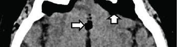
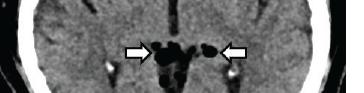





serious risks associated with defects to the skull base.7 Cerebrospinal fluid leakage creates a negative intracranial pressure that predisposes patients to pneumocephalus, while also increasing the risk of infections such as meningitis.7,8 In our patient, a sudden increase in intranasal pressure during sternutation may have allowed air to enter the intracranial space through a structurally weakened cribriform plate, resulting in pneumocephalus. This finding underscored the importance of preoperative imaging to evaluate the location and extent of the skull base defect and guide surgical planning, which ultimately led to successful repair and prevention of further complications.
The treatment of spontaneous pneumocephalus depends on the severity of symptoms.1 While many cases can be managed conservatively with observation and oxygen therapy, surgical intervention is indicated for symptomatic, recurrent, or tension pneumocephalus.1 Tension pneumocephalus is a neurosurgical emergency requiring immediate decompression due to the risk of brain herniation.1,9 Surgical options include burr hole trephination, needle aspiration and craniotomy.1,9 The primary goal of surgical intervention is to evacuate the air and repair any underlying defects in the skull base or dura.1 Intraoperative
techniques may include endoscopic or open approaches depending on the defect location and surgeon expertise.9 Patient education is also essential. Avoiding activities that increase intracranial pressure, such as nose blowing or the Valsalva maneuver, helps reduce the risk of recurrence.1 Emerging trends in the management of pneumocephalus include both non-invasive and surgical advancements. High-flow nasal cannula oxygen therapy has shown promise as a more effective and comfortable alternative to traditional oxygen delivery methods.10-11 High-flow nasal cannula delivers heated, humidified oxygen that reduces nasal irritation, improves patient comfort, and provides consistently high flow rates that help to maintain stable oxygen and fraction of inspired oxygen.10 This high oxygen delivery also creates a strong gradient for nitrogen to be washed out, helping to remove trapped intracranial air more efficiently.10 For surgical management, endoscopic endonasal approaches are increasingly being used for skull base repair, particularly for areas such as the cribriform plate that are difficult to access.9 Endoscopic assistance helps improve visualization and allows for more precise repairs, potentially reducing the risk of CSF leak.9
This case highlights a rare occurrence of non-traumatic pneumocephalus secondary to forceful sternutation. Although sternutation is a physiologic reflex, in rare instances it can lead to serious complications such as pneumocephalus. Emergency clinicians should consider this diagnosis in patients presenting with acute neurological symptoms following barotrauma-like events. Early
Tejpal et al. Pneumocephalus Secondary to Sternutation: A Case Report
recognition, timely imaging, and individualized management (ie, conservative or surgical), are essential to achieving optimal outcomes in these uncommon but serious cases.
The authors attest that their institution requires neither Institutional Review Board approval, nor patient consent for publication of this case report. Documentation on file.
Address for Correspondence: John Ashurst, DO, EdD, MS, Arizona College of Osteopathic Medicine, Midwestern University, 19555 North 59th Ave, Glendale, AZ 85308, USA. Email: jashur@ midwestern.edu.
Conflicts of Interest: By the CPC-EM article submission agreement, all authors are required to disclose all affiliations, funding sources and financial or management relationships that could be perceived as potential sources of bias. The authors disclosed none.
Copyright: © 2026 Tejpal et al. This is an open access article distributed in accordance with the terms of the Creative Commons Attribution (CC BY 4.0) License. See: http://creativecommons.org/ licenses/by/4.0/
1. Das JM, Munakomi S, Bajaj J. Pneumocephalus. [Updated 2024 Dec 11]. In: StatPearls [Internet]. Treasure Island (FL): StatPearls Publishing; 2025 Jan. Available from: https://www.ncbi.nlm.nih.gov/
books/NBK535412/. Accessed on April 4, 2025.
2. Tankersley A, Khan S, Estes K, et al. Non-surgical resolution of pneumocephalus following a sneeze. BMJ Case Rep 2024;17(9):e261096.
3. Setzen S and Platt M. The dangers of sneezing: a review of injuries. Am J Rhinol Allergy. 2019;33(3):331-337.
4. Songu M and Cingi C. Sneeze reflex: facts and fiction. Ther Adv Respir Dis. 2009;3(3):131-141.
5. Wu YH, Yeh IJ, Liu KT. Spontaneous pneumocephalus and subdural hemorrhage after sneezing. Am J Emerg Med. 2018;36(10):1928. e1-1928.e3.
6. Wankhade BS, Beniamein MMK, Alrais ZF, et al. What should an intensivist know about pneumocephalus and tension pneumocephalus? Acute Crit Care. 2023;38(2):244-248.
7. Komatsu F, Komatsu M, Ohmura T, et al. Endoscopic management of a traumatic meningo-encephalocele through a planum sphenoidale defect: case report. Neurol Med Chir (Tokyo). 2010;50(10):949-952.
8. Khurayzi T, Jan B, Bedaiwi R, et al. Otogenic pneumocephalus after tegmen bone reconstruction: a case report. Ear Nose Throat J 2020;99(10):NP152-NP156.
9. Dabdoub CB, Salas G, Silveira EN, et al. Review of the management of pneumocephalus. Surg Neurol Int. 2015;6:155.
10. Siegel JL, Hampton K, Rabinstein AA, et al. Oxygen therapy with high-flow nasal cannula as an effective treatment for perioperative pneumocephalus: case illustrations and pathophysiological review Neurocrit Care. 2018;29(3):366-373.
11. Lee DS, Cha JK, Lee DH, et al. Oxygen supplementation via high-flow nasal cannula is an effective treatment for pneumocephalus. J Neurocrit Care. 2019;12(2):98-101.
T. Neithiya, DM*
Jayan Jayapalan Nair, DM† Krishnadutt Chavali, MD*
Section Editor: Steven Walsh, MD
All India Institute of Medical Sciences, Department of Forensic Medicine and Toxicology, Raipur, India
ESIC Medical College & Hospital, Department of Forensic Medicine and Toxicology, Naroda-Bapunagar, India
Submission history: Submitted May 8, 2025; Revision received June 24, 2025; Accepted August 30, 2025
Electronically published February 25, 2026
Full text available through open access at http://escholarship.org/uc/uciem_cpcem DOI: 10.5811/cpcem.47366
Introduction: Neurotoxic envenomation often presents with non-specific neurological symptoms and minimal local signs, which can delay appropriate diagnosis and treatment. This is the first reported case of a neurotoxic snakebite presenting with an atypical symptom of unilateral neck pain.
Case Report: A 12-year-old girl was referred to our emergency centre with neck weakness progressing to quadriplegia, attributed to a fall while playing. A diagnosis of acute flaccid paralysis secondary to cervical trauma was made and treated at the first hospital; however, she developed respiratory distress and was transferred to our centre. Clinical examination and computed tomography ruled out cervical cord injury. A diagnosis of neurotoxic envenomation was considered, given our centre’s high snakebite burden and the symptom of descending flaccid paralysis. Despite initiating antivenom and supportive treatment, the patient died. As the death was sudden and unexplained, medicolegal autopsy was done. Meticulous examination revealed a suspicious mark over the right foot. Chemical analysis on a skin sample from the site tested positive for snake venom, confirming envenomation.
Conclusion: This case highlights the diagnostic challenge posed by atypical presentations of neurotoxic snakebite, especially in the absence of a clear history. In endemic areas, flaccid paralysis should prompt clinical suspicion of snakebite. Early recognition and timely administration of antivenom are crucial to prevent fatal outcomes. This case also underscores the need for strengthening diagnostic tools and forensic confirmation to avoid missed or delayed diagnoses, which carry serious medicolegal and public health implications. [Clin Pract Cases Emerg Med. 2026;10(2):128-131.]
Keywords: neurotoxic envenomation; flaccid quadriparesis; forensic toxicology; krait bite; case report.
Snakebites are recognized by the World Health Organisation as a neglected tropical disease, with an estimated 5.4 million bites annually leading to over 80,000 deaths in rural Asia and Africa.1 India accounts for nearly half of global snakebite mortality, with neurotoxic envenomation by kraits and cobras (Elapidae) as the leading cause.2 Children are especially vulnerable due to their smaller body mass and delayed access to care because of incomplete history.3 The descending paralysis seen in elapid bites can mimic other conditions such as Guillain-
Barré syndrome or cervical spinal cord injury, which can lead to misdiagnosis and delayed use of anti-snake venom.5-7 Neurotoxic snakebites usually present with minimal or absent local signs, complicating early clinical recognition.4 In fatal cases, lack of overt bite marks and non-specific autopsy findings complicate forensic confirmation.8
We present a case of a neurotoxic snakebite masquerading as cervical spine trauma in a child with ambiguous history and no clear bite evidence. To the best of our knowledge, this is the first reported case of a neurotoxic snakebite presenting
with the atypical symptom of unilateral neck pain, highlighting the diagnostic dilemma it can pose in the absence of clear bite evidence.
A 12-year-old girl was referred to our paediatric emergency department with right-sided neck pain for 12 hours, followed by altered sensorium for three hours. The pain reportedly began after she fell on her back and hit her neck while playing with friends. The pain restricted neck movement and was followed by tingling and weakness in all limbs. Initially seen by a local physician, she was sent home on medications. Later, due to worsening weakness and confusion, she was taken to a hospital, where cervical trauma was suspected.
No history of seizures, fever, bleeding, abdominal pain, vomiting or chronic illness was reported. She was initially managed for cervical spine injury and referred to our centre. At presentation, Glasgow Coma Scale (GCS) was as follows: eye response,2; verbal response 2; and motor response 1, with paradoxical breathing, pooling of oral secretions, hypotonia in all limbs, and absent reflexes. Pupils were normal. Due to the altered sensorium of the child, sensory loss could not be evaluated.
Given the fall history and focal neck symptoms, cervical trauma was initially suspected. The absence of bite history and local signs added to the diagnostic dilemma. Empirical management was initiated with cervical spine stabilization, tetanus toxoid, and 10 vials of anti-snake venom. The child was intubated under manual in-line stabilisation and ventilated. The 20-minute whole-blood clotting test was negative. One hour later, she developed hypotension with blood pressure of 74/50 millimetres of mercury (mm Hg) managed with fluid bolus. Central venous access was obtained, and blood pressure stabilized. The GCS had slight improvement. Ventilation required high pressures, raising concern for lung pathology, but auscultation was clear.
Six hours later, reflexes became elicitable although plantar reflexes remained mute, suggesting neuromuscular dysfunction. A second dose of 10 vials of anti-snake venom was given, along with atropine and neostigmine. After another hour, she again developed hypotension with cold peripheries. Noradrenaline was started. Hydrocortisone and chlorpheniramine were given to manage suspected anaphylaxis related to anti-snake venom. Fraction of inspired oxygen was increased to 100% due to desaturation (oxygen saturation, 86%). Point-of-care ultrasound showed B-lines suggesting pulmonary edema. Intravenous furosemide was given, and oxygenation improved.
Computed tomography showed no injury. Hyperthermia of greater than 105 °F was managed with antipyretics and fluids. Intravenous antibiotics were added empirically. Arterial blood gas revealed respiratory acidosis pH of 7.20 (reference range 7.35-7.45); partial pressure of carbon dioxide 62.4 mm Hg (35-45 mm Hg); and lactate 1.23 millimoles per liter (mmol/L) (0.5-1.6 mmol/L). Inotropes were escalated. Differential
CPC-EM Capsule
What do we already know about this clinical entity?
Neurotoxic snakebite causes descending paralysis and respiratory failure, often with minimal local signs. Atypical or non specific presentations causes delayed diagnosis.
What makes this presentation of disease reportable?
Unilateral neck pain with restricted neck movement and a misleading trauma history mimicked cervical injury, delaying diagnosis of neurotoxic envenomation in a child without bite history.
What is the major learning point?
Acute flaccid paralysis despite imaging status in endemic areas should prompt early consideration of neurotoxic snakebite, even without bite history or bite marks.
How might this improve emergency medicine practice?
Early suspicion and empiric antivenom in atypical paralysis can prevent fatal delays caused by anchoring bias toward trauma diagnoses.
diagnoses included the following: neurotoxic envenomation (possible snakebite); meningitis; brainstem dysfunction; Guillain-Barré syndrome; pulmonary edema/anaphylaxis related to anti-snake venom; and cervical spine injury.
More than 24 hours after presentation, the patient developed pulseless ventricular tachycardia. Resuscitation was unsuccessful, and she was declared dead. Due to the sudden unexplained death, medicolegal autopsy was performed. During autopsy, external examination revealed two scabbed puncture wounds over the right lateral malleolus, suspected to be the bite site. Internally, lungs were congested with haemorrhagic areas and frothy secretions with histopathology confirming diffuse pulmonary edema and pneumonitis. Histopathology also showed mild cerebral edema and neuronal swelling.
Skin tissue from the bite site along with control from opposite leg was preserved and sent to the forensic science laboratory. The lab report confirmed the presence of snake venom using immunodiffusion with polyvalent anti-snake venom antibodies. The final cause of death was certified as neurotoxic snakebite leading to respiratory failure and cardiac arrest.
Neurotoxic Snakebite Presenting with Early Neck Pain and Muscle Weakness: A Case Report
Neurotoxic snakebite, particularly from cobras and kraits, can present with minimal local signs and vague systemic symptoms.9 In endemic regions, atypical presentations can delay appropriate treatment, especially when misleading histories such as trauma are present. In the case of our patient, anchoring bias and limited history delayed recognition. Initially, cervical spine trauma was suspected based on the history of fall, neck pain, and progressive weakness. Neck symptoms, hypotonia, areflexia, and later respiratory distress suggested a high cervical lesion. Cranial nerve signs, including ptosis, were misinterpreted as Horner syndrome.
There were no systemic signs of infection, no gastrointestinal or seizure symptoms, and no local signs of envenomation. The whole-blood clotting test was negative. Classic signs such as ptosis and ophthalmoplegia were absent at onset. However, signs such as preserved pupillary reflexes, generalized areflexia, pooling of secretions, and symmetrical flaccid paralysis were more consistent with neuroparalysis. Reflex improvement following administration of anti-snake venom and neostigmine supported reversible neuromuscular blockade.
This is likely the first reported case of neurotoxic envenomation with isolated neck pain progressing to paralysis. While neck weakness has been documented in other case reports, localized unilateral neck pain as the first symptom of envenomation has not been previously reported. While the presenting clinical picture supported cervical trauma, the absence of sensory loss and the presence of symmetrical descending weakness suggested neurotoxic envenomation. Such subtle clues, even without bite history, should raise suspicion—especially in endemic settings.
Prior literature confirms that elapid venom acts at the neuromuscular junction, with venom of the krait (containing β-bungarotoxin) causing presynaptic acetylcholine blockade.10 The toxin leads to flaccid paralysis that may not respond to neostigmine if treatment is delayed. Kraits are nocturnal, and victims are often bitten during sleep, typically without local swelling or visible marks. Cobra venom, by contrast, is less potent and produces deeper bites, more prominent local signs, and earlier cranial nerve involvement.
This case emphasizes the need to suspect snakebite in children with acute flaccid paralysis—even with misleading histories. Antivenom should be administered early based on clinical judgment rather than awaiting confirmation. In this case, the anti-snake venom was given empirically 12 hours post-bite, but delayed diagnosis likely contributed to the fatal outcome. Delay in administration of anti-snake venom worsens outcomes. Antivenom is most effective in neurotoxic envenomation before full-blown paralysis sets in.11 Supportive
care, including mechanical ventilation, neostigmine, and atropine, was appropriately attempted. Pulmonary edema after anti-snake venom use, whether due to toxin-mediated capillary leak or anaphylaxis, has been previously described.12
Forensically, histopathology is non-specific (eg, pulmonary oedema).13 However, postmortem venom detection from skin near the suspected bite-site provides reliable confirmation, even without visible marks or history. Such confirmation is critical for legal certification and family compensation in India, where snakebite deaths are deemed “unnatural.”14, 15 Finally, this case reinforces the World Health Organisation’s call for improved snakebite prevention and response strategies: public education; elevated sleeping,;early health-seeking behaviour; and rapid access to anti-snake venom and critical care services.
Delayed presentation is common in rural India due to limited infrastructure and delayed referrals. In this case, critical time was lost due to misdiagnosis at the peripheral centre. Despite resuscitation, the late presentation and delayed treatment worsened the patient’s outcome. From a legal standpoint, snakebite deaths require inquest and autopsy. Diagnostic ambiguity can delay or deny family compensation. Thus, accurate confirmation through methods such as venom testing of skin from the bite-site is vital.
Key implications in this case study include the following: snakebites masquerading as spinal trauma; clinician awareness in endemic areas; forensic lab’s vital role in confirming envenomation in ambiguous or delayed cases; standard protocols for early sample collection; availability of point-ofcare venom-detection tools; public health education on early recognition; wearing protective clothing; and seeking immediate care.
Neurotoxic snakebite can mimic other emergencies, such as cervical spine injury, particularly when the history is ambiguous. Clinicians in endemic regions must maintain high suspicion for snakebite in any child presenting with acute flaccid paralysis, cranial nerve involvement, and normal imaging. Awareness of atypical presentations, as in this case, is vital to improve outcomes in endemic areas. This case also demonstrates the importance of forensic infrastructure to ensure accurate death certification, epidemiological reporting, and timely compensation for families.
The authors attest that their institution requires neither institutional review board approval, nor patient consent for publication of this case report. Documentation on file.
Neithiya et al.
Neurotoxic Snakebite Presenting with Early Neck Pain and Muscle Weakness: A Case Report
Address for Correspondence: T Neithiya, DM, 1st Floor, B Block, Administration Building, 5th Gate, AIIMS Raipur, GE Road, Tatibandh, Raipur, Chhattisgarh – 492099, India. Email: neithiyambbs27@gmail.com.
Conflicts of Interest: By the CPC-EM article submission agreement, all authors are required to disclose all affiliations, funding sources and financial or management relationships that could be perceived as potential sources of bias. The authors disclosed none.
Copyright: © 2026 Neithiya et al. This is an open access article distributed in accordance with the terms of the Creative Commons Attribution (CC BY 4.0) License. See: http://creativecommons.org/ licenses/by/4.0/
1. Snakebite Envenoming: A Strategy for Prevention and Control.; 2019. http://apps.who.int/bookorders. Accessed August 29, 2025.
2. Mohapatra B, Warrell DA, Suraweera W, et al. Snakebite mortality in India: a nationally representative mortality survey. PLoS Negl Trop Dis. 2011;5(4):e1018
3. Le Geyt J, Pach S, Gutiérrez JM, et al. Paediatric snakebite envenoming: recognition and management of cases. Arch Dis Child 2021;106(1):14-19.
4. Ariaratnam CA, Sheriff MHR, Theakston RDG, et al. Distinctive epidemiologic and clinical features of common krait ( Bungarus caeruleus ) bites in Sri Lanka. Am J Trop Med Hyg 2008;79(3):458-462.
5. Mehta V, Kumar R, Prabhakar R, et al. Dramatic neuromuscular paralysis following occult snakebites. J Family Med Prim Care 2022;11(1):386-389.
6. Agarwal S, Khanduri S, Kaeley N, et al. Brain dead presentation of
snake bite. Indian J Crit Care Med. 2018;22(7):541-543.
7. Alirol E, Sharma SK, Bawaskar HS, et al. Snake bite in South Asia: a review. PLoS Negl Trop Dis. 2010;4(1):e603.
8. Feola A, Marella GL, Carfora A, et al. Snakebite envenoming a challenging diagnosis for the forensic pathologist: a systematic review. Toxins (Basel). 2020;12(11):699.
9. Sahoo B, Mishra R, Jain M, et al. Early morning neuroparalytic syndrome – a diagnostic dilemma: a case report. J Pediatr Crit Care 2022;9(4):142.
10. Silva A, Maduwage K, Sedgwick M, et al. Neuromuscular effects of common krait (Bungarus caeruleus) envenoming in Sri Lanka. PLoS Negl Trop Dis. 2016;10(2):e0004368.
11. Ahmed S, Ahmed M, Nadeem A, et al. Emergency treatment of a snake bite: pearls from literature. J Emerg Trauma Shock 2008;1(2):97.
12. Singh AR and Aggarwal AP. Pulmonary oedema complicating snake bite due to Bungarus caeruleus Singapore Med J. 2007;48(8): e227-30.
13. Keche AS, Jahan A, Chaurasia JK, et al. Snake bite fatalityhistopathology of various organs. J Forensic Med Tox 2024;41(1):146-149.
14. Lee LP, Tan CH, Khomvilai S, et al. Characterizing and applying immunoglobulins in snakebite diagnostics: a simple and rapid venom detection assay for four medically important snake species in Southeast Asia. Int J Biol Macromol. 2023;236:123-727.
15. Dhananjaya BL, Menon JC, Joseph JK, et al. Snake Venom Detection Kit (SVDK): update on current aspects and challenges. In: Gopalakrishnakone P, Faiz A, Fernando R, Ariaranee Gnanathasan C, Habib A, and Chen-Chang Yang (eds.) Clinical Toxicology in Asia Pacific and Africa. Springer Science+Business Media B.V,, Dordrecht, Netherlands; 2015:379-400.
Reshvinder Dhillon, MD
Sarah McMullin, MD
Section Editor: Shadi Lahham, MD
University of South Alabama, Department of Emergency Medicine, Mobile, Alabama
Submission history: Submitted July 22, 2025; Revision received September 19, 2025; Accepted November 5, 2025
Electronically published March 10, 2026
Full text available through open access at http://escholarship.org/uc/uciem_cpcem DOI: 10.5811/cpcem.49037
Introduction: In this report we highlight the emerging role of pediatric cardiac point-of-care ultrasound (POCUS) in rapidly diagnosing infective endocarditis, using a clinical case as illustration.
Case Report: A six-year-old girl with a known ventricular septal defect presented with worsening respiratory symptoms, fevers, abdominal pain, and decreased oral intake. Initial POCUS, performed by an emergency physician, indicated a suspicious echogenic mass in the right atrium, prompting formal echocardiography. Further imaging and cultures confirmed infective endocarditis due to methicillin-sensitive Staphylococcus aureus
Conclusion: This case underscores the utility of pediatric cardiac POCUS as a rapid bedside diagnostic tool for infective endocarditis in emergency settings, leading to early diagnosis and management. Although POCUS cannot replace comprehensive echocardiography, its immediate availability significantly accelerates diagnosis and management initiation, particularly in pediatric patients with congenital heart conditions who are at increased risk for the condition. Ongoing training and standardized protocols will enhance its efficacy. Clinicians should recognize the strengths and limitations of POCUS, integrating it into broader diagnostic workflows for pediatric infective endocarditis. [Clin Pract Cases Emerg Med. 2026;10(2):132-136.]
Keywords: POCUS; pediatrics; endocarditis; bacteremia; case report.
INTRODUCTION
Point-of-care ultrasound (POCUS) has emerged as a transformative tool in pediatric emergency medicine, particularly in the evaluation of cardiac conditions.1 Over the past decade, technological advances and increasing physician expertise have expanded its applications from basic cardiac assessments to more complex diagnoses, including the detection of vegetations in infective endocarditis.3,4 In the context of infective endocarditis, POCUS can be used to detect valvular vegetations, although it is not as comprehensive as transthoracic echocardiography or transesophageal echocardiography.
Pediatric emergency physicians (EP) can reliably detect decreased left-sided systolic function and pericardial effusion
using POCUS. Case reports show that pediatric EPs have detected right-sided outflow tract obstruction, aortic root dilatation, and congenital cardiac disease using POCUS. Training for pediatric cardiac POCUS competency is feasible, and cardiac POCUS does not increase the burden of cardiology resources on the pediatric emergency department (ED).3,15
Point-of-care ultrasound presents unique advantages and challenges. Children generally have better acoustic windows than adults, allowing for clearer visualization of cardiac structures. However, the smaller size of cardiac structures and the need for patient cooperation can make image acquisition more challenging. Additionally, the presence of underlying congenital heart defects, which occur in 0.8-1% of live births, can complicate image interpretation.1,4
A six-year-old female with a history of unrepaired ventricular septal defect and eczema presented to the ED with a one-week history of worsening respiratory symptoms. Her illness began as mild cough and congestion but progressed to abdominal pain, poor oral intake, and fevers. Two months earlier, cardiology follow-up had documented a small, membranous, pressure-restrictive ventricular septal defect with a left-to-right shunt and trace aortic regurgitation.
On arrival to the ED, her temperature was 40.4 °Celsius, heart rate 178 beats per minute, respiratory rate 78 breaths per minute, and blood pressure 89/63 millimeters of mercury, with increased work of breathing and a holosystolic murmur.
Laboratory studies showed leukocytosis, elevated C-reactive protein, and elevated procalcitonin. A chest radiograph (Image 1) revealed bilateral infiltrates, supporting the ED team’s working diagnosis of pneumonia with sepsis, and she was admitted for antibiotic and respiratory support.
The patient was initially admitted to the pediatric floor but deteriorated rapidly, requiring escalation from high-flow nasal cannula to bilevel positive airway pressure support and transfer to the pediatric intensive care unit (PICU). While in the PICU, an emergency medicine resident rotating on the unit performed a POCUS exam, consistent with institutional training initiatives that encourage residents to integrate POCUS into evaluation of critically ill patients. Although the
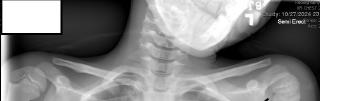
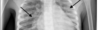
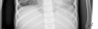
CPC-EM Capsule
What do we already know about this clinical entity?
Infective endocarditis is uncommon in children but risk rises with congenital heart disease; point-of-care ultrasound (POCUS) may show vegetations, yet echo is definitive.
What makes this presentation of disease reportable?
Emergency department cardiac POCUS incidentally found a right-sided vegetation in a child presumed septic pneumonia, triggering urgent echo and methicillin-sensitive Staphylococcus aureus infective endocarditis (IE).
What is the major learning point?
Cardiac POCUS can rapidly screen febrile kids with congenital heart disease for mobile echogenic vegetations and speed confirmatory echo/treatment.
How might this improve emergency medicine practice?
Using cardiac POCUS in sick children can shorten time to IE recognition, prompt early antibiotics/consults, and allow safe serial reassessment.
intent was to assess cardiac function as a possible cause of respiratory distress, the resident unexpectedly identified an echogenic structure in the right atria (Image 2, Image 3). The structure was described as hyperechoic floating between the right atria and the ventricle.
Formal echocardiography confirmed a 16-mm tricuspid valve vegetation consistent with infective endocarditis. Blood cultures and urine cultures were obtained, which subsequently grew methicillin-sensitive Staphylococcus aureus (MSSA). Following the MSSA blood culture results and POCUS findings, the patient was started on antibiotic therapy with cefazolin, gentamicin, and rifampin. Gentamicin and rifampin were added for synergistic effect with cefazolin, due to the patient’s critical condition and prolonged bacteremia.
During her 17-day stay in the PICU, the patient developed multiple complications, including septic pulmonary emboli confirmed by computed tomography (CT) angiography. She experienced recurrent episodes of coughing up blood, which required gastrointestinal prophylaxis with famotidine and








sucralfate. She also developed iron deficiency anemia that required supplementation. Serial monitoring was performed throughout her admission and included frequent assessments by cardiology with repeat echocardiograms demonstrating persistent but gradually decreasing vegetation size. An ophthalmology evaluation showed no evidence of retinal seeding, and CT of the brain revealed no evidence of cerebral emboli or abscess formation.
The patient completed a 50-day course of intravenous antibiotics via peripherally inserted central catheter line, 50 days of cefazolin and 24 days of rifampin and gentamicin. Final echocardiogram prior to discharge showed a small 6-mm hyperechoic lesion on the septal leaflet of the tricuspid valve, likely representing scarring. This was still present at the four-month cardiology follow-up. She was discharged home with close follow-up with infectious disease, cardiology, and her primary care physician. The patient’s uncontrolled eczema was identified as a possible source of the initial infection, and dermatology follow-up was arranged.
This case highlights the emerging role of POCUS in the early detection of endocarditis in pediatric patients. The rapid identification of a foreign hyperechoic structure by POCUS led to expedited formal imaging and appropriate treatment initiation. In pediatric patients with underlying cardiac conditions presenting with fever or respiratory symptoms, POCUS has become an increasingly valuable initial screening tool.
In suspected infective endocarditis, POCUS can demonstrate vegetations as mobile, irregular, echogenic masses adherent to the valvular surface and moving independently from the valve leaflet. The tricuspid valve is
particularly amenable to POCUS due to its anterior position, often best visualized in the apical four-chamber or parasternal short-axis view. In this case, the vegetation’s movement between the right atrium and ventricle was distinctly seen on both views, increasing diagnostic confidence.7 Recent studies have demonstrated promising diagnostic accuracy for POCUS in detecting endocarditis. A systematic review by Bai et al found that transthoracic echocardiography, including point-ofcare studies, had a pooled sensitivity of 61% and specificity of 94% for detecting vegetations when compared to transesophageal echocardiography.5
The diagnostic utility of POCUS in endocarditis varies by vegetation size and location. Vegetations larger than 10 mm are detected with higher sensitivity (> 90%), while those < 5 mm may be missed on initial POCUS examination.6,7 Rightsided structures, particularly the tricuspid valve, are generally better visualized than left-sided structures due to their anterior location and proximity to the chest wall. Studies have shown that the sensitivity of transthoracic echocardiography, including POCUS, for detecting vegetations in infective endocarditis to be 70-80% right-sided vegetations, such as those on the tricuspid valve. For left-sided vegetations, the sensitivity of transthoracic echocardiography is generally lower, ranging from 50-60%, depending on the valve involved and the quality of imaging.8,9
Point-of-care ultrasound is particularly valuable in the diagnosis and management of pediatric infective endocarditis due to its ability to provide rapid, bedside cardiac assessment without the delays associated with formal echocardiography. In
et al. Catching Silent Heart Killers—How Bedside Ultrasound Revealed Hidden Endocarditis: A Case Report
emergency settings, POCUS can be performed within minutes of presentation, expediting diagnosis by several hours compared with traditional imaging modalities, which is especially critical in pediatric patients in whom early intervention significantly improves clinical outcomes.10 Furthermore, POCUS allows for repeat examinations to monitor disease progression. This capability is especially advantageous in critically ill pediatric patients for whom transportation to the echocardiography laboratory may pose clinical risk or instability.11
The use of POCUS has also been demonstrated to be cost-effective. Studies have shown that initial screening with POCUS can reduce the need for unnecessary comprehensive echocardiograms in low-risk patients, resulting in substantial healthcare cost savings.12 Importantly, early detection and treatment initiation facilitated by POCUS have been associated with reduced morbidity in pediatric endocarditis, particularly in children with underlying structural heart disease.13
Despite these advantages, several limitations of POCUS must be acknowledged. The diagnostic accuracy of POCUS is highly dependent on operator skill and experience in cardiac imaging. Evidence demonstrates that accuracy improves with training, but novice users may miss subtle pathological findings.13 Additionally, the quality of POCUS images can be limited by patient-related factors such as respiratory motion, body habitus, or inability to remain still, all of which are common challenges in pediatric populations. Finally, while POCUS is highly effective as an initial screening modality, it should be viewed as complementary to comprehensive echocardiography rather than a replacement. Comprehensive echocardiography remains essential for detailed evaluation of cardiac valve function, precise measurement of vegetations, and identification of complications such as abscess formation or valvular perforation.13
The role of POCUS in pediatric endocarditis continues to evolve, with ongoing research focused on standardizing protocols and improving training methods. Recent technological advances, including artificial intelligenceassisted interpretation and three-dimensional imaging capabilities, may further enhance the diagnostic accuracy of POCUS in the future.14
Point-of-care ultrasound proved to be a valuable tool in the early diagnosis of tricuspid valve endocarditis in this pediatric patient. This case adds to the growing body of evidence supporting the use of POCUS in pediatric emergency medicine for rapid bedside diagnosis of cardiac conditions.2,4
Patient consent has been obtained and filed for the publication of this case report.
Address for Correspondence: Reshvinder Dhillon, MD, University of South Alabama, Department of Emergency Medicine, 1700 Center Street, Mobile, Alabama. Email: rdhillon@health. southalabama.edu.
Conflicts of Interest: By the CPC-EM article submission agreement, all authors are required to disclose all affiliations, funding sources and financial or management relationships that could be perceived as potential sources of bias. The authors disclosed none.
Copyright: © 2026 Dhillon et al. This is an open access article distributed in accordance with the terms of the Creative Commons Attribution (CC BY 4.0) License. See: http://creativecommons.org/ licenses/by/4.0/
1. Lu JC, Riley A, Conlon T, et al. Recommendations for cardiac point-of-care ultrasound in children: a report from the American Society of Echocardiography. J Am Soc Echocardiogr. 2023;36(3):265-277.
2. López Palmero S, López Zúñiga MA, Rodríguez Martínez V, et al. Point-of-care ultrasound (POCUS) as an extension of the physical examination in patients with bacteremia or candidemia. J Clin Med. 2022;11(13):3636.
3. Scheier E. Cardiac POCUS in pediatric emergency medicine: a narrative review. J Clin Med. 2023;12(17):5666.
4. Díez-Vidal A, Martínez-Martín P, González-Muñoz B, et al. Point-ofcare ultrasound in infectious diseases: current insights and future perspectives. Clin Infect Dis. 2024;79(2):420-429.
5. Díaz-Gómez JL, Mayo PH, Koenig SJ. Point-of-care ultrasonography. N Engl J Med.2021;385(17):1593-1602.
6. Malik SB, Hsu JY, Hurwitz Koweek LM, et al. ACR Appropriateness Criteria® infective endocarditis. J Am Coll Radiol. 2021;18(5S):S52-S61
7. Pathak NJ, Ng L, Saul T, et al. Focused cardiac ultrasound diagnosis of right-sided endocarditis. Am J Emerg Med. 2013;31(6):998.e3-4.
8. Bai AD, Steinberg M, Showler A, et al. Diagnostic accuracy of transthoracic echocardiography for infective endocarditis findings using transesophageal echocardiography as the reference standard: a meta-analysis. J Am Soc Echocardiogr. 2017;30(7):639-646.
9. Saric M, Armour AC, Arnaout MS, et al. Guidelines for the use of echocardiography in the evaluation of a cardiac source of embolism. J Am Soc Echocardiogr. 2016;29(1):1-42.
10. Spencer KT, Kimura BJ, Korcarz CE, et al. Focused cardiac
Catching Silent Heart Killers—How Bedside Ultrasound Revealed Hidden Endocarditis: A Case Report Dhillon et
ultrasound: recommendations from the American Society of Echocardiography. J Am Soc Echocardiogr. 2013;26(6):567-581.
11. Singh Y, Tissot C, Fraga MV, et al. International evidence-based guidelines on point of care ultrasound (POCUS) for critically ill neonates and children issued by the POCUS Working Group of the European Society of Paediatric and Neonatal Intensive Care (ESPNIC). Crit Care. 2020;24(1):65.
12. Labovitz AJ, Noble VE, Bierig M, et al. Focused cardiac ultrasound in the emergent setting: a consensus statement of the American Society of Echocardiography and American College of Emergency
Physicians. J Am Soc Echocardiogr. 2010;23(12):1225-1230.
13. Habib G, Lancellotti P, Antunes MJ, et al. 2015 ESC guidelines for the management of infective endocarditis: the Task Force for the Management of Infective Endocarditis of the European Society of Cardiology (ESC). Eur Heart J. 2015;36(44):3075-3128.
14. Dey D, Slomka PJ, Leeson P, et al. Artificial intelligence in cardiovascular imaging: JACC state-of-the-art review. J Am Coll Cardiol. 2019;73(11):1317-1335.
15. Nizami Z, Garcia P, Ahuja P, et al. Supraventricular tachycardia in children. Prog Pediatr Cardiol. 2024;73:101716.
Rochak Kansakar, MBBS*
Elijah J. Katz, MD†
Justin Zhao, MD†
Evan
Weldon, BS†
Section Editor: Shadi Lahham, MD
Bayalpata Hospital, Department of Emergency Medicine, Sanfebagar, Accham, Nepal University of Texas Southwestern Medical Center, Department of Emergency Medicine, Dallas, Texas
Submission history: Submitted August 23,2025; Revision received September 4,2025; Accepted October 20,2025
Electronically published March 29, 2026
Full text available through open access at http://escholarship.org/uc/uciem_cpcem
DOI: 10.5811/cpcem.50737
Introduction: Drowning is a significant cause of death in Nepal, especially in rural areas. Identifying pulmonary edema is important for management of cases of non-fatal drowning, and while radiograph is the standard of care, point-of-care ultrasound (POCUS) offers a rapid, accessible alternative.
Case Report: A 40-year-old woman presented to the emergency department after non-fatal drowning with respiratory distress and hypoxia. Chest radiograph was unavailable. Point-of-care ultrasound revealed diffuse B-lines consistent with pulmonary edema. She was stabilized and transported to a hospital with intensive care-level management.
Conclusion: Point-of-care ultrasound enabled rapid identification of pulmonary edema and guided timely referral. In resource-limited settings, POCUS is a valuable tool for managing drowning victims when other diagnostics are unavailable. [Clin Pract Cases Emerg Med. 2026;10(2):137-140.]
Keywords: point-of-care ultrasound; non-fatal drowning; rural emergency care; Nepal; case report.
Drowning represents a significant cause of injury and death in Nepal, with estimated mortality from drowning between 1.9 and 4.9 per 100,000 people.1,2 Children and men are the highest risk groups, and the burden from drowning is likely underestimated due to lack of surveillance systems and under-reporting.1,3 Rural areas in Nepal face further challenges from drowning because of greater access to bodies of water, flood susceptibility, and limited medical care.3
The hospital-based medical management of non-fatal drowning patients includes the assessment of respiratory and cardiovascular damage and the initiation of appropriate care, which can range from warming and low-flow oxygen to mechanical ventilation and extracorporeal membrane oxygenation.4 Best practice guidelines recommend pulse oximetry, arterial blood gases, chest radiograph, and basic metabolic panel as first-line diagnostics.5 However, in lowresource settings, point-of-care ultrasound (POCUS) represents a rapid, cost-effective, and versatile imaging
modality with high sensitivity and specificity for relevant pathologic findings such as B-lines and pulmonary edema.6-8 Ultrasound can be used for risk-stratification of non-fatal drowning patients by monitoring for the presence and abundance of B-lines, which correlates directly with the presence and severity of pulmonary edema.9 Early ultrasound findings can guide appropriate management and referral decisions even in the absence of other imaging and labs.
This case report follows a 40-year-old female no-fatal drowning victim who was managed at Bayalpata Hospital in rural Western Nepal. The case highlights the utility of ultrasound in guiding management and risk stratification of patient care in austere medical centers.
A 40-year-old female was brought to the emergency department (ED) following an accidental, non-fatal drowning in river water. The initial report stated the patient had been submerged for about one hour. The prolonged submersion in
Point-of-Care Ultrasound After Non-fatal Drowning in Rural Western Nepal: A Case Report Kansakar
high-current waters with rocky surfaces resulted in facial and superficial injuries, as well as injuries to both lower limbs. On presentation the patient exhibited labored breathing and signs of respiratory distress, with room air oxygen saturation measuring 72% on pulse oximeter.
Physical examination revealed bilaterally decreased breath sounds, with crackles on auscultation over the infraaxillary regions. Her respiratory rate was 26 breaths per minute, and she appeared confused. Her Glasgow Coma Scale (GCS) score was 13: eye opening 4, verbal 4, and motor response 5. Blood pressure was 100/60 millimeters of mercury, temperature 94.5 °Fahrenheit, and heart rate 95 beats per minute. Supplemental oxygen was administered via face mask, and gradual rewarming was initiated. Bedside glucose measurement was 72 milligrams per deciliter (mg/dL) (reference range: 70-99 mg/dL).
Initial laboratory tests, including a complete blood count, renal function test, and liver function test, yielded results within normal limits. Arterial blood gases were not available at the hospital, and radiograph was temporarily unavailable due to a power outage. A POCUS was performed at the bedside using the handheld Butterfly iQ (Butterfly Network, Inc, Burlington MA) with the patient sitting semi-recumbent at a 45-degree angle. The lung zones assessed included the bilateral superior anterior, inferior anterior, superior lateral, and inferior lateral. Multiple B-lines, some confluent, were observed across most of the lung zones.
Ultrasound images from three representative zones are shown in Images 1-3. Additional findings revealed normal ventricular function and no evidence of pericardial effusion. The inferior vena cava was non-plethoric and had 50% collapsibility, suggesting adequate volume status. Given the clinical presentation of respiratory distress and the sonographic evidence of diffuse B-lines, a diagnosis of non-cardiogenic pulmonary edema secondary to the near-



CPC-EM Capsule
What do we already know about this clinical entity?
Non-fatal drowning patients require assessment of pulmonary edema to be appropriately managed and risk- stratified.
What makes this presentation of disease reportable?
This case demonstrates the successful use of pointof-care ultrasound (POCUS) in an austere medical environment as the sole imaging modality.
What is the major learning point?
POCUS can rapidly identify pulmonary edema after non-fatal drowning, enabling timely stabilization and transfer in low-resource settings.
How might this improve emergency medicine practice?
Expanding POCUS training and access in rural facilities can enhance early diagnosis and guide appropriate referral for non-fatal drowning victims.
drowning event was made.
She was treated with supplemental oxygen via face mask. Gradual rewarming was initiated to correct hypothermia. Her blood pressure was maintained within a normal range with intravenous fluids. She was kept on non-invasive oxygenation






Image 3. Point-of-care ultrasound of the right lung, in the right anterior superior view, showing numerous B-lines in a non-fatal drowning patient.
specificity of 66.67%.12 It takes, on average, six minutes to accurately perform POCUS while the wait for an radiograph can vary widely in rural setting, if it is available at all.13 We believe that further ultrasound availability and training in rural areas of Nepal would be effective tools to enhance clinical management.
Limitations of this case report include a lack of follow-up after referral, initial radiograph confirmation for pulmonary edema, and additional supporting lab work. History, physical exam, basic labs, and POCUS were the driving factors of clinical management.
via face mask at six liters per minute. Due to ongoing respiratory distress, positive pressure ventilation with bilevel positive airway pressure was considered; however, due to facial injuries around the nasal area, she could not tolerate it. Subsequently, the patient was sent by ambulance to another hospital for intensive care-level management.’
Without access to radiograph or arterial blood gases, POCUS findings—specifically widespread B-lines suggesting severe pulmonary edema—helped guide the care of this patient and resulted in rapid transport to a center with mechanical ventilation capabilities. Pulmonary edema without clear signs of shock changed the patient from a grade 2 drowning victim, defined as rales in some pulmonary fields, to a grade 3. Grade 2 drowning patients can be managed in the ED with nasal cannula/non-rebreather oxygen, rewarming therapy, beta-agonists, and observation, all of which were available at the Bayalpata hospital.
In contrast, Grade 3 and above non-fatal drowning victims require intensive care-level management, arterial blood gas, advanced airway management and, in some cases, hemodynamic support, few of which were available.10 Given the evidence of likely aspiration and depressed mental status, supported by the abnormal POCUS findings, there was concern for potential deterioration requiring additional respiratory support.11 Therefore, the decision was made to rapidly transport the patient to Dadeldhura hospital by ambulance after the initiation of intravenous fluids, oxygen via nasal cannula, and rewarming therapy.
How does POCUS compare to radiograph in the detection of pulmonary edema? Ghauri et al reported that ultrasound has a sensitivity and specificity of 91.05% and 91.18%, respectively, for diagnosing pulmonary edema. In contrast, radiograph was shown to have a sensitivity of 60.16% and a
This case highlights the role of point-of-care ultrasound in managing non-fatal drowning victims in resource-limited settings. In the absence of radiograph imaging, arterial blood gases, or advanced laboratory support, POCUS enabled the early detection of diffuse pulmonary edema and guided the decision to escalate care and refer to a higher center of care. The rapid bedside assessment provided risk-stratification, based on the diffuse and abundant B-lines, helping the patient receive appropriate care and reducing further deterioration. This case supports the growing body of evidence demonstrating the diagnostic utility of POCUS in austere environments. Broader implementation of ultrasound training and access in rural Nepal could improve the quality and timeliness of care for drowning patients.
Patient consent has been obtained and filed for the publication of this case report.
Address for Correspondence: Evan Weldon, BS, University of Texas Southwestern Medical Center, Department of Emergency Medicine, 5323 Harry Hines Boulevard, E4.300, Dallas, TX 75390-8579. Email: evan.weldon@utsouthwestern. edu.
Conflicts of Interest: By the CPC-EM article submission agreement, all authors are required to disclose all affiliations, funding sources and financial or management relationships that could be perceived as potential sources of bias. The authors disclosed none.
Copyright: © 2026 Kansakar et al. This is an open access article distributed in accordance with the terms of the Creative Commons Attribution (CC BY 4.0) License. See: http://creativecommons.org/ licenses/by/4.0/
1. Sedain B and Pant PR. Status of drowning in Nepal: a study of central police data. F1000Res. 2018;7:576.
2. Nepal Health Research Council (NHRC), Ministry of Health and
Point-of-Care Ultrasound After Non-fatal Drowning in Rural Western Nepal: A Case Report Kansakar et al.
Population (MoHP), Institute for Health Metrics and Evaluation (IHME), Monitoring Evaluation and Operational Research (MEOR). Nepal Burden of Disease 2019: A Country Report based on the 2019 Global Burden of Disease Study. Kathmandu, Nepal: NHRC, MoHP, IHME, and MEOR; 2021. Accessed October 20, 2025. Available at: https://nhrc.gov.np/wp-content/uploads/2022/02/BoD-Report-Bookinclud-Cover-mail-6_compressed.pdf
3. Pradhan EK, West KP, Katz J, et al. Risk of flood-related mortality in Nepal. Disasters. 2007;31(1):57-70.
4. Szpilman D and Morgan P. Management for the drowning patient. Chest. 2020;159(4):1473-1483.
5. Szpilman D, Sempsrott J, Schmidt A. Drowning. BMJ Best Practice. Updated November 15, 2023. Accessed October 20, 2025. https:// bestpractice.bmj.com/topics/en-gb/657
6. Picano E, Pellikka PA. Ultrasound of extravascular lung water: a new standard for pulmonary congestion. Eur Heart J. 2016;37(27):20972104.
7. Liu RB, Donroe JH, McNamara RL, et al. The practice and implications of finding fluid during point-of-care ultrasonography. JAMA Intern Med. 2017;177(12):1818-1825.
8. Heldeweg MLA, Jagesar AR, et al. Effects of lung ultrasonographyguided management on cumulative fluid balance and other clinical outcomes: a systematic review. Ultrasound Med Biol. 2021;47(5):1163-1171.
9. Blanco PA and Cianciulli TF. Pulmonary edema assessed by ultrasound: impact in cardiology and intensive care practice. Echocardiography. 2016;33(5):778-787.
10. Szpilman D. Near-drowning and drowning classification. Chest 1997;112(3):660-665.
11. Van Berkel M, Bierens JJ, Lie RL, et al. Pulmonary oedema, pneumonia and mortality in submersion victims; a retrospective study in 125 patients. Intensive Care Med. 1996;22(2):101-107.
12. Ghauri SK, Mustafa KJ, Javaeed A, et al. Accuracy of lung ultrasound and chest X-rays in diagnosing acute pulmonary oedema in patients presenting with acute dyspnoea in emergency department. J Pak Med Assoc. 2021;71(10):2423-2425.
13. Patrick DP, Bradley XG, Wolek C, et al. Minutes matter: time it takes to perform point-of-care ultrasound. AEM Educ Train. 2023;7(4):e10901.
Armin Dehkordi, BS*
Daniel Aloise, MD†
Eric Scheppke, DO†
Mary Christodoulou, BS‡
Grayson Gigliotti, BS‡
Tony Zitek, MD†
Arkansas College of Osteopathic Medicine at Arkansas Colleges of Health Education, Fort Smith, Arkansas
Mount Sinai Medical Center Miami Beach, Department of Emergency Medicine, Miami Beach, Florida
Florida International University, Herbert Wertheim College of Medicine, Miami, Florida
Section Editor: Christopher San Miguel, MD
Submission history: Submitted June 26, 2025; Revision received September 14, 2025; Accepted October 20, 2025
Electronically published March 19, 2026
Full text available through open access at http://escholarship.org/uc/uciem_cpcem DOI: 10.5811/cpcem.47257
Introduction: Chalk-stick fractures are transverse spinal injuries seen in patients with ankylosing spondylitis due to chronic inflammation and spinal rigidity. These fractures may result from minor trauma and are associated with potentially fatal complications. While spinal fractures in ankylosing spondylitis are well recognized, thoracic chalk-stick fractures complicated by hemothorax from vascular injury remain exceedingly rare. We present a case of an elderly male with ankylosing spondylitis who sustained a thoracic chalk-stick fracture following a ground-level fall, complicated by hemothorax and hemorrhagic shock. This case highlights a rarely reported but life-threatening complication and emphasizes the importance of early imaging and high clinical suspicion in this highrisk population—even after minor trauma.
Case Report: A 90-year-old male with known history of ankylosing spondylitis presented to the emergency department after a ground-level fall associated with syncope. He had thoracic back pain, dyspnea, and hypotension. Computed tomography revealed a thoracic vertebra 11 chalk-stick fracture with interspinous vascular injury and a large, right-sided hemothorax. The patient underwent emergent chest tube placement, blood transfusion, and vasopressor support, which initially stabilized his condition. However, his hospital course was complicated by multiple comorbidities, and he ultimately died after prolonged critical care.
Conclusion: This case illustrates the potential for catastrophic vascular complications from minor trauma in patients with ankylosing spondylitis. Thoracic chalk-stick fractures may result in life-threatening hemothorax and hemorrhagic shock. Emergency physicians should maintain a high index of suspicion and obtain early radiographic imaging to facilitate timely diagnosis and intervention in this high-risk population. [Clin Pract Cases Emerg Med. 2026;10(2):141-145]
Keywords: chalk-stick fracture; ankylosing spondylitis; hemothorax; case report.
Chalk-stick fractures are complete, transverse disruptions of the vertebral body, intervertebral disc, and posterior elements, resembling a snapped piece of chalk.1 They are commonly seen in patients with ankylosing spondylitis (AS), a chronic inflammatory spondyloarthropathy strongly associated
with the HLA-B27 allele, where progressive enthesitis and ligamentous ossification result in a rigid yet brittle spine prone to fracture even after minor trauma.3,5-7 While these injuries are most common in the cervical spine, 10-11% occur in the thoracic region.2-5 Thoracic fractures in AS are particularly concerning due to their proximity to vital vascular structures.
Disruption of posterior intercostal arteries, coursing on the anterolateral vertebral body, can result in hemothorax or retroperitoneal bleeding, potentially leading to hemorrhagic shock and death if not promptly recognized.6-8
We present a case of an elderly male with AS who developed a large, right-sided hemothorax after a ground-level fall caused a thoracic vertebra 11 (T11) chalk-stick fracture and interspinous vascular injury. The case highlights how these fractures can present subtly and progress rapidly. Uniquely, this injury involved the thoracolumbar junction, was initially clinically subtle, and led to catastrophic vascular injury—an uncommon but life-threatening complication in AS. This case underscores the need for a high index of suspicion, early cross-sectional imaging, and prompt coordination of hemorrhage control and definitive stabilization, even in the context of low-energy trauma.
A 90-year-old male presented to our emergency department (ED) after experiencing a syncopal episode, resulting in a ground-level backward fall. The patient reported shortness of breath and pain in his upper back and the back of his head following the fall with brief witnessed loss of consciousness. Notably, the patient had a history of AS, hypertension, congestive heart failure, atrial fibrillation on apixaban without any rate-controlling agents, end-stage renal disease on dialysis, and polycystic kidney disease.
Upon arrival, the patient presented with the following: Glasgow Coma Scale of 15; body mass index of 29.9 kilograms per square meter; temperature of 36.8 degrees Celsius; heart rate of 71 beats per minute; respiratory rate of 20 respirations per minute; blood pressure of 88/59 millimeters of mercury (mm Hg), and oxygen saturation on pulse oximetry of 97% on room air. He appeared tachypneic with accessory muscle use. A cervical collar was placed immediately on arrival to the ED. Pertinent physical examination findings included decreased
CPC-EM Capsule
What do we already know about this clinical entity?
Chalk-stick fractures are unstable transverse spinal injuries seen in ankylosing spondylitis, often resulting from minimal trauma and associated with high morbidity and mortality.
What makes this presentation of disease reportable?
We describe a thoracic chalk-stick fracture with interspinous vascular injury causing massive hemothorax and hemorrhagic shock after lowenergy trauma.
What is the major learning point?
Timely imaging and high suspicion are critical. Even minor trauma in ankylosed spines can cause catastrophic vascular injury needing rapid management.
How might this improve emergency medicine practice?
Being vigilant for vascular injury in ankylosing spondylitis trauma will lead to rapid imaging and emergent intervention to prevent complications.
breath sounds on the right and midline thoracic spinal tenderness. The remainder of the physical examination was unremarkable. The patient became hypotensive to 75/46 mm Hg and was subsequently started on norepinephrine with
Tachypnea with accessory muscle use
Responded to norepinephrine
Recurrent hypotension despite vasopressor support
Improved hemodynamics and decreased need for vasopressors
*Values demonstrate relative heart rate stability despite progressive hypotension, with blood pressure improving only after definitive source control.
BP, blood pressure; HR, heart rate; RR, respiratory rate; SpO2, oxygen saturation; mm Hg, millimeters of mercury; RA, room air; bpm, beats per minute; rpm, respirations per minute.
Dehkordi et al.
Hemothorax from a Thoracic Chalk-Stick Fracture in Ankylosing Spondylitis: A Case Report
improvement of blood pressure (Table 1).
Point-of-care ultrasound (POCUS) was performed to evaluate for abdominal aortic aneurysm and to assess the lungs. The initial limited exam focused on the apices and demonstrated lung sliding bilaterally, suggesting absence of pneumothorax in those regions. The inferior, lateral, and posterior lung zones were not examined at that time and, therefore, the large hemothorax was not identified on POCUS. A chest radiograph obtained shortly thereafter revealed a large, right-sided pleural effusion without visible pneumothorax, consistent with the subsequent computed tomography (CT), which demonstrated a large hemothorax and only a small anterior pneumothorax.
A CT angiogram of the chest, abdomen, and pelvis identified a large, right-sided hemothorax secondary to an acute chalk-stick fracture of the T11 vertebra in the setting of AS with associated interspinous vascular injury (Image 1).
Thoracic and cardiovascular surgery were consulted and expressed concern about active extravasation of a spinal artery and an unstable thoracic spine given the chalk-stick fracture (Image 2).
A right femoral central venous catheter was placed for transfusion of two units of packed red blood cells, a right femoral arterial line was inserted, and a right-sided 36 French chest tube was placed with approximately 500 milliliters of blood return. Following chest tube placement, the patient’s work of breathing and hemodynamics improved, with stabilization of blood pressure and reduced vasopressor
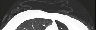
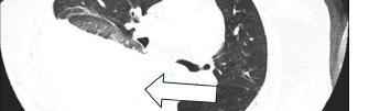

1. Axial computed tomography angiogram of the chest showing a large, right-sided hemothorax (arrow). Findings were secondary to an acute thoracic vertebra 11 chalk-stick fracture in the setting of ankylosing spondylitis. The fracture disrupted adjacent interspinous vasculature, resulting in hemorrhagic shock, although no active contrast extravasation was identified.
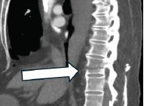
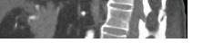
tomography angiogram of
to
ing an acute chalk-stick fracture (arrow) at thoracic
in a patient with ankylosing spondylitis. This unstable fracture at the thoracolumbar junction was associated with interspinous vascular injury and a large, right-sided hemothorax, underscoring the risk of catastrophic vascular complications in this population.
requirement. The patient had been on chronic anticoagulation with apixaban, but his last dose was more than 24 hours prior to presentation; therefore, pharmacologic reversal was not administered in the ED. In similar cases of life-threatening bleeding from factor Xa inhibitors, reversal agents such as andexanet alfa or four-factor prothrombin complex concentrate may be considered when immediately available and indicated. Key abnormalities included severe anemia (hemoglobin 8.1 g/L) and a markedly elevated lactate (8.0 mmol/L), consistent with lactic acidosis from profound tissue hypoperfusion in hemorrhagic shock and likely compounded by severe anemia and impaired lactate clearance from underlying end-stage renal disease. The complete laboratory panel is shown in Table 2.
The patient was admitted to the surgical intensive care unit (SICU) with a diagnosis of a chalk-stick fracture of the T11 vertebra, secondary to traumatic fall and an interspinous vascular injury resulting in a right unilateral hemothorax. Neurosurgery, nephrology, thoracic and cardiovascular surgery were involved in the patient’s multidisciplinary care throughout his prolonged SICU stay. Hospital course was complicated by subsequent right lower extremity deep venous thrombosis diagnosed by ultrasound, for which an inferior vena cava filter was placed. The patient underwent posterior
Table 2. Lab studies of elderly patient with history of ankylosing spondylitis who presented with large, right-sided hemothorax secondary to an acute chalk-stick fracture of the thoracic vertebra 11.
Lab Test Patient Value on Admission Reference Range
Lactic acid 8.0 mmol/L 0.4 - 2 mmol/L Hemoglobin 8.1 g/dL 14.0 - 18.0 g/dL
White blood cells 12.14 x 10³ cells/μL 4.80 - 10.80 x 10³ cells/μL
Red blood cells 2.75 x 10⁶ cells/μL 4.63 - 6.08 x 10⁶ cells/μL
Platelet count 295 x 10³ cells/μL 150 - 450 x 10³ cells/μL
Sodium 134 mmol/L 136 - 145 mmol/L
Chloride 93 mmol/L 98 - 107 mmol/L
BUN 28 mg/dL 7 - 18 mg/dL
Creatinine 5.9 mg/dL 0.7 - 1.3 mg/dL
BUN, blood urea nitrogen dL, deciliter; L, liter; mg, milligrams; mmol, millimoles; μL, microliter.
due to the lack of protective mediastinal organs on the right, compared to the left.8 Although hemothorax is typically linked with blunt or penetrating chest trauma, it can be an unexpected sign of spinal injury in patients with a rigid spine, even after minor trauma.
A few case reports discuss this rare complication. One case involved a 74-year-old woman with ankylosing spinal hyperostosis who developed massive hemothorax after a fall, caused by a T11 extension-distraction fracture.11 Another report described a 68-year-old man with AS and a T9 fracture extending to the ribs, resulting in right-sided hemothorax and paraplegia.12 These cases reinforce the need for early imaging and heightened suspicion for thoracic vascular injury in AS patients. A systematic review reported a 33.3% mortality rate among patients with spinal fracture-induced hemothorax, highlighting the importance of rapid diagnosis and aggressive management.8
thoracic vertebra 9 (T9) to first lumbar spinal instrumentation as well as video-assisted thoracoscopic surgery throughout his month-long stay in the SICU. On hospital day 31, the patient acutely decompensated. Resuscitation was initiated, resulting in return of spontaneous circulation. However, in accordance with the family’s wishes, the patient was later extubated and pronounced deceased.
Vertebral fractures are common in elderly patients after falls, but those with AS require particular attention. Individuals with AS experience spinal fractures at rates up to four times that of the general population, with a lifelong incidence ranging from 5-15%.4,9 These fractures often occur from low-energy trauma due to a rigid, osteoporotic spine commonly referred to radiographically as a “bamboo spine.” Most affect the cervical spine, especially cervical vertebrae 5 and 6 (81.2%), while thoracic and lumbar fractures account for 10.7% and 7.8%, respectively.5 Despite their lower incidence, thoracic fractures carry high morbidity and mortality due to spinal instability and neurologic compromise, present in up to 67.2% of cases.4
Thoracic fractures, although less frequently seen in AS, carry unique risks due to the region’s proximity to critical structures, including the pleural cavity and thoracic vessels. One rare but potentially fatal complication is hemothorax, which may result from disruption of intercostal vessels or adjacent vascular structures following spinal fracture.6,10 Right-sided hemothorax is more frequently reported, likely
While surgical intervention was the primary approach in previous cases, there is growing evidence that selective arterial embolization can be a life-saving adjunct in cases of active bleeding. In one reported case, a 68-year-old man with AS and thoracic spine dislocation at T11 underwent urgent angiography, revealing extravasation from the intercostal artery. Successful embolization stabilized his hemodynamics, permitting delayed spinal fixation.13 The case highlights the potential benefit of angiography and embolization in managing hemodynamically unstable patients with spinal fractures and suspected vascular injury.
Beyond hemorrhage, chalk-stick fractures can also lead to delayed spinal epidural hematomas, reported in 0.5-1.7% of patients.1 Hematomas may develop hours after injury, requiring ongoing monitoring and possible repeat imaging. In unstable patients, angiography should be strongly considered to assess for active extravasation and guide timely intervention. The absence of rate-controlling medications makes the patient’s relatively normal heart rate notable in the context of hemorrhagic shock, underscoring that elderly patients may manifest shock with blunted tachycardic responses even without rate-controlling medications.14
Diagnosis of chalk-stick fractures can be delayed due to overlapping injuries and masked symptoms. In cases of thoracic vertebral fractures with hemothorax, respiratory compromise may overshadow signs of spinal instability, leading to a delay in definitive imaging. Standard radiographs are often insufficient due to extensive spinal fusion and overlying ossification, necessitating CT or magnetic resonance imaging for proper evaluation.3 Given the risks of delayed neurologic or vascular complications, a low threshold for advanced imaging is warranted in AS patients—even after minor trauma and in the absence of overt neurological deficits. Prompt recognition and intervention remain critical in optimizing outcomes and preventing devastating sequelae.
In individuals with ankylosing spondylitis, even low-
Dehkordi et al.
Hemothorax from a Thoracic Chalk-Stick Fracture in Ankylosing Spondylitis: A Case Report
energy trauma should raise immediate concern for spinal fractures. A thorough physical examination, followed by prompt diagnostic imaging—including point-of-care ultrasound and radiographic imaging—is warranted to evaluate for structural injury. Additionally, it is essential to evaluate adjacent soft tissues, including vascular structures, even in the absence of significant pain or overt neurological deficits. A meticulous assessment of the spine, guided by a high index of suspicion, can help prevent delayed diagnosis, intervention, and associated complications. Timely recognition and appropriate management of chalk-stick fractures are critical to optimizing outcomes, particularly in patients with ankylosing spondylitis.
Patient consent has been obtained and filed for the publication of this case report.
Address for Correspondence: Armin Akbarpur Dehkordi, BS, Arkansas College of Osteopathic Medicine at ACHE, Fort Smith, AR 72916; Hollywood, FL 33020. Email: aakbarpurdehkordi@ achehealth.edu.
Conflicts of Interest: By the CPC-EM article submission agreement, all authors are required to disclose all affiliations, funding sources and financial or management relationships that could be perceived as potential sources of bias. The authors disclosed none.
Copyright: © 2026 Dehkordi et al. This is an open access article distributed in accordance with the terms of the Creative Commons Attribution (CC BY 4.0) License. See: http://creativecommons.org/ licenses/by/4.0/
2. Anwar F, Al-Khayer A, Joseph G, et al. Delayed presentation and diagnosis of cervical spine injuries in long-standing ankylosing spondylitis. Eur Spine J. 2011;20(3):403-407
3. Chaudhary SB, Hullinger H, Vives MJ. Management of acute spinal fractures in ankylosing spondylitis. ISRN Rheumatol. 2011;150484
4. Skarentzos K, Karamanou G, Chrysafis I, et al. Chalk-stick fracture in ankylosing spondylitis. Clin Rheumatol. 2020;39(8):2469-2470
5. Tomar SS. Chalkstick fracture: a catastrophic injury. Asian J Neurosurg. 2018;13(2):383-385
6. Masteller MA, Chauhan A, Musunuru H, et al. Haemothorax and thoracic spine fractures in the elderly. Case Rep Radiol. 2012;162064.
7. Tu PH, Liu ZH, Yeap MC, et al. Spinal cord injury and spinal fracture in patients with ankylosing spondylitis. BMC Emerg Med 2022;22(1):73.
8. Ninomiya K, Kuriyama A, Uchino H. Massive hemothorax due to bleeding from thoracic spinal fractures: a case series and systematic review. Scand J Trauma Resusc Emerg Med. 2020;28(1):92.
9. Kang KH, Seok SY, Cho JH. Concurrently occurring spinal cord cross-section and aortic injury after a chalk-stick fracture and dislocation in patient with ankylosing spondylitis: clinical image. World Neurosurg. 2024;184:149-151
10. Ozono K, Son K, Momii K, et al. Severe hemothorax due to traumatic fracture of thoracic vertebra. Surg Case Rep. 2024;10(1):26.
11. Hirota R, Irifune H, Takahashi N, et al. A case of ankylosing spinal hyperostosis with massive hemothorax due to thoracic vertebral fracture caused by minor trauma. Spine Surg Relat Res. 2018;3(3):274-276.
12. Lin YT, Chen TC, Yeh CM. Haemothorax caused by spinal fracture following minor trauma in a patient with ankylosing spondylitis. Br J Hosp Med (Lond). 2023;84(4):1-2.
13. Morita S, Tsuji T, Yamagiwa T, et al. Lethal hemothorax following thoracic dislocated spinal fracture: the usefulness of arterial embolization. Int J Emerg Med. 2009;2(3):177-178.
REFERENCES
1. Baldwin ML, Shufflebarger EF, Pacheco ZS. Chalk-stick fracture in ankylosing spondylitis: a case report of cervical spine injury following minor trauma. Cureus. 2024;16(9):e68931.
14. American College of Surgeons Committee on Trauma. Best Practices Guidelines: Geriatric Trauma. Chicago, IL: American College of Surgeons; 2019. Available at: https://www.facs.org/media/ubyj2ubl/ best-practices-guidelines-geriatric-trauma.pdf. Access on May 2, 2025.
Yoshihiro Miyake, MD*†
Tomohiro Abe, MD, PhD*‡
Keisuke Kubo, MD*†
Hideki Nagoshi, MD*†
Hidenobu Ochiai, MD, PhD*
Section Editor: Lev Libet, MD
University of Miyazaki, Department of Emergency and Critical Care Medicine, Miyazaki, Japan
Miyakonojo Medical Association Hospital, Department of Emergency and Critical Care Medicine, Miyazaki, Japan
Cardiovascular Biology Research Program, Oklahoma Medical Research Foundation, Oklahoma City, Oklahoma
Submission history: Submitted June 24, 2025; Revision received June 30, 2025; Accepted October 23,2025
Electronically published March 29, 2026
Full text available through open access at http://escholarship.org/uc/uciem_cpcem DOI: 10.5811/cpcem.48556
Introduction: Intracranial arterial injury is typically associated with high-energy trauma. Early diagnosis and treatment are essential for improving the patient’s functional prognosis.
Case Report: A 76-year-old woman complained of pulsatile tinnitus 15 days post motor vehicle collision. On presentation she was found to have sustained polytrauma, mostly to her abdominal and thoracic regions, as well as a facial contusion. She then developed ptosis, conjunctival congestion, and an ocular motility disorder in all directions in her right eye. Magnetic resonance angiography showed a direct high-flow shunt from the internal carotid artery to the cavernous sinus. Similar symptoms then developed on her left side. She underwent successful embolization to the left-sided fistula.
Conclusion: Fistulization of the carotid with the cavernous sinus is a rare complication of head or facial trauma. Our case indicates that this can occur even in patients with seemingly minor head or facial trauma. [Clin Pract Cases Emerg Med. 2026;10(2):146-149.]
Keywords: carotid-cavernous sinus fistula; delayed diagnosis; multiple trauma; case report.
Carotid-cavernous fistula involves arteriovenous communication between the carotid artery and the cavernous sinus and is secondary to trauma in 70-90% of all cases.1 While it a rare complication of head and neck trauma, when present it is typically associated with high-energy trauma such as that involving a skull base fracture.2 The pathogenesis of carotidcavernous fistula is markedly increased pressure in the cavernous sinus due to the influx of arterial blood into the cavernous sinus, followed by regurgitation and stasis of the ocular vein. The high pressure in the cavernous sinus damages the cranial nerves passing through the cavernous sinus (oculomotor, trochlear, ophthalmic, maxillary, and abducens nerves) causing various complex neuro-ophthalmic symptoms such as ptosis, conjunctival congestion, diplopia, and ocular motility disorders.2 In general, a direct shunt from the internal carotid artery into the cavernous
sinus is characterized by higher shunt flow and more severe symptoms compared with indirect shunts.3
Early recognition and treatment are crucial for the preservation of cranial nerve function.3 Here we describe a case of direct carotid-cavernous fistula associated with minimal head and facial trauma. Despite its potentially treatable nature, this case illustrates the difficulty making the early diagnosis.
A 76-year-old woman with past medical history of hypertension and dyslipidemia experienced a motor vehicle collision. On arrival to the emergency department, her vital signs were as follows: Glasgow Coma Scale score, 14 (Eye 4, Verbal 4, Motor 6); blood pressure, 115/61 millimeters of mercury; heart rate, 58 beats per minute; respiratory rate, 24 per minute; and oxygen saturation, 100% (oxygen 6 liters per minute via mask).
et al.
Carotid-cavernous Fistula in a Patient with Minimal Head and Facial Trauma: A Case Report
The patient complained of pain in the lumbar and pelvic regions. Mild periorbital ecchymoses were noted in her right eye (Image 1A). Pupils were equal and reactive to light, and no anisocoria was observed. No initial cranial nerve deficits were noted. Computed tomography (CT) showed no intracranial hemorrhagic lesions and no fractures to the skull or facial bones (Images 1B, 1C). Other findings on CT included fractures of the left fourth and fifth ribs, a liver injury (the American Association for the Surgery of Trauma liver injury grade III), a right iliac fracture, a left lower abdominal subcutaneous hematoma, and a compression fracture of the first lumbar vertebra. Transcatheter arterial embolization was performed to treat the hemorrhage around the right iliac fracture. She was also transfused four units each of packed red blood cells and fresh frozen plasma.
Her general condition was stable, but on hospital day 15 swelling increased around her right eye and she complained of pulsatile tinnitus near her right eye. Two days later, she developed ptosis, conjunctival congestion, and an ocular motility disorder of the right eye (Image 2A). Magnetic resonance angiography (MRA) of the head showed shunting of the right internal carotid artery and cavernous sinus, and carotid-cavernous fistula was diagnosed (Images 2B-D). Three days later similar symptoms developed on the left. Contrast angiography of the right internal carotid artery showed contrast injection directly into the right cavernous sinus, confirming the
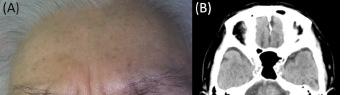
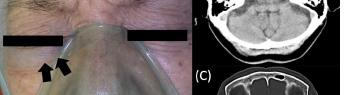
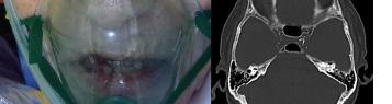

Image 1. Facial appearance and cranial computed tomography (CT) performed on arrival.
Mild periorbital ecchymoses was visible around the right eye (arrows) (A), and cranial CT shows no signs of cranial injury, skull fracture, or a carotid-cavernous fistula (B, C).
CPC-EM Capsule
What do we already know about this clinical entity?
Carotid-cavernous fistula is a rare but serious complication, most often associated with severe face and head trauma.
What makes this presentation of disease reportable?
This case demonstrates a direct carotid-cavernous fistula developing in the subacute phase in a patient with minimal facial trauma.
What is the major learning point?
Even minor head or facial trauma may complicate a carotid-cavernous fistula in the subacute phase, hindering a timely diagnosis.
How might this improve emergency medicine practice?
Emergency physicians should consider carotidcavernous fistula in patients presenting with neuro-ophthalmic symptoms, even after minor head or facial trauma.
exact location of the carotid-cavernous fistula, as well as regurgitation into the right superior ophthalmic vein and inferior ophthalmic vein. Regurgitation into the contralateral superior ophthalmic vein, inferior ophthalmic vein, and cortical vein via the intercavernous sinus was also evident (Images 3A, 3B). Contrast angiography of the left internal carotid artery showed no shunting into the cavernous sinus (Images 3C, 3D).
On Day 21, contrast angiography of the right internal carotid artery showed contrast influx from the internal carotid artery into the cavernous sinus (A) (white arrows) and reflux into the left cavernous sinus and cortical vein (B) (white arrows). Contrast angiography of the left internal carotid artery showed blood supply to both anterior arteries and the right middle cerebral artery with regurgitation into the right internal carotid artery (C, D) (white arrows), but no carotid-cavernous fistula directly into the cavernous sinus. After coil embolization on Day 23, contrast angiography of the right internal carotid artery showed disappearance of the shunt (E, F) (white arrows) and blood flow into the right middle cerebral artery.
The right carotid-cavernous fistula resolved post treatment noted on right internal carotid artery angiography (Images 3E, 3F). At the end of treatment, the neuro-ophthalmic symptoms in her left eye and the pulsatile tinnitus near the right eye resolved, but the symptoms in her right eye persisted.
Carotid-cavernous Fistula in a Patient with Minimal Head and Facial Trauma: A Case Report




Image 2. Ocular findings and magnetic resonance angiography (MRA) performed on Day 17: (A) Conjunctival congestion and an ocular motility disorder are evident in all directions of the right eye; (B) MRA showing bilateral cavernous sinuses and the right ophthalmic vein (arrows); and (C, D) The left transverse sinus and right middle cerebral vein (arrows) are also depicted.
Although MRA on Day 27 showed resolution of the carotidcavernous fistula on the right the ocular motility disorder, ptosis persisted.
This case, in which the patient suffered a direct carotidcavernous fistula during the subacute phase of trauma care, demonstrates the difficulty in making an early diagnosis of carotid-cavernous fistula because of the timing of its onset in a polytrauma patient. Treatment options include conservative management, surgery, and endovascular therapy. Spontaneous shunt closure is expected in only approximately 17% of direct carotid-cavernous fistula cases vs approximately 30% of indirect cases treated conservatively.4 Surgical interventions, such as suturing or clipping the fistula, packing the cavernous sinus, or ligating the internal carotid artery are invasive, although they can achieve shunt closure in 31-79% of carotidcavernous fistula cases.5 Endovascular embolization of the shunt is the first choice of treatment, as it can successfully manage the fistula in more than 80% of cases with fewer adverse events compared with surgical intervention.6
Early diagnosis and treatment minimizes damage to the involved cranial nerves. The earlier the intervention the higher the chance of reversibility. Post shunt closure symptoms generally resolve within hours to days.2 Making the early diagnosis of carotid-cavernous fistula is challenging, particularly in the acute phase after trauma, when ocular findings may be subtle or overshadowed by other severe injuries. Moreover, patients with carotid-cavernous fistula are less likely to have typical symptoms: in fact, a systematic
Image 3. Angiography findings on Day 21 and after coil embolization on Day 23. (A)-(D) angiography on Day 21 and (E)-(F) after coil embolization on Day 23. (A), (C), and (E) show the lateral view. (B), (D), and (F) are the anteroposterior view.
review reported that among patients with carotid-cavernous fistula, ptosis, conjunctival congestion, and diplopia occur in only 14%, 7%, and 2%, respectively.7 Furthermore, the onset of traumatic carotid-cavernous fistula varies widely, from immediately after injury to several weeks later, which adds complexity to the presentation.8 Our case adds to the literature that demonstrates carotid-cavernous fistula can develop in the subacute phase after injury and seemingly minor facial trauma. Carotid-cavernous fistula is a rare complication. In this case the patient’s angiogram was delayed based on her evolving symptoms. However, the lesson from this case is not limited to carotid-cavernous fistula; in the management of severe trauma, delayed or missed diagnoses can lead to long-term disability. As seen in our case during the treatment of torso trauma, the diagnosis of non-fatal but complicated
Miyake et al.
Carotid-cavernous Fistula in a Patient with Minimal Head and Facial Trauma: A Case Report
injuries can easily be delayed, particularly in patients with multiple trauma.9 Therefore, after addressing life-threatening conditions, careful and continued follow-up is essential to identify injuries that were not apparent during the initial assessment, and further diagnostic evaluation should be pursued as new symptoms emerge.
The early diagnosis and treatment of carotid-cavernous fistula, a rare complication of head and facial trauma, can prevent neuro-ophthalmic symptoms. Carotid-cavernous fistula should be considered when neuro-ophthalmic symptoms are present after trauma, even in patients with minimal facial and/or head trauma, even in the subacute phase.
Written informed consent for publication was obtained from the patient’s next of kin.
1. Chaudhry IA, Elkhamry SM, Al-Rashed W, et al. Carotid cavernous fistula: ophthalmological implications. Middle East Afr J Ophthalmol 2009;16(2):57-63.
2. Liang W, Xiaofeng Y, Weiguo L, et al. Traumatic carotid cavernous fistula accompanying basilar skull fracture: a study on the incidence of traumatic carotid cavernous fistula in the patients with basilar skull fracture and the prognostic analysis about traumatic carotid cavernous fistula. J Trauma. 2007;63(5):1014-1020.
3. Henderson AD and Miller NR. Carotid-cavernous fistula: current concepts in aetiology, investigation, and management. Eye (Lond) 2018;32(2):164-172.
4. Higashida RT, Hieshima GB, Halbach VV, et al. Closure of carotid cavernous sinus fistulae by external compression of the carotid artery and jugular vein. Acta Radiol Suppl. 1986;369:580-583.
5. Ellis JA, Goldstein H, Connolly ES Jr, et al. Carotid-cavernous fistulas. Neurosurg Focus. 2012;32(5):E9.
6. Lewis AI, Tomsick TA, and Tew JM Jr. Management of 100 consecutive direct carotid-cavernous fistulas: results of treatment with detachable balloons. Neurosurgery. 1995;36(2):239-344.
Address for Correspondence: Tomohiro Abe, MD, Department of Emergency and Critical Care Medicine, Faculty of Medicine, University of Miyazaki, 5200, Kihara, Kiyotake, Miyazaki, 8891692, Japan. Email: tomohiro_abe@med.miyazaki-u.ac.jp
Conflicts of Interest: By the CPC-EM article submission agreement, all authors are required to disclose all affiliations, funding sources and financial or management relationships that could be perceived as potential sources of bias. The authors disclosed none.
Copyright: © 2026 Miyake et al. This is an open access article distributed in accordance with the terms of the Creative Commons Attribution (CC BY 4.0) License. See: http://creativecommons.org/ licenses/by/4.0/
7. Al-Shalchy A, Al-Wassiti AS, Hashim MA, et al. Neuro-ophthalmic manifestations of carotid cavernous fistulas: a systematic review and meta-analysis. Cureus. 2024;16(7):e65821.
8. Shim HS, Kang KJ, Choi HJ, et al. Delayed contralateral traumatic carotid cavernous fistula after craniomaxillofacial fractures. Arch craniofacial Surg. 2019;20(1):44-47.
9. Lee WC, Chen CW, Lin YK, et al. Association of head, thoracic and abdominal trauma with delayed diagnosis of co-existing injuries in critical trauma patients. Injury. 2014;45(9):1429–1434.
Jordan Richardson, MD*†
Neha Raukar, MD, MS*
Luke Wood, DO*
Section Editor: Austin Smith, MD
Mayo Clinic Alix School of Medicine, Department of Emergency Medicine, Rochester, Minnesota
Regions Hospital, Department of Emergency Medicine, St. Paul, Minnesota
Submission history: Submitted March 18, 2025; Revision received June 18, 2025; Accepted October 15, 2025
Electronically published March 10, 2026
Full text available through open access at http://escholarship.org/uc/uciem_cpcem DOI: 10.5811/cpcem.47041
Introduction: Hyponatremia is a common and often vexing electrolyte abnormality seen in the emergency setting. The severity of a patient’s symptoms is often dictated by the acuity of hyponatremia development and degree of serum sodium deficit, with patients typically demonstrating more severe neurological symptoms in acute-onset severe hyponatremia. Patients prescribed chlorthalidone are at particular risk of developing hyponatremia, especially in the setting of a secondary insult.
Case Report: We describe two patients presenting to the emergency department with severe hyponatremia who were taking chlorthalidone. Both patients had clinical symptoms that were mild given the degree of their hyponatremia. Additionally, each patient had a secondary insult affecting their volume status that was an important contributing factor in the development of hyponatremia.
Conclusion: Thiazide diuretic use, particularly chlorthalidone, is an important consideration when evaluating patients with new-onset hyponatremia, especially in the setting of excess volume losses or increased consumption of water. These cases demonstrate how patients with severe hyponatremia can present with atypical or mild clinical symptoms and often do not demonstrate significant neurological symptoms such as seizures. [Clin Pract Cases Emerg Med. 2026;10(2):150-153.]
Keywords: hyponatremia; thiazide diuretic; diuretics; volume loss; case report.
Hyponatremia, defined as a serum sodium (SNa) concentration < 135 milliequivalents per liter (mEq/L), is one of the most common electrolyte abnormalities encountered in the emergency department (ED). It has a variety of underlying causes. The most frequent presentation is hypotonic hyponatremia, which can be sub-classified into three categories based on volume status: hypervolemic; hypovolemic; and euvolemic.1 Hypervolemic hypotonic hyponatremia occurs when water retention exceeds the body’s ability to excrete it, often due to kidney dysfunction, heart failure, cirrhosis, or nephrotic syndrome. Hypovolemic hypotonic hyponatremia results from sodium losses in the form of osmotic diuresis, adrenal insufficiency, vomiting, diarrhea, or excessive sweating. Euvolemic hypotonic
hyponatremia is often associated with thiazide diuretic use, severe hypothyroidism, low solute intake, syndrome of inappropriate antidiuresis, or primary polydipsia.1
The symptoms of hyponatremia are varied and typically correlate to both the severity and acuity of the electrolyte derangement. Chronic hyponatremia can be asymptomatic or present with mild symptoms including disorientation or lethargy.2 Acute mild hyponatremia can present as nausea, vomiting, decreased appetite, or headache, which can progress to altered mental status, seizures, and coma. The risks of inadequate or delayed treatment, including cerebral edema and demyelination, are significant and life-threatening.2 In this report of two patients taking chlorthalidone who presented with mild symptoms despite severe hyponatremia, we discuss acute and chronic hyponatremia in the context of
chlorthalidone use and share considerations for managing hyponatremia in the ED.
Case 1
A 65-year-old female presented to the ED with the chief complaint of increased depression. Further history revealed a one-week history of intermittent vomiting, sleep difficulties, and reduced appetite. The patient’s husband presented with her and described episodic gait instability and vague speech difficulties over the prior two days. Her medical history included hypothyroidism, depression, and resistant hypertension, for which chlorthalidone 25 mg had been initiated 16 days prior. The patient denied any associated headaches, vision changes, syncopal episodes, or seizures.
Upon arrival, the patient was afebrile and hemodynamically stable. Cardiopulmonary exam was unremarkable, and she had no focal abdominal tenderness. Neurological exam demonstrated no cranial nerve deficits with full and symmetrical strength and gross sensation. She had no past-pointing, dysmetria, or abnormal heel-to-shin testing. No dysarthric speech or expressive aphasia was noted. Her gait was described as slowed but stable and non-ataxic. She denied any suicidal ideations but had a withdrawn affect and endorsed feelings of increased depression with poor sleep and appetite.
The patient’s husband reiterated the intermittent nature of her gait difficulties and speech changes, noting her normal bedside neurological exam. He once again expressed concern for her worsening depression. The emergency care team suspected either a potential neurological or psychiatric etiology. Her initial workup included complete blood count, basic metabolic panel, magnesium level, point-of-care venous blood gas, coagulation panel, thyroid function cascade, electrocardiogram, computed tomography (CT) head, and CT angiography of the head and neck with planned psychiatric consultation if the medical workup was unrevealing.
Prior to imaging, the patient had point-of-care venous blood gas demonstrating pH 7.51. Partial pressure of carbon dioxide was 34 millimeters of mercury (mm Hg) (reference range: 41-51 mm Hg), and SNa < 100 mEq/L (135-145 mEq/L). Given the patient’s mild, intermittent symptoms, non-focal neurological exam and SNa < 100, this laboratory finding was presumed to be in error and management of hyponatremia was deferred until the patient’s basic metabolic panel (BMP) had resulted. Basic metabolic panel returned showing SNa of 98 mEq/L and, once again, the accuracy of this lab result was questioned. It seemed unlikely for a hyponatremia of this severity to develop acutely (with normal SNa 16 days prior) in the absence of seizures or other objective neurological changes. A repeat BMP confirmed the presence of profound hyponatremia (SNa 97 mEq/L). The patient was also found to be hypomagnesemic to a level of 1.1 mg per deciliter (dL) (1.7-2.3 mg/dL). She was started on fluid restriction, and we obtained serum osmolarity, urine osmolarity, and urine sodium
CPC-EM Capsule
What do we already know about this clinical entity?
Hyponatremia can present as a myriad of clinical symptoms. Typically, the severity of symptoms relates to the degree of sodium deficit and whether the onset was acute or chronic.
What makes this presentation of disease reportable?
Acute, severe hyponatremia should result in significant clinical symptoms and objective neurological findings. Both cases demonstrate how this can present in patients with minimal symptoms.
What is the major learning point?
Thiazide diuretics predispose patients to developing acute hyponatremia. A secondary insult that affects volume status often predisposes these patients to severe hyponatremia.
How might this improve emergency medicine practice?
Hyponatremia can present with symptoms discordant from the degree of sodium deficit and timing of onset. Thiazide diuretic use is an important risk factor to be considered in these patients.
to classify her new-onset hyponatremia.
The patient’s potassium and magnesium were repleted, and she received 50 mL of hypertonic saline in the ED. Chlorthalidone, a thiazide diuretic, was discontinued because of the known side effect of hyponatremia. She was transferred to the intensive care unit for further management, where she was found to have Clostridium difficile colitis contributing to her gastrointestinal (GI) symptomology and significant volume losses.
A 61-year-old male was referred to the ED after outpatient lab studies earlier in the day returned showing hyponatremia (SNa 116 mEq/L). Medications included metformin 2 grams, atorvastatin 20 mg, lisinopril 20 mg, and topical testosterone 40 mg, as well as chlorthalidone 25 mg for over five years. Given his metformin use, the patient had been encouraged to drink copious water in preparation for an outpatient abdominal CT for chronic nausea and diarrhea, and he had consumed four 20-ounce bottles of water. Following
Minimally Symptomatic Severe Hyponatremia: Two Case Reports
the patient’s CT, he drank an additional two 20-ounce bottles of water and then had blood work performed that revealed significant hyponatremia.
Upon arrival at the ED, the patient was hemodynamically stable and denied headache, confusion, syncope, seizures, dizziness, or weakness. The physical exam was unremarkable and non-focal. Lab work performed in the ED showed a SNa level of 116 mEq/L. Chlorthalidone was discontinued, and he was started on normal saline with free water restrictions. The patient was admitted to the hospital for further fluid and electrolyte management.
These cases both illustrate profound hyponatremia in the setting of an acute change in volume status while prescribed chlorthalidone. In both cases, the patients had relatively mild symptoms disproportionate to the degree of sodium deficit. In Case 1, the patient had a SNa level of 97 mEq/L yet was reporting only poor sleep and appetite loss. Her husband had noted mild speech slurring and gait instability on a subacute basis. The patient in Case 2 was asymptomatic.
In this discussion, we review the literature on chlorthalidone and hyponatremia, presenting symptoms of chronic and acute hyponatremia, and important clinical considerations for hyponatremia in the ED. Hyponatremia is a well-known side effect of thiazide diuretics. Risk factors include advanced age, female sex, inappropriate osmotic diuresis, and mild hypokalemia.3 The time between thiazide diuretic initiation and hyponatremia can range from a few days to over a year with metabolic derangements typically presenting within 2-3 weeks of initiation of treatment.4 Thiazide diuretics act by blocking the resorption of sodium chloride in the distal convoluted tubule, thus inhibiting osmotic water resorption. However, thiazide diuretics have varying propensities to cause hyponatremia. Several studies have found that chlorthalidone is associated with a higher risk of hyponatremia than hydrochlorothiazide when used in equal doses.5-7
Hydrochlorothiazide has a less potent effect on blood pressure and is often prescribed at a higher dosage compared to chlorthalidone. When adjusting for this difference, one study found that the risk of developing hyponatremia was not significantly different in patients taking chlorthalidone (12.5-25 mg/day) compared to patients taking a two-fold higher dose per day of hydrochlorothiazide.5 Currently, guidelines are lacking regarding monitoring for electrolyte derangements when thiazide diuretics are initiated; therefore, patients can present with hyponatremia after recent initiation of treatment or with long-term use of chlorthalidone.
These two cases demonstrate that while we typically associate hyponatremia secondary to thiazide use with euvolemic hyponatremia, the presence of a secondary insult in volume status can make the hyponatremia even more severe. In our first case, the patient was hypovolemic secondary to C difficile infection with several days of preceding GI volume
losses. Volume depletion can lead to increased release of antidiuretic hormone, resulting in the production of more concentrated urine.4 This can potentiate underlying hyponatremia from the diuretic with increased water retention, causing a dilutional effect with sodium and other electrolytes, thus resulting in a hypovolemic hypotonic hyponatremia.
In the second case, the patient had been instructed to increase his water intake significantly in the day leading up to his presentation, resulting in a dilutional hyponatremia. It has been noted that some patients with thiazide-induced hyponatremia have been found to have underlying polydipsia at baseline.8 In this case, the patient had hypervolemic hypotonic hyponatremia. Both patients were susceptible to hyponatremia due to their chlorthalidone use, but in both cases a secondary insult to their volume status was critical to the development of severe hyponatremia.
Hyponatremia can present as acute, subacute, and chronic. Acute hyponatremia, defined as hyponatremia that develops in < 48 hours, typically present with more pervasive and severe symptoms due to insufficient time for adaptive shifts in osmolality to protect from cerebral edema.1 Severe, acute hyponatremia can present with altered mental status, seizures, coma, and neurogenic shock.9 However, subacute or chronic hyponatremia, occurring over a period > 48 hours, is more often associated with milder symptoms of gait instability, dysarthria, disorientation, and somnolence, similar to the patient’s presenting symptoms in case 1.2
When evaluating hyponatremia in the ED, there are several important, specific considerations. To determine the etiology of hyponatremia, it is imperative to establish both volume status and osmolality to differentiate the type of hyponatremia, which ultimately guides clinical management. Point-of-care ultrasound, clinical exam, and serum creatinine can all be used to determine volume status.9 Osmolality, urine osmolality, and urine sodium should also be collected, with important consideration for collecting these labs before initiating any saline repletion. Additional considerations for the emergency physician include collecting serum glucose to rule out hyperglycemic hyponatremia, reviewing the patient’s medication list, and measuring serum potassium as this is also often concurrently depleted in hyponatremic patients. Emergency physicians should also be aware that there can be significant variations between point-of-care and standard laboratory measurements of SNa by up to 4 millimoles.10 While this may not be clinically meaningful for diagnostic purposes, it is important to ensure that the same method is being used for serial measurements during sodium repletion.
We present two cases illustrating severe hyponatremia occurring in patients on chlorthalidone with secondary insults. Given the acuity and severity of their hyponatremia, both patients had relatively mild clinical symptoms, and neither presented with seizures or significant alterations to mental
Richardson et al.
status. Chlorthalidone use is an important risk factor for developing hyponatremia, especially in patients who have a secondary insult affecting their volume status. These cases demonstrate how the emergency clinician must maintain a high degree of suspicion for potential hyponatremia when patients are prescribed chlorthalidone and are presenting with vague neurological symptoms. These cases also illustrate how patients can have profound hyponatremia and present asymptomatically or with mild clinical symptoms.
Patient consent has been obtained and filed for the publication of the case reports.
Address for Correspondence: Luke Wood, DO, Mayo Clinic Alix School of Medicine, Department of Emergency Medicine, 200 First Street SW Rochester, MN 55905. Email: wood.luke@mayo.edu
Conflicts of Interest: By the CPC-EM article submission agreement, all authors are required to disclose all affiliations, funding sources and financial or management relationships that could be perceived as potential sources of bias. The authors disclosed none.
Copyright: © 2026 Richardson et al. This is an open access article distributed in accordance with the terms of the Creative Commons Attribution (CC BY 4.0) License. See: http://creativecommons.org/ licenses/by/4.0/
Minimally Symptomatic Severe Hyponatremia: Two Case Reports
1. Adrogué HJ, Tucker BM, Madias NE. Diagnosis and management of hyponatremia: a review. JAMA. 2022;328(3):280-291.
2. Buffington MA, Abreo K. Hyponatremia: a review. J Intensive Care Med. 2016;31(4):223-236.
3. Barber J, McKeever TM, McDowell SE, et al. A systematic review and meta-analysis of thiazide-induced hyponatraemia: time to reconsider electrolyte monitoring regimens after thiazide initiation? Br J Clin Pharmacol. 2015;79(4):566-577.
4. Filippone EJ, Ruzieh M, Foy A. Thiazide-associated hyponatremia: clinical manifestations and pathophysiology. Am J Kidney Dis. 2020;75(2):256-264.
5. van Blijderveen JC, Straus SM, Rodenburg EM, et al. Risk of hyponatremia with diuretics: chlorthalidone versus hydrochlorothiazide. Am J Med. 2014;127(8):763-771.
6. Hripcsak G, Suchard MA, Shea S, et al. Comparison of cardiovascular and safety outcomes of chlorthalidone vs hydrochlorothiazide to treat hypertension. JAMA Intern Med. 2020;180(4):542-551.
7. Dhalla IA, Gomes T, Yao Z, et al. Chlorthalidone versus hydrochlorothiazide for the treatment of hypertension in older adults: a population-based cohort study. Ann Intern Med. 2013;158(6):447-455.
8. Frenkel N, Vogt L, De Rooij S, et al. Thiazide-induced hyponatraemia is associated with increased water intake and impaired ureamediated water excretion at low plasma antidiuretic hormone and urine aquaporin-2. J Hypertens. 2015;33(3):627-633.
9. Lindner G, Schwarz C, Haidinger M, Ravioli S. Hyponatremia in the emergency department. Am J Emerg Med. 2022;60:1-8.
10. Solak Y. Comparison of serum sodium levels measured by blood gas analyzer and biochemistry autoanalyzer in patients with hyponatremia, eunatremia, and hypernatremia. Am J Emerg Med. 2016;34(8):1473-1479.
Rochelle Kofman, BS*
Addison B. Smartt, BS*
Reginald J. Myles, BS*
Patrick Kishi, MD*†
Douglas Rappaport, MD*†
Kevin Drechsel, MD*†
Mayo Clinic Alix School of Medicine, Phoenix, Arizona Mayo Clinic Arizona, Department of Emergency Medicine, Phoenix, Arizona
Section Editor: Alexander John Scumpia, MD
Submission history: Submitted August 24, 2025; Revision received October 19, 2025; Accepted November 17, 2025
Electronically published March 13, 2026
Full text available through open access at http://escholarship.org/uc/uciem_cpcem DOI: 10.5811/cpcem.50741
Introduction: Central retinal artery occlusion (CRAO) is a neurological and ophthalmologic emergency that presents as sudden, painless, monocular vision loss. Central retinal artery occlusion can be classified as arteritic or non-arteritic. Most cases of non-arteritic CRAO are due to embolism, commonly from atherosclerosis of the ipsilateral carotid artery. More proximal sources of embolism are uncommon but can occur. Prompt recognition of CRAO is critical for vision preservation therapy and initiation of ischemic stroke diagnosis protocols.
Case Report: We present the case of a 66-year-old female who presented to the emergency department eight hours after sudden, painless, monocular vision loss. Her past medical history included type II diabetes, hypertension, and hyperlipidemia. She had previously undergone bilateral lens replacement for cataracts three years prior and had a history of intermittent floaters, which had been worsening over the previous six months. She denied any associated pain, headache, speech difficulty, focal weakness, and ocular trauma. Point-of-care ultrasound (POCUS) of the affected eye revealed the presence of a retrobulbar spot sign, which is associated with CRAO with a non-arteritic embolic etiology.
Conclusion: Point-of-care ultrasound is an efficient diagnostic tool for the assessment of acute, painless, monocular vision loss. The presence of a retrobulbar spot sign indicates central retinal artery occlusion, providing both diagnostic and prognostic information. Although not a definitive diagnostic tool, POCUS can expedite treatment for patients with central retinal artery occlusion, a diagnosis with a time-sensitive treatment window. [Clin Pract Cases Emerg Med. 2026;10(2):154-158.]
Keywords: point-of-care ultrasound; central retinal artery occlusion; retrobulbar spot sign; cardioembolic stroke; case report.
INTRODUCTION
Central retinal artery occlusion (CRAO) is a rare, visionthreatening neuro-ophthalmologic emergency that presents with sudden, painless, monocular vision loss. Without immediate identification and management, CRAO risks permanent vision loss, with studies showing that > 80% of cases resulted in visual acuity of 20/200 or worse. Better
visual outcomes were more common in cases of CRAO with cilioretinal artery sparing.1,2
The incidence of CRAO in the United States is approximately two cases per 100,000 person-years and increases with male sex and age to 10.1 cases per 100,000 person-years in individuals > 80 years of age.3. Central retinal artery occlusion can be classified as arteritic (5%),
Koffman et al. Central Retinal Artery Occlusion Diagnosed via Ocular Point-of-care Ultrasound: Case Report
such as in giant cell arteritis, or non-arteritic (95%).1 Most cases of non-arteritic CRAO are due to embolic occlusion of the central retinal artery, commonly from atherosclerosis of the ipsilateral carotid artery, with some studies reporting the presence of carotid artery plaques in up to 71% of cases and significant carotid stenosis in 31-40% of cases.4,5 Cardiac sources of emboli, such as atrial fibrillation or a cardiac mass, while prevalent, are less common.6 There are limited published reports describing cardiac masses, such as fibroelastomas or myxomas, presenting with CRAO as the initial symptom.7 This variant highlights the importance of maintaining a broad embolic differential, particularly in patients without known atrial fibrillation or significant carotid disease.
The American Heart Association characterizes CRAO as an acute ischemic stroke event that carries risks of recurrent vascular events.1,8 Early diagnosis and intervention are critical to avoid irreversible retinal damage and vision loss in addition to identifying and managing underlying vascular risk factors. On examination, fundoscopic findings may reveal a pale retina and a cherry-red spot at the macula. However, these can be subtle or absent in the early presentation. As CRAO is considered a stroke equivalent, standard workup includes immediate neuroimaging, typically with non-contrast head computed tomography (CT) to rule out hemorrhage, followed by CT angiography (CTA) or magnetic resonance imaging (MRI) of the head and neck to assess for vascular occlusion or ischemia. Cardiac telemetry and echocardiography are often used to evaluate for cardioembolic sources such as atrial fibrillation or structural cardiac abnormalities or masses.1,5
Point-of-care ultrasound (POCUS) plays a valuable role in the rapid evaluation of acute vision loss and has recently emerged as an important adjunct in CRAO diagnosis and management. In particular, the retrobulbar spot sign, a hyperechoic focus within the optic nerve sheath seen on ocular ultrasound, has been associated with CRAO, and its presence has been shown to confirm a thromboembolic cause and can exclude an arteritic etiology. Additionally, this finding also rules out other causes of sudden, painless, monocular vision loss, such as retinal detachment, optic neuropathy, or vitreous hemorrhage and can, therefore, guide further workup and management.9-12 The sensitivity and specificity of the for detection of an embolic CRAO are 83% and 100%, respectively.12
Here we present a case of CRAO in the emergency department (ED) setting, with a retrobulbar spot sign seen on ocular ultrasound. The embolic source was identified by transesophageal echocardiogram as a mass on the mitral valve.
A 66-year-old female presented to the ED eight hours after sudden loss of central vision in the right eye. Her past medical history included diabetes mellitus type II, hypertension, hyperlipidemia, hypothyroidism, and Charcot
CPC-EM Capsule
What do we already know about this clinical entity?
Central retinal artery occlusion (CRAO) is a neurological and ophthalmologic emergency that presents with painless monocular loss of vision.
What makes this presentation of disease reportable?
We present a case of CRAO diagnosed with bedside ocular ultrasound demonstrating retrobulbar spot sign; workup ultimately revealed a cardiac myxoma as the embolic source.
What is the major learning point?
CRAO can be diagnosed with ocular ultrasound demonstrating retrobulbar spot sign; further workup can include a bedside echocardiogram to search for an embolic source.
How might this improve emergency medicine practice?
Point-of-care ultrasound can aid in a timely diagnosis of CRAO.
arthropathy. She had undergone bilateral lens replacement for cataracts three years prior and had a history of intermittent floaters, which had been worsening over the prior six months.
Prior to the sudden and total loss of central vision while sitting at her desk at 9 am that morning, she endorsed seeing “bright lights” in her vision in the preceding few days. She described them as white or gray, occurring transiently. She denied any associated pain, headache, speech difficulty, unilateral weakness, or ocular trauma. On arrival to the ED at 5 pm, she was hypertensive with a blood pressure of 174/74 millimeters of mercury, but her vital signs were otherwise all within normal limits. A neurological examination yielded no focal deficits; she was alert and oriented, pupils were round and reactive, extraocular muscles were intact, and cranial nerves showed no gross deficits. Bilateral conjunctiva and eyelids showed no erythema or injection, and fundoscopic examination yielded poor visualization of the vessels and optic disc.
Point-of-care ultrasound (POCUS) of the eye showed no evidence of retinal detachment, vitreous hemorrhage, or posterior vitreous detachment; however, a retrobulbar spot sign was detected (Image 1). This finding in context of the clinical picture put CRAO) at the top of the differential. Based





Image 1. Findings on ocular point-of-care ultrasound using a linear probe demonstrate a retrobulbar spot sign (RBSS) in the transverse view, consistent with central retinal arterial occlusion.

on these findings, a CTA of the head and neck was ordered, and ophthalmology was consulted.
The CTA revealed no acute hemorrhage, ischemia, mass effect, or other gross abnormality. A partially calcified extraaxial mass, likely meningioma, on the left frontal convexity was discovered but was deemed unlikely to be the cause of her vision loss. An exam performed by ophthalmology confirmed central vision loss in the right eye with accompanying macular retinal whitening, with a perfused retinal periphery. This finding combined with the clinical picture pointed toward a CRAO, and the patient’s care was subsequently managed from the perspective of likely ischemic stroke.
The stroke neurology team recommended brain MRI with and without contrast for further evaluation of both the possibility of ischemic stroke and to further characterize the mass found on CTA. As part of this workup, cardiac telemetry and echocardiography were ordered to assess for a cardioembolic source of emboli. The MRI yielded evidence of multiple, tiny punctate multifocal embolic-appearing acute and subacute infarctions (Image 2). They were present in both the anterior and posterior circulation, indicating a probable proximal source of embolism. This patient was eligible for neither intravenous thrombolysis nor endovascular treatment for the CRAO given the long delay between symptom onset and presentation to the ED.
On cardiac workup, the electrocardiogram demonstrated normal sinus rhythm; however, a transesophageal echocardiogram performed by cardiology revealed a highly mobile pedunculated mass attached to the mitral valve protruding into the left ventricular outflow tract during systole (Images 3, 4). This finding revealed the source of emboli presenting both clinically and radiographically.
Subsequently, cardiothoracic surgery was consulted and recommended mitral valve replacement and resection of the
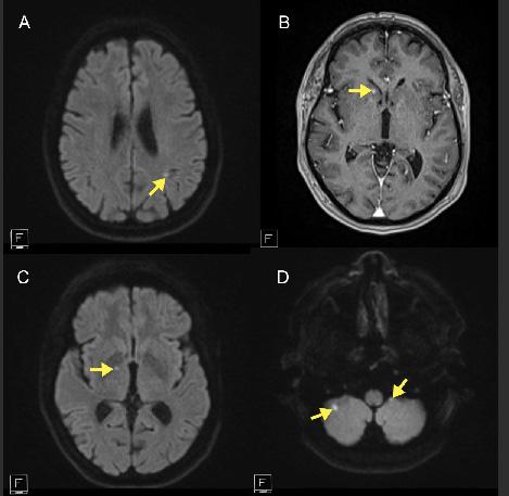
Image 2. Axial images of brain magnetic resonance imaging obtained with T2-weighted flair (A), T1-weighted post contrast (B), and diffusion-weighted (C, D) sequences showing punctate hyperintensities in the periventricular and deep white matter and cerebellum (yellow arrows) consistent with chronic, small-vessel ischemic changes.
mass due to its high embolic risk. Endocarditis was ruled out due to lack of fever, leukocytosis, negative blood cultures, and no other signs of infection. She elected to undergo mitral valve mass resection, mitral valve replacement with a tissue valve, and left atrial appendage excision. The operation occurred on day 7 of her admission, delayed for appropriate cessation of anticoagulation. After excision, the mitral valve was determined to have extensive myxoid degeneration and dystrophic calcification by a surgical pathologist.
Patients with CRAO typically present with sudden, painless vision loss. The differential for this presentation, however, can be wide, including retinal detachment, vitreous bleeding, temporal arteritis, and optic neuropathy. Central retinal artery occlusion visualized with ultrasound may show a hyperechoic retrobulbar spot sign, which represents embolic activity in the retinal artery. The retrobulbar spot sign can also show papilledema.
When assessing for a retrobulbar spot sign there is risk of optic nerve drusen presenting as a false positive for CRAO. This deposit of hyalin calcific material in the optic nerve head may cause similar symptoms but can be excluded given clinical context and accurate capture with ultrasound. Optic








nerve drusen is an insidious visual impairment that develops chronically. Because of this, optic nerve drusen is often found incidentally. Both size and location can differentiate optic nerve drusen from CRAO with retrobulbar spot sign, given that ultrasound visualization of optic nerve drusen is larger and present at the terminal optic nerve,13 while the retrobulbar spot sign is smaller and presents more centrally and further from the optic nerve ending.10
Point-of-care ultrasound can be an efficient diagnostic tool to narrow the differential of acute, painless, monocular vision loss. Non-ionizing imaging is safe and presents no risk of radiation exposure. The use of Doppler can indicate abnormalities in blood flow, aiding in differentiating CRAO from other diagnoses. As documented in McGuire et al, POCUS with the use of Doppler can also be used to monitor progression with serial Doppler measurements, tracking the patient’s progression over time.14 These functions make POCUS evaluation of CRAO especially useful in emergent cases and situations where ophthalmic specialists are not immediately available. Additionally, there is evidence that the presence of a retrobulbar spot sign predicts poor response to thrombolytic treatment for CRAO.15
However, POCUS has clear limitations. There can be discomfort associated with pressure applied to the eye. However, there is no evidence of ultrasound use causing damage to the retina or optic nerve when performed with low-intensity, non-contact techniques. Accuracy of Doppler evaluation is heavily dependent on user expertise, as there is a small margin of error in the angle of visualization. If the angle is off slightly, the signal and strength of the Doppler flow will decrease. Additionally, the absence of a retrobulbar spot sign
does not rule out a diagnosis of CRAO. For these reasons, ultrasound is not the definitive diagnostic tool for vision loss or CRAO, although it can provide timely, accurate, and inexpensive diagnostic information.
Given the negligible risk profile and significant diagnostic utility of ultrasound, especially in the acute setting where rapid differentiation from other causes of acute vision loss is essential, the benefits of ultrasound in diagnosing CRAO far outweigh any potential adverse effects. The use of POCUS in this patient’s workup revealed the retrobulbar spot sign of the right eye, indicating CRAO. Point-of-care ultrasound narrowed the differential and greatly expedited further exploration of the embolic source, leading to subsequent discovery and excision of this patient’s cardiac myxoma. Although in this case the patient was not eligible for thrombolytic treatment, expedited workup and diagnosis of CRAO with POCUS may preserve the possibility of thrombolysis for a patient presenting earlier with this diagnosis. This case affirms the utility of POCUS in patients presenting with acute, monocular vision loss, both for narrowing the diagnostic differential and expediting subsequent treatment.
Point-of-care ultrasound is an efficient and effective diagnostic tool for the assessment of acute, painless, monocular vision loss. The presence of a retrobulbar spot sign indicates central retinal artery occlusion, providing both diagnostic and prognostic information. Although not a definitive diagnostic tool, POCUS can expedite treatment for patients with CRAO, a diagnosis with a time-sensitive treatment window.
Central Retinal Artery Occlusion Diagnosed via Ocular Point-of-care Ultrasound: Case Report Koffman et al.
Patient consent has been obtained and filed for the publication of this case report.
Address for Correspondence: Douglas Rappaport, MD, Mayo Clinic Arizona, Department of Emergency Medicine, 5777 E Mayo Blvd. Phoenix AZ 85054. Email: Rappaport.Douglas@mayo.edu.
Conflicts of Interest: By the CPC-EM article submission agreement, all authors are required to disclose all affiliations, funding sources and financial or management relationships that could be perceived as potential sources of bias. The authors disclosed none.
Copyright: © 2026 Koffman et al. This is an open access article distributed in accordance with the terms of the Creative Commons Attribution (CC BY 4.0) License. See: http://creativecommons.org/ licenses/by/4.0/
5. Tiwari V, Bagga SSJ, Prasad R, Mathurkar S. A review of current literature on central retinal artery occlusion: its pathogenesis, clinical management, and treatment. Cureus. 2024;16(3):e55814.
6. Mac Grory B, Landman SR, Ziegler PD, et al. Detection of atrial fibrillation after central retinal artery occlusion. Stroke. 2021;52(9):2773-2781.
7. Ma JH, Gill MK. Calcified amorphous tumor: a rare cause of central retinal artery occlusion. Am J Ophthalmol Case Rep. 2018;10:25-27.
8. Chodnicki KD, Pulido JS, Hodge DO, Klaas JP, Chen JJ. Stroke risk before and after central retinal artery occlusion in a US cohort. Mayo Clin Proc. 2019;94(2):236-241.
9. Smith AT, Wilbert CD, Ferre RM. Using the retrobulbar spot sign to assist in diagnosis and management of central retinal artery occlusions. J Ultrasound Med. 2020;39(1):197-202.
10. Lottspeich C, Mackert MJ, Köhler A, Bauer A, Hoffmann U, Czihal M. Retrobulbar spot sign in metachronous bilateral central retinal artery occlusion of cardioembolic origin. J Neuroophthalmol. 2021;41(1):e105-e106.
REFERENCES
1. Mac Grory B, Schrag M, Biousse V, et al. Management of central retinal artery occlusion: a scientific statement from the American Heart Association [published correction appears in Stroke. 2021 ;52(6):e309]. Stroke. 2021;52(6):e282-e294.
2. Kim YH, Park KH, Woo SJ. Clinical manifestations and visual prognosis of cilioretinal artery sparing central retinal artery occlusion. Korean J Ophthalmol. 2020;34(1):27-34.
3. Leavitt JA, Larson TA, Hodge DO, Gullerud RE. The incidence of central retinal artery occlusion in Olmsted County, Minnesota. Am J Ophthalmol. 2011;152(5):820-3.e2.
4. Hayreh SS, Podhajsky PA, Zimmerman MB. Retinal artery occlusion: associated systemic and ophthalmic abnormalities. Ophthalmology. 2009;116(10):1928-1936.
11. Czihal M, Lottspeich C, Köhler A, et al. Transocular sonography in acute arterial occlusions of the eye in elderly patients: diagnostic value of the spot sign. PLoS One. 2021;16(2):e0247072.
12. Ertl M, Altmann M, Torka E, et al. The retrobulbar “spot sign” as a discriminator between vasculitic and thrombo-embolic affections of the retinal blood supply. Ultraschall Med. 2012;33(7):E263-E267.
13. Almog Y, Nemet A, Nemet AY. Optic disc drusen demonstrate a hyperechogenic artifact in B mode ultrasound. J Clin Neurosci. 2016;23:111-119.
14. McGuire D, Calleja R, Pai E, Bahl A. Emergency department Doppler assessment of a central retinal artery occlusion: case report. Clin Pract Cases Emerg Med. 2024;8(2):115-119.
15. Nedelmann M, Graef M, Weinand F, et al. Retrobulbar spot sign predicts thrombolytic treatment effects and etiology in central retinal artery occlusion. Stroke. 2015;46(8):2322-2324.
Russell Baker, DO
Section Editor: Jacqueline Le, MD
Submission history: Submitted August 9, 2025; Revision received September 30, 2025; Accepted October 20, 2025
Electronically published March 29, 2026
Full text available through open access at http://escholarship.org/uc/uciem_cpcem
DOI: 10.5811/cpcem.50606
Introduction: Radial collateral ligament injuries of the thumb are rare, especially in adolescent athletes. We present a case of a 17-year-old female cheerleader who sustained a complete radial collateral ligament tear while basing during a cheerleading stunt.
Case Report: The patient presented to the emergency department with pain in the right thumb after catching a falling flyer. Examination of the first metacarpophalangeal joint of the right thumb revealed tenderness and laxity. Radiographic imaging showed no fracture. Magnetic resonance imaging confirmed a complete radial collateral ligament tear. She underwent surgical repair with full recovery.
Conclusion: This case highlights an uncommon thumb ligament injury in a non-traditional mechanism. Emergency physicians should consider radial collateral ligament tears in patients with metacarpophalangeal joint tenderness, even when radiographs are normal. [Clin Pract Cases Emerg Med. 2026;10(2):159-161.]
Keywords: thumb injury; radial collateral ligament; cheerleading; adolescent; stunting; case report
Collateral ligament injuries of the thumb metacarpophalangeal joint are common in sports involving falls, torsional forces, or high-load gripping. The ulnar collateral ligament is involved in most of these injuries, typically due to valgus stress, and is classically referred to as “skier’s thumb” or “gamekeeper’s thumb.”1 Radial collateral ligament injuries are far less common, accounting for a small minority of cases, and are rarely reported in pediatric or adolescent athletes.2 Cheerleading is now recognized as a high-demand athletic activity with over 3.5 million participants in the United States.3 While injury surveillance data consistently identify high rates of musculoskeletal trauma, particularly to the upper extremities during stunting, radial collateral ligament injuries have not been specifically associated with cheerleading. This case describes an isolated, complete radial collateral ligament tear sustained by a base during a complex aerial stunt, emphasizing the diagnostic considerations and implications for emergency physicians.
A 17-year-old, right hand-dominant female competitive cheerleader presented to the emergency department (ED) with right thumb pain following a stunting injury. She was acting as a base during a “full twisting rewind” stunt. During the catch, the flyer descended off axis, and the patient’s right thumb was forced into abrupt hyperextension and radial deviation as she caught the flyer’s foot. Image 1 demonstrates proper catch and hand placement.
On physical examination of the thumb, there was localized tenderness over the radial aspect of the metacarpophalangeal joint, minimal swelling, and no ecchymosis. Varus stress testing revealed increased laxity compared to the contralateral thumb, with a soft endpoint. Sensation to light touch and two-point discrimination was intact in both the radial and ulnar digital nerve distributions, and capillary refill was normal. No associated wrist or scaphoid tenderness was present. Plain radiographs of the right thumb, including stress views, revealed no fracture,




subluxation, or joint space widening. The patient was immobilized in a thumb spica splint and referred for outpatient follow-up.
At one-month follow-up with orthopedics, the patient reported persistent pain and instability. In week five, 3.0 Tesla magnetic resonance imaging was recommended and demonstrated a complete tear of the radial collateral ligament at its metacarpal origin, along with mild dorsal subluxation of the proximal phalanx (Image 2). The patient underwent surgical repair within two weeks of confirmatory diagnosis, consisting of reattachment of the ligament to the metacarpal head using a Mitek suture anchor. Postoperatively, she completed a period of immobilization followed by supervised hand therapy. At 12 weeks, she demonstrated full range of motion, grip strength equal to the contralateral side, and return to non-contact cheerleading.
The radial collateral ligament of the thumb
CPC-EM Capsule
What do we already know about this clinical entity?
Injuries to the thumb radial collateral ligament are uncommon and frequently overlooked in initial assessment.
What makes this presentation of disease reportable?
A complete radial collateral ligament tear occurred in a teenage cheerleading base, representing a novel sports-related injury mechanism.
What is the major learning point?
Radial collateral ligament injury should be suspected in patients with metacarpophalangeal tenderness and normal imaging after high-energy trauma.
How might this improve emergency medicine practice?
The case promotes early recognition and referral of thumb instability to prevent chronic pain and functional loss.
metacarpophalangeal joint functions as the primary restraint to varus stress and contributes to joint stability during pinch and grip.4 While ulnar collateral ligament injuries are common due to abduction and valgus loading, radial collateral ligament injuries occur less frequently and are often overlooked during initial evaluation. Failure to recognize these injuries can result in chronic metacarpophalangeal instability, decreased pinch strength, and early degenerative arthropathy.5
This case represents an uncommon mechanism of radial collateral ligament disruption in a cheerleading base. Injury biomechanics involved a high-energy varus load transmitted axially through the thumb as the athlete attempted to control the flyer’s fall. Epidemiologic data from the National Electronic Injury Surveillance System and cheerleading injury registries demonstrate that stunting is the leading cause of upper extremity injury, accounting for approximately 29% of reported cases, but injury categorization rarely differentiates between collateral ligament types.6 This suggests that radial collateral ligament injuries may be under-reported or misclassified.
From an emergency medicine standpoint, evaluation of thumb trauma should include bilateral metacarpophalangeal joint stress testing to assess varus and valgus stability. The exam should document neurovascular status and compare findings to the uninjured hand. Targeted radiographs,




Image 2. T1- (left) and T2- (right) weighted magnetic resonance imaging of the right thumb metacarpophalangeal joint demonstrating a complete tear of the proximal radial collateral ligament (white arrows).
The author attests that their institution does not require Institutional Review Board approval. Patient consent has been obtained and filed for the publication of this case report.
Address for Correspondence: Russell Baker, NRP FACEP FAEMS FAWM, Department of Emergency Medicine, Texas Tech University Health Sciences Center, 130 Rick Francis St, MSC 41003, El Paso, Texas 79905. Email: Russell.baker@ttuhsc.edu
Conflicts of Interest: By the CPC-EM article submission agreement, all authors are required to disclose all affiliations, funding sources and financial or management relationships that could be perceived as potential sources of bias. The authors disclosed none.
Copyright: © 2026 Baker. This is an open access article distributed in accordance with the terms of the Creative Commons Attribution (CC BY 4.0) License. See: http://creativecommons.org/ licenses/by/4.0/
1. Patel DS, Lipman GS, Monto RR. Thumb injuries in athletes. Curr Sports Med Rep. 2011;10(3):132-138.
including stress views if tolerated, are useful to exclude associated avulsion fractures or subluxations. If significant instability is present despite normal radiographs, early referral for advanced imaging is recommended. Prompt identification is crucial, as surgical repair is typically indicated for complete radial collateral ligament ruptures, especially when joint subluxation or persistent instability is present.7 Delayed diagnosis is associated with inferior outcomes, including chronic pain and reduced grip strength.8
Isolated radial collateral ligament tears of the thumb metacarpophalangeal joint are rare and may be missed during the initial ED evaluation, particularly in sports not traditionally associated with this injury pattern. This case highlights an increasingly prevalent mechanism in cheerleading and underscores the importance of thorough ligamentous testing in any patient with metacarpophalangeal joint tenderness following high-energy upper extremity trauma. Early recognition, appropriate immobilization, and timely referral can optimize functional outcomes.9
2. Mitchell TW and deSa DL. Radial collateral ligament injuries of the thumb MCP joint: epidemiology, biomechanics, diagnosis, and treatment. Hand (N Y). 2022;17(2):235-242.
3. American Academy of Pediatrics, Council on Sports Medicine and Fitness. Cheerleading injuries: epidemiology and recommendations for prevention. Pediatrics. 2012;130(5):966-971.
4. Avery DM 3rd and Rodner CM. Thumb collateral ligament injuries in the athlete. Curr Sports Med Rep. 2017;16(1):42-47.
5. Shields BJ and Smith GA. Cheerleading-related injuries in the United States: a prospective surveillance study. J Athl Train. 2009;44(6):567-577.
6. Canty G and King J; Council on Sports Medicine and Fitness. Safety in cheerleading: epidemiology and recommendations: policy statement. Pediatrics. 2024;154(5):e2024068956.
7. Sochol KM, Charen DA, Kim J, et al. Upper extremity injuries in pediatric athletes: trends in soft tissue injury. Ann Joint. 2018;3:27.
8. American Medical Society for Sports Medicine. AMSSM position statement: cheerleading injuries and prevention. Clin J Sport Med. 2021;31(3):e143-e150.
9. Xu AL, Suresh KV, Lee RJ. Progress in cheerleading safety: update on the epidemiology of cheerleading injuries presenting to US emergency departments, 2010-2019. Orthop J Sports Med. 2021;9(10).
Tomoki Nagano, MD*
Ryo Sakuma, MD*
Wataru Horiguchi, MD*
Soi Jeong, MD*
Takaki Tanamoto, MD*
Yumi Yokota, MD*
Matthew Fowler, DO†
Jin Kim, MD‡
Section Editor: Steven Walsh, MD
Japanese National Physician Graduate Medical Education Program, United States Naval Hospital, Yokosuka, Japan
United States Naval Hospital, Department of Internal Medicine, Yokosuka, Japan
United States Naval Hospital, Department of Emergency Medicine, Yokosuka, Japan
Submission history: Submitted July 28, 2025; Revision received October. 13, 2025; Accepted November 9, 2025
Electronically published April 7, 2026
Full text available through open access at http://escholarship.org/uc/uciem_cpcem DOI: 10.5811/cpcem.50946
Introduction: The vasovagal reaction can lead to benign, self-limiting syncope triggered by stimuli such as pain or emotional stress. However, in rare and severe cases it may result in cardiac asystole. Previous episodes of vasovagal reactions could be a risk factor for cardiac asystole.
Case Report: We present a 39-year-old male with a previous episode of vasovagal syncope who developed an unexpected 15-second episode of asystole during venipuncture, for which we performed immediate chest compressions. Further evaluations revealed no apparent underlying cause. The patient was subsequently diagnosed with transient asystole secondary to vasovagal reaction.
Conclusion: While venipuncture is a common procedure in clinical practice, clinicians should be aware of the potential risk for cardiac asystole. Detailed medical history of previous episodes of vasovagal reactions could be helpful. [Clin Pract Cases Emerg Med. 2026;10(2):162-165.]
Keywords: vasovagal reaction; syncope; cardiac asystole; venipuncture; case report.
Venipuncture is the most common invasive procedure in hospitals and clinics. While typically associated with minor complications such as pain and bruising, it is regarded as safe and well-tolerated. However, a few studies have reported rare but serious complications, including sudden asystole secondary to vasovagal reaction.1-6 Herein, we present a case of unexpected asystole induced by venipuncture attributed to a severe vasovagal reaction. We also conducted a literature review to identify similar reported cases. We discuss the importance of detailed history-taking of previous vasovagal reactions as they may be potential red flags for predicting these events.
A 39-year-old male with no significant past medical
history presented with acute palpitations, which lasted for approximately 15 minutes. On arrival, his vital signs were as follows: heart rate, 89 beats per minute (bpm); respiratory rate, 12 breaths per minute; blood pressure, 113/74 millimeters of mercury; body temperature, 36.7 °Celsius; and oxygen saturation, 98% on room air. Initial 12-lead electrocardiogram (ECG) was unremarkable. The patient was placed on telemetry and a laboratory workup was initiated. During venipuncture, he complained of nausea and lightheadedness. The patient subsequently developed sinus bradycardia that eventually progressed to asystole, at which point he became unconscious. Asystole persisted for 15 seconds, and when no pulse was detected, chest compressions were initiated (Image 1). After 20 seconds of chest compression, he regained consciousness with return of spontaneous circulation. At that point, he was









Image 1. A and B: Telemetry records immediately before the asystolic event (lead Ⅱ and V1), with single arrowhead indicating the beginning of cardiac asystole. C: Double arrowheads indicate initiation of chest compressions.
alert and oriented but appeared cool, clammy, and poorly perfused. He was, therefore, given one liter of normal saline with a pressure bag.
Although his heart rate initially improved, it subsequently declined again to the mid-30s bpm. He was placed in the Trendelenburg position, and 0.5 mg of atropine was administered, which improved his heart rate to 100 bpm (Image 2). A repeat 12-lead ECG remained unremarkable. After regaining consciousness, the patient reported a previous syncopal episode during venipuncture that had resolved spontaneously, and he described experiencing intense anxiety after three failed venipuncture attempts during the episode. Further evaluation including lab tests and echocardiogram showed no identifiable causes. The patient did not report any orthostatic symptoms before or after the event. He was continuously monitored in the hospital for 24 hours with no events. The patient was diagnosed with transient asystole
CPC-EM Capsule
What do we already know about this clinical entity?
Vasovagal reactions usually cause syncope but in rare cases can progress to severe bradycardia or even cardiac asystole.
What makes this presentation of disease reportable?
A healthy adult man developed transient cardiac asystole during venipuncture, despite no underlying cardiac abnormality.
What is the major learning point?
A history of prior vasovagal episodes may indicate increased susceptibility to asystole during minor procedures.
How might this improve emergency medicine practice?
Recognizing patients with prior vasovagal reactions allows proactive monitoring and early intervention during procedures.
secondary to vasovagal reaction and was discharged without any symptoms.
This case suggests the following: 1) simple venipuncture can cause cardiac asystole secondary to a severe vasovagal reaction; and 2) previous episodes of vasovagal reactions could signify a risk. The differential diagnosis of transient asystole includes arrhythmogenic causes such as sinus node dysfunction, atrioventricular block, severe hyperkalemiainduced asystole, or acute myocardial infarction with conduction block. In this case, the patient’s baseline 12lead ECG, lab findings, echocardiogram, and continuous 24-hour telemetry monitoring revealed no abnormalities, making these alternative diagnoses unlikely. Given his prior syncopal episode during venipuncture, the event was most consistent with transient asystole secondary to an exaggerated vasovagal reaction triggered by repeated venipuncture attempts. The primary pathophysiology of vasovagal reaction is thought to involve a cardioinhibitory response that results from either increased parasympathetic activation, a vasodepressor response due to inhibited sympathetic activity, or a combination of both mechanisms.7-9 Emotional stress or
Unexpected Cardiac Asystole Caused by Vasovagal Reaction During Venipuncture: A Case Report Nagano




Image 2. Monitoring record of syncopal episode. The single arrowhead indicates the beginning of cardiac asystole. Double arrowheads show the initiation of chest compressions, while triple arrowheads mark the return of consciousness and cessation of chest compressions. The quadruple arrowheads denote the administration of atropine.
Table. Previously published reports of cardiac asystole during venipuncture.
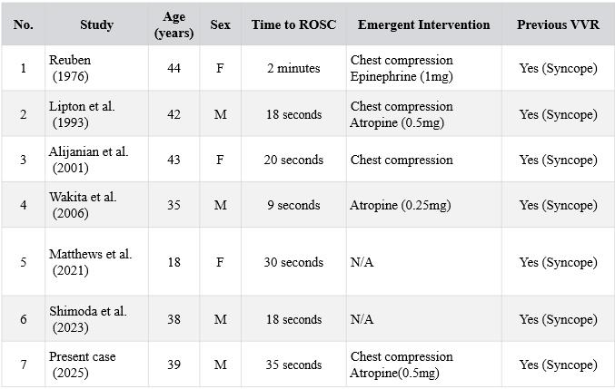
Abbreviations: F, female; M, male; N/A, not applicable; mg, milligram; ROSC, return of spontaneous circulation; VVR, vasovagal reaction.
somatic pain can induce this cardioinhibitory response, which may result in profound bradycardia or hypotension.10 In severe cases, the vasovagal reaction has been reported to lead to asystole. In this case, the pain and emotional stress caused by multiple failed venipunctures were thought to have triggered a severe vasovagal reaction, leading to bradycardia followed by transient cardiac asystole.
To our knowledge, our case represents the seventh reported adult case of cardiac asystole caused by a venipuncture-related vasovagal reaction to be reported in the literature (Table). Interestingly, all six previously reported cases described a prior episode of vasovagal reaction. In this case, a detailed history-taking following the asystolic event revealed a previous, patient-reported episode of vasovagal syncope, suggesting a possible predisposition to exaggerated vagal responses. Individuals with recurrent vasovagal reactions are known to have a lower threshold for parasympathetic activation, predisposing them to bradycardia or even asystole under stress or painful stimuli.10 Since the patient was on telemetry during the venipuncture, the asystole was promptly detected, and timely intervention was started. While reports of similar cases are limited, previous episodes of vasovagal reactions could possibly represent a potential risk factor for cardiac arrest during these procedures, particularly in individuals with underlying autonomic hypersensitivity.
Although venipuncture is a commonly performed procedure, clinical teams should be aware that it carries the rare potential of an exaggerated vasovagal reaction such as pronounced bradycardia and even asystole. History of previous venipuncture-related vasovagal symptoms should heighten awareness of potential adverse reactions. These patients can benefit from close observation and should trigger a low threshold for telemetry and escalation of care.
The authors attest that their institution requires neither Institutional Review Board approval, nor patient consent for publication of this case report. Documentation on file.
Address for Correspondence: Tomoki Nagano, MD, Japanese National Physician Graduate Medical Education Program, U.S. Naval Hospital Yokosuka, 82, Inaoka-cho, Yokosuka-city, Kanagawa 2380002, Japan. Email: tomoki.nagano.office@gmail.com
Conflicts of Interest: By the CPC-EM article submission agreement, all authors are required to disclose all affiliations, funding sources and financial or management relationships that could be perceived as potential sources of bias. The authors disclosed none. The views in this article are those of the authors only and do not represent the official policy or position of the Defense Health Agency, Department of Defense, or the U.S. Government.
Copyright: © 2026 Nagano et al. This is an open access article distributed in accordance with the terms of the Creative Commons Attribution (CC BY 4.0) License. See: http://creativecommons.org/ licenses/by/4.0/
Nagano et al. Unexpected Cardiac Asystole Caused by Vasovagal Reaction During Venipuncture: A Case Report
1. Reuben T. Cardiac arrest following routine venipuncture. JAMA. 1976;236(16):1846-7.
2. Lipton JD and Forstater AT. Recurrent asystole associated with vasovagal reaction during venipuncture. J Emerg Med. 1993;11(6):723-7.
3. Alijanian A, Bedrossian E, Feeney C, Devlin DH. Asystole secondary to venipuncture: report of case. J Oral Maxillofac Surg. 2001;59(4):461-2.
4. Wakita R, Ohno Y, Yamazaki S, et al. Vasovagal syncope with asystole associated with intravenous access. Oral Surg Oral Med Oral Pathol Oral Radiol. 2006;102(6):e28-32.
5. Matthews L and Kurukumbi M. Reflex anoxic seizures induced by needle stick and successfully treated with intranasal midazolam. Cureus. 2021;13(9):e18002.
6. Shimoda H, Yamauchi K, Takahashi T. Transient asystole associated with vasovagal reflex in an oral surgery patient: a case report. SAGE Open Med Case Rep. 2023;11:1-5.
7. Chen MY, Goldenberg IF, Milstein S, et al. Cardiac electrophysiologic and hemodynamic correlates of neurally mediated syncope. Am J Cardiol. 1989;63(1):66-72.
8. Morillo CA, Eckberg DL, Ellenbogen KA, et al. Vagal and sympathetic mechanisms in patients with orthostatic vasovagal syncope. Circulation. 1997;96(8):2509-13.
9. Shim SH, Park SY, Moon SN, et al. Baseline heart rate variability in children and adolescents with vasovagal syncope. Korean J Pediatr. 2014;57(4):193-8.
10. David LJ, Wouter W, Michele B, et al. Pathophysiology of the vasovagal response. Heart Rhythm. 2018;15(6):921-929.
Sabri Onur Çağlar, MD*
Hilal Çağlar, MD†
Serdar Hira, MD‡
Bandırma Royal Hospital, Department of Cardiology, Balıkesir, Türkiye
Bandırma Royal Hospital, Department of Physical Therapy, Balıkesir, Türkiye
Bandırma Royal Hospital, Department of Biochemistry, Balıkesir, Türkiye
Section Editor: Ezhilkugan Ganessane, MD
Submission history: Submitted September 16, 2025; Revision received October 9, 2025; Accepted October 10, 2025
Electronically published March 29, 2026
Full text available through open access at http://escholarship.org/uc/uciem_cpcem DOI: 10.5811/cpcem.52890
Introduction: Diclofenac sodium is a widely used medication for its analgesic and anti-inflammatory properties. Although the adverse effects of diclofenac are well described, diclofenac-associated newonset atrial fibrillation in a young, healthy adult has not been previously reported.
Case Report: A 22-year-old man with severe pain following an ankle injury used diclofenac sodium at a dose of 50 mg orally three times daily for one week. At the end of the week, he presented to the emergency department (ED) complaining of palpitations for a few hours. The patient had no past medical history. His physical examination revealed no pathologic signs except for tachycardia and an irregularly irregular pulse rate. An electrocardiogram showed an irregularly irregular rhythm with a ventricular rate of 128 beats per minute (bpm) (rapid ventricular response) and absent P waves, consistent with atrial fibrillation. The patient was monitored in the ED, and a standard regimen of intravenous amiodarone was initiated for rate control. Approximately three hours after initiation, sinus rhythm returned and his heart rate stabilized at 60 bpm.
Conclusion: This case report describes a possible association between diclofenac use and new-onset atrial fibrillation in a young, previously healthy individual. The findings highlight the need for awareness of the potential arrhythmogenic effects of diclofenac and underscore the importance of taking a careful drug history in patients presenting with unexplained atrial fibrillation. [Clin Pract Cases Emerg Med. 2026;10(2):166-169.]
Keywords: case report; diclofenac; atrial fibrillation; pain.
Diclofenac sodium is a nonsteroidal anti-inflammatory drug (NSAID) used to relieve pain by inhibiting cyclooxygenase (COX) enzymes, reducing the production of prostaglandins, precursors to inflammatory molecules.1 It is widely available over the counter in low-, middle-, and high-income countries and is even prescribed to patients with known cardiovascular comorbidities, under the assumption that low doses are safe, although this has not been proven. While the side effects of diclofenac are well known, there are few reports of diclofenac-induced atrial fibrillation in young patients with no previous cardiac history.2,3 Here we report a
young patient with new-onset atrial fibrillation that may have been related to high-dose oral diclofenac sodium.
A 22-year-old male experienced severe pain after spraining his ankle while playing football. He used diclofenacsodium at a dose of 50 mg orally three times daily for a week. At the end of the week, he presented to the emergency department (ED) complaining of palpitations for a few hours. The patient had no past medical history. He did not take any regular medications and further reported no use of alcohol, caffeine, stimulants, or drugs. After physical examination by
Atrial Fibrillation in a Young Patient Using High-dose Oral Diclofenac: A Case Report
the emergency physician the patient was then referred to a cardiologist. No pathologic signs were observed except for tachycardia and an irregularly irregular pulse rate. An electrocardiogram showed an irregularly irregular rhythm with a ventricular rate of 128 beats per minute (bpm) (rapid ventricular response) and absent P waves, consistent with atrial fibrillation (Image 1).
Cardiac point-of-care ultrasound demonstrated a preserved global and segmental left ventricular systolic function without valvular abnormalities or intracardiac thrombus. Laboratory results, including complete blood count and routine biochemical tests, were within normal limits. The patient was admitted to a monitoring bed, and the standard regimen of amiodarone was initiated: a 150 mg intravenous bolus over 10 minutes, followed by 900 mg (6 ampoules) diluted in 500 mL of 5% dextrose and infused over 24 hours—1 mg per minute for the first six hours and 0.5 mg per minute for the next 18 hours. Approximately three hours into the infusion, sinus rhythm returned and heart rate decreased to 60 bpm; the infusion was then discontinued to avoid further bradycardia (Image 2). Biochemical tests, including troponin and creatine kinase-MB, were monitored to evaluate for acute coronary syndrome and remained within reference ranges. During the 24 hours of observation, the patient had no additional cardiac problems and was discharged uneventfully with treatment recommendations.
Diclofenac has analgesic, anti-inflammatory, antipyretic, and anti-cancer effects and is widely used for the treatment of various conditions, including painful musculoskeletal disorders, dysmenorrhea, fever, and arthritis, as well as cancer




CPC-EM Capsule
What do we already know about this clinical entity?
Atrial fibrillation is a common arrhythmia often associated with structural or metabolic disorders.
What makes this presentation of disease reportable?
This case describes atrial fibrillation in a young patient following high-dose oral diclofenac use.
What is the major learning point?
Non-cardiac medications such as NSAIDs may trigger arrhythmias even in young individuals without comorbidities.
How might this improve emergency medicine practice?
Awareness of drug-related arrhythmias can help emergency physicians identify and manage unexpected atrial fibrillation cases promptly.
and neurodegeneration.4 Diclofenac is mainly associated with the occurrence of gastrointestinal side effects involving abdominal pain, diarrhea, dyspepsia, nausea, and gastrointestinal reflux with long-term use; other observed side effects include hepatic and renal toxicity.1 Cardiovascular toxicity has also become a growing concern, particularly with long-term or high-dose exposure.¹ Even in individuals without pre-existing cardiovascular disease, diclofenac has been associated with a small but measurable increase in events such as myocardial infarction, stroke, and thromboembolism.2 Atrial fibrillation is the most common arrhythmia whose prevalence has increased globally, particularly with age.5 It is known to contribute to heart failure, cerebrovascular events, coronary artery disease, and higher incidence of mortality.6 Some medications, such as corticosteroids, have been associated with an increased risk of atrial fibrillation, but data on the possible role of diclofenac in the development of AF remain sparse.7 Okuyan reported paroxysmal atrial fibrillation developing 20-30 minutes after intramuscular diclofenac administration in a 48-year-old patient.3 Epidemiological evidence indicates that initiating diclofenac is associated with an increased risk of developing cardiovascular disease, including atrial fibrillation or flutter, when compared with no drug use, paracetamol, or other conventional nonsteroidal





Taken together, these mechanisms suggest that diclofenac may increase susceptibility to atrial fibrillation through both direct electrophysiological changes and indirect systemic effects, particularly in predisposed individuals or during high-dose exposure. The current case highlights this possibility given the short-term use of high-dose oral diclofenac before the onset of atrial fibrillation in a previously healthy young adult. While the close timing of drug intake and symptom onset supports a possible temporal relationship, definitive conclusions about causality cannot be drawn from a single report. Additionally, given the relatively short time course of conversion (~3 hours) with amiodarone administration, spontaneous conversion of paroxysmal atrial fibrillation cannot be excluded. Large-scale studies and meta-analyses have demonstrated an association between NSAID exposure and atrial fibrillation risk, particularly in populations with cardiovascular comorbidities. Our observation provides further insight into this association.
anti-inflammatory drugs (NSAID), particularly within the first 30 days of treatment initiation 2
Large, population-based studies support this hypothesis. A Danish nationwide analysis found that both non-selective and COX-2 selective NSAIDs were linked to a transient rise in atrial fibrillation incidence during the first month of treatment.8 Similarly, a meta-analysis found a 1.2-fold increased risk of atrial fibrillation in users of non-aspirin NSAIDs compared to non-users, and this risk increased by 1.5-fold in new users, suggesting that this may represent a class effect rather than an isolated drug phenomenon. The study also found that COX-2 inhibitors, particularly diclofenac, were associated with higher risks than nonselective NSAIDs.9
The precise mechanisms linking diclofenac use to atrial fibrillation are still not fully understood. Several hypotheses have been proposed based on the known pharmacological properties of NSAIDs. Diclofenac exerts strong inhibition of COX-2, which alters prostaglandin and thromboxane synthesis and may impair vascular homeostasis and endothelial function, thereby promoting atrial remodeling and electrical instability.10 Experimental data indicate that diclofenac can modulate cardiac electrophysiology by inhibiting voltagegated potassium and calcium channels and altering repolarization dynamics, which may shorten atrial refractory periods and facilitate re-entry mechanisms.11 Furthermore, diclofenac has been shown to increase oxidative stress and systemic inflammation, processes that contribute to structural atrial remodeling and arrhythmogenesis.12,13 Volume retention and blood pressure elevation associated with NSAID use can also increase atrial wall tension and stretch, further facilitating abnormal conduction and ectopic activity.8
This case highlights the need for awareness of the potential arrhythmogenic effects of commonly used NSAIDs such as diclofenac and underscores the importance of taking a careful drug history in patients presenting with unexplained atrial fibrillation. Further epidemiological and mechanistic studies are needed to clarify whether this association reflects a true causal relationship or is a coincidental occurrence.
The authors attest that their institution does not require Institutional Review Board approval. Patient consent has been obtained and filed for the publication of this case report.
Address for Correspondence: Serdar Hıra, MD, Department of Biochemistry, Bandırma Royal Hospital, Sağlık St. No:4, Paşakonak, Bandırma, Balıkesir 10200, Türkiye. Email: serdar. hira@royalhastanesi.com.tr
Conflicts of Interest: By the CPC-EM article submission agreement, all authors are required to disclose all affiliations, funding sources and financial or management relationships that could be perceived as potential sources of bias. The authors disclosed none.
Copyright: © 2026 Çağlar et al. This is an open access article distributed in accordance with the terms of the Creative Commons Attribution (CC BY 4.0) License. See: http://creativecommons.org/ licenses/by/4.0/
1. Satar HA, Yousif E, Ahmed A. Diclofenac: a review on its synthesis, mechanism of action, pharmacokinetics, prospect and environmental
Çağlar et al.
Atrial Fibrillation in a Young Patient Using High-dose Oral Diclofenac: A Case Report impact. Al-Mustaqbal J Pharm Med Sci.2024;2(3):2.
2. Schmidt M, Sørensen HT, Pedersen L. Diclofenac use and cardiovascular risks: series of nationwide cohort studies. BMJ 2018;362:k3426.
3. Okuyan H. Diclofenac induced paroxysmal atrial fibrillation. J. Emerg. Med. Case Rep. 2018;9(4):80-2.
4. Amanullah A, Upadhyay A, Dhiman R, et al. Development and challenges of diclofenac-based novel therapeutics: targeting cancer and complex diseases. Cancers. 2022;14(18):4385.
5. Linz D, Gawalko M, Betz K, et al. Atrial fibrillation: epidemiology, screening and digital health. Lancet Reg Health Eur. 2024;37:100786.
6. Mekhael M, Marrouche N, El Hajjar AH, Donnellan E. The relationship between atrial fibrillation and coronary artery disease: understanding common denominators. Trends Cardiovasc Med. 2024;34(2):91-8.
7. Zhou Z, Long Y, He X, Li Y. Effects of different doses of glucocorticoids on postoperative atrial fibrillation: a meta-analysis. BMC Cardiovasc Disord. 2023;23(1):16.
8. Schmidt M, Christiansen CF, Mehnert F, et al. Non-steroidal anti-inflammatory drug use and risk of atrial fibrillation or flutter: population based case-control study. BMJ. 2011;343:d3450.
9. Liu G, Yan YP, Zheng XX, et al. Meta-analysis of nonsteroidal anti-inflammatory drug use and risk of atrial fibrillation. Am J Cardiol 2014;114(10):1523-9.
10. Ricciotti E, Haines PG, Chai W, FitzGerald GA. Prostanoids in cardiac and vascular remodeling. Arterioscler Thromb Vasc Biol 2024;44(3):558-83.
11. Gwanyanya A, Macianskiene R, Mubagwa K. Insights into the effects of diclofenac and other non-steroidal anti-inflammatory agents on ion channels. J Pharm Pharmacol. 2012;64(10):1359-75.
12. Thai PN, Ren L, Xu W, et al. Chronic diclofenac exposure increases mitochondrial oxidative stress, inflammatory mediators, and cardiac dysfunction. Cardiovasc Drugs Ther. 2023;37(1):25-37.
13. Taghdiri A. Inflammation and arrhythmogenesis: a narrative review of the complex relationship. Int J Arrhythmia. 2024;25(1):4.
Neil Wallace, MD*
Aila Hauger†
University of Arizona, Department of Emergency Medicine, Tucson, Arizona
University of Arizona College of Medicine - Tucson, Tucson, Arizona
Section Editor: Ezhilkugan Ganessane, MD
Submission history: Submitted August 24, 2025; Revision received October 25, 2025; Accepted December 9, 2025
Electronically published April 22, 2026
Full text available through open access at http://escholarship.org/uc/uciem_cpcem DOI: 10.5811/cpcem.50746
Introduction: Amyand hernia is a rare condition in which the appendix is found within an inguinal hernia sac, often mimicking incarcerated or strangulated hernias. Diagnosis is typically made intraoperatively, but increasing use of point-of-care ultrasound (POCUS) is enhancing preoperative recognition.
Case Report: A 46-year-old male presented with a chronic, partially reducible inguinal hernia. Pointof-care ultrasound revealed a bowel-containing hernia with fluid. Computed tomography confirmed an inflamed appendix within the sac. Laparoscopic appendectomy and open hernia repair were performed without complications.
Conclusion: Early use of POCUS can expedite diagnosis of Amyand hernia, improving surgical outcomes through timely intervention. [Clin Pract Cases Emerg Med. 2026;10(2):170-173.]
Keywords: Amyand hernia; inguinal hernia; point-of-care ultrasound; appendicitis; case report.
Amyand hernia is a rare diagnosis in which the vermiform appendix is found within the hernia sac of an inguinal hernia. First described in the 18th century by Claudius Amyand, who performed the first successful appendectomy during hernia repair, the condition remains rare in modern surgical practice.1 The overall incidence of Amyand hernia ranges from 0.191.7% of all inguinal hernia cases, with acute appendicitis within the hernia sac occurring in just 0.07-0.13% of cases.2,3 Most Amyand hernias are diagnosed intraoperatively, as their clinical presentation typically mimics that of the more common incarcerated or strangulated inguinal hernia. This overlap often makes preoperative identification difficult, especially in the absence of systemic symptoms such as fever, leukocytosis, or peritonitis.
With the increasing availability and use of imaging modalities in the emergency department (ED), particularly point-of-care ultrasound (POCUS) and computed tomography (CT), clinicians now have improved tools to recognize rare conditions such as Amyand hernia before surgery. This case report describes a middle-aged male who presented with a chronic, partially reducible inguinal hernia and was ultimately
diagnosed with Amyand hernia after POCUS and CT revealed an inflamed appendix within the hernia sac. The case highlights the growing importance of ultrasound in expediting the diagnosis and management of complex surgical conditions.
A 46-year-old White male with a body mass index of 34 (reference range: 18.5-24.9) and a history of substance use disorder presented to the ED for possible incarcerated hernia. The patient reported several months of progressive right groin swelling and intermittent pain, with worsening symptoms over the prior three months. He noted that he first noticed the hernia five years earlier, and it had been reducible. However, over the prior 48 hours he had begun to have more scrotal pain and was unable to reduce it, causing him to present to the ED. The patient denied systemic symptoms, including fever, chills, nausea, vomiting, constipation, or abdominal pain. He had no prior abdominal surgeries and denied any urinary complaints at presentation. He had recently been released from prison and had only recently established outpatient care.
On examination, the patient was afebrile without tachycardia and non-toxic in appearance. His right scrotum
was enlarged, tender, and contained a partially reducible mass. There was no overlying skin erythema or warmth. The abdomen was soft, non-tender, and non-distended. Laboratory evaluation revealed a mild leukocytosis, with white blood cells counted to 12,000 per microliter (µL) (reference range: 4,500-11,000/µL) with neutrophilic predominance. The remainder of the complete blood count was within normal limits. Electrolytes, renal function, liver enzymes, and urinalysis were unremarkable. A POCUS of the groin demonstrated a bowel-containing right inguinal hernia measuring 5 cm × 5 cm × 5 cm, with surrounding fluid extending into the scrotum, as well as a non-compressible, hyperemic, blind-ended tubular structure (Image).
The hernia was noted to be partially reducible during the scan. Given the ultrasound findings, CT of the abdomen and pelvis with contrast was obtained. The CT revealed a right inguinal hernia containing the distal ileum, cecum, and appendix. The appendix measured 9 mm in diameter and was surrounded by fat stranding and fluid within the hernia sac, concerning for acute appendicitis. There was no evidence of bowel obstruction or free air.
The patient was evaluated by the surgical team and admitted with a plan for operative intervention. He was made nil per os, started on intravenous fluids, and received preoperative antibiotics. In the operating room, laparoscopy revealed an acutely inflamed appendix within the hernia sac, along with the cecum and ileum. The hernia demonstrated
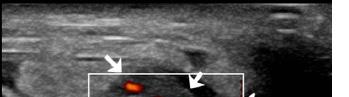
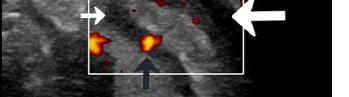

CPC-EM Capsule
What do we already know about this clinical entity?
Amyand’s hernia is an inguinal hernia containing the appendix, occurring in ~1% of inguinal hernias. It’s rarely diagnosed preoperatively, and may either be normal or inflamed.
What makes this presentation of disease reportable?
This case report shows an Amyand’s hernia diagnosed by emergency department performed point-of-care ultrasound, highlighting important sonographic features.
What is the major learning point?
Inguinal hernias may contain a variety of abdominal structures, including the appendix.
How might this improve emergency medicine practice?
Identifying an inguinal hernia at risk of complication from bedside reduction is important, and point-of-care ultrasound may help identify high risk features.
both direct and indirect components, and the floor of the inguinal canal was obliterated. A laparoscopic appendectomy was performed, followed by open right inguinal hernia repair with mesh placement. There were no intraoperative complications, and estimated blood loss was 100 mL.
Gross pathology confirmed acute appendicitis, with a 6.2-cm vermiform appendix showing congested serosa and fibrinous exudate. No perforation or fecaliths were identified. The patient was transferred to the surgical floor postoperatively and had an uncomplicated hospital course. He was discharged the following day with activity restrictions, instructions for follow-up, and return precautions.
At his two-week outpatient follow-up, the patient reported no fever, pain, or drainage from the surgical sites. On exam, his incisions were clean, dry, and intact with no signs of infection. He was tolerating his diet, having regular bowel movements, and voiding without difficulty.
Amyand hernia represents a rare surgical finding, characterized by the presence of the vermiform appendix within an inguinal hernia sac. The incidence of Amyand hernia
Preoperative Diagnosis of Amyand Hernia in the ED with Point-of-care Ultrasound Wallace et al. is reported to range from 0.19-1.7% of inguinal hernia cases and, when appendicitis is involved, it becomes even less common with estimates ranging from 0.07%-0.13%.2,3 A retrospective review by D’Alia et al identified Amyand hernia in 0.6% of 1,341 inguinal hernias, all occurring on the right side and exclusively in male patients.4 As previously mentioned, Amyand hernia holds a unique place in surgical history. In 1735, Claudius Amyand performed what is recognized as the second documented appendectomy on an 11-year-old patient whose inflamed appendix was located within an inguinal hernia sac, later giving rise to the eponymous condition.1 This operation marked both the initial description and successful surgical management of Amyand hernia, as well as one of the earliest documented appendectomies in medical literature.
In this case, the chronicity of symptoms without overt signs of systemic infection made the clinical picture more ambiguous, underscoring the value of early imaging, particularly POCUS, in the ED. Ultrasound is increasingly used in the ED as a first-line modality for evaluating scrotal and inguinal pathology due to its accessibility, lack of radiation, and ability to provide dynamic assessment. The American College of Radiology recommends ultrasound as the preferred initial imaging modality for suspected groin hernia, citing sensitivities as high as 97% when performed by skilled operators.5 While there are varying levels of competency with POCUS, there have been several studies that show emergency physicians can perform POCUS to reliably confirm the presence of a hernia sac, identify features of incarceration or strangulation (such as non-reducible bowel, free fluid, or absent Doppler flow), and detect associated small bowel obstruction, thus expediting surgical decision-making and risk stratification in the emergency setting.6,7 However, it is understandable that identifying Amyand hernia on POCUS is unlikely expected of novice sonographers. Additional systematic reviews and meta-analyses support the reliability of ultrasound in diagnosing both overt and occult groin hernias, as well as in accurately distinguishing between inguinal and femoral types.8
Of note: The chart abstractors were not blinded to the patient outcomes and this report. In our patient, POCUS was instrumental in identifying a bowel-containing inguinal hernia with associated fluid and signs of inflammation, prompting further investigation with CT and early surgical consultation. The majority of Amyand hernia cases are identified intraoperatively, and this pattern has been well documented in the medical literature. Multiple systematic reviews and case series consistently report that the clinical presentation of Amyand hernia is indistinguishable from that of an incarcerated or strangulated inguinal hernia, making preoperative diagnosis challenging. One systematic review found that only 23.1% of cases were diagnosed preoperatively, most often with the aid of imaging such as ultrasound or CT,
while the remainder were discovered during surgery for presumed incarcerated hernia.9 Similarly, other series and reviews emphasize that intraoperative identification is the norm, with preoperative suspicion being low due to nonspecific clinical findings.2,9,10
While our patient ultimately underwent CT for further characterization of the rare finding of Amyand hernia, POCUS assisted with expediting surgical referral and confirming the diagnosis of an incarcerated hernia early in the clinical course. Ultimately, the surgical team decided to pursue operative management given the free fluid in the hernia sac and risk of perforation with bedside reduction. In patients with an inconclusive or atypical physical exam, POCUS can facilitate timely surgical consultation when positive and may help avoid unnecessary radiation exposure from CT imaging when negative. In this case, the diagnostic utility of POCUS was highlighted by the early surgical consultation, which ultimately allowed for early surgical intervention. This is important as delays in emergent hernia repair have been associated with increased complication rates, including prolonged operative time, extended postoperative hospital stay, and higher rates of reoperation.11
The hernia sac itself is formed by peritoneum protruding through an abdominal wall defect, and the herniated contents are typically mobile and reducible unless incarcerated or strangulated.12 Typical hernia contents include small bowel, which is identified by its characteristic peristalsis and fluid- or gas-filled loops. In contrast, preperitoneal fat appears as a homogeneous, moderately hyperechoic, non-vascular, nonperistaltic structure that does not share the layered appearance of bowel and typically shows minimal or no response to Valsalva maneuvers. Omental fat is also hyperechoic but generally more heterogeneous and lobulated than preperitoneal fat. It may contain small vessels visible on color Doppler and lacks the tubular morphology and peristaltic motion of bowel.12 Less commonly, the urinary bladder may herniate, presenting as a fluid-filled, anechoic, and non-peristaltic structure that may change with voiding.13 In female patients, the ovary or fallopian tube can herniate and may be seen as complex adnexal structures with internal follicles and varying Doppler flow; the ovary may appear hypoechoic or heterogeneous, depending on vascular compromise.14 Rarely, a Meckel diverticulum (Littre hernia) may be present within the sac.
Point-of-care ultrasound plays additional roles in the emergency evaluation of inguinal hernias, particularly in differentiating hernia contents and identifying high-risk features. The decision to reduce a hernia is often a clinical one, incorporating history and physical exam. Point-of-care ultrasound is an adjunct that helps identify high-risk hernias that should not be reduced. These include hernias with features of strangulation or ischemic bowel. While success of non-operative reduction of Amyand hernia is unknown due to its rare presentation, care should be taken with certain high-
Wallace et al.
Preoperative Diagnosis of Amyand Hernia in the ED with Point-of-care Ultrasound
risk features that might predispose to rupture, such as a hyperemic appendix with surrounding free fluid.10 Signs concerning for strangulation include non-compressible bowel, bowel wall thickening, free fluid in the hernia sac, and absent or reduced Doppler flow. These findings should prompt immediate surgical consultation rather than manual reduction, as reduction of non-viable bowel may result in perforation, peritonitis, or sepsis.15
Our case illustrates how the early integration of POCUS into the evaluation of groin complaints can both narrow the differential and expedite appropriate surgical care. Emergency physicians should maintain a high index of suspicion and incorporate ultrasound early in the evaluation of groin complaints, particularly in cases with chronicity, atypical features, or inconclusive physical exam findings. Incorporating early ultrasound can facilitate earlier recognition, better communication with surgical teams, and improved patient outcomes.
The authors attest that their institution requires neither Institutional Review Board approval, nor patient consent for publication of this case report. Documentation on file.
1. Amyand C. VIII. Of an inguinal rupture, with a pin in the appendix coeci, incrusted with stone; and some observations on wounds in the guts. Phil Trans R Soc. 1736; 39 (443):329-342.
2. Sharma H, Gupta A, Shekhawat NS, et al. Amyand’s hernia: a report of 18 consecutive patients over a 15-year period. Hernia 2007;11(1):31-35.
3. Ivanschuk G, Cesmebasi A, Sorenson EP, et al. Amyand’s hernia: a review. Med Sci Monit Int 2014;20:140-146.
4. D’Alia C, Lo Schiavo MG, Tonante A, et al. Amyand’s hernia: case report and review of the literature. Hernia. 2003;7(2):89-91.
5. Garcia EM, Pietryga JA, Kim DH, et al. ACR Appropriateness Criteria® Hernia. J Am Coll Radiol. 2022;19(11):S329-S340.
6. Al Jalbout N, Liteplo A, Shokoohi H. Point-of care ultrasound in stratifying clinically evolving inguinal and femoral hernias. J Emerg Med. 2022;63(2):257-264.
7. Jacobson JA, Khoury V, Brandon CJ. Ultrasound of the groin: techniques, pathology, and pitfalls. Am J Roentgenol 2015;205(3):513-523.
8. Kwee RM and Kwee TC. Ultrasonography in diagnosing clinically occult groin hernia: systematic review and meta-analysis. Eur Radiol 2018;28(11):4550-4560.
9. Papaconstantinou D, Garoufalia Z, Kykalos S, et al. Implications of the presence of the vermiform appendix inside an inguinal hernia (Amyand’s hernia): a systematic review of the literature. Hernia 2020;24(5):951-959.
Address for Correspondence: Neil Wallace, University of Arizona Department of Emergency Medicine, 1501 N Campbell Ave, PO Box 245057, Tucson, AZ 85724. Email Address: ndw1297@ arizona.edu.
Conflicts of Interest: By the CPC-EM article submission agreement, all authors are required to disclose all affiliations, funding sources and financial or management relationships that could be perceived as potential sources of bias. The authors disclosed none.
Copyright: © 2026 Wallace et al. This is an open access article distributed in accordance with the terms of the Creative Commons Attribution (CC BY 4.0) License. See: http://creativecommons.org/ licenses/by/4.0/
10. Michalinos A, Moris D, Vernadakis S. Amyand’s hernia: a review. Am J Surg. 2014;207(6):989-995.
11. Leeds IL, Jones C, DiBrito SR, et al. Delay in emergency hernia surgery is associated with worse outcomes. Surg Endosc. 2020;34(10):4562-4573.
12. Fitzgibbons RJ and Forse RA. Clinical practice. Groin hernias in adults. N Engl J Med. 2015;372(8):756-763.
13. Ogbetere FE and Imoisili U. Recurrent inguinal hernia containing the urinary bladder: a rare occurrence. Ann Afr Med. 2022;21(3):288-290.
14. Maqbool S, Huma A, Anwar MI, et al. Ovary and fallopian tube as contents of indirect inguinal hernia: a case report. Int J Surg Case Rep. 2022;100:107733.
15. Pastorino A, Alshuqayfi AA. Strangulated Hernia(Archived). In: StatPearls. Treasure Island (FL): StatPearls Publishing; December 19, 2022. Available from: https://www.ncbi.nlm.nih. gov/books/NBK555972/. Accessed August 15, 2025.
Andrew J. Gonedes, DO*
Alexandra Martinez, BS†
Allan M. Greissman, MD‡
Hanan Atia, MD*
Eric Boccio, MD, MPH§
Section Editor: Anna McFarlin, MD
Memorial Healthcare System, Department of Emergency Medicine, Hollywood, Florida
Florida International University, Herbert Wertheim College of Medicine, Miami, Florida
Joe DiMaggio Children’s Hospital, Department of Pediatric Critical Care Medicine, Hollywood, Florida
Mount Sinai Medical Center of Florida, Department of Emergency Medicine
Miami Beach, Florida
Submission history: Submitted September 21, 2025; Revision received November 2, 2025; Accepted December 4, 2025
Electronically published April 7, 2026
Full text available through open access at http://escholarship.org/uc/uciem_cpcem DOI: 10.5811/cpcem.52835
Introduction: Dense deposit disease, also known as C3 glomerulopathy, is a rare renal disorder caused by abnormal complement deposition in the glomerular basement membrane. Patients often require long-term immunosuppressive therapy and, in some cases, complement inhibitors such as ravulizumab. While effective at limiting renal damage, complement blockade significantly increases susceptibility to invasive infections from encapsulated bacteria, particularly Neisseria meningitidis Despite immunization and antimicrobial prophylaxis, these patients remain incompletely protected. We describe a case of meningococcemia in a fully vaccinated adolescent with dense deposit disease on ravulizumab therapy.
Case Report: A 17-year-old male with a history of dense deposit disease on mycophenolate mofetil and ravulizumab presented to the pediatric emergency department with fever, vomiting, altered mental status, and a rapidly evolving petechial-purpuric rash. He was fully immunized with both meningococcal conjugate and serogroup B vaccines. Initial evaluation revealed fever, hypotension, and altered level of consciousness. Laboratory studies showed leukocytosis, elevated inflammatory markers, and blood cultures subsequently confirmed N. meningitidis. Empiric ceftriaxone and vancomycin were initiated, later narrowed to ceftriaxone. Supportive management included intravenous (IV) fluids, vasopressors, IV immunoglobulin, and dexamethasone. The patient demonstrated rapid improvement, with resolution of hemodynamic instability and normalization of kidney function. He was discharged on hospital day eight with prophylactic penicillin and close outpatient follow-up.
Conclusion: This case underscores the risk associated with complement inhibition, even in fully vaccinated individuals. Clinicians must maintain high vigilance for meningococcal disease in immunocompromised patients and initiate early aggressive therapy to optimize outcomes. [Clin Pract Cases Emerg Med. 2026;10(2):174-177.]
Keywords: dense deposit disease; meningococcemia; C5 complement inhibitor; ravulizumab; case report.
Dense deposit disease (DDD), also known as C3 glomerulopathy, is a rare kidney disease caused by abnormal deposition of complement component C3 in the glomerular
basement membrane. It can result in significant renal dysfunction and has a poor prognosis if left untreated. It is typically treated with immunosuppressive therapy. In certain cases, complement inhibitors such as ravulizumab are used to
modulate the immune system and prevent further kidney injury. However, these therapies leave patients vulnerable to encapsulated organisms such as Neisseria meningitidis, which can cause severe and life-threatening conditions such as meningococcal meningitis and meningococcemia. Despite vaccinations and prophylactic antimicrobials, these patients remain incompletely protected. We present the case of a 17-year-old male with a history of DDD and taking the C5 complement inhibitor ravulizumab who developed meningococcemia despite having received both the meningococcal conjugate vaccine (MCV4) and serogroup B vaccine (MenB).
A 17-year-old male with past medical history significant for DDD presented to the pediatric emergency department (ED) with fever and altered mental status. The initial diagnosis of DDD was made when the patient was eight years of age following workup that revealed proteinuria, hematuria, and progressive renal impairment. Genetic testing was confirmatory for the disease, and the patient was started on immunosuppressive therapy which included mycophenolate mofetil monotherapy initially and mycophenolate mofetil and rovelizumab dual therapy subsequently. At the time of ED presentation, the patient was up-to-date with age- and riskspecific vaccinations including MCV4 and MenB.
One week prior to presentation, the patient developed a fever, headache, nausea, and vomiting. Upon arrival to the pediatric ED, the patient was lethargic and altered, unable to respond to questions or participate in the physical examination. Initial ED vital signs were remarkable for hypotension (blood pressure 90/54 millimeters (mm) of mercury); tachycardia (heart rate 164 beats per minute); tachypnea (respiratory rate 20 breaths per minute); and fever (temperature 39.4 °C, oral). The patient was able to localize noxious stimuli, and the initial Glasgow Coma Scale score was seven. Physical examination revealed nuchal rigidity and a positive Brudzinski sign. A petechial rash developed, which rapidly coalesced into larger purpuric lesions across the chest, upper and lower extremities, and bilateral hands (Image).
Given the patient’s altered mental status, rapid sequence intubation was considered for airway protection; however, the procedure was deferred initially until hemodynamics could be optimized. Initial laboratory workup revealed the following: leukopenia, 3,600 cells per microliter (µL) [reference range: 4,500-11,000 cells/µL]; elevated C-reactive protein, 4.99 milligrams (mg) per deciliter (dL) [< 0.9 mg/dL]; elevated procalcitonin, > 200 nanograms (ng) per µL [< 0.05 ng/µL]; elevated serum creatinine, 1.99 mg/dL [0.7-1.3 mg/dL]; anion gap metabolic acidosis, 20 milliequivalents (mEq) per liter (L) [8-12 mEq/L]; and decreased serum bicarbonate, 12 mEq/L [22-29 mEq/L]. Peripheral blood cultures were obtained. The performance of a lumbar puncture in the ED was deferred due to the patient’s hemodynamic instability.
CPC-EM Capsule
What do we already know about this clinical entity?
Meningococcemia is a rapidly progressive and lifethreatening infection with greatly increased risk in patients receiving C5 complement inhibitors.
What makes this presentation of disease reportable?
We present a case of meningococcemia in a fully vaccinated adolescent on ravulizumab, illustrating meningococcal risk despite vaccination and prophylaxis.
What is the major learning point?
Vaccination does not fully protect patients on C5 inhibitors, making early recognition and prompt aggressive treatment essential for patient survival.
How might this improve emergency medicine practice?
High clinical suspicion and timely initiation of empiric therapy for meningococcemia is paramount in immunocompromised patients, even if vaccinated.
Given the high clinical suspicion for meningococcemia, aggressive fluid resuscitation and empiric antimicrobial therapy consisting of ceftriaxone and vancomycin was initiated. Additionally, intravenous immunoglobulin (IVIG) was administered to support the immune response in the setting of immunosuppressive therapy. Following aggressive fluid resuscitation, the patient remained hypotensive, and dexamethasone was given as adjunctive therapy for refractory septic shock. Blood cultures grew N. meningitidis, and the antibiotic regimen was narrowed to ceftriaxone. The patient was admitted to the pediatric intensive care unit where vasopressors were initiated, and infectious disease and nephrology services were consulted.
Within 72 hours of initiating targeted therapy, the patient’s clinical condition began to improve. The patient defervesced and his mental status improved, returning to baseline on hospital day three. Following stabilization of his hemodynamics, vasopressors were discontinued. Repeat laboratory test results revealed declining inflammatory markers and normalization of kidney function. Doses of mycophenolate and ravulizumab were held during the entirety of the hospital length of stay. On hospital day eight, the patient





Image. Lesions ranging from small red and purple flat petechia (< 3 millimeters (mm) in diameter) to large red and purple purpura (310 mm in diameter) noted across the skin of the chest (A), upper extremities (B), lower extremities (C), and palms of the bilateral hands (D-F) concerning for meningococcemia.
The skin rashes associated with meningococcemia are a sign of severe systemic involvement and imminent multisystem organ failure and indicates a poor prognosis if not treated promptly.3-4 According to the U.S. Centers for Disease Control and Prevention (CDC) Health Alert Network, meningococcal disease carries a case-fatality rate of 10-15%. An increase in invasive meningococcal disease cases has been noted with 143 cases reported in the first quarter of the 2024 calendar year and 422 cases reported in calendar year 2023.5
Neisseria meningitidis is a gram-negative bacterium that can cause meningococcal meningitis and meningococcemia in susceptible individuals. The bacterium’s polysaccharide capsule contributes to its virulence interfering with phagocytosis and antibody-mediated killing. In immunocompromised patients, particularly those on complement inhibitors such as ravulizumab, the ability to clear encapsulated organisms such as Neisseria meningitidis, Streptococcus pneumoniae, and Haemophilus influenzae is significantly impaired.6 Ravulizumab inhibits complement component C5 which is cleaved by C5 convertase into C5a and C5b thus preventing formation of the membrane attack complex which is essential for the perforation and osmotic cell lysis of invading pathogens.7 According to the CDC, the risk of meningococcal disease is up to 2,000 times greater for people receiving complement inhibitors compared to healthy controls.
was discharged home, and prophylactic penicillin V was added to his medication regimen. Outpatient follow-up with nephrology and infectious diseases were arranged, and the patient was scheduled to receive vaccine boosters at his next outpatient visit.
Meningococcal meningitis is a serious bacterial infection that causes inflammation of the membranes surrounding the brain and spinal cord. Symptoms may include fever, headache, nuchal rigidity, nausea, vomiting, photophobia, and altered mental status. In some cases, N. meningitidis may infect the bloodstream, a condition called meningococcemia. In the post-Haemophilus influenzae type b era, the presence of rash in cases of meningococcemia has declined; however, when present, it is often pathognomonic.1-2 The rash typically starts as petechiae – red or purple lesions caused by bleeding capillaries under the skin - < 3 mm in diameter and may rapidly coalesce into larger purpura (3-10 mm in diameter) or ecchymoses (> 10 mm in diameter).
In the most severe cases, the pro-inflammatory response triggered by the release of bacterial endotoxins may result in a hypercoagulable state known as disseminated intravascular coagulation that results in overactivation of the clotting cascade in the microvasculature. This results in the consumption of platelets and clotting factors leading to a hypocoagulable state that may precipitate bleeding across various sites including the skin, nose, mouth, and solid organs.
The U.S. Food and Drug Administration-approved prescribing information for complement inhibitors includes a black box warning for increased meningococcal disease risk and recommends administration of meningococcal vaccines to patients receiving complement inhibitors.8 Despite appropriate vaccination and antimicrobial prophylaxis, substantial risk remains, as CDC data suggest meningococcal vaccines likely provide incomplete protection against invasive meningococcal disease in patients receiving C5 inhibitors.9 The patient had received both the MCV4 and MenB vaccines. While nongroupable N. meningitidis caused most infections identified in patients receiving eculizumab, experts believe that patients on other C5 inhibitors may be susceptible to similar strains.10 Of note, in addition to ravulizumab, the patient was also taking mycophenolate mofetil, which likely further suppressed the immune system’s proliferation of T and B cell lymphocytes.
Given the potential for rapid deterioration in immunocompromised individuals, management of meningococcal meningitis requires early identification, prompt initiation of appropriate antibiotics, and continued supportive care. Empiric therapy for suspected meningococcal disease should include an extended-spectrum cephalosporin, such as cefotaxime or ceftriaxone. Once the diagnosis is established, definitive treatment can be continued with an extendedspectrum cephalosporin, or, alternatively, if susceptibility to penicillin is confirmed, treatment can be switched to penicillin G or ampicillin.11-12 Adjunctive therapies, such as corticosteroids to reduce inflammation and IVIG to support the immune
Gonedes et al. Meningococcemia in Boy with DDD Receiving the C5 Complement Inhibitor Ravulizumab system, may also be beneficial in improving outcomes.13 Preventive strategies, including vaccination and prophylactic antibiotics, are staples of care in at-risk populations.14
This case highlights the increased risk of invasive infections in pediatric patients on immunosuppressants. Despite being fully vaccinated, the patient developed meningococcemia, underscoring the limitations of vaccinations in providing complete protection in patients on C5 complement inhibitors such as ravulizumab. The patient presented clinically with the characteristic petechial rash, which rapidly progressed to purpura, pathognomonic for meningococcemia when other clinical symptoms of meningococcal meningitis are present. Early identification, prompt treatment with empiric and then targeted antimicrobial therapy, and aggressive resuscitation were essential to the patient’s survival and recovery. This case emphasizes the importance of initiating preventive strategies while recognizing their limitations, counseling patients about vigilant surveillance and monitoring, and maintaining a high index of clinical suspicion for meningococcal disease in patients who receive complement inhibitors.
The authors attest that their institution requires neither Institutional Review Board approval, nor patient consent for publication of this case report. Documentation on file.
Address for Correspondence: Eric Boccio, Mount Sinai Medical Center, Department of Emergency Medicine, 4300 Alton Road, Miami Beach, FL 33140, USA. Email: eric.boccio@msmc.com.
Conflicts of Interest: By the CPC-EM article submission agreement, all authors are required to disclose all affiliations, funding sources and financial or management relationships that could be perceived as potential sources of bias. The authors disclosed none.
Copyright: © 2026 Gonedes et al. This is an open access article distributed in accordance with the terms of the Creative Commons Attribution (CC BY 4.0) License. See: http://creativecommons.org/ licenses/by/4.0/
1. Karanika M, Vasilopoulou VA, Katsioulis AT, et al. Diagnostic clinical and laboratory findings in response to predetermining bacterial pathogen: data from the Meningitis Registry. PLoS One 2009;4(7):e6426.
2. van de Beek D, Brouwer MC, Koedel U, et al. Community-acquired bacterial meningitis. Lancet. 2021;398(10306):1171-1183.
3. Rivacoba MC, Villena R, Hormazabal JC, et al. Hypervirulent strains of Neisseria meningitidis and clinical manifestations in children with invasive meningococcal disease. Pediatr Infect Dis J 2023;42(8):660-666.
4. Pace D and Pollard AJ. Meningococcal disease: clinical presentation and sequelae. Vaccine. 2012;30 Suppl 2:B3-9.
5. Harris E. CDC warns of surge in meningococcal disease in US. JAMA. 2024;331(19):1614.
6. Galli N, Pettine L, Panigada M, et al. Non-capsulated Neisseria meningitidis sepsis in a paroxysmal nocturnal hemoglobinuria patient treated with ravulizumab: case report and review of the literature. Front Immunol. 2023;14:1269325.
7. Navarro-Carrera P, García-Rodríguez J, Cendejas-Bueno E. Detection of Neisseria meningitidis in a patient receiving ravulizumab by the FilmArray® Meningitis/Encephalitis panel - a case report. J Infect. 2021;82(6):e22-e23.
8. Kang C. Ravulizumab: a review in generalised myasthenia gravis. Drugs. 2023;83(8):717-723.
9. McAlpine A and Sadarangani M. Meningitis vaccines in children: What have we achieved and where next? Curr Opin Infect Dis 2019;32(5):510-516.
10. Parikh SR, Campbell H, Bettinger JA, et al. The everchanging epidemiology of meningococcal disease worldwide and the potential for prevention through vaccination. J Infect. 2020;81(4):483-498.
11. Leclerc F, Noizet O, Dorkenoo A, et al. Treatment of meningococcal purpura fulminans. Arch Pediatr. 2001;8 Suppl 4:677s-688s.
12. Haas H and Caseris M. Antibiotic treatment of invasive meningococcal infections. Rev Prat. 2024;74(8):s15-s18.
13. Ciofi degli Atti M, Esposito S, Parola L, et al. In-hospital management of children with bacterial meningitis in Italy. Ital J Pediatr. 2014;40:87.
14. Brady RC. Meningococcal infections in children and adolescents: update and prevention. Adv Pediatr. 2020;67:29-46.
Jacob Lenning, MD
Caleb Halfill, MD
Justin Rountree, MD
Section Editor: Joel Moll, MD
Western
Submission history: Submitted November 4, 2025; Revision received November 26, 2025; Accepted December 30, 2025
Electronically published April 7, 2026
Full text available through open access at http://escholarship.org/uc/uciem_cpcem DOI: 10.5811/cpcem.53083
Introduction: Reversible cerebral vasoconstriction syndrome is a recently defined disease entity classically presenting with recurrent thunderclap headache. The pathology involves triggered cerebral arterial vasoconstriction, which can lead to complications including seizure, ischemic stroke, and intracranial hemorrhage. Diagnosis requires angiography, and treatment consists of vasodilatory therapy.
Case Report: We describe a case of reversible cerebral vasoconstriction syndrome following glucocorticoid burst therapy in a patient on multiple vasoactive medications, suggesting the possibility of compounding risk factors and triggers. As is common with this syndrome, the patient in our case required multiple hospital visits for diagnosis but ultimately experienced a positive outcome upon treatment.
Conclusion: The presentation of reversible cerebral vasoconstriction syndrome can vary. Diagnosis requires a high degree of suspicion in patients with potential triggers to ensure early treatment and avoidance of poor outcomes. [Clin Pract Cases Emerg Med. 2026;10(2):178-181.]
Keywords: reversible cerebral vasoconstriction syndrome; thunderclap headache; glucocorticoids; case report
Reversible cerebral vasoconstriction syndrome is an under-recognized cause of headache often requiring multiple medical encounters before diagnosis.1 Recurrent thunderclap headache—a sudden headache with maximal intensity upon onset—is the classic chief complaint; associated features such as focal neurological deficits are not uncommon.2,3 The pathophysiology involves triggered cerebral arterial vasoconstriction.4,5 Reversible cerebral vasoconstriction syndrome is often diagnosed on computed tomography angiography (CTA) or magnetic resonance angiography (MRA) after other causes of thunderclap headache such as subarachnoid hemorrhage are ruled out. The gold standard for diagnosis is cerebral digital subtraction angiography.6,7
Most patients with reversible cerebral vasoconstriction syndrome have an excellent prognosis. However, sequelae may include cerebral infarction and hemorrhage leading to
increased morbidity and mortality.6,7 Therefore, awareness of this syndrome and potential triggers is imperative. We present the second reported case of reversible cerebral vasoconstriction syndrome following glucocorticoid therapy. The first reported case was thought to be related to steroid burst therapy.8 In the case we describe here the temporal relationship alone does not prove glucocorticoid therapy was the trigger. While studies have demonstrated worse outcomes when glucocorticoids were used in the treatment of the syndrome, none have confirmed a triggering effect. Further investigation is needed.7 However, this case does prompt consideration of a compounding risk factor and trigger effect.
A 49-year-old female with a past medical history including Crohn disease, hypertension, and insulin-dependent diabetes, prescribed venlafaxine for mood disorder and estradiol for
vasomotor menopausal symptoms, presented to the emergency department (ED) with four days of intermittent headaches. The patient described each episode as a sudden “boom” with severe intensity. Review of the medical record revealed that she had completed a three-day burst of methylprednisolone 1 gram daily prior to symptom onset and was continued on a prednisone taper for pyoderma gangrenosum of the lower extremity. Her initial blood pressure was 179/77 millimeters of mercury with otherwise normal vital signs. She was lying on the stretcher with the lights off and appeared uncomfortable. The physical examination revealed no focal neurological deficits, and the lower extremity wound appeared to be non-infected.
Subarachnoid hemorrhage was considered to be the most likely diagnosis. Computed tomography brain without contrast revealed no acute intracranial hemorrhage and was grossly normal. The CTA head and neck revealed a possible 2-mm aneurysm arising from the right posterior cerebral artery (not imaged) without evidence of rupture but was otherwise normal (Image 1A). Despite diphenhydramine, metoclopramide, and acetaminophen, the headache remained unchanged. Therefore, the neurointerventionalist was consulted and took the patient for diagnostic cerebral digital subtraction angiography, which demonstrated no actual aneurysm. Further inpatient workup included a lumbar puncture with normal opening pressure and unremarkable cerebrospinal fluid analysis. The headaches improved with blood pressure control, and the patient was discharged home.
Two days later, she was brought back to the ED by the family for confusion. The family stated the patient was “slow to respond” during conversation and was no longer independent with her activities of daily living. The patient endorsed the return of her headache as well. She was alert and oriented without focal neurological deficits but was clearly slow in her verbal responses and had difficulty providing history.
Repeat CT brain without contrast identified acute infarctions in the right cerebellum and left parietal lobe. Upon admission, repeat CTA head and neck revealed extensive vasoconstriction of the intracranial vasculature with diminished flow through the cerebral arteries compared to the previous study (Image 1B). Differential diagnosis included acute vasculitis and reversible cerebral vasoconstriction syndrome. The neurointerventionalist performed a repeat cerebral digital subtraction angiography demonstrating the classical segmental vasoconstriction of the syndrome (Image 1C). An intra-arterial infusion of verapamil was administered, resulting in the reversal of vasoconstriction and confirming the diagnosis of reversible cerebral vasoconstriction syndrome (Image 1D).
The following day, the patient reported that her headache had completely resolved. She was placed on extended-release verapamil by mouth daily, and she was switched from venlafaxine to lamotrigine for her mood disorder. Additionally, she was taken off estrogen, although neurology consult felt the final few low-doses of the oral prednisone taper could be continued for the pyoderma gangrenosum. The patient was
CPC-EM Capsule
What do we already know about this clinical entity?
Reversible cerebral vasoconstriction syndrome is a recently defined disease entity classically presenting with recurrent thunderclap headache.
What makes this presentation of disease reportable?
This case of a patient who developed the syndrome following glucocorticoid burst therapy highlights multiple possible risk factors and triggers.
What is the major learning point?
Diagnosis requires a high degree of suspicion in patients with potential triggers to ensure early treatment and avoidance of poor outcomes.
How might this improve emergency medicine practice?
Increased awareness of reversible cerebral vasoconstriction syndrome as a possible cause of thunderclap headache may hasten diagnosis and treatment.





Computed tomography angiography (CTA) of the head upon initial presentation demonstrating the normal-appearing middle cerebral artery (black arrows) compared to the repeat CTA head; (B) demonstrating segmental vasoconstriction (black arrows); (C) cerebral digital subtraction angiography (DSA) demonstrating segmental vasoconstriction (white arrows) compared to DSA immediately following intra-arterial verapamil; and (D) resolution of the vasoconstriction (white arrows).
Reversible Cerebral Vasoconstriction Syndrome Following a Steroid Burst: A Case Report
discharged two days after the therapeutic intra-arterial verapamil infusion. Upon follow-up a few months later, she was headache-free and back to her normal daily activities.
As in the case presented here, reversible cerebral vasoconstriction syndrome most commonly presents with thunderclap headache in female patients 40-60 years of age,2,3,9 although the syndrome has been recognized in men and children as well.3 In addition to female sex, use of vasoactive medications is one of the most commonly reported triggers.3,5 Others include blood transfusions, immunosuppressants, surgery, trauma, catecholamine secreting tumors, autonomic dysfunction, recent pregnancy, arterial disorders, arterial dissections, systemic lupus erythematosus, migraine headaches, and even lifestyle changes.5,6,9,10
The patient in our case had multiple possible triggers for the syndrome. She was taking venlafaxine and estrogen therapy, both vasoactive medications. She had also just completed a steroid burst, and although no studies provide direct evidence of glucocorticoids triggering the syndrome, one prior case report describes a similar temporal relationship..Steroids are known to worsen the condition7,8 ; therefore, even if not the trigger, it is possible the steroid burst therapy exacerbated the disease process in this patient. Further study is necessary to determine whether glucocorticoids trigger reversible cerebral vasoconstriction syndrome. The multiple potential triggers in this case raise the possibility of a compounding risk factor and trigger effect, although this hypothesis would require further investigation.
The exact pathophysiology of reversible cerebral vasoconstriction syndrome is unknown; there are multiple proposed mechanisms including disturbances in cerebral vascular tone, sympathetic overactivity, endothelial dysfunction, oxidative stress, and blood-brain barrier breakdown.5,11 Previous studies report no histologic or inflammatory changes in brain or arterial biopsies of patients diagnosed with the syndrome and, therefore, biopsy is not routinely recommended if it is suspected.4 Catecholamines, endothelin-1, serotonin, nitric acid, and prostaglandins are associated with vasospasm in aneurysmal subarachnoid hemorrhage, and these factors may have similar role in reversible cerebral vasoconstriction syndrome, although further investigation is needed.11 Posterior reversible encephalopathy syndrome shares many features with reversible cerebral vasoconstriction syndrome, Some authors hypothesize the entities to reside on the same spectrum of disease.4 The current expert consensus considers it to be the endpoint of multiple disease states, which is reflected in previous terms for the syndrome including migrainous vasospasm, Call-Fleming syndrome, and benign and idiopathic thunderclap headache.1,2,4
Thunderclap headache is the predominant presenting feature of reversible cerebral vasoconstriction syndrome. Patients may experience multiple, recurrent thunderclap headaches, although this historical feature is not completely sensitive nor is it specific
for diagnosis of the syndrome.3,5,9 One study estimated that up to 8.8% of thunderclap headaches presenting to the ED are due to reversible cerebral vasoconstriction syndrome, although actual incidence may be higher due to overall low awareness among clinicians.12 Photophobia, vomiting, seizures, focal neurological deficits, vision changes, encephalopathy, and acute hypertension are other possible presenting features of the syndrome but are even less sensitive and specific.4,5,11 Physical exams are typically non-focal but may occasionally demonstrate neurological deficits Generally, the syndrome is characterized by variable presentations making recognition difficult for clinicians.9,11
The differential diagnosis for reversible cerebral vasoconstriction syndrome includes other causes of thunderclap headaches such as subarachnoid hemorrhage, intracerebral hemorrhage, cerebral venous sinus thrombosis, cervical artery dissection, cerebral infarctions, central nervous system (CNS) tumors, and CNS infections.5 Bloodwork and cerebrospinal fluid analysis can evaluate for these other causes but will not directly make the diagnosis of this syndrome. As in our case, symptoms often precede identifiable radiological changes. Initial CT and magnetic resonance imaging are initially normal in many patients: 55% in one study, with most patients (81%) demonstrating abnormal findings upon repeat imaging.3 Radiographic studies may exhibit subarachnoid hemorrhage, intracranial hemorrhage, and cerebral infarction. Cerebral edema with mass effect and herniations are less commonly seen.11,13 Cerebral infarctions in reversible cerebral vasoconstriction syndrome often appear bilaterally, especially in watershed regions. Intracerebral hemorrhage tends to be multifocal as well.11,13
Cerebral digital subtraction angiography is the gold standard diagnostic tool with 100% sensitivity for the typical cerebral vasoconstrictive pattern of the syndrome, if present. Less invasive CTA and MRA studies are around 80% sensitive.6 The typical angiography findings include alternating areas of vasoconstriction and dilation forming a “sausage on a string” or “string of beads” pattern as seen in this case (Image 1B, C).6,10,11 Diagnostic confirmation of reversible cerebral vasoconstriction syndrome requires observed reversibility of vasospasm on imaging.11 To aid in the diagnosis, a few scoring tools have been developed, but none have been validated.6 Some studies suggest that progression of the syndrome can be monitored with transcranial Doppler ultrasound by evaluating maximum mean flow velocity in the middle cerebral artery, although diagnostic value is limited with sensitivity of 42-67%.7
Urgent recognition of reversible cerebral vasoconstriction syndrome is necessary to withdraw triggers and limit severe effects of complications that can be associated with significant morbidity and even mortality.1,5 Life-threatening conditions such as seizure, ischemic stroke, and intracranial hemorrhage can be caused by cerebral vasoconstriction associated with the syndrome.1,2 The reported complication rates vary, with up to 43% of patients experiencing intracranial hemorrhage, 33% ischemic stroke, and 28% posterior reversible encephalopathy
Lenning et al. Reversible Cerebral Vasoconstriction Syndrome Following a Steroid Burst: A Case Report syndrome.6 Morality rates up to 6.8% have been reported.6
Treatment of reversible cerebral vasoconstriction syndrome is largely supportive and includes symptom control, management of complications, withdrawal of triggers, and initiating vasodilator therapy with calcium channel blockers and magnesium sulfate.5,6 First-line treatment with calcium channel blockers such as nimodipine have been shown to improve headache intensity, although they have not been proven to alter duration or mitigate the risk of complications.5,6,13 Treatment with calcium channel blockers must be done carefully to avoid associated hypotension, which may increase the risk of infarction in vascular watershed areas.10
Additional therapies include intra-arterial treatment with vasodilators and balloon angiography, which improve artery caliber; however, no studies have shown clear clinical benefit.5,6 Treatment with high-dose glucocorticoids was historically common; however, multiple studies have described poor outcomes with steroid treatment.5–7,11 No studies report evidence of reversible cerebral vasoconstriction syndrome triggered by glucocorticoids, although the temporal relationship in our case and the one prior case report suggests further investigation is warranted.8 Ultimately, most patients diagnosed with the syndrome will have an excellent prognosis following supportive care and removal of triggers, making awareness of it and the potential triggers imperative.
Reversible cerebral vasoconstriction syndrome is a recently defined disease entity and should be routinely included in the differential diagnosis for thunderclap headache. The presentation is often variable, requiring emergency clinicians to maintain high suspicion in patients with possible triggers, especially if multiple risk factors and potential triggers are present. Early identification will ensure patients receive timely care with management of any complications to limit morbidity and mortality.
Patient consent has been obtained and filed for the publication of this case report.
1. Kim T, Ahn S, Sohn CH, et al. Reversible cerebral vasoconstriction syndrome at the emergency department. Clin Exp Emerg Med 2015;2(4):203-209.
2. Cheng YC, Kuo KH, Lai TH. A common cause of sudden and thunderclap headaches: reversible cerebral vasoconstriction syndrome. J Headache Pain. 2014;15(1):1-10.
3. Singhal AB, Hajj-Ali RA, Topcuoglu MA, et al. Reversible cerebral vasoconstriction syndromes: analysis of 139 cases. Arch Neurol 2011;68(8):1005-1012.
4. Miller TR, Shivashankar R, Mossa-Basha M, et al. Reversible cerebral vasoconstriction syndrome, part 1: Epidemiology, pathogenesis, and clinical course. AJNR AM J Neuroradiol. 2015;36(8):1392-1399.
5. Ducros A and Wolff V. The typical thunderclap headache of reversible cerebral vasoconstriction syndrome and its various triggers. Headache. 2016;56(4):657-673.
6. Spadaro A, Scott KR, Koyfman A, et al. Reversible cerebral vasoconstriction syndrome: a narrative review for emergency clinicians. Am J Emerg Med. 2021;50:765-772.
7. Singhal AB and Topcuoglu MA. Glucocorticoid-associated worsening in reversible cerebral vasoconstriction syndrome. Neurology 2017;88(3):228-236.
8. Erhart DK, Ludolph AC, Althaus K. Cerebral infarctions following an increase in corticosteroids: an atypical case of reversible cerebral vasoconstriction syndrome. J Neurol. 2022;269(10):5655-5659.
9. Choi HA, Lee MJ, Choi H, et al. Characteristics and demographics of reversible cerebral vasoconstriction syndrome: a large prospective series of Korean patients. Cephalalgia. 2018;38(4):765-775.
10. Skandhan AK, Ramakrishnan K, Anand R. Call-Fleming syndrome. Indian J Radiol Imaging. 2013;23(3):198-201.
11. Calabrese LH, Dodick DW, Schwedt TJ, et al. Narrative review: reversible cerebral vasoconstriction syndromes. Ann Intern Med 2007;146(1):34-44.
Address for Correspondence: Jacob Lenning, MD, Western Michigan Homer Stryker M.D. School of Medicine, Department of Emergency Medicine,1000 Oakland Drive, Kalamazoo, MI 49008. Email: jacob.lenning@wmed.edu
Conflicts of Interest: By the CPC-EM article submission agreement, all authors are required to disclose all affiliations, funding sources and financial or management relationships that could be perceived as potential sources of bias. The authors disclosed none.
Copyright: © 2026 Lenning et al. This is an open access article distributed in accordance with the terms of the Creative Commons Attribution (CC BY 4.0) License. See: http://creativecommons.org/ licenses/by/4.0/
12. Grooters GS, Sluzewski M, Tijssen CC. How often is thunderclap headache caused by the reversible cerebral vasoconstriction syndrome? Headache. 2014;54(4):732-735.
13. Singhal AB and Bernstein RA. Opinions and arguments postpartum angiopathy and other cerebral vasoconstriction syndromes. Neurocrit Care. 2005;3(1):91-97
Elizabeth Welsch, BS*
Jerome Evans, BS†
Alexander Yoxall, MD‡
Anna Culhane, MD‡
Medical College of Wisconsin, Milwaukee, Wisconsin
University of Wisconsin, Madison, Wisconsin
Medical College of Wisconsin, Department of Emergency Medicine, Milwaukee, Wisconsin
Section Editor: Ezhilkugan Ganessane, MD
Submission history: Submitted August 18, 2025; Revision received October 6, 2025; Accepted December 30, 2025
Electronically published April 7, 2026
Full text available through open access at http://escholarship.org/uc/uciem_cpcem
DOI: 10.5811/cpcem.50572
Introduction: Metformin, a first-line type two diabetes medication, is generally considered safe and effective. However, it is rarely associated with life-threatening lactic acidosis. This generally presents in patients with gastrointestinal upset as a primary complaint. It is most common in patients with underlying chronic kidney disease. Prevention of associated mortality requires early diagnosis and intervention with fluids, bicarbonate, vasopressors, and hemodialysis.
Case Report: This is a notable presentation of metformin toxicity, as the 68-year-old male patient presented with an atypical chief complaint of dyspnea and no history of kidney disease. Physical exam was notable for tachypnea and clear breath sounds. Labs revealed anion gap metabolic acidosis from an accumulation of lactic acid and acute renal failure. Other causes of lactic acid metabolic acidosis were considered and ruled out. Nephrology was consulted and pharmacological therapies started as the patient transitioned to the intensive care unit for emergent hemodialysis. He eventually regained renal function and was discharged. His metformin level returned several weeks later significantly elevated.
Conclusion: Emergency physicians should maintain metformin toxicity on the differential for patients on metformin presenting with dyspnea and anion gap metabolic acidosis with elevated serum lactic acid concentrations. Other sources of lactic acidosis should be ruled out, and corrective therapies, including renal replacement therapies, should be started immediately. Additionally, it is essential to determine a patient’s kidney function before being prescribed metformin and to have the patient serially monitored as an outpatient. [Clin Pract Cases Emerg Med. 2026;10(2):182-186.]
Keywords: metformin; lactic acidosis; case report.
The American Diabetes Association identifies metformin as a safe, first-line drug for the treatment of type two diabetes mellitus, a condition that affects over 34 million people in the United States.1,2 However, patients with decreased kidney function are at risk of developing metformin toxicity, the most severe form of which causes an elevated anion gap metabolic acidosis known as metformin-associated lactic acidosis, characterized by elevated blood lactate levels (> 5 millimoles per liter [mmol/L]) and decreased blood pH (< than 7.35).1,3 While rare, metformin-associated lactic acidosis has a
mortality rate of up to 50%.8 However, early diagnosis and intervention with fluids, bicarbonate, vasopressors, and hemodialysis can lead to more positive outcomes.3 For this reason, it is important to consider this diagnosis early in a patient’s presentation to begin prompt treatment. While metformin-associated lactic acidosis is a rare diagnosis, this case is unique for its presentation with an atypical chief complaint and lack of predisposing risk factors. Prior case reports cite gastrointestinal (GI) symptoms as the patient’s primary complaint.4 In this case the patient’s chief concern was shortness of breath, with GI upset only being elicited through
thorough history taking. Additionally, the majority of patients in prior case reports had existing renal pathology that predisposed them to metformin-associated lactic acidosis.5 Our patient was at his baseline creatinine of 1.03 milligrams per deciliter (mg/dL) (0.70-1.30 mg/dL) three months prior to his presentation.
A 68-year-old male presented to the emergency department (ED) with a chief complaint of dyspnea. On interview, the patient stated he had dyspnea and cough since the day before. He also had an episode of emesis one night prior. This occurred before the shortness of breath. He did not have further emesis and had no abdominal pain. He had not attempted to eat or drink much since the emesis occurred. The patient was blind and was unable to describe his sputum or emesis. He denied any chest pain, orthopnea, dysuria, diarrhea, constipation, fever, chills, or sick contacts. He reported no difficulties with urinating. He denied any alcohol, drug, or tobacco use. He additionally denied any history of asthma, chronic obstructive pulmonary disease, renal disease, or heart failure. Vital signs were as follows: heart rate, 69 beats per minute; blood pressure, 138/58 millimeters of mercury (mm Hg); respiratory rate, 18 breaths per minute; pulse oximetry, 97% on room air; and temperature, 37.0 °C.
The patient had not received care at our facility since 2016; therefore, chart review was limited. He had a past medical history significant for congenital blindness, type 2 diabetes with a hemoglobin A1c of 7.1% from three months prior, hyperlipidemia, hypertension, and venous stasis ulcers. There was no familial or social history on file. The patient’s last primary care visit had been three months prior to his ED presentation. At that time, his metformin dose was increased from 500 mg twice daily to 1000 mg twice daily. He was also started on 20 mg of furosemide daily for bilateral lower extremity lymphedema. He had not had his renal function checked since these medication changes. His blood pressure was noted to be 128/82 mm Hg. He had been taking 12.5 mg of hydrochlorathiazide daily for many years, and this medication had not changed.
On physical exam in the ED, the patient was in marked respiratory distress with tachypnea. His respiratory rate appeared significantly higher than the 18 breaths per minute recorded in the triage note. Pertinently, he had clear breath sounds bilaterally on auscultation. He was normocephalic and atraumatic. He had normal heart sounds with normal rate and rhythm. His abdominal exam was benign, with no distension, guarding, or tenderness. His capillary refill was normal. He was neurovascularly intact, with no focal neurological deficits, apart from known blindness. His skin was notable for bilateral, lower extremity ulcers with minor brown skin discoloration, but without redness or purulent discharge. He was alert and oriented to person, place, and time.
Given that the patient’s primary concern was dyspnea but with clear lung sounds on exam, a broad workup was performed to evaluate for possible causes. This included
CPC-EM Capsule
What do we already know about this clinical entity?
Metformin toxicity is a rare but serious complication of metformin use that requires swift intervention to prevent severe complications.
What makes this presentation of disease reportable?
Our patient had no previous renal dysfunction or severe gastrointestinal symptoms, presenting only with dyspnea.
What is the major learning point?
Metformin toxicity should be considered in any patient with anion gap metabolic acidosis with no other clear cause even with no known renal dysfunction.
How might this improve emergency medicine practice?
This case demonstrates the importance of considering metformin toxicity for any patient taking the medication and to begin prompt treatment.
troponin to evaluate for acute coronary syndrome, brain naturetic peptide (BNP) for possible new-onset heart failure, and a D-dimer as we were considering a pulmonary embolism on the initial differential diagnosis. His initial electrocardiogram (ECG) showed normal sinus rhythm with no arrhythmia or evidence of ischemia.
His primary labs were significant for severe anion gap metabolic acidosis. His pH was 6.89 (reference range: 7.327.42); venous partial pressure of carbon dioxide, 9 mm Hg (42-55 mm Hg); venous bicarbonate level 2 mmol/L (23-33 mmol/L); and anion gap, 44 milliequivalents per liter (mEq/L) (4-12 mEq/L). He was hyperkalemic to 6.2 mmol/L (3.4-5.1 mmol/L) (Table 1). His kidney function was also found to be vastly reduced, with a creatinine of 11.25 mg/dL (0.70-1.30 mg/ dL) and blood urea nitrogen level, 89 mg/dL (6-23 mg/dL).
Nephrology was emergently consulted, which recommended initiating fluid resuscitation, sodium bicarbonate, and furosemide, as well as calcium gluconate and insulin for hyperkalemia. The patient was given fluid resuscitation with 1 liter lactated Ringer’s; 150 mEq sodium bicarbonate; 40 mg intravenous (IV) Lasix push; 1 gram calcium gluconate; and 5 units IV regular insulin with one amp dextrose 50% to prevent hypoglycemia, as per hospital policy. Given his critically ill state, he was given 2 g of ceftriaxone in the ED. Of note, the troponin, BNP, and D-dimer that had been ordered as part of the
Atypical Presentation of Metformin-associated Lactic Acidosis: A Case Report
Table 1. Initial laboratory workup of a 68-year-old male diagnosed with metformin-associated lactic acidosis.
PH CO2, venous (mm Hg) 9 42-55
PH O2, venous (mm Hg) 78 25-40
HCO3, venous (mmol/L) 2 23-33
Base
Mean corpuscular volume (fL) 110 79-98
Platelet count (10e3/µL) 155 160-400
Neutrophil % 86 43-74
Lymphocyte % 8 17-46
Monocyte % 3 4-13
Eosinophil % 0 0-6
Basophil % 1 0-1
Blood urea nitrogen (mg/dL) 89 6-23 Sodium (mmol/L) 138 136-145
(mmol/L)
3.4-5.1 Chloride (mmol/L) 92 98-107 CO2, total (mmol/L) 2 22-29
Anion gap (mmol/L) 44 7-15
Glucose (mg/dL) 121 70-180
Creatinine (mg/dL) 11.25 0.70-1.30
eGFR (mL/min/1.73m2) 4 ≥ 60
Albumin (g/dL) 4.2 3.8-5.0
Protein, total (g/dL) 7.2 6.1-8.2
Alkaline phosphatase (U/L) 101 40-129
AST (U/L) 24 < 50
ALT (U/L) 16 < 42
Bilirubin, total (mg/dL) 0.3 0.2-1.2
Troponin (ng/L) 100 ≤ 16
D-dimer (mg/L FEU) 2.24 0.0-0.50
BNP (pg/mL) 3,232 ≤ 125
AST, aspartate aminotransferase; ALT, alanine transaminase; BNP, brain naturetic peptide; CO2, carbon dioxide; dL, deciliter; eGFR, estimated glomerular filtration rate; FEU, fibrinogen equivalent unit; g, gram; L, liter; 10e3, 10,000; uL, microliter; µ10e6, 1 million; mg, milligram; mmol, millimole; ng, nanogram; pg, picogram; RBC, red blood cell count; WBC, white blood cell count.
corrected. The patient was admitted to the intensive care unit (ICU) for further care following the return of the initial results. On reassessment, we conducted a directed interview for potential causes of his anion gap metabolic acidosis. The patient specifically denied any ingestion of non-steroidal antiinflammatories or acetaminophen, antifreeze, or ethanol. He reported a normal appetite and no unusual ingestions. He reported he had been taking furosemide, hydrochlorathiazide, and metformin as prescribed. His serum levels of salicylates and acetaminophen were undetectable. His secondary labs were significant for severely elevated lactic acid levels to 17.3 mmol/L (reference range: 0.5-2.0 mmol/L) and a minor betahydroxybutyrate elevation to 6.65 mmol/L (0.1-0.27 mmol/L) (Table 2), which led to the determination that this was primarily a lactic acid anion gap metabolic acidosis with minor ketoacidosis.
Once admitted to the medical ICU, the patient was started on emergent hemodialysis with norepinephrine vasopressor support (.05-0.1 micograms [mcg] per kilogram per minute for a mean arterial pressure goal > 65). Two days after admission, he was transferred to the floor. His workup revealed acute renal failure with unknown cause. Computed tomography of the abdomen and pelvis demonstrated severe right-sided hydronephrosis with ureteropelvic junction obstruction and severe parenchymal thinning. Urology deemed the ureteropelvic junction obstruction chronic due to the corresponding renal atrophy and a nuclear medicine evaluation showing 6% of renal function on the right. Urology consult did not feel these findings contributed to the patient’s acute presentation. His infectious workup returned with negative blood cultures at five days. Given this finding in combination with no infectious symptoms it was thought unlikely that sepsis contributed to the presentation. Nephrology consult
initial dyspnea workup were all found to be elevated. However, given the patient’s severe metabolic acidosis, we believed the most likely cause for his dyspnea and tachypnea was respiratory compensation. Therefore, we deferred further cardiopulmonary evaluation to the inpatient team after the acidosis had been
Table 2: Secondary laboratory workup of 68-year-old male diagnosed with metformin-associated lactic acidosis
Lab Value Reference Interval
Acetone (mg/dL) Not detected Not detected
Ethanol, serum (g/dL) Not detected Not detected
Isopropanol (mg/dL) Not detected Not detected
Methanol (mg/dL) Not detected Not detected
Ethylene glycol (mg/dL) Not detected Not detected
Salicylate (mg/dL) < 0.3 ≤ 29.9
Acetaminophen (mg/L) < 5 ≤ 30
Beta-hydroxybutyrate (mmol/L) 6.65 0.1-0.27
Lactic acid (mmol/L) 17.3 0.5-2.0
Blood cultures In process No growth detected
Metformin (mcg/mL) In process 1-2
dL, deciliter; g, grams; mg, milligram; mmol, millimoles; mcg, microgram.
Welsch et al.
Atypical Presentation of Metformin-associated Lactic Acidosis: A Case Report
favored the lactic acidosis to be metformin-associated lactic acidosis; however, the inciting renal insult remained unclear.
The patient underwent continuous renal replacement therapy for one night and then received one additional hemodialysis session two days later. He ultimately regained renal function before being discharged on day 10 of hospitalization with no need for outpatient dialysis. His metformin, hydrochlorathiazide, and furosemide were discontinued at time of discharge. The patient’s serum metformin levels resulted several weeks later, measuring to be 27 mcg/mL by high-performance liquid chromatography-tandem mass spectrometry. The reported therapeutic range was 1-2 mcg/mL. It was also reported that metformin-associated lactic acidosis was generally associated with plasma concentrations > 5 mcg/mL.
Metformin toxicity is a rare but serious complication of metformin use with high morbidity and mortality. Since the 1970s when cases of metformin toxicity were first reported, the broad term metformin-associated lactic acidosis has been used to describe metformin toxicity. However, in 2017 it was suggested that this toxicity exists as a spectrum of diseases.8 This spectrum was developed to differentiate lactate accumulation due to metformin vs other causes vs a combination of the two.9
The spectrum of metformin toxicity has three subcategories of lactic acidosis. Metformin-induced lactic acidosis, metformin-associated lactic acidosis, and metforminunrelated lactic acidosis. Metformin-induced lactic acidosis occurs when metformin is the cause of the patient’s illness. Patients will typically have metformin concentrations significantly greater than normal levels. This is commonly associated with an acute metformin overdose or a subacute accumulation of a standard metformin dose due to renal dysfunction.10 Metformin-unrelated lactic acidosis is on the opposite end of the spectrum, where patients who happen to be on metformin have another clear, underlying cause for their lactic acidosis (eg, sepsis, stroke, cardiogenic shock). The metformin levels of these patients, if measured, will be within normal range. Metformin-associated lactic acidosis lies between the two. This is thought to occur when a patient has an underlying illness, and metformin use amplifies the degree of lactic acidosis the patient experiences. While metformin levels will be elevated, generally they will not be as elevated as in cases of metformin-induced lactic acidosis.8,10
Differentiating between diseases on this spectrum in the ED is challenging, particularly between metformin-induced and metformin-associated lactic acidosis as serum metformin levels are not often readily available. It is, therefore, recommended that emergency clinicians treat as metformin-associated lactic acidosis, as this is the most severe form of metformin toxicity and has the highest associated mortality on the spectrum.10
Metformin-associated lactic acidosis is a rare but lifethreatening cause of anion gap metabolic acidosis. It is estimated there are < 10 true events of metformin-associated
lactic acidosis per 100,000 patient years of exposure.6,7 This means that while metformin-associated lactic acidosis should be on the differential for all patients taking metformin with severe anion gap metabolic acidosis, it is important to rule out other potential causes. Specifically, it is important to rule out glycol ingestion, acetaminophen overdose, methanol intake, aspirin overload, uremic renal failure, and ketosis.
Once a lactic acidosis has been identified, it can be classified as one of two types, A and B. Type A occurs from tissue hypoperfusion, which shifts cellular metabolism toward anaerobic glycolysis and results in increased lactic acid production. Type A includes shock states, regional ischemia, and cardiopulmonary arrest.11 Type B is due to overwhelming pyruvate levels and is the mechanism of action for metforminassociated lactic acidosis.11 Its specific mechanism includes the inhibition of complex one of the mitochondrial respiratory chain, which shifts the body toward anaerobic metabolism and increases lactic acid generation.12
Type A lactic acidosis due to infection or tissue ischemia was considered in this case. The source of the lactic acid seemed unlikely to be due to an acute ischemic event, as the patient had a normal ECG and no chest pain or abdominal pain, with a benign abdominal exam. It was similarly less likely to be from an infectious source, as he was afebrile and had no signs or symptoms of a focal infection. However, given limited information in the ED, the patient was given ceftriaxone, and blood cultures were obtained, as metforminassociated lactic acidosis often occurs in combination with sepsis. It is important to rule these sources of lactic acid out before attributing it to metformin, and in the ED setting it is important to cover the patient with antibiotics in case an occult infection is contributing to the presentation.
If other causes of lactic acidosis have been reasonably ruled out, the emergency clinician should suspect metformininduced and metformin-associated lactic acidosis. It is essential to begin correcting the electrolyte imbalances as well as giving fluid resuscitation if no contraindications exist. Progression of acidosis can lead to conditions such as shock that further reduce lactate clearance, causing a positive feedback loop, which underlines the importance of early intervention. In our literature review, renal replacement therapy was associated with favorable outcomes even in patients with lower pH levels and higher lactate or metformin levels.13 Current literature also shows no correlation between plasma levels of metformin or lactate with early mortality.14
It should be noted that this patient’s workup and metformin levels could be argued to fit a diagnosis closer to metformininduced rather than metformin-associated lactic acidosis. We chose the terminology metformin-associated lactic acidosis based on our working ED diagnosis, and the suspected diagnosis throughout the patient’s hospital stay. Nephrology classified the patient as likely metformin-associated lactic
Atypical Presentation of Metformin-associated Lactic Acidosis: A Case Report Welsch et al.
acidosis at the time of discharge; however, the significantly elevated metformin levels that later returned indicate our patient’s condition could have been classified as metformininduced lactic acidosis. The patient had no history of metformin overdose or renal dysfunction; however, given the mortality associated with metformin-associated lactic acidosis it was treated as such in the ED. Our case shows that the distinction between diseases on the metformin toxicity spectrum can be nearly impossible to determine while hospitalized; however, resuscitation and treatment in the ED remains the same.
Metformin toxicity is an anion gap metabolic acidosis secondary to elevated lactate levels in patients taking metformin. It classically presents with GI symptoms in a patient with a history of renal disease; however, as this case demonstrates, it is important to maintain metformin toxicity on the differential diagnosis for patients taking metformin who present with shortness of breath and a lactic acidosis without an alternate identifiable etiology. Although metformin levels will not be confirmed until long after the patient has left the ED, excluding other potential causes of lactic acidosis creates a reasonable suspicion for metformin-induced or metformin-associated lactic acidosis in patients on metformin. The emergency physician has an essential role in this workup and is positioned to expedite proper treatment.
The authors attest that their institution requires neither Institutional Review Board approval, nor patient consent for publication of this case report. Documentation on file.
Address for Correspondence: Anna Culhane, MD, Medical College of Wisconsin, Department of Emergency Medicine, 8701 Watertown Plank Rd, Milwaukee, WI 53226. Email: aculhane@ mcw.edu
Conflicts of Interest: By the CPC-EM article submission agreement, all authors are required to disclose all affiliations, funding sources and financial or management relationships that could be perceived as potential sources of bias. The authors disclosed none.
Copyright: © 2026 Welsch et al. This is an open access article distributed in accordance with the terms of the Creative Commons Attribution (CC BY 4.0) License. See: http://creativecommons.org/ licenses/by/4.0/
1. Inzucchi SE, Bergenstal RM, Buse JB, et al. Management of hyperglycemia in type 2 diabetes: a patient-centered approach. Diabetes Care. 2012;35(6):1364-1379.
2. Fang M, Wang D, Coresh J, et al. Trends in diabetes treatment and control in U.S. adults, 1999–2018. N Engl J Med 2021;384(23):2219-2228.
3. Dyatlova N, Tobarran NV, Kannan L, et al. Metformin-associated lactic acidosis (MALA). In: StatPearls. StatPearls Publishing; 2025. Available at: http://www.ncbi.nlm.nih.gov/books/NBK580485/ Accessed December 28, 2025.
4. Pasquel FJ, Hinedi Z, Umpierrez GE, et al. Metformin-associated lactic acidosis. Am J Med Sci 2015;349(3):263-267.
5. Biradar V, Moran JL, Peake SL, et al. Metformin-associated lactic acidosis (MALA): clinical profile and outcomes in patients admitted to the intensive care unit. Crit Care Resusc. 2010;12(3):191-195.
6. DeFronzo R, Fleming GA, Chen K, et al. Metformin-associated lactic acidosis: current perspectives on causes and risk. Metabolism 2016;65(2):20-29.
7. van Berlo-van de Laar IRF, Vermeij CG, Doorenbos CJ. Metformin associated lactic acidosis: incidence and clinical correlation with metformin serum concentration measurements. J Clin Pharm Ther. 2011;36(3):376-382.
8. Lalau JD, Kajbaf F, Protti A, et al. Metformin-associated lactic acidosis (MALA): moving towards a new paradigm. Diabetes Obes Metab. 2017;19(11):1502-1512.
9. Metformin toxicity. EMCrit Project. Available at: https://emcrit.org/ibcc/ metformin/. Accessed November 26, 2025.
10. Rivera D, Onisko N, Cao JD, et al. High risk and low prevalence diseases: metformin toxicities. Am J Emerg Med. 2023;72:107-112.
11. Baddam S and Tubben RE. Lactic acidosis. In: StatPearls. StatPearls Publishing; 2025. Available at: http://www.ncbi.nlm.nih.gov/books/ NBK470202/. Accessed December 28, 2025.
12. Owen MR, Doran E, Halestrap AP. Evidence that metformin exerts its anti-diabetic effects through inhibition of complex 1 of the mitochondrial respiratory chain. Biochem J. 2000;348(3):607-614.
13. Juneja D, Nasa P, Jain R. Metformin toxicity: a meta-summary of case reports. World J Diabetes. 2022;13(8):654-664.
14. Vecchio S, Giampreti A, Petrolini VM, et al. Metformin accumulation: lactic acidosis and high plasmatic metformin levels in a retrospective case series of 66 patients on chronic therapy. Clin Toxicol (Phila). 2014;52(2):129-135.
Nital Vaghela, MD*
Mohamad K. Abou Chaar, MD*
Steven C. Mahnke, MD†
Ceylan Colak, MD‡
Daniel Stephens, MD*
Tobias Kummer, MD†
Section Editor: Jacqueline Le, MD
Mayo Clinic, Division of Trauma, Critical Care and General Surgery, Rochester, Minnesota Mayo Clinic, Department of Emergency Medicine, Rochester, Minnesota. Mayo Clinic, Department of Radiology, Rochester, Minnesota
Submission history: Submitted September 24, 2025; Revision received November 29, 2025; Accepted December 30, 2025
Electronically published April 7, 2026
Full text available through open access at http://escholarship.org/uc/uciem_cpcem DOI: 10.5811/cpcem.52940
Introduction: Foreign body-induced pancreatitis is rare and diagnostically challenging, often presenting with non-specific symptoms and no clear history, unlike typical causes.
Case Report: A 70-year-old man presented with vomiting and abdominal tenderness. Imaging revealed a 4-cm sharp foreign body near the pancreatic head causing inflammation. Endoscopy and endoscopic ultrasound failed to locate the object. Surgical exploration with intraoperative ultrasound identified and removed the foreign body at the pylorus-duodenal junction. The patient recovered without complications.
Conclusion: Early diagnosis, multimodal imaging, and surgical collaboration are essential for optimal management of foreign body-induced pancreatitis. [Clin Pract Cases Emerg Med. 2026;10(2):187-190.]
Keywords: foreign body; pancreatitis; surgery; case report.
Foreign body-induced pancreatitis is a rare cause of acute pancreatitis, with few cases described in the literature.1-5 Sharp objects like toothpicks or fish bones can perforate the gastrointestinal wall, and in about 1% of the cases they migrate to the pancreas or liver, causing inflammation that mimics acute pancreatitis.6-8 Diagnosis is frequently delayed due to the non-specific clinical presentation and the inability of patients to recall foreign body ingestion, highlighting the importance of early multimodal imaging and multidisciplinary evaluation.9,10 This case of a patient with foreign body-induced pancreatitis further demonstrates the diagnostic value of advanced imaging modalities and the integral role of collaborative management in guiding appropriate therapeutic intervention.1,2
A 70-year-old male presented to the emergency
department with vomiting, syncope, and abdominal pain that started two days earlier, associated with tenderness localized to the right upper quadrant and epigastric region. His medical history was notable for chronic hepatitis B without cirrhosis and recurrent episodes of pancreatitis in 2021 and 2022, with no identifiable cause. On physical examination, tenderness was noted in these areas. Vital signs were as follows: temperature, 37.8 °C; heart rate, 98 beats per minute; respiratory rate, 18 breaths per minute; blood pressure 151/73 mmHg; and oxygen saturation, 97%.
Laboratory investigations revealed a complete blood count showing hemoglobin at 10.6 grams per deciliter (g/dL) (reference range: 13.5-17.5 g/dL); hematocrit 34.1% (4153%); leukocyte count 23.7 x10^3/microliter (µL) (4.0-11.0 ×10³/µL) with neutrophils 19.91 x10^3/µL (1.5-8.0 ×10³/µL). Basic metabolic panel results included sodium 133 millimoles per liter (mmol/L) (135–145 mmol/L); bicarbonate 19 mmol/L (22-29 mmol/L), serum calcium 8.4 milligrams per deciliter
CPC-EM Capsule
What do we already know about this clinical entity?
A contrast-enhanced computed tomography (CT) of the abdomen confirmed the sharp foreign body with surrounding inflammatory changes near the pancreatic head and the antroduodenal region suspicious for foreign body-induced focal pancreatitis (Image 2).
The patient denied any history of ingesting foreign objects. Shortly after admission, he developed fever and neutrophilia and was started on broad-spectrum antibiotics (piperacillintazobactam). Pain was effectively managed with oral oxycodone and acetaminophen as needed. An esophagogastroduodenoscopy performed the following day was unremarkable, with no foreign body visualized. The patient was kept nil per os with intravenous fluids maintained for potential surgical intervention. A subsequent abdominal radiograph confirmed the persistence of the foreign body consistent with the initial ultrasound and CT findings (Image 3).
Laboratory tests showed continued elevated lipase (91 U/L). Endoscopic ultrasound was attempted but failed to identify or retrieve the foreign object. After multidisciplinary discussion and informed consent, the patient was scheduled for open abdominal exploration to remove the foreign body. Intraoperatively, ultrasound was used to localize the foreign object near the pancreas but initially failed to detect it. Further


Foreign body-induced pancreatitis is rare, often mimics other causes, and is hard to diagnose without clear ingestion history.
What makes this presentation of disease reportable?
The use of multimodal imaging and surgical intervention after endoscopic failure highlights challenges that can be encountered in diagnosis and management.
What is the major learning point?
Early suspicion and multimodal imaging, including point-of-care ultrasound (POCUS), combined with multidisciplinary collaboration are key.
How might this improve emergency medicine practice?
Recognizing foreign body-induced pancreatitis and using POCUS early can expedite diagnosis and guide timely multidisciplinary management. (mg/dL) (8.5-10.5 mg/dL); glucose 213 mg/dL (70-99 mg/dL (fasting) or < 140 mg/dL (random); and creatinine 1.62 mg/dL (0.6-1.3 mg/dL), with other electrolytes within normal limits. Liver function tests showed alanine aminotransferase 27 units per liter (U/L) (7-56 U/L); aspartate aminotransferase 21 U/L (10-40 U/L); alkaline phosphatase 118 U/L (40-129 U/); and serum lipase was 91 U/L (0-160 U/L). Given the right upper quadrant pain, a point-of care ultrasound (POCUS) was performed, which showed an unremarkable gallbladder but a 4.2-cm foreign body in close proximity to the pancreatic head with peripancreatic edema (Image 1, Video).
dissection revealed severe tissue edema and induration around the gastrohepatic ligament. Repeat intraoperative ultrasound identified the foreign body anteriorly at the junction of the pylorus and the first portion of the duodenum, which was successfully removed (Image 4).
The postoperative course was uneventful, and the patient recovered well, eventually being discharged in stable condition.
Foreign body-induced pancreatitis is rare and diagnostically challenging, often presenting without a clear history of ingestion and with symptoms that mimic other causes of acute pancreatitis.1 While CT is consistently described as the key diagnostic modality in foreign bodyinduced pancreatitis,11 the combination of ultrasound, CT, endoscopy, endoscopic ultrasound, and intraoperative ultrasound was essential in our patient’s care. While both POCUS and CT identified the foreign body, both endoscopy
Point-of-care ultrasound demonstrating a linear echogenic structure consistent with a foreign body in the pancreas on a grayscale transverse image.


computed tomography, with contrast (A), and without contrast (B), showing hyperdense linear foreign body (arrow) centered between the pancreatic head and antrum/duodenal region with adjacent inflammation.

and endoscopic ultrasound failed to retrieve it, highlighting the limitations of any single imaging modality in complex cases (Table). This case demonstrates that POCUS, a widely accessible tool in emergency and acute care settings, is a feasible adjunctive modality for raising early suspicion of pancreatic pathology. Due to the fixed position of the foreign body in our case, endoscopic retrieval was not feasible, which aligns with published literature that success rates decline when foreign bodies are fixed, extra-luminal, or associated with marked inflammatory changes.8,10
Surgical exploration remains the definitive treatment when less invasive methods fail. The literature, although limited to case reports and small series, consistently supports
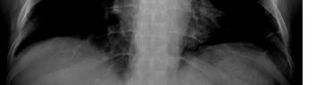
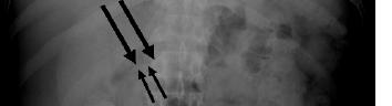
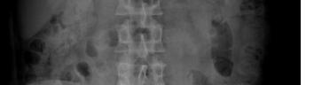


surgical intervention as necessary for safe foreign body removal, especially in the context of complications or persistent symptoms. Intraoperative ultrasound, as used here, can facilitate precise localization and minimize trauma to surrounding tissues.10 Multidisciplinary collaboration involving emergency medicine, radiology, gastroenterology, and surgery is critical for timely diagnosis and optimal management. Our patient’s favorable outcome reflects the value of teamwork and the integration of multiple imaging and therapeutic modalities.12
Foreign body-induced pancreatitis is a rare and diagnostically challenging condition that often mimics other causes of acute pancreatitis. This case highlights the crucial role of multimodal imaging and multidisciplinary collaboration in achieving accurate diagnosis and effective management. Surgical exploration remains essential when less invasive methods fail, underscoring the importance of early recognition and a team-based approach to optimize patient outcomes.
Foreign Body-induced Pancreatitis—Multimodal Imaging and Multispecialty Collaboration Vaghela et al.
Table. Description of advanced imaging modalities, and their respective findings, in case of foreign-body-induced pancreatitis.
Imaging Modality Findings
Point-of care ultrasound
Contrast-enhanced abdominal computed tomography
Esophagogastroduodenoscopyendosonography
4.2-cm foreign body near the pancreatic head with peripancreatic edema.
Approximate 4-cm linear structure slightly abuts the superior pancreatic body and partially abuts/ along the mildly thickened submucosa of the duodenal bulb/distal antrum, where there is mild adjacent stranding but without drainable fluid collection or overt pneumoperitoneum.
Localized moderate inflammation characterized by edema, erosion and erythema was found on the medial wall of the duodenal bulb. There was no foreign body noted intraluminally.
Abdominal radiograph Thin linear radiopaque object projecting over the right paramedian upper abdomen at the level of the first lumbar vertebral body.
Endoscopic ultrasound No discrete foreign object could be identified in the pancreatic head.
Intraoperative ultrasound Foreign body lying anteriorly at the junction of the pylorus and first portion of the duodenum.
The authors attest that their institution requires neither Institutional Review Board approval, nor patient consent for publication of this case report.
Address for Correspondence: Tobias Kummer, MD, Mayo Clinic, Department of Emergency Medicine, 200 First St SW, Rochester, MN 55905, United States.. Email: kummer.tobias@mayo.edu
Conflicts of Interest: By the CPC-EM article submission agreement, all authors are required to disclose all affiliations, funding sources and financial or management relationships that could be perceived as potential sources of bias. The authors disclosed none.
Copyright: © 2026 Vaghela et al. This is an open access article distributed in accordance with the terms of the Creative Commons Attribution (CC BY 4.0) License. See: http://creativecommons.org/ licenses/by/4.0/
1. Jaan A and Mulita F. Gastrointestinal foreign body. In: StatPearls. StatPearls Publishing LLC; 2025. Available from: https://www.ncbi. nlm.nih.gov/books/NBK562203/. Accessed September, 2025.
2. Pereira B, Santos A, Pereira E, et al. Foreign body-induced acute pancreatitis: ultrasonographic diagnosis. Rev Esp Enferm Dig 2013;105(7):439-40.
3. Hao F, Feng Q, Li J, et al. An ingested metallic wire migrating from stomach to pancreas treated by laparoscopic surgery: A case report.
Front Surg. 2023 6;9:927637.
4. Wang WG, Zhang Y, Wang L, et al. Chronic pancreatic inflammatory granuloma caused by foreign body presenting as a pancreatic pseudotumor: a case report and literature review. Pancreatology 2015;15(5):573-575.
5. Garment AR, Schwartz MB, Axsom KM. Foreign body-induced abscess resembling pancreatic neoplasia. J Gen Intern Med 2012;27(11):1561-4.
6. Yasuda T, Kawamura S, Shimada E, et al. Fish bone penetration of the duodenum extending into the pancreas: report of a case. Surg Today. 2010;40(7):676-8.
7. Kim KH, Woo EY, Rosato EF, et al. Pancreatic foreign body: ingested toothpick as a cause of pancreatitis and hemorrhage. Gastrointest Endosc. 2004;59(1):147-50.
8. Ehrsam JP, Meier Adamenko O, Schlumpf RB, et al. Foreign glass bodies in pleura and pancreas: systematic review for entry hypotheses and treatment options in an unresolved case. Surg Laparosc Endosc Percutan Tech. 2024;34(2):206-221.
9. Young CA, Menias CO, Bhalla S, et al. CT features of esophageal emergencies. Radiographics. 2008;28(6):1541-53.
10. Sun MR, Brennan DD, Kruskal JB, et al. Intraoperative ultrasonography of the pancreas. Radiographics 2010;30(7):1935-53.
11. Banks PA and Freeman ML. Practice Parameters Committee of the American College of Gastroenterology. Practice guidelines in acute pancreatitis. Am J Gastroenterol. 2006;101(10):2379-2400.
12. Wu C and Hungness ES. Laparoscopic removal of a pancreatic foreign body. JSLS. 2006;10(4):541-3.
Chad R. Sethman, PhD*
Jessica R Sethman, PA-C†
Bradley M End, MD, FPD-AEMUS‡
West Virginia University, Department of Microbiology, Immunology and Cell Biology, Morgantown, West Virginia
West Virginia University, Department of Emergency Medicine, Morgantown, West Virginia West Virginia University, Department of Emergency Medicine, Department of Medical Education, Morgantown, West Virginia
Section Editor: Melanie Heniff, MD, JD
Submission history: Submitted August 16, 2025; Revision received October 25, 2025; Accepted November 11, 2025
Electronically published April 21, 2026
Full text available through open access at http://escholarship.org/uc/uciem_cpcem
DOI: 10.5811/cpcem.50676
Introduction: Kawasaki disease is a vasculitis most commonly affecting children under five years of age but can also occur in older children and adults. When not sufficiently treated, Kawasaki disease can lead to cardiac complications such as myocarditis and coronary artery aneurysms, with aneurysms being the most serious long-term complication as it poses a risk for acute coronary syndrome.
Case Report: A 19-year-old with remote history of Kawasaki disease presented to the emergency department with chest pain, diaphoresis, and emesis after being struck in the chest by another player during a basketball game. Despite his young age and reported mild musculoskeletal trauma, an electrocardiogram and troponin were ordered. Electrocardiogram findings were concerning for ischemia, and troponin was elevated, confirming myocardial infarction and prompting a cardiology consult. Urgent percutaneous coronary intervention of the occluded aneurysm with balloon angioplasty resulted in significantly improved distal blood flow.
Conclusions: Kawasaki disease is widely recognized as a cause of cardiac complications in childhood, but the risk does not end there. Adults with a history of childhood Kawasaki disease remain at risk for complications of coronary artery aneurysm, even when they received appropriate medical treatment. Thrombosis or dissection of a coronary artery aneurysm can lead to acute coronary syndrome in otherwise healthy individuals. Therefore, emergency physicians must maintain a high level of suspicion for cardiac complications in both children and adults with a history of Kawasaki disease. In this case, prompt diagnosis and intervention were essential to achieving the best possible outcome. [Clin Pract Cases Emerg Med. 2026;10(2):191-194.]
Keywords: Kawasaki disease; coronary artery aneurysm; acute coronary syndrome; electrocardiogram; case report.
In 1967, a report by Tomisaku Kawasaki described a distinct type of vasculitis in 50 infants in Japan.1 Now commonly known as Kawasaki disease, the incidence of this disease has been steadily increasing and has now emerged as the most common cause of acquired heart disease in children in developed countries.2-4 The exact etiology is unknown; however, inflammatory mediators in the blood lead to
significant vascular inflammation that can cause the vessel walls to thicken, narrow, or weaken.5,6 Furthermore, the vasculitis associated with Kawasaki disease tends to localize to coronary arteries and can lead to cardiac complications such as myocarditis and coronary artery aneurysms, with aneurysms being the most serious long-term complication.7 Thrombus formation within the coronary artery aneurysm can result from a combination of abnormal blood flow and platelet
Myocardial Infarction in a 19-year-old with a History of Kawasaki Disease: A Case Report Sethman et al.
aggregation at the damaged endothelium leading to cardiac ischemia and myocardial infarction (MI).8
Various treatment options are available to mitigate the severity, duration and sequelae of Kawasaki disease. The primary objectives in treatment include the reduction of systemic vascular inflammation and inhibition of thrombosis formation. Therefore, treatment typically involves the administration of a single intravenous immunoglobulin infusion and low-dose acetylsalicylic acid.9 More aggressive treatments include antiplatelet and anticoagulant therapies such as clopidogrel, enoxaparin, and alteplase for higher risk patients and those who develop large aneurysms.3,9,10
The incidence of Kawasaki disease can be 25% or higher in untreated or delayed-treatment cases compared to an estimated 4% in those who are treated in a timely manner.7,11
Acute coronary syndrome most commonly occurs within the first two years following Kawasaki disease diagnosis, but cases have also been reported in adults long after the initial diagnosis especially in individuals with persistent coronary artery aneurysm.12-14 Therefore, vigilant monitoring of patients for aneurysm formation during the acute phase of illness, along with lifelong assessment for thrombotic occlusion, is crucial for effective treatment and long-term management.8,15
A 19-year-old male college student presented to the emergency department (ED) for chest pain after being struck in the chest by another player during a basketball game. He reported experiencing nausea and vomiting for two hours with associated mild dyspnea, diaphoresis, and light-headedness. He reported having had Kawasaki disease at age five, but it was undetermined whether he had treatment at that time. He did have a known right coronary artery aneurysm for which he routinely saw cardiology in his hometown and was prescribed daily acetylsalicylic acid and clopidogrel to reduce the risk of thrombus formation. Echocardiogram and cardiac stress tests were performed one month prior to his ED visit, which showed retained ejection fraction and no signs of coronary artery occlusion at that time, but images were not available for review at the time of his encounter.
On examination he was well appearing, resting comfortably, and conversing with an accompanying basketball player. Vital signs were as follows: blood pressure, 92/55 millimeters of mercury; heart rate, 62 beats per minute; respiratory rate, 18 breaths per minute; temperature, 36.2 °C, and oxygen saturation 95% on room air. Auscultation of the heart demonstrated a regular rhythm with no murmurs, gallops, or rubs. He had no tachypnea, respiratory distress, or adventitious lung sounds.
An electrocardiogram (ECG) was performed (Image 1)
CPC-EM Capsule
What do we already know about this clinical entity?
Kawasaki disease, a vasculitis most commonly affecting children, can lead to coronary artery aneurysms and subsequent thrombus formation when left untreated.
What makes this presentation of disease reportable?
An otherwise healthy 19-year-old with a history of Kawasaki disease presented with chest pain following musculoskeletal trauma. Cardiac assessment revealed myocardial infarction.
What is the major learning point?
Emergency physicians should maintain a heightened level of suspicion for acute coronary syndrome in patients with a history of Kawasaki disease.
How might this improve emergency medicine practice?
This case highlights the long-term complications of Kawasaki disease, and the need for timely cardiac assessment and intervention.
Image 1. Electrocardiogram upon emergency department arrival of a young patient with chest pain that demonstrates sinus rhythm with a first-degree atrioventricular block (double-sided arrow). Q waves (dashed arrows) and ST-segment depressions (open arrows) are observed in the inferior leads (II, III, and aVF). ST-segment elevations (solid arrows) are present in V2, aVR, and aVL.
Sethman et al. Myocardial Infarction in a 19-year-old with a History of Kawasaki Disease: A Case Report
and revealed sinus rhythm with first-degree atrioventricular block, Q waves, and ST-segment depressions in the inferior leads as well as nonspecific ST- segment elevations in noncontiguous leads. The patient’s cardiac troponin was 328 nanograms per liter (ng/L) (reference range: < 30 ng/L). Repeat troponin was not performed prior to intervention. His white blood cell count was 12,100 per liter (L) (< 11,000/L), and his basic metabolic panel was within normal limits. There was no acute cardiopulmonary process identified on chest radiograph. Given the heightened level of concern due to the patient’s history, continued chest pain, ischemic morphology of the ST-segment elevation in V2 with other ECG changes, and elevated troponin, cardiology was consulted and recommended urgent cardiac catheterization despite the patient not having met ST-segment elevation myocardial infarction (STEMI) criteria.
The catheterization revealed an acute near-total thrombotic occlusion of the aneurysm in the proximal portion of the right coronary artery (Image 2A). Balloon angioplasty and thrombectomy were performed at the time of catheterization (Image 2B), followed by stent placement.
The patient had an uncomplicated clinical course following percutaneous coronary intervention. Subsequent echocardiogram showed mildly depressed left ventricular function and moderately depressed right ventricular function with an ejection fraction of 45%. The patient was discharged in stable condition on postoperative day three with the diagnosis of non-STEMI (NSTEMI). It was
recommended that he continue taking 81 mg acetylsalicylic acid once daily as an outpatient and begin taking 20 mg rivaroxaban once daily. The previously prescribed clopidogrel was discontinued.
Although this patient was being managed by cardiology to prevent complication of a known right coronary artery aneurysm from childhood Kawasaki disease he still ultimately developed a near-complete occlusion of the right coronary artery leading to acute MI. Treatment principles for MI in Kawasaki disease patients are derived from the same guidelines established for the adult population with atherosclerotic coronary artery disease. Accordingly, indications for urgent cardiac catheterization in Kawasaki disease patients mirror those recommendations.3 In this case, the presence of Q waves and ST-segment depressions in leads II, III, and aVF indicated an age-undetermined infarction. The ST-segment elevations observed in V2, aVR, and aVL are considered nonspecific as there are no two contiguous leads of elevation.
Although this patient did not technically meet the criteria for STEMI, he did fulfill the diagnostic criteria for NSTEMI and cardiology determined that expedited percutaneous intervention was necessary. Further workup revealed a newly decreased ejection fraction, validating the need for urgent intervention. Given these findings, it is imperative that emergency physicians maintain a heightened level of suspicion for acute coronary syndrome in patients with a history of Kawasaki disease.
2. Cardiac catheterization images pre/post angioplasty of young man with history of Kawasaki disease: A) Initial angiography image revealing a visible cardiac artery aneurysm (solid black arrow) and diminutive distal flow due to occlusion/thrombus (dashed black arrow); and B) angiography following percutaneous coronary intervention with balloon angioplasty showing the aneurysm (solid white arrow) with significantly improved distal flow (dashed white arrow).
Because acute coronary syndrome is infrequently detected in young adults presenting with chest pain, emergency physicians may be less inclined to expand the cardiac workup beyond an ECG. While it is widely understood that Kawasaki disease can cause cardiac complications in childhood, it is important to recognize that even with appropriate medical therapy, these patients continue to carry the risk of coronary artery aneurysm and, thus, acute coronary syndrome, throughout adulthood. These late complications often present acutely, and emergency physicians are frequently the first point of contact when they do. Therefore, if a patient with a history of Kawasaki disease in childhood presents for chest pain, emergency physicians should strongly consider a full cardiac evaluation to rule out acute coronary syndrome even if the patient appears well and the history of present illness suggests a potential noncardiac etiology such as, in this case, musculoskeletal trauma.
Myocardial Infarction in a 19-year-old with a History of Kawasaki Disease: A Case Report Sethman et al.
The authors attest that their institution requires neither Institutional Review Board approval, nor patient consent for publication of this case report. Documentation on file.
Address for Correspondence: Chad R. Sethman, PhD. Department of Microbiology, Immunology and Cell Biology, West Virginia University, Morgantown, 64 Medical Center Drive, P.O. Box 9177, Morgantown, WV 26506-9177. E-mail: chad.sethman@ hsc.wvu.edu.
Conflicts of Interest: By the CPC-EM article submission agreement, all authors are required to disclose all affiliations, funding sources and financial or management relationships that could be perceived as potential sources of bias. The authors disclosed none.
Copyright: © 2026 Sethman et al. This is an open access article distributed in accordance with the terms of the Creative Commons Attribution (CC BY 4.0) License. See: http://creativecommons.org/ licenses/by/4.0/
1. Burns JC. Translation of Dr. Tomisaku Kawasaki’s original report of fifty patients in 1967. Pediatr Infect Dis J. 2002;21:993-995.
2. Huang SK, Lin MT, Chen HC, et al. Epidemiology of Kawasaki disease: prevalence from national database and future trends projection by system dynamics modeling. J Pediatr. 2013;163(1):12631.
3. McCrindle BW, Rowley AH, Newburger JW, et al. Diagnosis, treatment, and long-term management of Kawasaki disease: a scientific statement for health professionals from the American Heart Association. Circulation. 2017;135:e927.
4. Jone PN, Tremoulet A, Choueiter N, et al. Update on diagnosis and management of Kawasaki disease: a scientific statement from the
American Heart Association. Circulation. 2024;150(23):e481-e500.
5. Philip S, Jindal A, Krishna Kumar R. An update on understanding the pathophysiology in Kawasaki disease: possible role of immune complexes in coronary artery lesion revisited. Int J Rheum Dis 2023;26(8):1453-1463.
6. Leung DYM and Schlievert PM. Kawasaki syndrome: role of superantigens revisited. FEBS J. 2021;288(6):1771-1777.
7. Newburger JW, Takahashi M, Burns JC. Kawasaki disease. J Am Coll Cardiol. 2016;67(14):1738-1749.
8. Thangathurai J, Kalashnikova M, Takahashi M, et al. Coronary artery aneurysm in Kawasaki disease: coronary CT angiography through the lens of pathophysiology and differential diagnosis. Radiol Cardiothorac Imaging. 2021;3(5):e200550.
9. Gao L, Wang W, Wang H, et al. The safety and effectiveness of clopidogrel versus aspirin in Kawasaki disease with mild-to-moderate liver injury. Sci Rep. 2023;13(1):18324.
10. Johnson PN and Kuhn RJ. Combination thrombolytic and anti-platelet therapies in an infant with incomplete Kawasaki disease and coronary aneurysms. J Pediatr Pharmacol Ther. 2008;13(4):242-50.
11. Mossberg M, Mohammad AJ, Kahn F, et al. High risk of coronary artery aneurysm in Kawasaki disease. Rheumatology (Oxford). 2021;60(4):1910-1914.
12. Tsuda E, Hamaoka K, Suzuki H, et al. A survey of the 3-decade outcome for patients with giant aneurysms caused by Kawasaki disease. Am Heart J. 2014;167(2):249-258.
13. Sekimoto T, Shiraki T, Tanaka T, et al. Long-term Kawasaki disease complication: thrombotic coronary aneurysm leading to acute myocardial infarction. JACC Case Rep. 2024;29(17):102503.
14. Lee J, Seo J, Shin YH, Jang AY, Suh SY. ST-segment elevation myocardial infarction in Kawasaki disease: a case report and review of literature. World J Clin Cases. 2022;10(26):9368-9377.
15. Brogan P, Burns J, Cornish J, et al. Lifetime cardiovascular management of patients with previous Kawasaki disease. Heart (British Cardiac Society). 2020;106(6):411-420.
Mert Gültekin, MD*
Ayça Erinmez, MD†
Yunus Emre Karpuz, MD‡
Section Editor: Melanie Heniff, MD, JD
Şanlıurfa Training and Research Hospital, Department of Emergency Medicine, Şanlıurfa, Türkiye
Gaziantep University Faculty of Medicine, Department of Pediatric Surgery, Gaziantep, Türkiye
Gaziantep University Faculty of Medicine, Department of Emergency Medicine, Gaziantep, Türkiye
Submission history: Submitted September 22, 2025; Revision received December 12, 2025; Accepted December 19, 2025
Electronically published April 21, 2026
Full text available through open access at http://escholarship.org/uc/uciem_cpcem DOI: 10.5811/cpcem.52931
Introduction: Trichobezoar is a rare gastrointestinal condition typically caused by ingestion of hair, which most often affects adolescent females. Its clinical presentation is frequently nonspecific, with symptoms such as abdominal pain, constipation, or early satiety, which can delay recognition until severe complications such as obstruction or perforation develop.
Case Report: We present the case of a 14-year-old girl who developed a massive trichobezoar resulting in gastric perforation and death. She had a three-month history of intermittent constipation and multiple healthcare visits without definitive diagnosis. On arrival to the emergency department, she was in cardiopulmonary arrest. Computed tomography revealed a large intragastric mass with associated pneumoperitoneum. Emergency laparotomy confirmed a trichobezoar with gastric perforation and diffuse peritonitis. Despite prompt surgical and resuscitative intervention, the patient could not be revived.
Conclusion: This case underscores the importance for emergency physicians to maintain clinical vigilance when adolescents present with persistent, unexplained gastrointestinal symptoms. Trichobezoar should be considered in the differential diagnosis, even in the absence of psychiatric history. A low threshold for advanced imaging is warranted, as early recognition and intervention may prevent fatal complications such as gastric perforation and septic shock.
[Clin Pract Cases Emerg Med. 2026;10(2):195-199.]
Keywords: trichobezoar; gastric perforation; cardiac arrest; adolescent; case report.
Bezoars are solid masses formed by indigestible foreign materials within the gastrointestinal (GI) system. The taxonomy of bezoars reflects their diverse origins, with phytobezoars arising from undigested plant matter, trichobezoars developing from accumulated hair fibers, lactobezoars forming from milk protein precipitates, and pharmacobezoars resulting from medication deposits 1-3 Trichobezoars constitute a small subset of GI bezoars, representing approximately 6% of documented bezoar cases in
the medical literature.1,2 It typically occurs in adolescent girls, particularly those with developmental disabilities and underlying psychiatric disorders. Trichobezoars are associated with trichotillomania, a compulsive disorder characterized by repetitive hair-pulling behavior that causes significant distress, which is classified under obsessive-compulsive and related disorders in the Diagnostic and Statistical Manual of Mental Disorders, 5th Edition.1,5,6 Additional precipitating factors include depression, anxiety, neglect, and sudden emotional disruptions within the family.
Fatal Gastric Perforation Caused by Undiagnosed Trichobezoar in an Adolescent: A Case Report Gültekin et
Trichobezoars may present with nonspecific symptoms such as abdominal pain, constipation, bloating, and nausea, and their clinical presentation may simulate common GI pathologies including functional constipation, gastritis, peptic ulceration, or even abdominal tuberculosis, creating diagnostic uncertainty. Without timely recognition, these masses can cause various complications including protein-losing enteropathy, obstructive jaundice, intussusception, intestinal obstruction, and even gastric perforation, with potential for fatal outcomes.3,4,6,7 We present a fatal case involving a 14-year-old girl who developed gastric perforation secondary to a large trichobezoar. Despite multiple emergency department (ED) visits over a three-month period with GI complaints, trichobezoar was never considered in the differential diagnosis. This case demonstrates the diagnostic challenges of adolescent patients and emphasizes the necessity for comprehensive clinical assessment when standard treatments repeatedly fail.
A 14-year-old adolescent girl with no known chronic medical issues was brought to the ED by her family after a sudden collapse at home. On arrival, the patient was unresponsive and in cardiopulmonary arrest. She appeared pale and cyanotic, with no spontaneous respirations and no palpable pulse. Her Glasgow Coma Scale was three. Advanced Life Support was initiated immediately. Endotracheal intubation and chest compressions were performed, and due to difficulty obtaining peripheral intravenous access, an intraosseous line was placed in the right tibia for medications including epinephrine. Return of spontaneous circulation was achieved at the fifteenth minute of resuscitation. Postresuscitation examination noted fixed, dilated pupils unreactive to light. Point-of-care ultrasound revealed substantial free intraperitoneal fluid in the abdomen (measuring up to 130 mm in depth), and focused cardiac ultrasound showed normal cardiac contractility and no pericardial effusion.
Once the patient was stabilized, further history was obtained from the family. Medical, family, and psychosocial history was unremarkable. No chronic medical conditions, psychiatric disorders, or unusual behavioral patterns were reported by the family. When specifically questioned, the family denied any history of hair-pulling, hair-chewing, or pica-related behaviors. They reported that the patient had experienced intermittent abdominal pain and intractable constipation for the prior three months. During that period, she had been evaluated at multiple healthcare facilities. Each time, her symptoms were managed conservatively or symptomatically, with laxatives, enemas, or herbal remedies, but no definitive diagnosis was established. Notably, just two hours prior to presentation at our hospital, she had been seen at another ED for worsening abdominal discomfort and was
CPC-EM Capsule
What do we already know about this clinical entity?
Trichobezoars are gastric masses formed from ingested hair, typically affecting adolescent females with psychiatric comorbidities.
What makes this presentation of disease reportable?
In this fatal case of gastric perforation in an adolescent the diagnosis was missed despite recurrent emergency department visits, due to diagnostic anchoring.
What is the major learning point?
Recurrent, nonspecific abdominal pain in adolescents warrants advanced imaging and consideration of bezoars, even without known psychiatric history.
How might this improve emergency medicine practice?
Recognizing specific red flags and using computed tomography prevents diagnostic delays and catastrophic complications in cryptic abdominal pain cases.
given a large-volume enema, with no imaging performed. On our examination, the patient’s abdomen was markedly distended and firm. Bowel sounds were absent. There were no external signs of acute trauma. Genitourinary exam was unremarkable for injury.
Laboratory studies revealed a severe inflammatory and metabolic derangement consistent with abdominal sepsis: C-reactive protein, 331 milligrams per liter (mg/L) (reference range: 0-5 mg/L); markedly elevated white blood cell count, 17,350/mm³ (5,000-10,000/mm3); leukocytosis; hemoglobin 9.6 grams per deciliter (g/dL) (12-16 g/dL); anemia, and metabolic panel notable for creatinine 2.0 mg/dL (0.5-1.1 mg/ dL); acute kidney injury; sodium 128 millimoles (mmol)/L (135-145 mmol/L); and potassium 5.8 mmol/L (3.5-5 mmol/L). Arterial blood gas demonstrated a high anion-gap metabolic acidosis, pH 6.70 (7.35-7.45); bicarbonate, 5.9 mmol/L (21.8-26.2); and lactate, 17 mmol/L (0.5-1.6), consistent with profound shock.
Computed tomography (CT) was obtained. Head and chest CT were unremarkable; however, abdominal CT revealed massive pneumoperitoneum and free fluid throughout the peritoneal cavity (Image 1). Impressively, a large bezoar-
Gültekin et al. Fatal Gastric Perforation Caused by Undiagnosed Trichobezoar in an Adolescent: A Case Report
obtained immediately. The patient was started on broadspectrum antibiotics and taken emergently to the operating theater for exploratory laparotomy. During surgery, a large trichobezoar was found filling the stomach. A perforation approximately 2 cm in diameter was confirmed in the gastric wall at the site where the trichobezoar had likely pressed and eroded through (Image 3). Gross spillage of intestinal contents and pus was present in the peritoneal cavity, consistent with diffuse peritonitis. However, during the procedure the patient suffered a second cardiac arrest, likely due to refractory septic shock. Despite immediate resuscitative efforts, the patient could not be revived and was pronounced dead in the operating room. In accordance with ethical protocols and out of respect for the deceased patient, the surgical intervention was discontinued at that point, and complete trichobezoar removal was not undertaken.
like foreign body was visible, occupying the stomach and extending through the duodenum, measuring approximately 100 mm at its widest diameter on radiological assessment (Image 2). A full thickness defect was noted along the greater curvature of the stomach, consistent with gastric perforation, and multiple dilated small bowel loops with air-fluid levels suggested secondary intestinal obstruction by the mass.
Surgical and pediatric specialty consultations were
2.
computed tomography (axial plane) images at the level of the gastric outlet. (A) Free fluid (red arrow), pneumoperitoneum (blue arrow), and a large intragastric mass (yellow stars) are visible. (B) Radiological measurement of the intragastric mass demonstrates a diameter of approximately 100 millimeters.
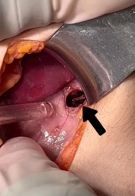
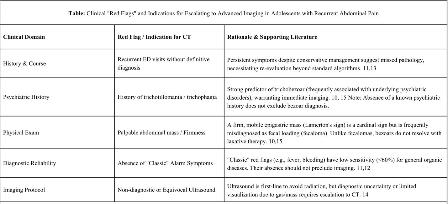
Trichobezoars are rare but serious conditions. They mostly affect teenage girls and are linked to hair-pulling and eating hair.1,5 Even though this condition is known, it is hard to diagnose early because symptoms are vague and there is often no clear mental health history. Several fatal trichobezoar cases have been reported in the literature.2,8,9 The main problem in diagnosing early is the unclear symptoms. Patients often have general complaints like stomach pain and constipation. In a study of 21 children, Wang et al2 reported that stomach pain (90.5%) and vomiting (76.2%) were common symptoms. Mirza et al4 saw these symptoms in 88% of 17 cases.
A key sign of trichobezoar is a firm, movable lump in the upper stomach area, known as the Lamerton sign.10 The presence of this lump varies; Wang et al 2 reported it in 61.9% of patients and Mirza et al4 in 41%. Without a known mental health history, these signs can be misunderstood. Wang et al noted that lumps were wrongly diagnosed as stool or normal stomach contents in four patients.2 Gomez-Suarez warned that telling the difference between physical and functional causes based on signs alone is often unreliable.11 Obtaining a history of eating hair is hard. Mewa Kinoo and Singh10 reported that patients often hide this behavior due to shame or not knowing, making the history “not easily forthcoming.” Therefore, not having a reported mental health history should not rule out the diagnosis.
Relying on traditional “red flags” like bleeding and weight loss for advanced imaging can be misleading. Delin and Berglund12 showed that while red flags are good for inflammatory bowel disease, they are not very sensitive (59.4%) for other conditions. Adeniyi et al13 found that among
children with repeated stomach pain, only 12.6% showed warning symptoms, but 87.4% had identifiable physical problems. Our case demonstrates this issue; the lack of “classic” warning symptoms gave false reassurance, causing deadly delays. For imaging, ultrasound is the first choice in a young patient to limit exposure to radiation, but it has limitations.14 Elghazeery and Hassan15 noted that hair’s high echogenicity and shadowing can hide the diagnosis or be mistaken for gas. Thus, if ultrasound is unclear, especially with a lump or repeated symptoms, CT is needed, as it is superior for a clear diagnosis.2,14 The critical red flags and indications for advanced imaging in adolescent patients with recurrent abdominal pain are summarized in the Table.
This case report has several strengths and limitations that warrant discussion. The primary strength lies in the detailed documentation of a rare but fatal presentation of trichobezoar, providing important educational value for emergency physicians as well as pediatricians and surgeons. The timeline illustrates the diagnostic challenges encountered across multiple healthcare settings, highlighting how nonspecific presentations can obscure recognition of life-threatening conditions in the ED. This report also emphasizes key considerations for children who may present repeatedly with refractory GI complaints, reminding emergency clinicians to re-evaluate prior assumptions when symptoms persist. However, several limitations must be acknowledged. The retrospective nature of the report restricts full reconstruction of the decision-making process during prior healthcare encounters. The absence of detailed documentation from earlier visits prevents comprehensive analysis of missed opportunities for earlier diagnosis.
Gültekin et al. Fatal Gastric Perforation Caused by Undiagnosed Trichobezoar in an Adolescent: A Case Report
This case highlights critical lessons for clinical practice, particularly for emergency physicians who encounter adolescents with non-specific abdominal complaints. Trichobezoar should be considered in the differential diagnosis of persistent or recurrent gastrointestinal symptoms unresponsive to conventional therapy, even in the absence of psychiatric or behavioral history. Emergency clinicians should maintain a low threshold for advanced imaging when standard treatments fail, as early recognition and intervention are essential to prevent perforation and other fatal complications. More broadly, this case underscores the need for systematic diagnostic approaches in the emergency setting, moving beyond repeated symptomatic management to uncover rare but life-threatening causes of abdominal pain in pediatric patients.
The authors attest that their institution requires neither Institutional Review Board approval, nor patient consent for publication of this case report. Documentation on file.
Address for Correspondence: Mert Gültekin, MD, Department of Emergency Medicine, Şanlıurfa Training and Research Hospital, Yenice Mah, Yenice Yolu No:1 Eyyübiye / Şanlıurfa / Türkiye, 63200. Email: mertgultekin27@gmail.com
Conflicts of Interest: By the CPC-EM article submission agreement, all authors are required to disclose all affiliations, funding sources and financial or management relationships that could be perceived as potential sources of bias. The authors disclosed none.
Copyright: © 2026 Gültekin et al. This is an open access article distributed in accordance with the terms of the Creative Commons Attribution (CC BY 4.0) License. See: http://creativecommons.org/ licenses/by/4.0/
1. Mezoff EA and Mezoff AG. Bezoars. In: Wyllie R, Hyams JS, Kay M, eds. Pediatric Gastrointestinal and Liver Disease, 6th ed. Amsterdam, the Netherlands: Elsevier; 2021:303-306.
2. Wang L, Chen Y, Chen S, et al. Gastrointestinal trichobezoars in the pediatric population: a retrospective study. BMC Pediatr. 2024;24(1):124.
3. Shrestha N, Regmee S, Kharel A, et al. Ileal perforation secondary to bowel obstruction caused by foreign body bezoar: a case report. Ann Med Surg (Lond). 2022;82:104564.
4. Mirza MB, Talat N, Saleem M. Gastrointestinal trichobezoar: an experience with 17 cases. J Pediatr Surg. 2020;55(11):2504-2509.
5. American Psychiatric Association. Diagnostic and Statistical Manual of Mental Disorders. 5th ed. Arlington, VA: American Psychiatric Association; 2013:235-236.
6. Sivasubramanian BP, Ashokkumar M, Afzal M, et al. Rapunzel syndrome in a teenage girl: a case report. Cureus 2022;14(10):e29975.
7. Ahmad Z, Sharma A, Ahmed M, et al. Trichobezoar causing gastric perforation: a case report. Iran J Med Sci. 2016;41(1):67-70.
8. Zyla A and Jacques R. Fatal Rapunzel syndrome: gastric and intestinal trichobezoars in a child with trichotillomania. Am J Forensic Med Pathol. 2023;44(3):210-215.
9. Ventura DE, Herbella FA, Schettini ST, et al. Rapunzel syndrome with a fatal outcome in a neglected child. J Pediatr Surg 2005;40(10):1665-1667.
10. Mewa Kinoo S and Singh B. Gastric trichobezoar: an enduring intrigue. Case Rep Gastrointest Med. 2012;2012:136963.
11. Gomez-Suarez R. Difficulties in the diagnosis and management of functional or recurrent abdominal pain in children. Pediatr Ann 2016;45(11):e388-e393.
12. Delin M and Berglund SK. Validation of red flags in the workup of children with long-term abdominal pain. A retrospective study. Acta Paediatr. 2024;113(5):1095-1102.
13. Adeniyi OF, Odeghe EA, Lawal MA, et al. Recurrent abdominal pain and upper gastrointestinal endoscopy findings in children and adolescents presenting at the Lagos University Teaching Hospital. PLoS One. 2019;14(5):e0216394.
14. Wolfe C, Halsey-Nichols M, Ritter K, et al. Abdominal pain in the emergency department: how to select the correct imaging for diagnosis. Open Access Emerg Med. 2022;14:335-345.
15. Elghazeery MA, Hassan AM. Gastrointestinal bezoars in paediatrics: case series and literature review. Afr J Paediatr Surg 2024;21(2):101-106.
Dago Berckmans, MD
AZ Sint-Maarten, Department of Emergency Medicine, Mechelen, Belgium
Section Editor: Ezhilkugan Ganessane, MD
Submission history: Submitted September 18, 2025; Revision received November 12, 2025; Accepted December 30, 2025
Electronically published April 21, 2026
Full text available through open access at http://escholarship.org/uc/uciem_cpcem DOI: 10.5811/cpcem.52911
Introduction: Paradoxical coronary embolism is a rare cause of myocardial infarction.
Case Report: A 57-year-old man presented with acute chest pain after a recent non-ST elevation myocardial infarction, during which a patent foramen ovale was identified. On readmission, the electrocardiogram showed an inferior ST-elevation myocardial infarction, and angiography revealed a distal thrombotic occlusion in otherwise normal coronary arteries. No venous thromboembolism was found, but thrombophilia testing revealed heterozygous factor V Leiden. He was managed conservatively and underwent successful patent foramen ovale closure.
Conclusion: This case highlights paradoxical embolism as a diagnostic consideration in acute myocardial infarction without coronary artery disease. [Clin Pract Cases Emerg Med. 2026;10(2):200-203.]
Keywords: paradoxical embolism; coronary embolism; myocardial infarction; patent foramen ovale; case report
Most myocardial infarctions result from atherosclerotic plaque rupture, but in rare cases embolic occlusion is the culprit. When an embolus traverses a patent foramen ovale into the arterial circulation, the mechanism is termed paradoxical embolism. Coronary embolism is estimated to account for 3% of acute myocardial infarctions, most often due to infective endocarditis, atrial fibrillation, or valvular heart disease.1,2 Paradoxical embolism represents only a small fraction of these cases and is considered exceedingly rare.1,3 While patent foramen ovale is a well-established risk factor in cryptogenic stroke, its role in acute coronary syndromes is seldom reported.
We present a patient with recurrent myocardial infarction in the absence of coronary artery disease, in whom paradoxical embolism was strongly suspected. The case was further complicated by a transient neurological deficit, underscoring the systemic manifestations of paradoxical embolism and the importance of considering this diagnosis in the emergency department (ED).
A 57-year-old man with a medical history of asthma and erosive gastritis had been admitted one week earlier with non-ST elevation myocardial infarction (NSTEMI). His cardiovascular risk profile included no smoking history, no hypertension, and no diabetes mellitus. He did have hypercholesterolemia and a strong family history, as his father had died of acute myocardial infarction at the age of 50. During the first admission, coronary angiography revealed no obstructive disease, but transesophageal echocardiography revealed a long-tunnel patent foramen ovale with color Doppler flow directed from the right atrium into the left atrium (Image 1, Video).
An agitated saline contrast study confirmed a large right-to-left shunt, with microbubbles crossing from the right to left (Image 2). Left ventricular function was normal, and only minimal aortic root atheroma was seen.
The patient was discharged on aspirin and ticagrelor but presented again to the ED several days later with sudden retrosternal chest pain. He also described transient weakness
Image 1. Transesophageal echocardiography, color Doppler mid-esophageal bicaval view. A long-tunnel patent foramen ovale (PFO) is seen with flow directed from the right atrium (RA) to the left atrium (LA). A white arrow marks the color Doppler jet across the PFO tunnel.
of the left arm lasting approximately 30 minutes, which had resolved before arrival. He denied speech disturbance or visual symptoms. Upon arrival, his vitals were as follows: heart rate, 58 beats per minute; blood pressure, 185/99 millimeters of mercury; oxygen saturation, 98% on room air; and temperature normal. On examination, he was alert, oriented, and in no acute distress. Cardiopulmonary examination was unremarkable. Neurological evaluation revealed no residual deficits, with intact cranial nerves, full motor strength and sensation, normal coordination, symmetric reflexes, and no pronator drift.
Electrocardiogram showed sinus rhythm at 58 beats per minute with ST-segment elevation in leads II, III, and aVF, accompanied by reciprocal ST depression in aVL, consistent with inferior STEMI (Image 3).
Coronary angiography on this presentation revealed thrombotic occlusion of distal circumflex branches with an embolic morphology; the remainder of the coronary arteries were normal. No intervention was performed given the distal location. Left ventricular systolic function was overall preserved and appeared hyperdynamic, with a mid-inferior akinetic
Video. Transesophageal echocardiography, color Doppler midesophageal view, with bicaval orientation demonstrating a longtunnel patent foramen ovale with right-to-left shunting from the right to the left atrium.
CPC-EM Capsule
What do we already know about this clinical entity?
Paradoxical coronary embolism is a rare cause of myocardial infarction, typically linked to intracardiac shunts such as a patent foramen ovale.
What makes this presentation of disease reportable?
The absence of coronary atherosclerosis in recurrent infarction highlights an uncommon, nonatherosclerotic mechanism of myocardial injury.
What is the major learning point?
Normal coronary angiography does not exclude myocardial infarction and should prompt evaluation for nonatherosclerotic causes.
How might this improve emergency medicine practice?
Recognition of embolic mechanisms facilitates timely diagnostic evaluation, targeted management, and prevention of recurrent ischemic events.
segment on left ventricular angiography, consistent with a recent inferior myocardial infarction. Noncontrast computed tomography of the brain was normal; magnetic resonance imaging (MRI) performed later revealed no acute ischemic lesions. Carotid duplex showed no significant stenosis. Lower extremity venous duplex revealed no deep vein thrombosis.
The patient was treated with dual antiplatelet therapy (aspirin, ticagrelor) and therapeutic low-molecular-weight heparin. His hospital course was uncomplicated. At discharge, he was transitioned to combination therapy with clopidogrel and rivaroxaban, with cardiology overseeing patent foramen ovale closure evaluation and hematology monitoring thrombophilia. Hematology later identified heterozygosity for factor V Leiden with an additional methylenetetrahydrofolate reductase variant. While the latter is of uncertain relevance in arterial events, the findings supported a prothrombotic predisposition.
This case illustrates paradoxical coronary embolism as a rare but important cause of myocardial infarction. Several features support this diagnosis: angiographically normal proximal coronary arteries; abrupt distal occlusion with embolic morphology; and a known patent foramen ovale with
Image 2. Transesophageal echocardiography, mid-esophageal bicaval view with agitated saline contrast. Microbubbles are visualized crossing from the right atrium (RA) to the left atrium (LA), confirming a large right-to-left shunt. A white arrow highlights the bubbles entering the left atrium.
The transient left-arm weakness raised suspicion of a concomitant cerebral embolic event. However, the brain MRI did not demonstrate any ischemic changes. Based on the clinical presentation and the absence of infarction on MRI, the episode most likely represented a transient ischemic attack.
The absence of deep vein thrombosis on duplex ultrasound raised the possibility of occult venous thrombosis (eg, pelvic veins), transient clot formation, or an underlying hypercoagulable state. A hematologic workup for thrombophilia demonstrated the presence of factor V Leiden and a methylenetetrahydrofolate reductase variant. Although the direct role of these abnormalities in arterial disease remains debated, they suggested a predisposition to thromboembolism.
a large right-to-left shunt. The patient’s cardiovascular risk profile included hypercholesterolemia and a strong family history of premature myocardial infarction, but he lacked other major risk factors such as smoking, hypertension, or diabetes. The first angiogram showed completely normal coronary arteries. On the second, the only abnormality was a thrombotic occlusion of distal circumflex branches with an appearance typical of embolization, while the proximal segments remained normal. This pattern made atherosclerotic plaque rupture unlikely.
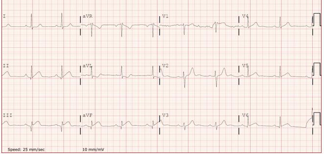
Image 3. Electrocardiogram at presentation showing sinus rhythm with ST-segment elevation in leads II, III, and aVF, and reciprocal depression in aVL, consistent with acute inferior ST-elevation myocardial infarction.
The immediate priority is stabilization and treatment of the acute coronary syndrome. When the culprit thrombus is accessible, percutaneous coronary intervention (PCI)— including aspiration thrombectomy—may be considered, while systemic thrombolysis has occasionally been reported when PCI is not feasible.4 In distal lesions not amenable to intervention, as in our patient, conservative management with antiplatelet therapy and anticoagulation is reasonable, after which the focus should shift to identifying and addressing the underlying cause of the embolism. Secondary prevention may include a tailored duration of anticoagulation and, when a right-to-left shunt is implicated, consideration of patent foramen ovale closure. Although evidence is extrapolated from stroke trials, randomized data such as the Patent Foramen Ovale Closure or Anticoagulants versus Antiplatelet Therapy to Prevent Stroke Recurrence trial support closure in carefully selected patients.5 In the absence of formal guidelines, decisions should be individualized and coordinated through multidisciplinary consultation with cardiology, neurology, and hematology.
Comparable cases in the literature share a recurring pattern: a documented venous source or acute pulmonary embolism (PE), a right-to-left shunt across a patent foramen ovale, and angiographic evidence of coronary embolization. Hakim et al described paradoxical coronary embolism in the setting of upper-extremity deep vein thrombosis from a peripherally inserted central catheter with concomitant PE.6 Ferreira et al reported an inferior STEMI occurring in the setting of PE in a patient with a proven patent foramen ovale.7 Boberg et al similarly presented the triad of acute pulmonary embolism, a large patent foramen ovale with right-to-left shunt, and subsequent embolic occlusion of the right coronary artery.8 In contrast, our patient had neither deep vein thrombosis nor PE but was found to have an underlying thrombophilia, suggesting paradoxical coronary embolism may occur even in the absence of demonstrable venous thromboembolism.
Paradoxical coronary embolism should be considered in patients presenting with STEMI and angiographically normal coronary arteries, particularly when a patent foramen ovale or
Berckmans Paradoxical Coronary Embolism as a Cause of Recurrent Myocardial Infarction: A Case Report
transient neurologic symptoms are present. In the emergency department, priorities are stabilization and guideline-based acute coronary syndrome care with early multidisciplinary involvement to pursue the embolic source, tailor antithrombotic therapy, and consider patent foramen ovale closure for secondary prevention.
The author attests that their institution does not require Institutional Review Board approval. Patient consent has been obtained and filed for the publication of this case report.
Address for Correspondence: Dago Berckmans, MD, Department of Emergency Medicine, AZ Sint-Maarten, Liersesteenweg 435, 2800 Mechelen, Belgium. Email: dago.berckmans@emmaus.be
Conflicts of Interest: By the CPC-EM article submission agreement, all authors are required to disclose all affiliations, funding sources and financial or management relationships that could be perceived as potential sources of bias. The authors disclosed none.
Copyright: © 2026 Berckmans. This is an open access article distributed in accordance with the terms of the Creative Commons Attribution (CC BY 4.0) License. See: http://creativecommons.org/ licenses/by/4.0/
1. Raphael CE, Heit JA, Reeder GS, et al. Coronary embolus: an underappreciated cause of acute coronary syndromes. JACC Cardiovasc Interv. 2018;11(2):172-180.
2. Lacey MJ, Raza A, Rehman H, et al. Coronary embolism: a systematic review. Cardiovasc Revasc Med. 2020;21(3):367-374.
3. Shibata T, Kawakami S, Noguchi T, et al. Prevalence, clinical features, and prognosis of acute myocardial infarction attributable to coronary artery embolism. Circulation. 2015;132(4):241-250.
4. Alenezi AR, Alanbaei M, Abouelenein I. Acute myocardial infarction secondary to paradoxical embolism. Eur J Case Rep Intern Med. 2020;7(12):001951.
5. Mas JL, Derumeaux G, Guillon B, et al. Patent foramen ovale closure or anticoagulation vs antiplatelets after stroke. N Engl J Med 2017;377:1011-1021.
6. Hakim FA, Kransdorf EP, Abudiab MM, et al. Paradoxical coronary artery embolism - a rare cause of myocardial infarction. Heart Views 2014;15(4):124-126.
7. Ferreira AR, Freitas A, Magno P, et al. Acute coronary syndrome of paradoxical origin. Rev Port Cardiol. 2013;32(10):817-21.
8. Boberg E, Hedman A, Hollenberg J. Pulmonary embolism with paradoxical embolization to right coronary artery in the presence of a large patent foramen ovale: a case report. Eur Heart J Case Rep 2024;8(4):ytae133.
Megan McLin-Evans, MD
Jenner Tiscareno, MD
Laura Lee Beneke, MD
University of Mississippi Medical Center, Department of Emergency Medicine, Jackson, Mississippi
Section Editor: Ezhilkugan Ganessane, MD
Submission history: Submitted September 15, 2025; Revision received November 20, 2025; Accepted January 13, 2026
Electronically published April 21, 2026
Full text available through open access at http://escholarship.org/uc/uciem_cpcem DOI: 10.5811/cpcem.51542
Introduction: Kratom (Mitragyna speciosa) is an unregulated herbal supplement increasingly associated with severe toxicity. Concentrated liquid formulations pose risks, with emerging reports of seizures, hepatotoxicity, and arrhythmias.9
Case Report: A previously healthy 24-year-old woman ingested a highly concentrated kratom extract and developed seizure-like activity followed by pulseless monomorphic ventricular tachycardia. She underwent approximately 45 minutes of resuscitation, including multiple defibrillations, dual-sequential shocks, amiodarone, lidocaine, magnesium, calcium, sodium bicarbonate, potassium repletion, epinephrine, and esmolol. Persistent instability prompted consultation with cardiology and cardiothoracic surgery, and she was cannulated for venoarterial extracorporeal membrane oxygenation (ECMO) in the emergency department. Lab studies showed profound hypokalemia, acidosis, and elevated lactate. Urine toxicology confirmed mitragynine. She stabilized on ECMO, was decannulated on hospital day two, extubated on day three, and discharged home neurologically intact on day seven.
Conclusion: Concentrated kratom extracts can precipitate life-threatening ventricular arrhythmias in previously healthy individuals. Emergency physicians should consider kratom in unexplained cardiac arrests and recognize the role of advanced support, including ECMO, in refractory toxicologic arrests. [Clin Pract Cases Emerg Med. 2026;10(2):204-207.]
Keywords: kratom; ventricular tachycardia; ECMO; cardiac arrest; toxicology; case report.
Kratom, derived from the Southeast Asian tree Mitragyna speciosa, has gained increasing popularity in the United States as an over-the-counter supplement marketed for pain relief, mood elevation, and opioid withdrawal.1 Its psychoactive alkaloids, primarily mitragynine and 7-hydroxymitragynine, exert both stimulant and opioid-like effects depending on the dose taken.¹ Despite its widespread availability and natural origin, kratom is not regulated by the U.S. Food and Drug Administration, and commercial preparations often vary significantly in potency and purity.2 Reports of toxicity have
increased in recent years, including cases involving seizures, hepatotoxicity, QT interval prolongation, and cardiac arrest.3,4 Concentrated liquid kratom formulations present a particularly high-risk profile due to their potent alkaloid content and ease of ingestion.5 Although kratom has not historically been considered arrhythmogenic, growing evidence points to potential pro-arrhythmic properties, particularly in young patients without preexisting cardiac disease.6,7 Here we describe a case of refractory monomorphic ventricular tachycardia and cardiac arrest following ingestion of a concentrated kratom extract, successfully managed with
emergency department (ED)-initiated venoarterial extracorporeal membrane oxygenation (VA-ECMO).
A previously healthy 24-year-old woman was found unresponsive by her boyfriend after reportedly consuming nearly an entire 15-mL bottle of MIT45 Super K Extra Strong (MIT45, Draper, UT), a highly concentrated kratom extract. She reported that she had not slept in several days and developed generalized shaking movements with urinary incontinence before becoming unresponsive. Emergency medical services were called and administered naloxone and midazolam en route to the ED. She was intubated in the field due to ongoing seizure-like activity and altered mental status. Shortly after intubation in the prehospital setting, she became pulseless with a wide-complex rhythm. On arrival to the ED, the patient remained pulseless with a widecomplex monomorphic rhythm consistent with ventricular tachycardia. High-quality cardiopulmonary resuscitation (CPR) was continued. She underwent multiple rounds of standard defibrillation with brief episodes of return of spontaneous circulation on approximately four occasions, each time degenerating back into pulseless ventricular tachycardia within seconds. At no point did she achieve sustained sinus rhythm.
The patient received multiple pharmacologic interventions, including a 300-mg amiodarone bolus with continuous infusion, a lidocaine infusion at 2 mg/minute, 4 grams of magnesium sulfate, 4 grams of calcium chloride followed by 2 grams of calcium gluconate, 175 milliequivalents (mEq) of sodium bicarbonate, aggressive potassium repletion, and multiple 1-mg epinephrine boluses. Due to ongoing electrical instability and concern for electrical storm, an esmolol bolus was given. Despite these measures and continued CPR, she remained in refractory ventricular tachycardia.
Given the lack of sustained return of spontaneous circulation after multiple standard shocks, three dual-sequential defibrillation attempts were performed. None resulted in conversion to a stable perfusing rhythm. After the third unsuccessful dual-sequential shock, cardiology and cardiothoracic surgery were emergently consulted, and the decision was made to proceed with VA-ECMO cannulation in the ED. Laboratory evaluation revealed severe hypokalemia with a potassium level of 1.9 mEq per liter (reference range: 3.5-5.1 mEq/L), sodium 150 mEq/L (135-145 mEq/L), arterial pH 7.18 (7.35-7.45), lactate 12.2 millimoles (mmol) per liter (0.5-2.0 mmol/L), glucose 240 mg per deciliter (dL) (70-110 mg/dL), aspartate aminotransferase 210 units/L (10-40 U/L), alanine aminotransferase 180 U/L (7-56 U/L), and creatine kinase 912 U/L (30-200 U/L). A urine drug screen was positive only for benzodiazepines, consistent with prehospital midazolam administration. Urine toxicology by liquid chromatography-mass spectrometry confirmed mitragynine and its metabolite, along with caffeine, cotinine, fentanyl, lidocaine, midazolam, phenylephrine, and trazodone. Analysis of two MIT45 Super K Extra Strong bottles from the patient’s
CPC-EM Capsule
What do we already know about this clinical entity?
Kratom is an unregulated supplement associated with seizures and toxicity, but life-threatening ventricular arrhythmias are rarely reported.
What makes this presentation of disease reportable?
This case describes refractory ventricular tachycardia after kratom ingestion requiring EDinitiated venoarterial extracorporeal membrane oxygenation (ECMO).
What is the major learning point?
High-potency kratom can cause malignant arrhythmias, and early consideration of ECMO may be lifesaving in refractory toxicologic arrest.
How might this improve emergency medicine practice?
Awareness of kratom cardiotoxicity could lead to early multidisciplinary activation and ECMO consideration in select arrest patients.
possession demonstrated mitragynine concentrations of 8.6 mg/ mL and 48.3 mg/mL.
Computed tomography of the head showed no acute intracranial process. Computed tomography of the chest, abdomen, and pelvis demonstrated an acute nondisplaced sternal fracture and fat stranding about the pancreas and duodenum concerning for acute interstitial edematous pancreatitis. Electrocardiography on arrival demonstrated wide complex tachycardia consistent with ventricular arrhythmia in the setting of electrolyte derangement (Images 1 and 2).
The patient was emergently cannulated for ECMO via the left femoral artery and vein at bedside in the ED, with placement of a distal reperfusion cannula after loss of Dopplerable signals in the left foot. Following ECMO initiation, she achieved hemodynamic stability and vasopressor requirements gradually decreased. She was transferred to the cardiovascular intensive care unit (ICU) on multiple vasopressors along with amiodarone and lidocaine infusions. Given concern for vasoplegia, she received methylene blue. N-acetylcysteine and broad-spectrum antibiotics were started for possible toxin-induced organ injury and post-cannulation prophylaxis. Over the first 24 hours in the ICU, she exhibited intermittent generalized myoclonus and perioral facial twitching. Neurology was consulted, and she was



Image 1 Initial electrocardiogram on arrival of a young woman who ingested kratom, showing wide complex tachycardia with right bundle branch block morphology and a QT interval of 602 milliseconds.
commands, and oriented, with no focal neurologic deficits. Her thrombocytopenia and coagulopathy, attributed to ECMO and critical illness, gradually improved. She completed a short course of broad-spectrum antibiotics, participated in physical therapy, and transitioned to an oral diet. She was discharged home on hospital day seven neurologically intact, with close cardiology, neurology, and toxicology follow-up arranged.
This case illustrates the life-threatening cardiac toxicity that may result from ingestion of high-potency kratom products. While mitragynine does not classically exhibit arrhythmogenic properties, case reports and animal studies suggest that it may prolong the QT interval and lead to ventricular dysrhythmias under certain conditions.2,3,5,6 Proposed mechanisms include direct sodium and potassium channel inhibition, mitochondrial toxicity, and sympathetic overdrive.4,6
treated with benzodiazepines and a levetiracetam loading dose followed by maintenance dosing.
Continuous electroencephalography demonstrated severe diffuse encephalopathy but no epileptiform discharges or electrographic seizures. Her myoclonus resolved over the next 48 hours. Serial laboratory studies showed improvement in lactic acidosis and liver enzyme elevations, with creatine kinase peaking and then downtrending. On hospital day two, after a successful ECMO clamp trial and normalization of hemodynamics, she was taken to the operating room for ECMO decannulation with femoral artery and vein repair. Transthoracic echocardiography performed around this time demonstrated normal left ventricular size with preserved systolic function, with an estimated ejection fraction of 50-55% (normal 50-70%) and mildly reduced right ventricular function.
She remained intubated postoperatively but was weaned to pressure support ventilation and extubated on hospital day three. Following extubation, the patient was awake, following



Image 2. Electrocardiogram demonstrating a ventricular escape rhythm with bigeminal premature ventricular complexes (arrows) and a markedly prolonged QRS interval of 262 milliseconds, consistent with progressive metabolic derangement.
In our patient, profound hypokalemia likely played a critical role in creating a vulnerable myocardial substrate for arrhythmogenesis. Although the cause of the hypokalemia is unclear, it may have been related to vomiting, catecholamine surge, or renal wasting induced by kratom’s adrenergic effects.9 Kratom may also have had a direct impact on pancreatic beta-cell function or insulin sensitivity, which could have further altered electrolyte dynamics and contributed to the development of metabolic acidosis and hypokalemia.8
While cardiac arrest in young patients is often attributed to congenital or structural causes, clinicians should also consider toxicologic etiologies, particularly in the absence of underlying heart disease. The increasing availability and use of kratom among young adults underscores the need for greater clinical awareness of its toxic potential. MIT45 Super K Extra Strong is advertised as containing 150-200 mg of mitragynine per bottle, which far exceeds traditional use levels. Additionally, due to the unregulated nature of these supplements, contamination with synthetic substances or adulterants is possible and may enhance toxicity.5
Venoarterial extracorporeal membrane oxygenation is not routinely available in all EDs, but its use in select cases of refractory toxicologic cardiac arrest has shown promise.7 In this case, VA-ECMO served as a bridge to recovery, allowing for correction of acidosis and electrolyte abnormalities while maintaining end-organ perfusion. Emerging evidence supports the use of extracorporeal life support in poisoned patients with potentially reversible causes of cardiac arrest who do not respond to conventional resuscitation.7 Criteria for ECMO consideration often include patients < 65 years of age, witnessed arrest, immediate initiation of CPR with acceptable low-flow duration, and absence of significant comorbidities.7 Early activation of the ECMO team is critical when patients meet these criteria.
Given the unpredictable content and potency of commercial kratom products, emergency physicians must maintain a high index of suspicion in cases of unexplained cardiac arrest or seizure, particularly in younger individuals. This case highlights both the dangers of high-potency kratom ingestion and the importance of advanced resuscitative strategies such as VA-
McLin-Evans et al.
VT Following Kratom Ingestion Requiring ECMO in A Young Woman
ECMO in select patients. Improved public health regulation and product oversight are urgently needed to reduce the risk of life-threatening toxicity from kratom.
Kratom toxicity can manifest with devastating cardiac consequences, particularly when consumed in concentrated liquid formulations. Emergency physicians should consider kratom as a potential etiology in young patients presenting with unexplained seizures, arrhythmias, or cardiac arrest. Profound hypokalemia, severe acidemia, and ventricular dysrhythmias may signal kratom-induced toxicity. Prompt recognition, aggressive supportive care, and early ECMO consideration can be lifesaving. As kratom use continues to rise, broader awareness and regulatory action are necessary to address its growing impact on public health.
The authors attest that their institution requires neither Institutional Review Board approval, nor patient consent for publication of this case report. Documentation on file.
Address for Correspondence: Megan McLin-Evans, MD, Department of Emergency Medicine, University of Mississippi, Medical Center, 2500 North State Street, Jackson, MS 39216. Email: mmclin2@umc.edu.
Conflicts of Interest: By the CPC-EM article submission agreement, all authors are required to disclose all affiliations, funding sources and financial or management relationships that could be perceived as potential sources of bias. The authors disclosed none.
Copyright: © 2026 McLin-Evans et al. This is an open access article distributed in accordance with the terms of the Creative Commons Attribution (CC BY 4.0) License. See: http:// creativecommons.org/licenses/by/4.0/
1. Warner ML, Kaufman NC, Grundmann O. The pharmacology and toxicology of kratom: from traditional herb to drug of abuse. Int J Legal Med. 2016;130(1):127-138.
2. Post S, Spiller HA, Chounthirath T, et al. Kratom exposures reported to United States poison control centers: 2011-2017. Clin Toxicol (Phila). 2019;57(10):847-854.
3. Eggleston W, Stoppacher R, Patel N, et al. Kratom use and toxicities in the United States. Pharmacotherapy 2019;39(7):775-777.
4. Hemby SE, McIntosh S, Leon F, et al. Abuse liability and therapeutic potential of the Mitragyna speciosa alkaloids mitragynine and 7-hydroxymitragynine. Addict Biol. 2019;24(5):874-885.
5. Gershman MD, Timm K, Frank M, et al. Deaths in Colorado attributed to kratom. N Engl J Med. 2019;380(1):97-98.
6. Kerrigan S and Basiliere S. Kratom: a systematic review of toxicological issues. WIREs Forensic Sci. 2022;4(1):e1420.
7. Hughes A, Johnson NJ, Mazor SS. Extracorporeal life support: indications and use in severely poisoned patients. Clin Pediatr Emerg Med. 2017;18(3):227-232.
8. Zhang P, Wei W, Zhang X, et al. Antidiabetic and antioxidant activities of Mitragyna speciosa (kratom) leaf extract in type 2 diabetic rats. Biomed Pharmacother. 2023;162:114689
9. Patel T, Karle E, Krvavac A. Kratom: an unusual cause of rhabdomyolysis and cholestasis. Crit Care Med. 2020;48(1):676
Kiveum Kim, DO
Taylor Craig, MD
Lucas Delicio, MD
Alexander John Scumpia, DO, MHA, MSc
Section Editor: Christopher San Miguel, MD
Submission history: Submitted June 15, 2025; Revision received July 31, 2025; Accepted September 10, 2025
Electronically published February 24, 2026
Full text available through open access at http://escholarship.org/uc/uciem_cpcem DOI: 10.5811/cpcem.48554
Case Presentation: A 64-year-old Black female presented to the emergency department following a new-onset tonic-clonic seizure. The patient had been given 2 milligrams of lorazepam by emergency medical services with cessation of seizure activity. On physical exam she was lethargic and had clear discharge from the right nare. Computed tomography of the brain initially demonstrated findings consistent with sinusitis versus ethmoidal mass. Magnetic resonance imaging of the brain demonstrated a right frontal ethmoidal encephalocele.
Discussion: Basal encephaloceles occur due to a defect in the skull base. Location of the defect and extracranial herniation of brain tissue can cause neurologic sequelae. This case illustrates the importance of maintaining a broad differential diagnosis and for emergency physicians to obtain imaging when evaluating seizures and/or chronic rhinorrhea in adults. [Clin Pract Cases Emerg Med. 2026;10(2):208-210.]
Keywords: basal encephalocele; atraumatic; primary (congenital) encephalocele; atraumatic encephalocele.
A 64-year-old Black female presented to the emergency department following a new-onset tonic-clonic seizure. The patient endorsed chronic rhinorrhea from the right nare intermittently for 25 years and increased frequency of rhinorrhea over that time frame, but she denied acute changes. No alleviating or exacerbating factors were associated with her rhinorrhea. On physical exam, the patient had clear nasal discharge from the right nare, with otherwise normal facial features. She initially presented post-ictal, but after returning to her baseline she had no focal neurological deficits, with cranial nerves II-XII grossly intact. Magnetic resonance imaging of the brain demonstrated an ethmoidal encephalocele (Images A and B). Beta-2 transferrin testing of rhinorrhea was performed, confirming cerebrospinal fluid leak.
The patient was admitted and evaluated by neurology. Subsequent electroencephalogram demonstrated diffuse encephalopathy consistent with post-ictal state with right frontal encephalocele assumed to be the epileptic focus. She was started on phenytoin and subsequently discharged with
instructions to follow up with neurosurgery. After undergoing endoscopic repair, a six-month follow-up deemed the repair successful and she was symptom-free. The patient was instructed to stop taking phenytoin. The patient has had no subsequent seizures post-resection of the anterior cranial fossa encephalocele.
Basal encephalocele is a type of primary (congenital) encephaloceles that occur due to a primary defect in the skull base. They can be further classified based on the location of the defect. A defect in the dura mater permits the extracranial herniation of brain tissue, and subsequent leptomeninges and cerebrospinal fluid into the extracranial space. The pathogenesis is believed to be a defective closure of the anterior portion of the neural tube early in the developmental process.1 Consequently, basal encephaloceles often present with other clinical features of patients with neural tube defects including broadened nasal bridge, hypertelorism, and other midfacial anomalies.2 They are primarily diagnosed in the
prenatal period with serum alpha fetoprotein levels and ultrasonography or early in childhood in patients who have associated phenotypic facial features.
It is much more difficult to diagnose primary encephalocele in adults, especially in those who lack the classical facial features, as was the case with our patient.3 Secondary (acquired) encephalocele is associated with cranial defects from injuries, such as those that occur in post-traumatic or post-surgical patients. The lack of associated facial features common in congenital encephalocele along with a lack of the classic presentation (post-traumatic and post-surgical history) made this a unique presentation and difficult to diagnose. Once the diagnosis is made, prompt treatment is critical to prevent complications including meningitis, tension pneumocephalus, and seizures.4 Treatment includes resection of the anterior cranial fossa encephalocele.5
The authors attest that their institution requires neither Institutional Review Board approval, nor patient consent for publication of this case report. Documentation on file.
CPC-EM Capsule
What do we already know about this clinical entity?
Basal encephaloceles are rare congenital skull base defects causing cerebral spinal fluid (CSF) leaks, seizures, or meningitis, usually diagnosed in infancy.
What is the major impact of the image(s)?
The magnetic resonance images confirm ethmoidal encephalocele and CSF leak, distinguishing it from sinus disease and guiding neurosurgical management.
How might this improve emergency medicine practice?
Emergency clinicians should be aware that chronic rhinorrhea with seizures may signal encephalocele, prompting imaging and early referral.
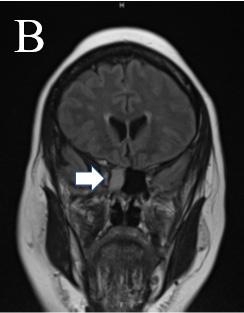
Address for Correspondence: Taylor Craig, MD, HCA Florida Aventura Hospital, Department of Emergency Medicine, 20900 Biscayne Blvd, Aventura, Florida 33180. Email: Taylor.Craig@ hcahealthcare.com.
Conflicts of Interest: By the CPC-EM article submission agreement, all authors are required to disclose all affiliations, funding sources and financial or management relationships that could be perceived as potential sources of bias. The authors disclosed none.
Copyright: © 2026 Kim et al. This is an open access article distributed in accordance with the terms of the Creative Commons Attribution (CC BY 4.0) License. See: http://creativecommons.org/ licenses/by/4.0/
1. Siffel C, Wong LY, Olney RS, et al. Survival of infants diagnosed with encephalocele in Atlanta, 1979-98. Paediatr Perinat Epidemiol. 2003;17(1):40-8.
2. Hoving EW and Vermeij-Keers C. Frontoethmoidal encephaloceles, a study of their pathogenesis. J Neurosurg Pediatr. 1997;27(5):246-256.
3. Harada, N, Nemoto, M, Miyazaki, C et al. Basal encephalocele in an adult patient presenting with minor anomalies: a case report. J Med Case Reports. 2014;8:24.
4. Lo BW, Kulkarni AV, Rutka JT, et al. Clinical predictors of developmental outcome in patients with cephaloceles. J Neurosurg Pediatr. 2008;2(4):254-257.
5. Ogiwara H and Morota N. Surgical treatment of transsphenoidal encephaloceles: transpalatal versus combined transpalatal and transcranial approach. J Neurosurg Pediatr. 2013;11(5):505- 510.
Christopher Allen, MD
Alexandra Gubbels, MD
Youyou Duanmu, MD, MPH
Jody Vogel, MD
Section Editor: Shadi Lahham, MD
Stanford University, Department of Emergency Medicine, Palo Alto, California.
Submission history: Submitted June 26, 2025; Revision received August 23, 2025; Accepted October 2, 2025
Electronically published March 13, 2026
Full text available through open access at http://escholarship.org/uc/uciem_cpcem
DOI: 10.5811/cpcem.48812
Case Presentation: A 20-year-old female with no past medical history presented to the emergency department (ED) after an episode of exertional syncope. Physical examination, vital signs, and electrocardiogram were unremarkable. Point-of-care ultrasound revealed abnormal invaginations in the interventricular septum. Laboratory evaluation was significant for markedly elevated troponin concerning for cardiac arrest. She was admitted to cardiology with suspicion for genetic cardiomyopathy. The patient underwent placement of an implantable cardioverter defibrillator after cardiac magnetic resonance imaging redemonstrated the septal invaginations known as myocardial crypts. Genetic studies later revealed sarcomere gene mutations associated with hypertrophic cardiomyopathy.
Discussion: Myocardial crypts, which are invaginations within the myocardium, are considered early morphological markers for hypertrophic cardiomyopathy and may precede the development of overt hypertrophy. The presence of myocardial crypts and syncope is highly concerning for evolving hypertrophic cardiomyopathy. In this case, identifying myocardial crypts on ED point-of-care ultrasound, in conjunction with clinical context, facilitated further confirmatory diagnostics and timely intervention with placement of an implantable cardioverter defibrillator. [Clin Pract Cases Emerg Med. 2026;10(2):211-213.]
Keywords: myocardial crypts; genetic cardiomyopathy; hypertrophic cardiomyopathy; point-of-care ultrasound; syncope.
A 20-year-old female presented to the emergency department (ED) after an episode of exertional syncope. She denied chest pain, prodromal symptoms, and past medical history, although she had reported a similar episode of exertional syncope years prior and a family history of sudden cardiac death. Physical examination and vital signs were unremarkable. Electrocardiogram (ECG) showed normal sinus rhythm without evidence of ischemic or dysrhythmic morphologies. Prior to laboratory results, point-of-care ultrasound (POCUS) echocardiogram (echo) revealed abnormal invaginations in the interventricular septum on parasternal long-axis view (Image 1).
While no obvious ventricular septal hypertrophy, left ventricular outflow tract (obstruction, or other structural or kinetic abnormalities were appreciated on POCUS, the septal invaginations raised suspicion for a structural cardiac cause of this patient’s syncope. Laboratory evaluation demonstrated markedly elevated troponin (1,961 nanograms per liter (ng/L) (reference range 0-10 ng/L for females) with leukocytosis, transaminitis, and acute kidney injury concerning for preceding cardiac arrest as etiology of syncope. These findings, including POCUS echo, were conveyed to the inpatient cardiology team who admitted the patient for telemetry and further diagnostics including cardiac magnetic resonance imaging (MRI). Cardiac MRI redemonstrated






CPC-EM Capsule
What do we already know about this clinical entity?
Myocardial crypts are invaginations in the myocardium, that may indicate early disease in hypertrophic cardiomyopathy. They are usually detected on magnetic resonance image.
What is the major impact of the image(s)?
Image 1. Point-of-care ultrasound echocardiogram on parasternal long-axis view demonstrating an invagination within the anteroseptal wall of the interventricular septum, known as a myocardial crypt (arrow). No septal hypertrophy, left ventricular outflow tract obstruction, or other structural or kinetic abnormalities were appreciated.
previously seen septal invaginations on echo, and formal radiographic interpretation confirmed these to be myocardial crypts (Image 2) without ventricular hypertrophy, wall motion abnormalities, or other abnormal findings.
Ultimately, the patient underwent implantable cardioverter defibrillator placement for presumed diagnosis of genetic





The ultrasound images demonstrate myocardial crypts can be seen on ultrasound, raising suspicion for an underlying cardiomyopathy prior to genetic confirmation. Ultrasound images show myocardial crypts can be seen on point-of-care ultrasound, raising suspicion for cardiomyopathy before MRI or genetic confirmation.
How might this improve emergency medicine practice?
Few reports describe myocardial crypts in emergency medicine. Our case highlights their detection on point-of-care ultrasound and the value of awareness for risk stratification and diagnosis.
cardiomyopathy and was discharged after an uneventful postoperative course. Genetic studies later confirmed this diagnosis, with results demonstrating sarcomere mutations associated with hypertrophic cardiomyopathy.
Image 2. Cardiac magnetic resonance imaging confirming myocardial crypt within the anteroseptal wall (arrow). On final impression, there were two additional myocardial crypts noted within the inferoseptal wall. Otherwise, there was normal left ventricle and right ventricle wall thickness, wall motion, and ejection fraction.

Genetic cardiomyopathy refers to a group of disorders caused by inherited mutations affecting the myocardium. The most common type is hypertrophic cardiomyopathy, characterized by ventricular septal hypertrophy leading to impaired hemodynamics from left ventricular outflow tract obstruction. Myocardial crypts are narrow, blood-filled invaginations within the myocardium that while not pathognomonic for hypertrophic cardiomyopathy, can serve as an early morphological marker for hypertrophic cardiomyopathy and may precede the development of overt hypertrophy.1-3 Myocardial crypts can be visualized on cardiac MRI in approximately two thirds of individuals carrying hypertrophic cardiomyopathy-related gene mutations prior to development of phenotypic hypertrophic cardiomyopathy, and in over half of these cases myocardial crypts precede detectable ECG abnormalities.4,5
Myocardial crypts can also be visualized on echocardiography. This case is, to our knowledge, the first description of myocardial crypts found on ED-performed POCUS. While myocardial crypts may occasionally be benign, in the appropriate clinical context, their finding on any imaging modality should raise suspicion for genetic cardiomyopathy as cardiology case reports have demonstrated.6-7 Although hypertrophic cardiomyopathyrelated sudden cardiac death occurs at an estimated 1% per year, early detection in the ED is challenging, with approximately 10% of patients lacking characteristic ECG abnormalities.8,9 This increases the utility of POCUS; unlike cardiac MRI, POCUS is widely available to the emergency physician when there is concern for hypertrophic cardiomyopathy. For this patient, recognizing myocardial crypts on POCUS, in conjunction with clinical context and laboratory data, raised suspicion for an underlying myocardial disorder leading to further confirmatory diagnostics and timely intervention with placement of an implantable cardiac defibrillator for prevention of sudden cardiac death.
This case highlights the potential role of POCUS in identifying subtle structural cardiac abnormalities in patients presenting with high-risk syncope. The detection of myocardial crypts on ED POCUS, even in the absence of septal hypertrophy or ECG abnormalities, raised suspicion for an underlying genetic cardiomyopathy and prompted expedited diagnostic evaluation. In patients with exertional syncope and concerning historical features such as a family history of SCD, recognition of myocardial crypts may represent an early imaging clue for evolving HCM. Incorporating careful assessment of myocardial morphology during ED POCUS may therefore help identify patients who warrant further cardiology evaluation and risk stratification.
The authors attest that their institution requires neither institutional review board approval, nor patient consent for publication of this case report. Documentation on file.
Address for Correspondence: Christopher Allen, MD, Department of Emergency Medicine, Stanford School of Medicine, 900 Welch Rd., Suite 300, Palo Alto, CA. 94304. Email: allencw@stanford. edu
Conflicts of Interest: By the CPC-EM article submission agreement, all authors are required to disclose all affiliations, funding sources and financial or management relationships that could be perceived as potential sources of bias. The authors disclosed none.
Copyright: © 2026 Allen et al. This is an open access article distributed in accordance with the terms of the Creative Commons Attribution (CC BY 4.0) License. See: http://creativecommons.org/ licenses/by/4.0/
1. Maron MS, Rowin EJ, Lin D, et al. Prevalence and clinical profile of myocardial crypts in hypertrophic cardiomyopathy. Circ Cardiovasc Imaging. 2012;5(4):441-447.
2. Germans T, Wilde AA, Dijkmans PA, et al. Structural abnormalities of the inferoseptal left ventricular wall detected by cardiac magnetic resonance imaging in carriers of hypertrophic cardiomyopathy mutations. J Am Coll Cardiol. 2006;48(12):2518-2523.
3. Child N, Muhr T, Sammut E, et al. Prevalence of myocardial crypts in a large retrospective cohort study by cardiovascular magnetic resonance. J Cardiovasc Magn Reson. 2014;16(1):66.
4. Efthimiadis GK, Zegkos T, Pagourelias ED, Karvounis H. Myocardial crypts as a preclinical sign of hypertrophic cardiomyopathy. Hippokratia. 2014;18(4):359-361.
5. Moon JC and McKenna WJ. Myocardial crypts: a prephenotypic marker of hypertrophic cardiomyopathy? Circ Cardiovasc Imaging 2012;5(4):431-432.
6. Alhatemi G, Sood A, Aldiwani H, et al. Description and clinical implications of myocardial clefts using echocardiography. Cureus 2021;13(6):e15407.
7. Spigno FD, Caraffini A, Canu E, et al. Myocardial crypts and ventricular fibrillation: two case reports. EMJ Cardiol. 2024;12(1):98-105.
8. Hong Y, Su WW, Li X. Risk factors of sudden cardiac death in hypertrophic cardiomyopathy. Curr Opin Cardiol 2022;37(1):15-21.
9. Rowin EJ, Maron BJ, Appelbaum E, et al. Significance of false negative electrocardiograms in preparticipation screening of athletes for hypertrophic cardiomyopathy. Am J Cardiol. 2012;110(7):1027-1032.
Atsuhito Tanaka, MD*
Yuka Kamitani, MD†
Tokyo Bay Urayasu Ichikawa Medical Center, Department of Emergency and Critical Care Medicine, Chiba, Japan
Kobe City Medical Center General Hospital, Emergency Care Center/Emergency Department, Kobe, Japan
Section Editor: Ezhilkugan Ganessane, MD
Submission history: Submitted July 22, 2025; Revision received September 21, 2025; Accepted November 17, 2025
Electronically published March 29, 2026
Full text available through open access at http://escholarship.org/uc/uciem_cpcem DOI: 10.5811/cpcem.49084
Case Presentation: An 88-year-old man was brought to our emergency department due to altered mental status and hemodynamic shock due to a urinary tract infection. Computed tomography showed an incarcerated ureter in the Petit triangle. Urology was consulted, and the hernia was reduced back into the retroperitoneal cavity.
Discussion: Petit hernia is rare; moreover, there is no literature to our knowledge discussing the ureter as the herniated structure. Interventional radiology can be considered as a reductive option. [Clin Pract Cases Emerg Med. 2026;10(2):214-216.]
Keywords: ureter herniation; Petit triangle.
CASE PRESENTATION
An 88-year-old man was transferred to our emergency department with a decreased level of consciousness and elevated inflammatory markers. His past medical conditions included osteomyelitis and cerebral infarction. No history of surgery or trauma was noted. On arrival, he was in septic shock for which vasopressor infusion and antibiotics were administered. Blood tests revealed elevated creatinine, and point-of-care ultrasound demonstrated dilatation of the right renal pelvis. Computed tomography (CT) of the abdomen was performed to determine the etiology of altered mental status and possible post-renal obstruction. Abdominal CT revealed an incarcerated right ureter that had herniated through the Petit triangle in the inferior lumbar region, accompanied by right renal pelvic dilatation (Images 1-3). Urology was consulted, and the ureter was retrogradely reduced back into the


















CPC-EM Capsule
What do we already know about this clinical entity?
Petit lumbar hernia is a rare type of lumbar hernia.
What is the major impact of the image(s)?
The images confirm a rare ureteral herniation into the Petit triangle while validating a successful endourological treatment option.
How might this improve emergency medicine practice?
Ureteral lumbar hernia should be on the differential for acute kidney injury and validates endourological reduction as a minimally invasive approach.
retroperitoneal cavity via cystoscopy. Renal function improved after the intervention, with a decrease in serum creatinine from 2.57 milligrams per deciliter (mg/dL) to 1.83 mg/dL (reference range: 0.61-1.04 mg/dL). The patient recovered and was discharged on day 10.
Petit hernia, a rare type of lumbar hernia, occurs through the space formed by the iliac crest, external oblique muscle, and latissimus dorsi muscle in the posterior abdomen, representing 1.5-2% of all hernias.1 Most reported cases discuss herniation of the colon or small bowel, often requiring surgical intervention. Ureteral hernias are uncommon clinical entities, making this specific combination unprecedented.2,4,5
Considering the patient’s age and clinical status, a lessinvasive approach was chosen over surgical intervention. Zandrino et al described a percutaneous anterograde approach to resolve a lumbar hernia in the first case report of a hernia retrogradely reduced back into the retroperitoneal cavity via cystoscopy and guidewire manipulation. This suggests that minimally invasive radiological or endourological techniques could be a primary therapeutic option in such rare cases.
The authors attest that their institution requires neither Institutional Review Board approval, nor patient consent for publication of this case report. Documentation on file.
Address for Correspondence: Atsuhito Tanaka, MD, Department of Emergency and Critical Care Medicine, Tokyo Bay Urayasu, Ichikawa Medical Center, 3-4-32 Todaijima, Urayasu-City, Chiba 279-0001. Email: atsuhito.tanaka17@gmail.com
Conflicts of Interest: By the CPC-EM article submission agreement, all authors are required to disclose all affiliations, funding sources and financial or management relationships that could be perceived as potential sources of bias. The authors disclosed none.
Copyright: © 2026 Tanaka et al. This is an open access article distributed in accordance with the terms of the Creative Commons Attribution (CC BY 4.0) License. See: http://creativecommons.org/ licenses/by/4.0/
1. Munoz-Munoz M, Torres-Mansur R, Torres-Ramirez MA. The rarity of Petit’s hernia: a case report on diagnosis and treatment. Int J Surg Case Rep. 2023;111:108886.
2. Yahya Z, Al-Habbal Y, Hassen S. Ureteral inguinal hernia: an uncommon trap for general surgeons. BMJ Case Rep 2017;2017:bcr2017219288
3. Zandrino F, Di Guardia G, Gallesio C. Inferior lumbar (Petit’s) hernia with ureteral incarceration treated with percutaneous repair. J Radiol Rev. 2022;9(3):158-60.
4. Ben Ghezala H, Feriani N. Hernia of the Jean Louis Petit triangle in the emergency department: a rare image. Pan Afr Med J. 2020;37:166.
5. Strange CD, Birkemeier KL, Sincleair ST. Atypical abdominal hernias in the emergency department: acute and non-acute. Emerg Radiol. 2009;16(2):121-8.
Taylor Stephens*
Bryan Imhoff, MD, MBA†
Janak Patel, DO, MBA†
Section Editor: Steven Walsh, MD
* † The University of Kansas School of Medicine, Kansas City, Kansas The University of Kansas Health System, Department of Emergency Medicine, Kansas City, Kansas
Submission history: Submitted October 14, 2025; Revision received November 26, 2025; Accepted December 4, 2025
Electronically published April 14, 2026
Full text available through open access at http://escholarship.org/uc/uciem_cpcem DOI: 10.5811/cpcem.53141
Case Presentation: We present the case of a 53-year-old female with darting eye movements and difficulty walking who was found to have opsoclonus-myoclonus syndrome only after multiple presentations to emergency departments over five days.
Discussion: Adult-onset opsoclonus-myoclonus syndrome is a rare central nervous system disease typically associated with paraneoplastic or idiopathic etiologies. With non-specific symptom presentation, this condition is commonly misdiagnosed in adults, leading to diagnostic delays and long-term motor and cognitive sequelae. [Clin Pract Cases Emerg Med. 2026;10(2):217-218.]
Keywords: opsoclonus; myoclonus; eye movements; case report.
A 53-year-old woman with a history of migraine headaches presented to her third emergency department (ED) in five days for suspected nystagmus and difficulty walking. She presented to the first ED reporting that her vision had “trouble focusing” and was documented to have eyes “darting back and forth when talking.” No acute findings on computed tomography head without contrast and magnetic resonance imaging head with and without contrast prompted discharge with a diagnosis of nystagmus with instructions to follow up with her ophthalmologist. Her ophthalmologist found no primary vision problems and referred her to otolaryngology (ENT) for “vestibular concerns.” The ENT office indicated she would be better served by a neurologist. Without improvement in her symptoms, she presented to a second ED two days after her primary encounter. There, her physical exam noted “visible horizontal nystagmus” and an unstable gait. Given previous negative imaging and established neurology follow-up, she was discharged. Two days later, she presented to our ED with worsening vision and gait that required significant familial assistance. On physical exam, she demonstrated persistent bilateral opsoclonus (video), significant left lower extremity myoclonus, and ataxia.
Video. Finding of bilateral opsoclonus on physical exam of 52-year-old woman who was diagnosed only after presentation to three emergency departments.
Neurology was consulted in our ED, and she was admitted to their service for further workup. While admitted, she received additional advanced imaging, lumbar puncture, and paraneoplastic and autoimmune panels with unremarkable results. Neurology diagnosed the patient with idiopathic opsoclonus-myoclonus syndrome and initiated treatment with intravenous (IV) methylprednisolone and IV immunoglobulin (IG) infusions followed by an oral prednisone taper at discharge, along with monthly IVIG and steroid infusions for 12 months. After three months of treatment, the patient reported feeling 80% back to baseline. Her myoclonus and ataxia had resolved, and opsoclonus had improved. Her cerebellar symptoms, including end point tremor, had improved with the addition of propranolol, providing a secondary anxiolytic benefit.
Opsoclonus-myoclonus syndrome is a central nervous system disease primarily affecting toddlers and is often associated with underlying malignancy.1 Adult onset is rare (< 0.2 cases per million) and typically associated with paraneoplastic or idiopathic (commonly para-infectious) etiologies.2,3 Our patient’s abnormal, erratic eye movements prompted a wider differential diagnosis to include opsoclonusmyoclonus syndrome as referenced in the original ED history and physical. With non-specific symptom presentation, this condition is commonly misdiagnosed in the adult population, leading to problematic diagnostic delays averaging 11 weeks following symptom onset.3
A common misdiagnosis is nystagmus, which consists of horizontal rhythmic and slow oscillation, whereas opsoclonus involves horizontal, vertical, and torsional saccades.1 This delay in diagnosis can result in long- term motor and cognitive deficits, commonly ataxia and residual dysarthria along with possible progression of underlying small-cell lung cancer, breast cancer, and ovarian cancer in paraneoplastic cases.1,2 With suspicion for opsoclonus-myoclonus syndrome, physicians should engage an integrated team of specialists including neurology, oncology, and immunology. Moreover, involvement of occupational and mental health therapists may improve prognosis and residual deficits.4
To initiate the process of healthcare team collaboration, emergency physicians should have clinical suspicion for opsoclonus when abnormal eye movements appear more erratic, rapid, and/or bidirectional than nystagmus, suggesting a pathologic presentation. Diagnosis can be difficult and is purely based on physical exam findings, given that the patient’s imaging and lab tests are typically negative. Nonetheless, prompt initiation of treatment is necessary to minimize the risk of long-term motor and cognitive sequelae.1
The authors attest that their institution does not require Institutional Review Board approval. Documentation on file. Patient consent has been obtained and filed for the publication of this case report.
Address for Correspondence: Bryan Imhoff, MD, MBA, The University of Kansas Health System, Department of Emergency Medicine, 4000 Cambridge Street Mailstop 1019, Kansas City, Kansas. Email: bimhoff@kmc.edu.
Conflicts of Interest: By the CPC-EM article submission agreement, all authors are required to disclose all affiliations, funding sources and financial or management relationships that could be perceived as potential sources of bias. The authors disclosed none.
CPC-EM Capsule
What do we already know about this clinical entity?
While opsoclonus-myoclonus syndrome most commonly affects children, it should remain on the differential for adults to avoid delay in diagnosis and treatment.
What is the major impact of the image(s)?
Video shows the characteristic nondirectional eye movements in opsoclonusmyoclonus syndrome as opposed to nystagmus, a common misdiagnosis.
How might this improve emergency medicine practice?
Understanding the nuances of eye movement disorders prevents diagnostic delays and progression of symptoms to functional impairment.
Copyright: © 2026 Stephens et al. This is an open access article distributed in accordance with the terms of the Creative Commons Attribution (CC BY 4.0) License. See: http://creativecommons.org/ licenses/by/4.0/
1. Zhang X, Yan W, Song Y, et al. Adult-onset idiopathic opsoclonusmyoclonus syndrome. Arq Bras Oftalmol. March 24, 2023. Available at: https://pmc.ncbi.nlm.nih.gov/articles/PMC11619083/. Accessed May 5, 2025.
2. Santos AB, Hong A, Hong I, Villegas JD. Exploring the symptoms of and insights into idiopathic opsoclonus-myoclonus-ataxia syndrome in adults. Cureus. 2024;16(10):e71568. Available at: https://pubmed. ncbi.nlm.nih.gov/39553020/. Accessed May 5, 2025.
3. Oh SY, Kim JS, Dieterich M. Update on opsoclonus-myoclonus syndrome in adults. J Neurol. 2019;266(6):1541-1548.
4. Margolin E, Kaur K, Jeeva-Patel T. Opsoclonus. In: StatPearls Treasure Island (FL): StatPearls Publishing; May 4, 2025. Available at: https://www.ncbi.nlm.nih.gov/books/NBK564353/. Accessed on May 5, 2025.
Bhargavesh Gottam, MD, MS
C. Eric McCoy, MD, MPH
Section Editor: Austin Smith, MD
University of California, Irvine, Department of Emergency Medicine, Irvine, California
Submission history: Submitted June 30, 2025; Revision received September 21, 2025; Accepted November 24, 2025
Electronically published April 21, 2026
Full text available through open access at http://escholarship.org/uc/uciem_cpcem
DOI: 10.5811/cpcem.48848
Case Presentation: A 52-year-old man involved in a high-speed car crash presented with hypotension, abdominal and back pain, and seatbelt bruising. Imaging revealed a mesenteric bucket-handle injury with active bleeding. He received resuscitation and was taken emergently to the operating room for a sigmoid colectomy with primary anastomosis.
Discussion: In patients with blunt abdominal trauma, 1-6% are diagnosed with mesenteric or hollow visceral injuries; the bucket handle injury is a subtype of these injuries. These injuries often present subtly and may be missed on initial evaluation, particularly when the extended focused assessment with sonography for trauma is negative. Unexplained hemodynamic instability should prompt further investigation, as delayed diagnosis can lead to bowel ischemia or infarction. Early recognition and surgical intervention are critical to reducing morbidity and mortality in these patients. [Clin Pract Cases Emerg Med. 2026;10(2):219-221].
Keywords: bucket handle injury; blunt abdominal trauma; case report
A 52-year-old male presented to the emergency department via ambulance following a motor vehicle collision in which he struck a tree at a high rate of speed. He was hypotensive in the field (systolic blood pressures in the 40s millimeters of mercury [mmHg], improving to the 80s mmHg after a fluid bolus). His chief complaint was abdominal and back pain. Initial vitals were as follows: temperature 34.7 oC; blood pressure, 78/44 mm Hg; heart rate, 98 beats per minute; respiratory rate, 17 breaths per minute; and oxygen saturation, 94% room air. His exam was significant for seatbelt bruising to the chest and abdomen along with diffuse abdominal tenderness. His extended focused assessment with sonography for trauma (eFAST) was negative, and he received a blood transfusion for persistent hypotension. Abdominopelvic computed tomography (CT) with contrast revealed an acute mesenteric bucket handle injury with active bleeding in the lower abdomen. He was emergently taken to the operating room (OR) where he received a sigmoid colectomy with primary anastomosis.





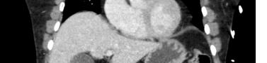
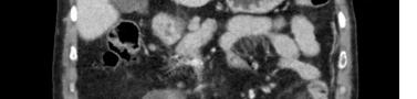
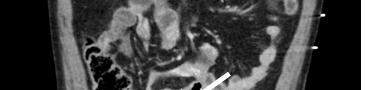
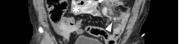

Hollow viscus or mesenteric injuries occur in approximately 1-6% of patients with blunt abdominal trauma.3 A bucket handle injury is a type of mesenteric injury in which the intestine separates from the mesentery due to shearing forces in blunt trauma, creating a devascularized segment of bowel that resembles the handle of a bucket.1 The diagnosis is often delayed and can lead to devastating consequences, including ischemic bowel, sepsis, bowel perforation, and death.1,4 Specific signs of bowel injury, such as pneumoperitoneum, extraluminal enteric contrast, or a focal wall defect are rarely seen in acute bucket handle injuries.1,2 While the eFAST is valuable for detecting free intraperitoneal fluid, it is limited in identifying mesenteric or bowel injuries, particularly in the early stages with only localized bleeding. In this patient, hypotension was likely multifactorial, related to both early septic physiology from evolving bowel ischemia and hypovolemia due to vascular injury. However, predictors of a surgically significant bowel injury—including
CPC-EM Capsule
What do we already know about this clinical entity?
A bucket handle injury is a type of mesenteric injury where the intestine separates from the mesentery due to shearing forces in blunt trauma, creating a devascularized segment of bowel.
What is the major impact of the image(s)?
This radiograph shows an uncommon pathology seen in blunt abdominal trauma.
How might this improve emergency medicine practice?
Enhancing awareness of this injury type can improve early recognition and intervention, thereby reducing morbidity and mortality rates.
active bleeding within the mesentery, interloop fluid, bowel wall perfusion defects, and traumatic abdominal wall hernias—can be seen on CT images of bucket-handle tears (Images 1 and 2).1,2 Computed tomography detection of traumatic bowel or mesenteric injuries remains challenging (sensitivity 59-95%).1 Surgical consultation is indicated for this condition; once diagnosed, bowel resection with anastomosis is usually required.2
The authors attest that their institution requires neither Institutional Review Board approval, nor patient consent for publication of this case report. Documentation on file.
Address for Correspondence: C. Eric McCoy, MD, MPH, University of California, Irvine Medical Center, Department of Emergency Medicine, 3800 W. Chapman Ave., Suite 3200, Orange, CA 92868 Email: cmccoy@hs.uci.edu.
Conflicts of Interest: By the CPC-EM article submission agreement, all authors are required to disclose all affiliations, funding sources and financial or management relationships that could be perceived as potential sources of bias. The authors disclosed none.
Copyright: © 2026 Gottam et al. This is an open access article distributed in accordance with the terms of the Creative Commons Attribution (CC BY 4.0) License. See: http://creativecommons.org/ licenses/by/4.0/
1. Extein JE, Allen BC, Shapiro ML, et al. CT findings of traumatic bucket-handle mesenteric injuries. Am J Roentgenol. Available at: https://www.ajronline.org/doi/10.2214/AJR.17.17927. Accessed August 9, 2024.
2. Sharma R. Bucket handle mesenteric injury. Radiopaedia. Published January 2, 2023. Available at: https://radiopaedia.org/articles/ bucket-handle-mesenteric-injury?lang=us. Accessed August 9, 2024.
3. Deng F, Altadill A, Silverstone L. Bucket handle mesenteric injury: radiology reference article. Radiopaedia. August 17, 2024. Available at: https://radiopaedia.org/articles/bucket-handle-mesentericinjury?lang=us. Accessed August 14, 2025.
4. What is: bucket handle injury of the intestine. The Trauma Pro. November 2, 2017. Available at: https://thetraumapro. com/2017/11/02/what-is-bucket-handle-injury-of-the-intestine/ Accessed August 14, 2025.
Luis A. Roque
Miller School of Medicine, University of Miami, Miami, Florida
Arácnido Taxonomy, Independent Researcher, Miami, Florida
Section Editor: Rick McPheeters, DO, FAAEM
Submission history: Submitted July 27, 2025; Revision received July 27, 2025; Accepted July 27, 2025
Electronically published March 10, 2026
Full text available through open access at http://escholarship.org/uc/uciem_westjem
DOI 10.5811/63007
Dear Editor,
I would like to respectfully bring to the authors’ attention a potentially significant issue concerning the identification of the spider species referenced in the published case report, “A Case Report of Delayed, Severe, Paroxysmal Muscle Cramping after Chilean Rose Tarantula (Grammostola rosea) Envenomation.” Upon close examination of the photograph included in the paper, it appears that the specimen may have been misidentified. Based on morphological features visible in the image, the spider in question bears a strong resemblance to Tliltocatl albopilosus, rather than the species currently cited.
While I understand that this article is a clinical case report and not primarily a taxonomic study, the accurate identification of the species involved is critical. Misidentification can have meaningful consequences, not only for the broader scientific record but also for the community of arachnid keepers and enthusiasts, as well as for healthcare professionals who rely on correct species identification to assess potential medical risks, treatment strategies, and prognoses in cases involving spider envenomation or exposure.
I therefore urge the authors to consult with a specialist in arachnology or seek confirmation from an authoritative taxonomic source to verify the species designation.
Addressing this matter would enhance the scientific rigor of the report and ensure its utility for both clinical and zoological reference.
Address for Correspondence: Luis A. Roque, University of Miami, Miller School of Medicine, 1450 NW 10th Ave, Miami, FL 33136, USA. Email: lroque@med.miami.edu.
Conflicts of Interest: By the CPC-EM article submission agreement, all authors are required to disclose all affiliations, funding sources and financial or management relationships that could be perceived as potential sources of bias. The authors disclosed none.
Copyright: © 2026 Roque et al. This is an open access article distributed in accordance with the terms of the Creative Commons Attribution (CC BY 4.0) License. See: http://creativecommons.org/ licenses/by/4.0/
1. Gooley BT, Hughes K, Gooley M, et al. A Case Report of Delayed, Severe, Paroxysmal Muscle Cramping after Chilean Rose Tarantula (Grammostola rosea) Envenomation. Clin Pract Cases Emerg Med. 2025 (Article in Press).
Jon B Cole, MD*
Brian T Gooley, MD*
Kirk A Hughes, RN*
Mark P Gooley, PharmD†
Daniel E Keyler, PharmD‡
Richard S Vetter, MS§
Section Editor: Lev Libet, MD
Minnesota Poison Control System, Minneapolis, Minnesota
Mayo Clinic, Rochester, Minnesota
University of Minnesota, Minneapolis, Minnesota
University of California Riverside, Riverside, California
Submission history: Submitted June 12, 2025; Revision received November 26, 2025; Accepted November 26, 2025
Electronically published March 7, 2026
Full text available through open access at http://escholarship.org/uc/uciem_cpcem
DOI: 10.5811/cpcem.48543
To the Editor:
We appreciate the author’s interest in our case report. We agree with his assertion that species identification is important in spider envenomation cases. Indeed, we sought to follow best-practice guidelines in reporting our case, as outlined in the article by Kong and Hart. Regarding the spider in our case, we took the following steps to identify the spider.
1. First, the spider was identified a priori by both the patient and the spider’s owner as Grammostola rosea
2. Second, we had additional images to help us identify the spider, not provided for conciseness of the manuscript.
3. Third, one of our co-authors, KAH, who has over 25 years of experience as a certified specialist in poison information as well as personal experience with tarantulas, agreed with the identification of the spider in the photos.
4. To ensure the identification of the spider was correct, and to ensure proper scientific context for spider envenomations, we recruited two additional authors with decades of experience as recognized international experts in envenomations, DEK and RSV, who also agreed with the species identification based on the photos. Richard Vetter, in particular, is an international expert in spider envenomation.
While we feel reasonably confident in our identification, given the feedback from this letter-writer, we sent the photo in the article to three additional experts in arachnology, who had differing opinions, and noted the spider could be Grammostola rosea as we wrote, but also Grammostola albopilosus, Tliltocatl albopilosus, or a Lasiodora species.
Given that there is some uncertainty with the identification of the spider, we agree that a limitation of this case report is that the tarantula could have been misidentified. The spider was identified by its keeper and several experts as Grammostola rosea, but also has features of Grammostola albopilosus, Tliltocatl albopilosus, and Lasiodora species. Nevertheless, emergency clinicians should remain aware of the possibility of delayed severe paroxysmal muscle cramping with tarantula envenomation from any species.
The authors attests that the patient provided consent for publication of this case report. Documentation on file.
Address for Correspondence: Jon B Cole, MD, Minnesota Poison Control System, Minnesota Regional Poison Center, 701 Park Ave, Minneapolis, MN, USA 55415. Email: jon.cole@hcmed.org.
Conflicts of Interest: By the CPC-EM article submission agreement, all authors are required to disclose all affiliations, funding sources and financial or management relationships that could be perceived as potential sources of bias. The authors disclosed none.
Copyright: © 2026 Cole et al. This is an open access article distributed in accordance with the terms of the Creative Commons Attribution (CC BY 4.0) License. See: http://creativecommons.org/ licenses/by/4.0/
1. Gooley BT, Hughes K, Gooley M, et al. A Case Report of Delayed, Severe, Paroxysmal Muscle Cramping after Chilean Rose Tarantula (Grammostola rosea) Envenomation. Clin Pract Cases Emerg Med. 2025 (Article in Press)
2. Roque LA. Letter to the editor: A Case Report of Delayed, Severe, Paroxysmal Muscle Cramping after Chilean Rose Tarantula (Grammostola rosea) Envenomation. Clin Pract Cases Emerg Med. 2025 (Article in Press)
3. Kong EL and Hart KK. Tarantula Spider Toxicity. In: StatPearls. Treasure Island (FL): StatPearls Publishing; May 1, 2023. Available at: https://www.ncbi.nlm.nih.gov/books/NBK557667/.
Vimoltip Luksanapisitakul, MD
Anas Alojayli, MD
Section Editor: Rick A. McPheeters, DO
London North West University Healthcare NHS Trust, Department of Obstetric and Gynaecology, London, Borough of Brent, United Kingdom
Submission history: Submitted November 14, 2025; Revision received November 21, 2025; Accepted December 4, 2025
Electronically published April 7, 2026
Full text available through open access at http://escholarship.org/uc/uciem_cpcem DOI: 10.5811/cpcem.53965
Abstract [Clin Pract Cases Emerg Med. 2026;10(2):225-226.]
Dear Editor:
We read with great interest “The Complexity of Weak Rhesus Positivity in Pregnancy: Challenges and Management --A Case Report,” which was published in Clinical Practice and Cases in Emergency Medicine, Volume 9 Issue 4.
We had a recent case in our maternity unit that shared similarities with the one reported above in its presentation and highlights the recurring challenges faced in clinical management in emergency contexts. A 37-year-old gravida four para three woman presented with second trimester vaginal bleeding to our hospital. During her three previous pregnancies in Canada, she had been classified as Rhesus (Rh)-positive and had not received any Rh immune globulin (RhIG) prophylaxis. In this pregnancy, in her first trimester, group and screen blood demonstrated a weak D reaction, and her Rh type was initially reported as Rh-positive. The sample was subsequently sent for further analysis in accordance with protocol, which confirmed a partial-D, and her status was later amended to Rh-negative. Identification of variant D blood type is clinically significant in obstetrical care, as these patients remain at risk of developing anti-D antibody, which can lead to the potentially severe and sometimes fatal complication of hemolytic disease of the fetus and newborn.
According to the Royal College of Obstetrics and Gynaecology Green-top Guideline No. 63: Antepartum Haemorrhage, RhIG prophylaxis should be administered to all non-sensitised Rh-negative women following any potential sensitising event, including second-trimester vaginal bleeding, and this should be given within 72 hours to minimise the risk
of alloimmunization.1 This aligns with the recommendations of the British Committee for Standards in Haematology, which advise that a maternal blood group and antibody screen should be tested to establish or confirm Rh status and to detect the presence of immune anti-D.2 The Committee also recommends that women with anomalous or indeterminate Rh typing status should be managed as Rh-D negative until definitive confirmatory testing has been completed.2
Understandably, this patient was perplexed and concerned regarding the advice to receive RhIG, particularly as she had not been offered this in her previous pregnancies. Approximately 15% of the British population are Rh-D negative, which means their red blood cells lack a protein called the Rh-D antigen on their surface.3 Although precise data on the prevalence of Rh-D variant are under-documented, estimates suggest a frequency of 0.2-1% among White populations.4 Despite the relatively low prevalence, individuals with Rh-D variant may present substantial clinical challenges, particularly when discrepant laboratory findings occur or when urgent management is required in emergency settings.
Although differing in context, both cases highlight the challenges associated with managing variant D blood groups including weak D and partial D in emergency settings. The implementation of globally standardised laboratory testing for variant D antigens is essential, as it plays a critical role in guiding the safe and effective administration of RhIG and ensuring appropriate clinical management, particularly in urgent scenarios.
The Complexity of Weak Rhesus Positivity in Pregnancy: Challenges and Management Luksanapisitakul et al.
Address for Correspondence: Vimoltip Luksanapisitakul, MD, Department of Obstetric and Gynaecology, London North West University Healthcare NHS Trust, Watford Road , London, Borough of Brent, HA1 3UJ, United Kingdom. Email: v.luksanapisitakul1@nhs.net
Conflicts of Interest: By the CPC-EM article submission agreement, all authors are required to disclose all affiliations, funding sources and financial or management relationships that could be perceived as potential sources of bias. The authors disclosed none.
Copyright: © 2026 Luksanapisitakul et al. This is an open access article distributed in accordance with the terms of the Creative Commons Attribution (CC BY 4.0) License. See: http:// creativecommons.org/licenses/by/4.0/
1. Royal College of Obstetricians and Gynaecologists (2011) Antepartum haemorrhage. Green–top Guideline No. 63. [Online]. Available from: https://www.rcog.org.uk/en/guidelines-researchservices/guidelines/gtg63/ [Accessed 8 December 2020]
2. Qureshi H, Massey E, Kirwan D, et al. BCSH guideline for the use of anti-D immunoglobulin for the prevention of haemolytic disease of the fetus and newborn. Transfus Med. 2014;24(1):8-20.
3. Booth C, Allard S, Robinson S. Blood transfusion. Medicine 2021;49(4):238-242.
4. Sandler SG, Chen LN, Flegel WA. Serological weak D phenotypes: a review and guidance for interpreting the RhD blood type using the RHD genotype. Br J Haematol. 2017;179(1):10-19.


BENEFITS
- Western Journal of Emergency Medicine Subscription -CAL/AAEM News Service email updates
-Discounted AAEM pre-conference fees -And more!
CAL/AAEM NEWS SERVICE
-Healthcare industry news
-Public policy
-Government issues
-Legal cases and court decisions