
welcome
The notion of intervention is at the very heart of the relationship between doctor and patient, serving as a bridge between illness and quality of life. It often comes in the form of a partnership that includes the patient, medical specialists and loved ones. Intervention is fundamental to receiving the exceptional care we all deserve.
In this issue of Curiosus, you’ll find intervention at work in stories about pre-cancer screening and genetic testing, prenatal surgery and research studies. You’ll also read about work we are doing with other providers in the community to care for those who don’t have easy access to treatment they need.
Compassion, an essential part of medical intervention, is built on understanding and trust. I invite you to see these qualities come to life in the story about our hospital’s language interpreters. These professionals not only help caregivers and patients communicate, they create the connection and sense of belonging that make intervention possible.

John Lynch, MD President, Barnes-Jewish Hospital
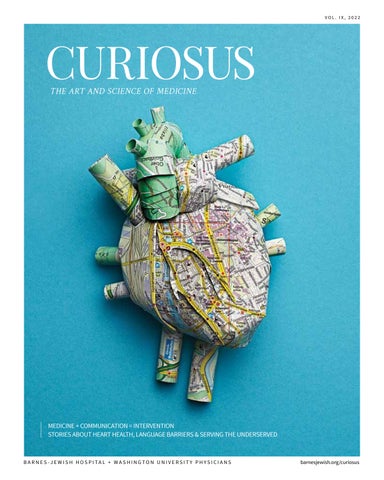
on the cover |
IN THIS ISSUE, READ STORIES THAT REVEAL THE WAYS COMMUNICATION, PREVENTION AND INTERVENTION AFFECT HEALTH AND QUALITY OF LIFE.
TABLE OF CONTENTS
kyoor-ee-OH-sus; Latin; adjective Eager to learn or know; inquisitive
4 |
6 | bench breakthroughs from the lab

RECURRENT UTIS AND THE GUT MICROBIOME
STUDY LOOKS AT DUAL NICOTINE USE
policy health care and the community
COVID-19 VACCINE BOOSTERS AND SOLID ORGAN TRANSPLANTS

8 |
RISKY DRIVING BEHAVIORS AND COMMON SLEEP DISORDERS
history milestones from the archives
18 |
A HISTORY OF HEART FAILURE— AND RECOVERY q & a interviews from the inside
12 |
FIERCE ADVOCACY: DENTAL CARE FOR THE UNDERSERVED
22 |
34 | 10 |
BRIDGING COMMUNICATION AND CULTURAL GAPS in depth featured stories
28 |
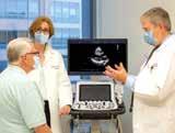
NEW DRUG, POSITIVE RESULTS FOR HYPERTROPHIC CARDIOMYOPATHY

A TWO-STEP INTERVENTION FOR LUNG CANCER
40 |
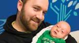
SPINA BIFIDA: MAKING REPAIRS BEFORE BIRTH
CONTRIBUTORS
ANNE MAKEEVER
The aim of medicine is to prevent disease and prolong life, the ideal of medicine is to eliminate the need of a physician. — William J. Mayo
Anne Makeever is the editor of this magazine, a poet and writer.
PAM MCGRATH
Based in St. Louis, Pam McGrath is a communications professional with 39 years of experience, the past 28 as a freelance writer and editor. Her articles for both physician and consumer audiences have appeared in magazines published by the area’s leading health-care systems, as well as its schools of medicine and nursing. Pam also is a member of Allegro: A St. Louis Choral Community and volunteers with Second Chance Ranch, a rescue dedicated to caring for homeless, neglected and sick senior dogs.
BARNES-JEWISH HOSPITAL, a nonprofit academic institution and the largest hospital in Missouri, is ranked No. 11 in the U.S., No. 1 in St. Louis and No. 1 in Missouri by U.S. News & World Report. Barnes-Jewish Hospital was the first adult hospital in Missouri to be certified as a Magnet hospital for its nursing excellence.
Barnes-Jewish Hospital is a member of BJC HealthCare, one of the largest nonprofit health-care organizations in the United States.
STEPHANE STEMMLER
Stephanie Stemmler is a St. Louis-based freelance writer with more than 35 years of experience in communications, including work as a television reporter and magazine writer, and as a public relations professional for hospitals and academic medical institutions. Always curious, she approaches story-writing as she does guitar-playing and quilt-making, by seeking unexpected and surprising insights.
WASHINGTON UNIVERSITY
SCHOOL OF MEDICINE is a leader in medical research, teaching and patient care, and is among the top recipients of research funding from the National Institutes of Health.
The 1,790 specialty and primary care clinicians who make up Washington University Physicians—the School of Medicine’s physician practice group— comprise the medical staff at Barnes-Jewish Hospital and St. Louis Children’s Hospital. Washington University Physicians also provides comprehensive medical care at multiple locations throughout the St. Louis region.
Barnes-Jewish Hospital and Washington University School of Medicine are nonprofit organizations and do not endorse commercial products or services.
Curiosus is published biannually by Barnes-Jewish Hospital.
Executive editor Michael Lourie, Barnes-Jewish Hospital
Editor-in-chief Anne Makeever, Werremeyer Creative
Contributing editor Juli Leistner, Washington University School of Medicine
Featured contributing writers
Tamara Bhandari, Jim Dryden, Pam McGrath, Kristina Sauerwein, Stephanie Stemmler Design Werremeyer Creative
MANAGE YOUR SUBSCRIPTION
Address changes | Subscribe |
Unsubscribe Send an email to: anne@werremeyer.com
WASHINGTON UNIVERSITY SCHOOL OF MEDICINE SCIENCE WRITERS
Tamara Bhandari, Jim Dryden, Kristina Sauerwein
RECURRENT UTIS AND THE GUT MICROBIOME
One of the greatest frustrations regarding urinary tract infections, also called UTIs, is that they so often recur. UTIs are caused by bacteria in the urinary tract and characterized by frequent and painful urination. A round of antibiotics usually clears up the symptoms, but the relief is often temporary: A quarter of women go on to develop a second UTI within six months. Some unfortunate individuals get UTIs over and over, and require antibiotics every few months.
A new study—published May 2 in Nature Microbiology—suggests that women who get recurrent UTIs may be caught in a vicious cycle in which antibiotics given to eradicate one infection predispose them to develop another. The study, by researchers at Washington University School of Medicine and the Broad Institute of MIT and Harvard, showed that a round of antibiotics eliminates disease-causing bacteria from the bladder but not from the intestines. Surviving bacteria in the gut can multiply and spread to the bladder again, causing another UTI.
At the same time, repeated cycles of antibiotics wreak havoc on the community of helpful bacteria that normally live in the intestines, the so-called gut microbiome. Women in the study with recurrent UTIs had less diverse microbiomes, and those microbiomes were deficient in an important group of bacteria that helps regulate inflammation. These study participants also had a distinct immunological signature in their blood indicative of inflammation.
“It’s frustrating for women who are coming in to the doctor with recurrence after recurrence after recurrence, and the doctor, who’s typically male, gives them advice about hygiene,” says co-senior author and Washington University researcher Scott Hultgren, PhD. “That’s not necessarily what the problem is. It’s not necessarily poor hygiene that’s causing this. The problem lies in the disease itself, in this connection between the gut and the bladder and levels of inflammation. Basically,
physicians don’t know what to do with recurrent UTI. All they have is antibiotics, so they throw more antibiotics at the problem, which probably just makes things worse.”
Most UTIs are caused by Escherichia coli (E. coli) bacteria from the intestines that get into the urinary tract. To understand why some women get infection after infection and others get one or none, Hultgren teamed up with Broad Institute scientists Ashlee Earl, PhD, the senior group leader for the Bacterial Genomics Group at Broad and the paper’s co-senior author, and Colin Worby, PhD, a computational biologist and the paper’s lead author.
The researchers studied 15 women with histories of recurrent UTIs and 16 women without. All participants provided urine and blood samples at the start of the study and monthly stool samples. The team analyzed the bacterial composition in the stool samples, tested the urine for the presence of bacteria, and measured gene expression in blood samples.
Over the course of a year, 24 UTIs occurred, all in participants with histories of repeated UTIs. When participants were diagnosed with a UTI, the team took additional urine, blood and stool samples.
The difference between the women who got repeated UTIs and those who didn’t, surprisingly, didn’t come down to the kind of E. coli in their intestines or even the presence of E. coli in their bladders. Both groups carried E. coli strains in
OUR STUDY CLEARLY DEMONSTRATES THAT ANTIBIOTICS DO NOT PREVENT FUTURE INFECTIONS OR CLEAR UTI-CAUSING STRAINS
FROM THE GUT, AND THEY MAY EVEN MAKE RECURRENCE MORE LIKELY BY KEEPING THE MICROBIOME IN A
DISRUPTED STATE.
— COLIN WORBY, PHD, COMPUTATIONAL BIOLOGIST, BROAD INSTITUTE
their guts capable of causing UTIs, and such strains occasionally spread to their bladders.
” “
The real difference was in the makeup of their gut microbiomes. Patients with repeat infections showed decreased diversity of healthy gut microbial species, which could provide more opportunities for disease-causing species to gain a foothold and multiply. Notably, the microbiomes of women with recurrent UTIs were particularly scarce in bacteria that produce butyrate, a short-chain fatty acid with anti-inflammatory effects.
“We think that women in the control group were able to clear the bacteria from their bladders before they caused disease, and women with recurrent UTI were not, because of a distinct immune response to bacterial invasion of the bladder potentially mediated by the gut microbiome,” Worby says.
The findings highlight the importance of finding alternatives to antibiotics for treating UTIs.
“Our study clearly demonstrates that antibiotics do not prevent future infections or clear UTI-causing strains from the gut, and they may even make recurrence more likely by keeping the microbiome in a disrupted state,” Worby notes.
Hultgren has long worked on finding innovative therapies to eradicate disease-causing strains of E. coli from the body while sparing the rest of
the bacterial community. His research forms the basis of an experimental drug based on the sugar mannoside and an investigational vaccine, both of which are being tested in people. Another strategy would be to rebalance the microbiome through fecal transplants, probiotic foods or other means.
“This is one of the most common infections in the United States, if not the world,” Hultgren says. “A good percentage of these UTI patients go on to get these chronic recurrences, and it results in decreased quality of life. There is a real need to develop better therapeutics that break this vicious cycle.”
— TAMARA BHANDARI
Originally published by Washington University School of Medicine at medicine.wustl.edu/news
Find this story and others online: barnesjewish.org/curiosus
Image courtesy of Shutterstock
COVID-19 VACCINE BOOSTERS AND SOLID ORGAN TRANSPLANTS
People who undergo solid organ transplants are at high risk of developing severe COVID-19 infections due to their need to take immunosuppressive medications. These medications, aimed at preventing the body from rejecting a transplanted organ, have the unintended consequence of making immune systems more open to the virus that causes COVID-19.
A new multicenter study—published online in The Journal of Infectious Diseases—has identified a strategy to lessen that risk. It shows that transplant recipients who receive three doses of a COVID-19 mRNA vaccine
have greater protection than that provided with two doses. (The Pfizer-BioNTech and Moderna vaccines are mRNA.) The study, by researchers at Washington University School of Medicine, the Centers for Disease
Image courtesy of Shutterstock
“
OUR RESEARCH ILLUSTRATES THAT BOOSTER VACCINE DOSES ARE PARTICULARLY IMPORTANT FOR IMMUNOSUPPRESSED PEOPLE.
— JENNIE KWON, DO, MSCI, INFECTIOUS DISEASES SPECIALIST
”
Control and Prevention (CDC), and Vanderbilt University Medical Center, examined 10,425 patients hospitalized across 21 hospitals in the U.S. Of those, 440 had solid organ transplants, 1,684 had immunocompromising conditions, and 8,301 had healthy immune systems. The findings showed that a regimen of two doses of the mRNA vaccine was 29% effective at preventing COVID-19 hospitalizations among transplant patients, while a three-dose regimen was 77% effective. (Solid organ transplants are defined as transplants of the kidney, liver, intestines, heart, lung or pancreas.)
Currently, three doses are widely adopted for people who have had a solid organ transplant. With increasing evidence of the benefits of additional vaccine doses, the CDC now recommends a fourth dose of the mRNA vaccine for people with moderate to severe immunocompromising conditions, including patients who have undergone solid organ transplants.
“Our research confirms that two doses of a SARS-CoV-2 mRNA vaccine do not provide as much protection against hospitalization in solid organ transplant recipients as compared to those with healthy immune systems,” says Jennie Kwon, DO, MSCI, one of the study’s first co-authors and a Washington University infectious diseases specialist. “But the good news is that additional vaccine doses appear to significantly increase effectiveness for solid organ transplant recipients.” Kwon also is a health-care epidemiologist at Barnes-Jewish Hospital, where she treats patients.
“The study’s results indicate that solid organ transplant recipients benefit from three doses of mRNA COVID-19 vaccines and support the CDC’s recommendations for this vulnerable population,” she says.
Wesley Self, MD, one of the study’s senior co-authors and an emergency medicine specialist at Vanderbilt, notes: “We believe these results demonstrate that solid organ transplant recipients remain at risk for COVID-19 despite vaccination and support the need for continued efforts to mitigate the risk of COVID in this population.”
“Throughout the COVID-19 pandemic, there has been a concern that immunocompromised people, such as those with a solid organ transplant, may not benefit from vaccination as much as immunocompetent people,” Self says. “However, our research illustrates that booster vaccine doses are particularly important for immunosuppressed people.”
The study also points to other measures to reduce the risk of COVID-19 among solid organ transplant recipients, including vaccination of close contacts, individual immune system monitoring and infectionprevention strategies, including wearing a mask in public spaces and social distancing.
— KRISTINA SAUERWEIN
Originally published by Washington University School of Medicine at medicine.wustl.edu/news
Find this story and others online: barnesjewish.org/curiosus
The CDC now recommends a fourth dose of the mRNA vaccine for people with moderate to severe immunocompromising conditions.
RISKY DRIVING BEHAVIORS AND COMMON SLEEP DISORDERS
People with sleep apnea wake up tired in the morning, no matter how many hours they actually sleep. The condition causes them to briefly stop and restart breathing dozens or even hundreds of times a night. Even though such breathing interruptions often don’t awaken those with apnea, they prevent them from sinking into deep, refreshing sleep.
A new study puts a number on how dangerous such chronic tiredness can be, at least in regard to driving. For every eight additional breathing interruptions per hour, the odds of making a dangerous driving move such as speeding, braking hard or suddenly accelerating increase by 27%, according to a study by researchers at Washington University School of Medicine.
Older adults are more likely to develop sleep apnea. They also are more likely to be seriously injured or killed in a car accident. The study’s findings, available online in the journal Sleep, suggest that screening older adults for sleep apnea and for treatment, if needed, may help older people continue driving safely for longer.
“The percentage of older adults with mild sleep apnea is 30% to 50%, but if such adults don’t have daytime sleepiness or other evidence of impairment, they may not come to medical attention,” says the study’s co-senior author Brendan Lucey, MD, Washington University neurologist at Barnes-Jewish Hospital and director of Washington University’s Sleep Medicine Center. “However, these findings suggest that we might want a lower threshold to evaluate older adults for sleep apnea and track their breathing interruptions. If their conditions worsen by just eight interruptions an hour, that could have significant adverse effects on their driving and their risk of suffering serious injury.”
People 65 and over are the most responsible drivers on the road. They obey speed limits.
They drive defensively. They avoid driving at night, in bad weather and in unfamiliar places. But the changes that often come with advancing age—such as deteriorating vision, slower reflexes and, yes, difficulty sleeping—can undermine even the safest habits.
Lucey teamed up with driving researcher Ganesh Babulal, PhD, OTD, Washington University neurology scientist and co-senior author of the study, to investigate the relationship between sleep apnea and risky driving behaviors. Participants were recruited from ongoing studies at Washington University’s Charles F. and Joanne Knight Alzheimer Disease Research Center, also known as the Knight ADRC.
Babulal and Lucey monitored the driving and sleep habits of 96 older adults under real-world conditions. They used a commercially available take-home test to identify people with sleep apnea and measured its severity. Less than five breathing interruptions per hour is considered normal, five to 15 is mild sleep apnea, 15 to 30 is moderate, and greater than 30 is severe.
To assess driving habits, the researchers installed a chip developed by Babulal and colleagues into participants’ personal vehicles and monitored their driving for a year, focusing on episodes of hard braking, sudden acceleration and speeding. In total, they collected data on more than 100,000 trips. Participants also were evaluated by researchers at the Knight ADRC for cognitive impairments and molecular signs of early Alzheimer’s disease.

Even though all participants were cognitively normal, about a third had brain changes indicative of early Alzheimer’s disease. The researchers found that the frequency with which drivers made dangerous moves behind the wheel rose in parallel with the frequency with which their sleep was interrupted at night, regardless of whether their brains bore the marks of early Alzheimer’s.
“We didn’t have cameras in the vehicles, so we don’t know exactly what happened that caused someone to, say, brake hard suddenly,” Babulal says. “But it could be something like a stoplight that they didn’t realize was red until they got close and had to stomp on the brakes. The more tired you are, the less attention you have to deploy to the task at hand, especially if it is novel and constantly changing.”
The study helps untangle the ways aging-associated risk factors such as poor sleep and Alzheimer’s disease put older adults in danger while driving,
and it could aid efforts to find ways to maximize years of safe driving, the researchers say.
“Driving always carries the risk of crashing, and older adults are at risk of more severe injury than younger adults if they experience a crash,” Babulal says. “But we can’t just tell them to give up their keys. When older people stop driving, they lose a lot of their independence and mobility, which is often associated with negative health and social outcomes. What we want to understand is what puts them at a higher risk so we can intervene and help them stay behind the wheel, safely, for as long as possible.”
— TAMARA BHANDARI
Originally published by Washington University School of Medicine at medicine.wustl.edu/news
Find this story and others online: barnesjewish.org/curiosus
CHANGES THAT OFTEN COME WITH ADVANCING AGE, INCLUDING SLEEP APNEA, CAN UNDERMINE EVEN THE SAFEST DRIVING HABITS.
Photo courtesy of Shutterstock
STUDY LOOKS AT DUAL NICOTINE USE
Most of the 40 million Americans who smoke cigarettes say they want to quit, and some move to e-cigarettes as a step toward quitting. However, a growing number of such people become dual nicotine users: They smoke traditional cigarettes and vape e-cigarettes, researchers at Washington University School of Medicine have found.
Rather than discontinuing their addiction, many end up substantially increasing the amount of nicotine they consume. The researchers found, however, that smoking-cessation treatments approved by the Food and Drug Administration (FDA) that focus on nicotine replacement and counseling can help dual users quit. In fact, such treatments seem to be effective in dual users as well as those who smoke traditional cigarettes exclusively. These findings were published in the journal Thorax.
“We recommend FDA-approved treatment such as nicotine replacement, the drug varenicline and counseling for cigarette smokers,” says Li-Shiun Chen, MD, MPH, ScD, psychiatrist at Siteman Cancer Center at Barnes-Jewish Hospital and Washington University School of Medicine, and senior investigator of the study. “But we know that a growing number of people are using both cigarettes and e-cigarettes. The good news is that tobacco treatment still can help these people stop smoking and defeat their nicotine addiction.”

Photo courtesy of Shutterstock
“
WE RECOMMEND FDA-APPROVED TREATMENT SUCH AS NICOTINE REPLACEMENT, THE DRUG VARENICLINE AND COUNSELING FOR CIGARETTE SMOKERS.
— LI-SHIUN CHEN, MD, MPH, SCD, PSYCHIATRIST
In an average year, about 70% of those who smoke will make an attempt to quit. Another 20% say they want to smoke less. But without treatment, such as nicotine replacement therapy and counseling, only about 5% quit successfully.
For the study, Chen and colleagues reviewed de-identified electronic health records of more than 110,000 smokers who were seen in outpatient clinics at Barnes-Jewish Hospital between 2018 and 2020. They found that the number of e-cigarette users tripled. Some apparently started vaping as a potential step to quitting, but many seemed to get stuck. Instead of quitting, they started vaping while also continuing to smoke traditional cigarettes.
A relatively small but growing percentage of the smokers who were studied became dual users during the course of the study. Some 0.8% reported dual use in the study’s first year, but that number had grown to 2.3% when data collection had concluded. The authors noted that the actual number of dual users was likely higher.
The researchers found that about one in five (20.8%) of the dual users quit smoking within 12 months. That compared to a 16.8% quit rate among those who smoked only traditional cigarettes.
Treatment still seemed to be the key for dual users. When they received smoking-cessation treatment, almost one-third (29%) became nonsmokers
12 months later. About 17% of dual users who didn’t get treatment were able to quit.
The study’s first author, Brendan Heiden, MD, a surgical resident and cardiothoracic surgery research fellow, says that although quit rates were higher among dual users, about two-thirds remained smokers a year later, even after treatment. That, combined with the fact that little is known about the long-term health effects of e-cigarette use, means that he and Chen don’t recommend that people who want to quit smoking should start vaping.
“The current scientific consensus is that using both cigarettes and e-cigarettes is bad for you,” Heiden says. “Although current guidelines do not recommend vaping for smoking cessation, we did find that among the growing number of people who use both of these products, traditional FDA-approved tobacco treatment—such as nicotine-replacement therapy and behavioral support therapy—can help them quit.”
— JIM DRYDEN
Originally published by Washington University School of Medicine at medicine.wustl.edu/news
Find this story and others online: barnesjewish.org/curiosus
policy | health care and the community
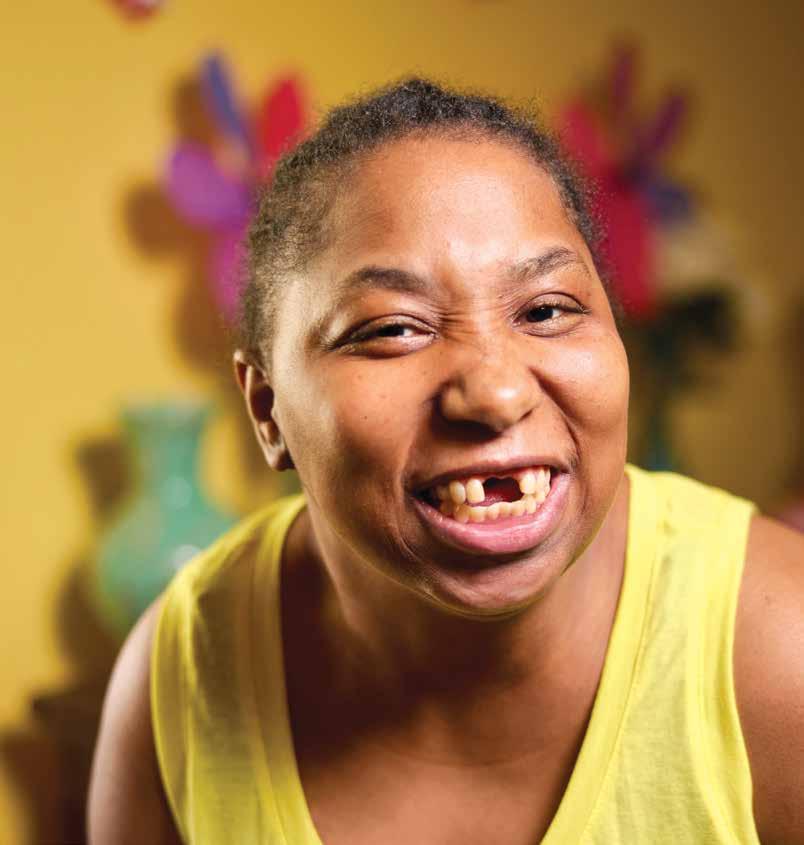
CASSANDRA HOLLAND WAS THE FIRST TO RECEIVE TREATMENT FROM A DENTISTRY PROGRAM DESIGNED TO HELP PEOPLE WITH SPECIAL NEEDS.

Brushing. Flossing. Regular dental checkups. Many of us take these actions for granted to keep our teeth healthy and our smiles bright and intact. But what if you have a medical complication that makes it difficult, if not impossible, to receive routine dental services?
“We fall through the cracks,” says St. Louis resident Sallye Holland. “And that has kept my daughter in pain for years and cost her almost half her teeth.”
FIERCE ADVOCACY: dental care for the underserved
BY STEPHANIE STEMMLER
BY STEPHANIE STEMMLER
Photo by Matt Miller
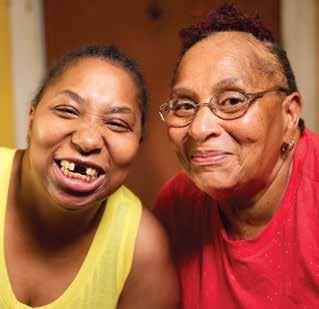
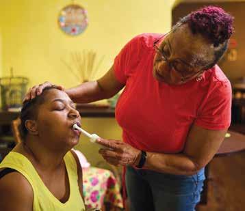
Holland’s daughter, Cassandra, 42, was diagnosed with cerebral palsy and an accompanying seizure disorder at birth. Her muscle spasticity means that she might suddenly move and bite down hard, creating a risk for any dentist who tries to treat her. Beyond trying to brush Cassandra’s teeth at home, Holland was left scrambling whenever serious dental concerns arose.
“For years, I kept a folder of phone numbers that I would call to try to get a dental appointment,” says Holland. “Every dentist I called was just not equipped to handle a patient like Cassandra. The closest place was probably Kansas City, and I couldn’t get there.”
Over time, Cassandra developed abscesses and infections in her mouth. Her teeth, decaying one by one, turned black. She was in daily pain.
“I even brought her to a hospital emergency room, but the only thing the doctors there could do was give her medication to temporarily stop the pain,” says Holland. “They couldn’t do anything to help with the actual problem because hospitals don’t do dental procedures.”
Addressing the need
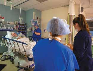
But now, at Barnes-Jewish Hospital, a team of specialists does just that. Thanks to a unique partnership forged by four health-care groups, people with conditions that had once made standard dental care impossible now can receive the care they need through the High Acuity Adult Special Needs Dental Program. Twice a month at the hospital, a collaborative team of specialists uses an operating room reserved for people with special needs. Before the work begins, general anesthesia is administered, which makes treatment possible.
“People weren’t getting the care they required,” says Jackie Martin, MD, MBA, vice president of perioperative services at Barnes-Jewish Hospital, “and if we weren’t able to help, then who? We’re here to serve the community, so we formed a team that aligns around a common vision.”
That vision—making essential dental care available to those without—is shared by all involved in the partnership: Affinia Healthcare, A.T. Still University –Missouri School of Dentistry and Oral Health, Barnes-Jewish Hospital and Washington University
Photos by Matt Miller
CASSANDRA HOLLAND AND HER MOTHER, SALLYE HOLLAND
IF NOT US, THEN WHO? NO ONE ELSE IS DOING THIS.
— JACKIE MARTIN, MD, MBA , VICE PRESIDENT OF PERIOPERATIVE SERVICES, BARNES-JEWISH HOSPITAL
School of Medicine. These four teams worked through the inherent complexities in any such collaboration over the course of three years, then debuted the program as a pilot project in October 2021.
Two months after start-up, the team declared it a success and, in January 2022, the High Acuity Adult Special Needs Dental Program became part of the official operating-room schedule at Barnes-Jewish Hospital.
“I believe this is the most significant relationship of its kind in the country,” says Alan Freeman, DMgt, FACHE, president and chief executive officer of Affinia Healthcare, a federally qualified health-care provider in St. Louis. “It’s a landmark achievement.”
A closer look at the problem
Before the high-acuity dental program was founded, administrators at Barnes-Jewish Hospital were aware of an increase in the number of patients seeking emergency dental services in its emergency department. Some of these people had special needs and were seeking care they couldn’t find elsewhere; others had limited access to dental care and, often, no dental insurance.
The Missouri Department of Health and Human Services noted in its 2020 Oral Health in Missouri report that in 2017 there were more than 56,000 dental-related visits to hospital emergency departments across the state. But an emergency room is not equipped to provide this kind of care, Martin notes, and patients with dental problems often are treated with short-term pain relief and then referred to a dental clinic.
” “
Many of the those who arrive at an emergency room looking for dental care are there because they don’t have access to a dentist—because they lack dental insurance or don’t have a dental office nearby, or both. In fact, the American Dental Association notes that Missouri has one of the highest levels of unmet need in the country. For people with special needs, the difficulty in accessing dental care is further complicated: The majority of dentists practicing in the state are not equipped to care for those with multiple medical, behavioral and physical complications.
The partners
Before their collaboration, the entities that formed the high-acuity program were working to offer dental services to those who, for a variety of reasons, don’t have easy access to care.
Dwight McLeod, DDS, MS, dean of the A.T. Still –Missouri School of Dentistry, notes that the school was founded in 2013 to help meet the needs of the underserved. Two years after it opened its doors to students, it partnered with Affinia Healthcare— the largest community health center in Missouri— to create the St. Louis Dental Education and Oral Health Center, also known as the St. Louis Dental Center. A.T. Still students spend the last two years of their dental education working at this clinic, which offers affordable dental services for people of all ages.
Before collaborating with A.T. Still to open the dental center, Affinia was working to meet the need by operating dental services clinics in the region— and providing services through a mobile dental clinic to children in under-resourced areas.
Despite these efforts, the number of dental-related visits to emergency rooms indicated the need for more intervention. In response, the Centene Charitable Foundation, working with A.T. Still and Affinia, funded the creation of the Centene Charitable Foundation Urgent Dental Care Center in 2017. This center treats people who don’t have dental insurance. In 2018, Barnes-Jewish Hospital made a $1.5 million grant over five years to A.T. Still to support expansion of the Urgent Dental Care Center’s hours. To date, the center has treated more than 20,000 people.
Additionally, the St. Louis Dental Center established a special-needs dentistry clinic within its existing program. It was designed to treat people with intellectual, physical and behavioral differences that make standard dental care difficult. “We offer these patients teeth cleanings, sealants, fillings, crowns, bridges—even oral surgery,” says Robert Schmidt, DDS, MS, former director , Special Care UnitSpecial Needs Dentistry, and a professor of clinical education. “But some high-acuity patients can’t or won’t open their mouths or sit down.” These patients, he notes, “need to be fully sedated during procedures, an option the St. Louis Dental Center can’t provide.”
The High Acuity Adult Special Needs Dental Program was created to help solve that specific problem.
A tailor-made solution
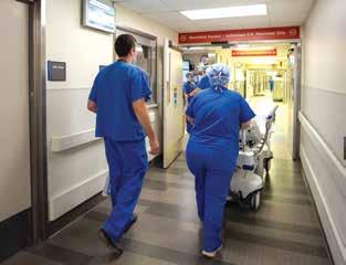
General anesthesia—and the safety protocols it requires—make dental procedures possible for people who otherwise couldn’t receive care.
Beginning in 2019, Washington University and Barnes-Jewish Hospital administrators met regularly with A.T. Still and Affinia partners to develop protocols for dental procedures to be performed in the hospital’s operating room. “We use the same safety procedures and quality-of-care measures for all our patients, whether they are being treated in the OR for dental problems or needing some other procedure,” says Washington University anesthesiologist Ivan Kangrga, MD, PhD, one of the specialists involved in establishing the program.
That commitment to safety means that all dentists, oral surgeons and dental assistants treating patients in a Barnes-Jewish operating room have received credentials from the Washington University School of Medicine’s Department of Otolaryngology. To further ensure and maintain quality, all dental and X-ray equipment was purchased by the hospital and donated to the program.
“We left no stone unturned,” says Kangrga. “Equipment, pharmacy services, patient transport,
Photos by Gregg Goldman
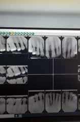
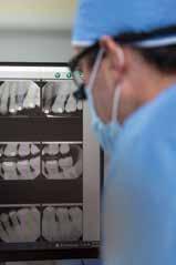
THE HIGH ACUITY ADULT SPECIAL NEEDS DENTAL PROGRAM TREATS PATIENTS IN AN OPERATING ROOM AT BARNES-JEWISH HOSPITAL.
training of certified nurse anesthetists, emergency contingencies: We carefully considered each aspect of the program before the first patient was scheduled.”
How the program works
People requiring high-acuity dental care—and general anesthesia to receive that care—are first evaluated at the St. Louis Dental Center; preadmission paperwork is completed at Barnes-Jewish Hospital. The hospital provides a dedicated operating room two days each month to help make scheduling easier. “We’re a busy hospital,” says Kangrga, “so having an established schedule helps ensure we meet the ongoing need.”
Martin adds: “Every person we treat through this program has value. Every partner supporting the program is committed to making our patients’ needs a priority.”
Schmidt notes that, currently, the High Acuity Adult Special Needs Dental Program has nearly 100 people on its waiting list. He adds: “By the time those patients receive treatment, we’ll have added 100 more to the list.”
Treating the first patient
“I remember it well,” says Schmidt, referring to the day Cassandra Holland, the first person treated
within the high-acuity program, arrived at Barnes-Jewish Hospital for dental care. “We didn’t know what to expect because we weren’t able to examine her until after sedation.”
For Holland, treatment required 13 extractions and nine fillings. But the team managed to eliminate the source of her pain—and preserve much of her smile.
“I was actually surprised she didn’t lose more of her teeth,” recalls Sallye Holland. After the procedure was done, she says, “I was singing everyone’s praises and thanking the Lord because my daughter was no longer suffering like she had been for years.”
Freeman sums up the effort to develop the highacuity dental program this way: “Fierce advocacy recognizes that some people have less than others. Collaborative efforts by willing partners can make a difference. This program is a superb example of fierce advocacy by everyone involved.” Y
For more information about the High Acuity Adult Special Needs Dental Program, including payment and insurance options, contact Affinia Healthcare at 314-833-2700.
Find this story and others online: barnesjewish.org/curiosus
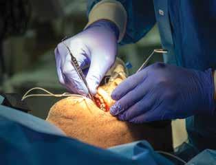
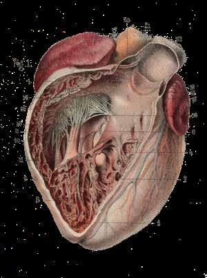
Illustration of the heart by French anatomists
Constantin Bonamy and Paul Broca, circa 1866
Illustration courtesy of Science Photo Library
A history of heart failure— and recovery
BY PAM MCGRATH
Just a few hundred years ago, the heart and its function were a bit of a mystery. And when the heart malfunctioned, treatment options were few and often ineffective. Today’s cardiologists offer their patients treatments that can lead to recovery.
To date, an Egyptian man named Nebiri, also known as Chief of Stables, is the oldest-known case of heart failure. Thanks to a German pathologist’s examination of 3,500-year-old mummified remains, it’s believed Nebiri died of pulmonary edema caused by heart failure.
In Nebiri’s day, physicians practicing medicine in Egypt, Greece, China and India recorded symptoms of heart failure that are similar to those we recognize today—but they didn’t have an accurate understanding of how the heart functioned and malfunctioned.
It was then understood that the heart served as a pump, contracting as it worked. But it was believed that the body’s arteries were filled with air and its veins with blood. The heart’s job was to pump air throughout the body, thereby distributing heat.
It wasn’t until the 17th century, when an English physician named William Harvey began studying the heart, that the organ’s true functions were more fully understood. Harvey’s work correctly identified the heart’s functions in relation to the body’s circulatory system.
And his observation that a dilated ventricle could cause the heart to fail was the first glimmer of an understanding of heart failure.
In the late 18th century, William Withering, another British physician, noted that a patient with dropsy— what we now call edema—greatly improved after taking a traditional herbal remedy whose active ingredient came from the foxglove plant (of the genus Digitalis). Withering’s work led to the development of a drug called digoxin or digitalis, which was used to treat heart failure into the 1980s.
When breakthrough medical tools—including X-ray technology, the stethoscope and electrocardiography— were developed in the 19th century, they proved effective in furthering heart-failure research. By the early 1900s, physicians knew that diuretics could reduce fluid buildup in the body—a contributing factor in heart failure. In the 1940s to 1960s, heart specialists refined techniques in cardiac catheterization—a procedure to clear blocked arteries—and other heart surgeries. The first human heart transplant happened in 1967.
Then in the 1970s, says Gregory Ewald, MD, Washington University cardiologist at Barnes-Jewish Hospital, medical science gained further insight into heart failure, its causes, effects and treatment.
Ewald also is director of the cardiovascular division’s section of heart failure and cardiac transplantation at Washington University School of Medicine.
It was in that decade, Ewald notes, that medicine developed an important understanding of the ways pre-existing conditions can cause heart dysfunction.
“And when the heart isn’t working effectively, the body may try to compensate. That effort may work for a while but, ultimately,” Ewald says, “the result is heart failure.”
Causes and symptoms of heart failure
Contemporary medicine has defined a number of conditions that can cause the heart to fail. Among them are:
> coronary artery disease and heart attack
> high blood pressure
> faulty heart valves
> damage to and inflammation of the heart muscle
> congenital heart defects
> abnormal heart rhythms
> other diseases, including diabetes, HIV and an over- or underactive thyroid
“Any of these can result in a heart that doesn’t efficiently supply blood to the body,” says Ewald. “That inefficiency produces a variety of symptoms that signal heart failure.”
The symptoms of heart failure include:
> shortness of breath during activity or when lying down
> exercise intolerance
> fatigue and weakness
> swelling in the legs, ankles and feet
> chest pain (when caused by a heart attack)
> rapid or irregular heartbeat
“Sometimes people think their allergies are acting up or that they have pneumonia or bronchitis, because those conditions can cause symptoms that are similar to those of heart failure,” says Ewald. “Fortunately, we can offer patients a number of tests that provide a definitive diagnosis.”
Diagnosis and treatment
To learn about the heart’s health and ability to function, a cardiologist may order one or more tests. An ultrasound can be used to reveal the heart’s activities and show blood flow patterns. An angiogram or cardiac catheterization can help a specialist understand how blood is flowing through the heart, arteries and veins. A cardiac MRI produces detailed images of the beating heart. And a blood test can reveal an elevated level of natriuretic peptide, an indication of heart failure.
“At the Washington University Heart Failure Center at Barnes-Jewish Hospital, our method of diagnosing encompasses a constellation of factors in addition to testing, including how the person appears during a physical exam and the overall medical history,” says Ewald. “We now recognize that some cardiomyopathies—chronic diseases of the
heart muscle—are genetic in nature, so we also take a thorough family medical history.”
After diagnosis
Ewald admits that if a newly diagnosed patient Googles the term “heart failure,” the results can be scary. He notes that some research shows that in a large population of people with heart failure, the five-year mortality rate approaches 30%-50%.
The American Heart Association estimated the prevalence of heart failure in the United States to be 6 million individuals in 2021, with approximately 960,000 new cases diagnosed each year. “The really good news, however, is that over the past 30 to 40 years, excellent studies have defined medication therapies that can significantly improve heart function for some.”
Heart failure can cause some people to have a low ejection fraction, which means the heart is not pumping an adequate amount of blood out of its lower chambers. Treatment guidelines for this condition include use of a combination of drugs, as well as a diuretic to help prevent edema.
“As heart failure specialists, my colleagues and I focus on determining what is best for the individual. That means adjusting dosages until we are sure the medications are effective,” Ewald says. And, when some of the standard drugs aren’t safe or effective for a particular patient, “we develop a drug regimen they can tolerate.”
Washington University cardiologists participate in the Heart Failure Clinical Network, a national, multidisciplinary community of investigators engaged in clinical research aimed at understanding and treating the condition. Ewald notes that his team’s association with this network helps offer patients the possibility of enrolling in ongoing trials of new drugs, some of which may prove more effective than those in current use. “This option gives our patients a distinct advantage over many other heart-failure programs.”
For some people with heart failure, medical management is not enough to protect them from
the effects of irregular heartbeats called cardiac arrhythmias. In these cases, implantable devices can help.
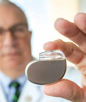
Ewald says a pacemaker is one such option. This small, battery-operated device is implanted in the chest, where it helps the heart maintain a regular rhythm. Other implantable devices include the cardiac defibrillator, which monitors heart rate and delivers an electrical shock to restore a heartbeat to normal in the case of tachycardia—a heart rate of more than 100 beats a minute.
Another option is a recently developed implantable device that can be used to continually track the heart’s rhythms. “The device sends readings via Bluetooth technology to our Heart Failure Center, allowing us to remotely keep constant watch while people go about their daily lives,” says Ewald.
For those with severe heart failure, a left ventricular assist device, or LVAD, can be implanted. This small, rotary pump is attached to the heart to help move blood from the lower-left heart chamber to the rest of the body.
“Implanting an LVAD is an open-heart procedure,” Ewald says. “However, for patients whose conditions aren’t improved with medications or heart-pacing devices, LVADs can provide support.” In some cases, an LVAD sustains life during the wait for heart transplantation. And for those who are ineligible for transplant, Ewald says, “this device can extend their lives, sometimes for many years.”
Washington University cardiologists have enrolled a large number of their patients in clinical trials for LVADs, pacemakers and implantable defibrillators. “Trials like these are another opportunity for us to offer our patients access to the latest technological advances for heart failure treatment,” Ewald says.
In some cases, the best option is heart transplantation. “The surgical techniques for heart transplantation are well established,” Ewald says. And in recent years, significant advances have been made in managing potential rejection of the new heart and
other post-transplant complications. Ewald notes that, thanks to surgical and post-surgical expertise, the Washington University and Barnes-Jewish Transplant Center offers survival rates that surpass national statistics.
People with heart failure can also do a lot to help themselves, Ewald says, by changing aspects of their lifestyles. “If they smoke, they need to stop. If they don’t exercise, they need to start.”
From 2003 to 2007, Washington University researchers participated in the largest exercise trial to date, called HF-ACTION, which enrolled more than 2,000 people with heart failure in the U.S., Canada and France. “We found that regular exercise positively impacts heart-failure symptoms—plus, people feel better and their quality of life improves,” Ewald says. In fact, he notes, these findings influenced the Centers for Medicare and Medicaid Services to offer insurance coverage for cardiac rehabilitation to patients with heart failure.
Life after heart failure
Ewald says he and his colleagues at the Heart Failure Center find it deeply rewarding to help people who, at risk of dying from heart failure, make a full recovery after having an LVAD implanted or receiving a heart transplant.
“These are the people who tell us about the grandkids who have been born since they underwent their procedures—children they otherwise never would have met,” he says.
Equally rewarding is watching people improve after taking one of the latest drug therapies. “The implantable devices and heart transplantation are the ‘cool’ treatments that receive a lot of attention,” says Ewald. “But many more lives are impacted by medical management. Today’s advanced drug therapy can improve heart function so dramatically that patients seemingly destined to receive a heart transplant never reach that point. Instead, they lead active, fulfilling lives by taking four pills a day.” Y
Find this story and others online: barnesjewish.org/curiosus
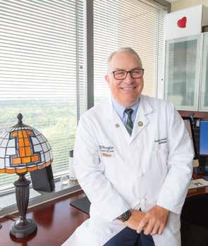
Cardiologist Gregory Ewald, MD; Ewald with a pacemaker device
Photos by Gregg Goldman
Image courtesy of Shutterstock
Bridging communication and cultural gaps
BY PAM MCGRATH | PHOTOS BY GREGG GOLDMAN
Of the nearly 2.81 million people living in the St. Louis metropolitan area in 2019-2021, an estimated 140,000 came from a different country to make St. Louis home. Their nations of origin are spread across the globe: North and South America, the Caribbean, Africa, Asia’s five regions, and Europe (east, west, south and central). The greatest number of foreign-born St. Louisans are from India, Mexico, China, Bosnia and Herzegovina, Vietnam, the Philippines, South Korea, Germany, the United Kingdom and Canada.
The first languages of many of these individuals are myriad. Upon arrival, many don’t speak fluent English, if any at all. Their cultures and backgrounds vary widely. What they have in common, however, is their need for health care.
BJC HealthCare’s Language Services, a department within the BJC Office of Diversity, Equity and Inclusion, provides free, 24/7 access to interpreters for patients and their family members who need help with spoken language or with sign-language communication. In the interview that follows, you’ll meet the service’s director, Christopher Fan, MSW, MPH, MBA, who leads a core staff of interpreters that services the Washington University Medical Campus and Barnes-Jewish Hospital. You’ll also meet Pilar Kellogg, MAIA, and Brandi Evans, BS/ASLEI, NIC, BEI Master, two interpreters who specialize in, respectively, Spanish and American Sign Language,
or ASL. I spoke with them recently about the ways interpreter services can bridge communication and cultural gaps that may prevent immigrants, refugees, the deaf/hard-of-hearing community and blind individuals from accessing essential medical services.
Q: Christopher, can you provide some background on BJC Language Services?
During the 1960s and ’70s, St. Louis experienced a first wave of immigrants and refugees from Eastern Europe, predominantly people from what was then the Soviet Union. When these immigrants required health services, it became clear that the language gap between them and their health-care providers was a critical barrier to good outcomes. That’s when Interpreter Services’ precursor, Refugee and Immigrant Services, was established at Barnes-Jewish Hospital.

Q: How has the service changed over time?
The federal government views St. Louis as a desirable location for refugees, and political leaders in both city and county governments are supportive of this assessment because diversity brings energy to the cultural and economic growth of the region. Examples of that benefit include the revitalization of the Bevo Mill area by Bosnian immigrants and the Hispanic business district that has flourished along Cherokee Street in south St. Louis.
In 2006, the original service became Language Services, offering expanded capabilities. Now a part of BJC HealthCare, a nonprofit health-care organization to which Barnes-Jewish Hospital belongs, Language Services has 25 full-time, medically qualified and certified interpreters specializing in languages as diverse as Arabic, Farsi, Spanish, Vietnamese, Swahili, Bosnian and ASL. Since it’s not possible to have staff members who can interpret for every language possibility, we also contract with interpreter agencies across the
St. Louis region. These relationships add 300 to 500 additional interpreters and many more languages to our in-person interpreter capabilities. In addition, we contract with virtual interpreter agencies that provide services over the phone or video. On any given day, we can support approximately 300 different languages and dialects.
Q: How does the service work?
First and foremost, we offer language support that is free to all patients and their family members. When a patient’s preferred language is identified as other than spoken English, we receive a call or email from a clinician or other team member with a request for an interpreter. If we don’t have an interpreter on staff for the requested language, we contact the appropriate agency.
If someone needs interpreter services at a location other than the medical center, designated staff make arrangements with an interpreter agency.
INTERPRETER PILAR KELLOGG, MAIA, TALKS WITH CHRISTOPHER FAN, MSW, MPH, MBA, DIRECTOR OF INTERPRETER SERVICES.

Beyond interpretation, what does the service offer?
We also work to address the differences in customs and cultures. For instance, in many Arabic-speaking Muslim communities the word “cancer” is taboo and should be interpreted as “tumor” or “growth.” And in some cultures, removing shoes before entering an exam room is considered good manners. When we share these kinds of insights into cultural differences with physicians, nurses and other caregivers, we’re helping to ensure patients are comfortable when receiving care.
To aid us in this endeavor, we have completed a pilot program in which we developed formal educational resources focusing on the top 10 language/cultural groups seen in Barnes-Jewish Hospital outpatient clinics. We also covered cultural expectations, the historical traumas some communities have experienced, dietary preferences and differences between the U.S. health system and the country of origin. The resources we developed are proving to be exceptionally useful tools for nurse educators during training sessions.
ON
ANY GIVEN DAY, WE CAN SUPPORT APPROXIMATELY 300 DIFFERENT LANGUAGES AND DIALECTS.
— CHRISTOPHER FAN, MSW, MPH, MBA, DIRECTOR OF INTERPRETER SERVICES
”Q: Christopher, as the director of Interpreter Services, what qualifications do you look for in a good interpreter? And Brandi and Pilar, as experienced interpreters, what are the important qualities that help you do your jobs?
Christopher: We look for individuals who are passionate about people. We want interpreters who believe in BJC HealthCare’s aim to improve the health and well-being of the community and who will embrace our values of safety, courtesy, expertise and efficiency.
“
I’VE BEEN PRESENT WHEN PATIENTS RING THE BELL UPON COMPLETING THEIR LAST CANCER TREATMENT, WHICH IS A SPECIAL PRIVILEGE BECAUSE I’VE BEEN PART OF THEIR JOURNEY.
—
PILAR KELLOGG, MAIA, INTERPRETER
”
We also look for experienced medical interpreters who have undergone 40 hours of sanctioned training and have passed an assessment of their linguistic skills. That assessment measures the ability to interpret from English to another language and from another language into English on the spot, in real time.
Pilar: I think there are two essential qualities. The first is effective listening using in-depth knowledge of the language and culture. This is important in transmitting the message accurately, acting mainly as a conduit but, when necessary, as a clarifier. Active listening also includes paying attention to cues like tone, inflection and volume of the message from both the provider and the patient, because there is significance in how ideas are expressed.
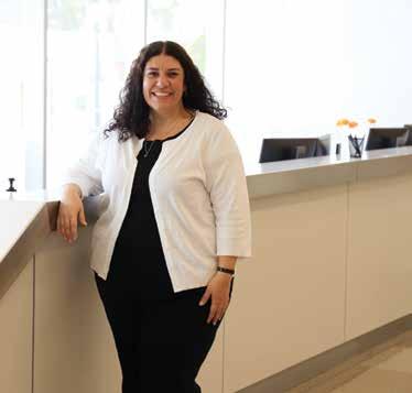
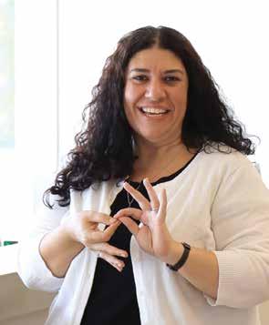
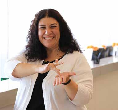
INTERPRETER BRANDI EVANS DEMONSTRATES THE AMERICAN SIGN LANGUAGE SIGNS FOR “INTERPRETER” (CENTER) AND “MEDICINE” (RIGHT).
The second quality is trust. An interpreter has to trust that the patient will receive exceptional care from every team member, starting at the front desk and moving on to the physicians, nurses, medical assistants, financial counselors and social workers.
Brandi: In ASL, as with any language, there are not always direct translations of words and phrases. An ASL interpreter needs to have an excellent grasp of the English language. Otherwise, it’s impossible to interpret information accurately into the target language.
Patience is another important quality. A good interpreter should take the time required to accurately communicate a deaf person’s concerns to hearing doctors and nurses, and then use the best ASL interpretation to accurately convey the medical professionals’ responses.
Photos by Werremeyer Creative
Q: Can each of you give me an example of an incident that brought home to you the importance of what you do?
Christopher: As a second-generation Taiwanese American, I grew up watching my highly educated parents, as well as friends and colleagues, try to navigate the American health-care system. I completed master’s degrees in social work and public health so that I could focus my career on reducing health disparities. Language interpretation is vital to reaching that goal.
Brandi: For a number of years, I’ve worked with a deaf person who initially had limited sign vocabulary and whose family didn’t use ASL. I needed to meet this person where he was, letting go of any expectation that he had the same language usage I and his providers have. But over time and working with an ASL colleague, I’ve helped this person understand enough about sign language that he can tell us when he is hurting or how he is feeling. He couldn’t have done that 10 years ago.
Pilar: My father died of cancer, and I was diagnosed with breast cancer in 2013. Through these experiences I have a deeper understanding of and compassion for people with cancer who need my services. I’ve been present when patients ring the bell upon completing their last cancer treatment, which is a special privilege because I’ve been part of their journey. At those moments, I know patients have received the care they deserve and achieved their best outcomes. They are ringing the bell of victory. Y
Find this story and others online: barnesjewish.org/curiosus
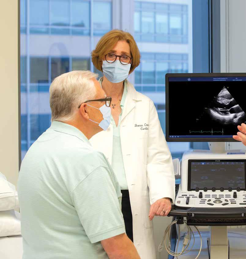
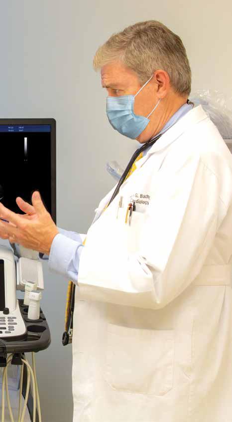
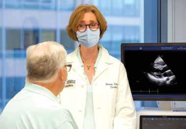
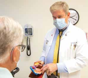
CARDIOLOGISTS SHARON CRESCI, MD, (LEFT) AND RICHARD BACH, MD, FACC, (RIGHT) DISCUSS
TREATMENT OPTIONS WITH A PATIENT.
An estimated 1 million people in the U.S. have hypertrophic cardiomyopathy—a heart condition also known as HCM—and many don’t know they have the condition until it’s too late. A genetic disease, HCM often goes undetected until someone in the family experiences symptoms, which can range from irregular heart rhythms and stroke to heart failure and death in people of any age. And HCM is the most common cause of sudden cardiac death in young people.
New drug, positive results for hypertrophic cardiomyopathy
BY STEPHANIE STEMMLER
PHOTOS BY GREGG GOLDMAN
in depth | new drug, positive results for hypertrophic cardiomyopathy
“It’s an autosomal dominant disorder,” says Washington University cardiologist Sharon Cresci, MD.
“That means if just one parent has an abnormal genetic variant linked to HCM, there’s a 50-50 chance that the variant will be passed down to a child or be found in a parent or sibling.” Cresci has special expertise in genetics and advanced imaging for HCM and is the associate director of the Washington University Hypertrophic Cardiomyopathy Center.
HCM is characterized by a thickening, or hypertrophy, of the heart muscle; over time, HCM can cause the heart to stiffen. It also can cause cellular changes that affect the heart’s electrical signals, which may lead to dangerous heart arrhythmias. In approximately 70% of people with HCM, the thickened heart muscle causes an obstruction of blood flow as it exits the heart, a phenomenon known as left ventricular outflow tract obstruction. This condition is called obstructive HCM.
Treatment options for HCM and obstructive HCM include medication, surgery and—for some—heart transplantation. Additionally, some people treated at the center may have the option to participate in clinical trials focused on evaluating investigational drugs and other treatments. One such trial has resulted in a newly approved drug for obstructive HCM that offers positive results.
New treatment for HCM
Washington University cardiologist Richard Bach, MD, FACC, and his team had exhausted non-invasive treatments for a patient suffering from HCM. Surgery seemed to be the only remaining option. Until, that is, Bach enrolled the man, who was in his 50s, in a clinical trial evaluating a drug called mavacamten. “He felt dramatically better after taking the drug. And three years later,” Bach says, “he continues to feel well, which has postponed or eliminated his need for surgery or other interventions.”
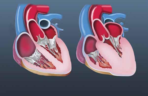
ILLUSTRATED CROSS SECTIONS OF THE HEART SHOW A NORMAL HEART (LEFT)
HEART WITH MUSCLE ENLARGEMENT CAUSED BY HCM (RIGHT).
Image courtesy of Science Photo Library / Catherine Twomey
THIS IS A ONCE-A-DAY PILL. THE KEY, HOWEVER, IS THAT PATIENTS WHO ARE PRESCRIBED THIS DRUG WILL NEED TO BE CLOSELY SUPERVISED AT AN EXPERIENCED CENTER.
— RICHARD BACH, MD, FACC, CARDIOLOGIST
No longer an investigational drug, mavacamten—also known by its brand name, Camzyos—was approved by the U.S. Food & Drug Administration in 2022 for treatment of obstructive HCM. This approval came after a landmark, multicenter clinical trial found it was an effective treatment option for obstructive HCM. Washington University physicians at Barnes-Jewish Hospital were the first in the region to offer mavacamten to qualified patients.
“We were among the 68 medical centers worldwide that participated in clinical trials evaluating the drug,” says Bach, who served as principal investigator of the study conducted at Washington University. “As a result, we now have the experience to identify and treat patients who may benefit from this new medication.”
Mavacamten, taken orally, reduces the obstruction in the heart caused by HCM. In the international drug trial for this medication, more than half of the patients taking the drug showed complete relief from outflow tract obstruction. In a second multicenter study, the majority of patients who were being considered for a surgical intervention but first were treated with mavacamten no longer required the invasive procedure.
Targeting the problem
Inside the heart, two proteins called myosin and actin work in tandem to control heart-muscle contractions, allowing the heart to pump blood throughout the body. “Myosin acts like a lever, and actin is like a filament or string,” explains Bach.
“Myosin grabs the actin and pushes it back and forth, causing the heart cells to contract.
People with HCM have an over-abundance of active myosin, which causes the heart to have excessive or “hyperdynamic” contraction. That contraction, coupled with a thickened heart muscle in the outflow tract, contributes to blockage of blood flow. As a result, excessive pressure builds within the heart. Mavacamten specifically targets myosin, inhibiting that protein so the heart can relax; obstruction and pressure are reduced.
“This is a once-a-day pill,” says Bach. “The key, however, is that patients who are prescribed this drug will need to be closely supervised at an experienced center.”
The FDA, in fact, mandates close supervision. Because mavacamten relaxes heart muscle, it can, in rare cases, inhibit the heart’s pumping function. Given this risk, the FDA approved the drug through its Risk Evaluation and Mitigation Strategy (REMS) program. The REMS qualification means the FDA requires that health-care providers undergo training and certification before they can prescribe the drug—and must monitor its effects. It also requires that patients receive initial and regular follow-up echocardiograms to monitor dosage and heart function.
“An echocardiogram helps us determine the correct dosage because it measures the gradient of extra pressure across the left ventricular outflow tract.
WE
CAN IDENTIFY WHETHER OTHER FAMILY MEMBERS CARRY A DISEASE-CAUSING GENETIC VARIANT OF HCM BEFORE THE DISEASE DEVELOPS.
—
SHARON CRESCI, MD, CARDIOLOGIST
“That gradient needs to be visualized carefully and interpreted accurately. It requires clinical expertise to properly acquire and interpret the echocardiogram,” Cresci says.
Bach adds: “This new drug gives us an important new treatment option for patients who have failed more traditional medications, such as beta blockers or calcium channel blockers.”
More treatment options
In addition to mavacamten, the Hypertrophic Cardiomyopathy Center offers people with HCM a range of medical, interventional and surgical options.
Surgery to treat obstructive HCM works by thinning the thickened heart muscle. This treatment, first available in the 1960s, has been the standard of care for more than 20 years. Washington University cardiothoracic surgeons Ralph Damiano Jr., MD, and Kunal Kotkar, MD, specialize in this surgical procedure, called septal myectomy. A newer, less invasive treatment, called mini-sternotomy, is another option and may offer some people a speedier recovery.
“The incision used in a mini-sternotomy is less than half the length of that required for traditional septal myectomy,” notes Kotkar. “But both procedures can offer good outcomes.”
” “
Kotkar also specializes in a more complex procedure called apical myectomy, available at the Washington University and Barnes-Jewish Heart & Vascular Center and at only a few other centers in the U.S.
“Some patients with HCM have an obstruction inside one of the lower chambers of the heart,” Kotkar says, “so we work inside the ventricle to remove the blockage and create a larger cavity for blood flow.”
A less invasive procedure called alcohol septal ablation is an option for people who can’t have surgery. In alcohol septal ablation, Bach says, “we use a balloon catheter to block the septal artery that moves blood into the area of thickened muscle.”
Once the balloon is in place, alcohol is injected into the area, which destroys the thickened tissue. “As a result, some of the obstruction is relieved almost immediately, and, as the treated area heals, the septum is thinned without requiring open-heart surgery,” Bach adds.
HCM also can cause atrial fibrillation, an abnormal heart rhythm that may lead to heart failure if untreated. In 1987, James Cox, MD, former chief of cardiothoracic surgery at Washington University, and colleagues developed what’s now known as the Cox-Maze procedure. During Cox-Maze surgery, specialists create a series of tiny incisions in the heart that block abnormal electrical signals.
Since then, Damiano has introduced less invasive advances to this procedure that have further improved outcomes for patients. He also performs a combined treatment that includes septal myectomy and a modified Cox-Maze procedure to treat HCM and atrial fibrillation simultaneously.
Not all people with HCM develop obstruction. For people with non-obstructive HCM, treatment includes medication and, for those who aren’t helped by medication, heart transplantation. Physicianresearchers at the Hypertrophic Cardiomyopathy Center are involved in clinical trials evaluating new medications to treat non-obstructive HCM, and they collaborate closely with the Washington University and Barnes-Jewish Transplant Center to care for people needing transplantation.
HCM and genetic testing
An inherited disease, HCM can run in families—and it can develop at any age. In many instances, HCM is undiagnosed until a significant heart problem occurs. When that happens, genetic testing for the patient and the immediate family can help to clarify the cause of symptoms and offer care options for others in the family who may be at risk. Screening often includes an echocardiogram as well.
Cresci says, “We can identify whether other family members carry a disease-causing genetic variant of
HCM before the disease develops.” She notes that the decision to undergo screening should be a shared decision among family members. “We work with the family so they understand the genetic nature of the disease and the possible implications of any test results.”
Currently, Cresci and colleagues are engaged in research to better diagnose and understand HCM. She is the principal investigator of an ongoing Washington University observational study that to date has enrolled more than 1,000 patients and family members. The aim is to better identify markers of the onset, progression and prognosis of HCM, as well as to identify other disease-associated genetic variants.
A brighter future
Bach, who was one of the investigators in the myosin inhibitor clinical trials, has several patients being treated at the Hypertrophic Cardiomyopathy Center who are registered in the FDA’s mavacamten REMS program. He says, “As an interventional cardiologist who sees many HCM patients each year, I’m very happy to have a new oral medication available for treatment. For those who may benefit from this option, it’s a new treatment with great promise to improve not only their symptoms but also their quality of life.” Y
Find this story and others online: barnesjewish.org/curiosus


Most of us have seen the warnings: Every cigarette pack sold in the United States since 1965 carries a label that identifies the health hazards of smoking. Yet despite these mandated labels and a host of public awareness campaigns in print, broadcast and social media that encourage people to stop smoking, the Centers for Disease Control and Prevention, or CDC, notes that in 2020, more than 30 million people in the U.S. smoke cigarettes. And every day, an estimated 1,600 young people light up a cigarette for the first time.
A two-step intervention for lung cancer
BY STEPHANIE STEMMLER ILLUSTRATION BY ABIGAIL GOH

80%-90% of lung cancer deaths are directly linked to smoking. American Cancer Society
The 2020 statistics are significant because they represent the first time in 20 years that annual cigarette sales have gone up, not down. While a direct correlation to the COVID-19 pandemic hasn’t been made, the recent increase in the sale of cigarettes, as well as the popularity of other nicotine products such as e-cigarettes, causes concern among health-care specialists who are on the frontlines trying to lower the incidences of lung cancer.
The correlation between smoking and lung cancer is well documented. What’s worrisome is that lung cancer remains the leading cause of cancer deaths in the U.S. According to the American Cancer Society, more people die of lung cancer each year than the number of people who die from breast, colon and prostate cancers combined. This year alone, it estimates 237,000 new lung cancer diagnoses—and 130,000 deaths.
Here’s the conundrum: While health experts say deaths from lung cancer are mostly preventable if people would stop smoking, that’s a complicated proposition. The nicotine in cigarettes is highly addictive. Quitting is really hard to do. And there’s where researchers are focusing their efforts.
Easier said than done
“For years, we’ve had it backwards,” says Li-Shiun Chen, MD, MPH, ScD, psychiatrist at Siteman Cancer Center at Barnes-Jewish Hospital and Washington University School of Medicine. “We were treating the consequences of lung cancer and not addressing the major cause, which is smoking. But cancer-care strategies must include steps that can mitigate or even prevent cancer from occurring. We need to get in front of this disease.”
Chen, a nationally recognized researcher on smoking-cessation strategies, says there’s a “huge misalignment” in what doctors believe and what patients really want. “Many doctors think that patients don’t really want to quit smoking, but the reality is that nearly 70% of adults who smoke say they want to quit,” she says. “Another 20% want to reduce the number of cigarettes they use.”
The discrepancy Chen points out was vividly apparent during a recent focus group, during which she, colleagues and patient participants searched for ways to engage people in smoking-cessation programs. “We had a patient in one of our groups tell us: ‘Everyone asks me about smoking, but no one offers anything to help me quit,’” recalls Chen. “Clearly, it’s not enough for a doctor or nurse to tell a patient that they should quit, then make a checkmark on a medical-history form. We need to offer proven smoking-cessation treatment options, just like we do with cancer or any life-shortening condition.”
In 2017, as part of the National Cancer Institute’s (NCI) Cancer Moonshot program, the Cancer Center Cessation Initiative was established to help cancer centers across the country conduct research, and create and implement tobaccocessation treatment programs that were proven to work. Toward that end, in a clinical research study, Chen and psychiatrist Laura Bierut, MD; implementation scientist Alex Ramsey, PhD; medical oncologist Ramaswamy Govindan, MD; and other Washington University researchers at The Alvin J. Siteman Cancer Center at Barnes-Jewish Hospital and Washington University School of Medicine
Cigarette
smoking is the No. 1 risk factor for lung cancer.
and Prevention
used electronic medical records (EMRs) to identify patients with cancer who were smokers. Then, whenever one of those patients came in for a clinic visit, they were offered immediate help to quit smoking. The smoking-cessation options offered included:
> Immediate referral for free, text-based counseling from the NCI
> Onsite help to download free phone apps to access the NCI’s QuitGuide and quitSTART smoking-cessation platforms
> FDA-approved medications to stop or reduce smoking
> Referral to a free smoking-cessation program
In short, people were offered several point-of-care treatment programs that were easily accessible. The result? The number of patients receiving smokingcessation treatment increased from 2% at the start of the program to nearly 30%. Overall, about one-third of patients participating in the program stopped smoking. The findings were published in the May 2022 issue of the Journal of the National Comprehensive Cancer Network.
Of significance, the researchers also found that those who quit smoking after a cancer diagnosis doubled their survival rate and lowered their risk of recurrence, no matter the severity of the cancer.
The early success of Siteman’s point-of-care tobacco treatment program has prompted health-care leaders to deploy the program at many other hospitals
within BJC HealthCare, the network to which Barnes-Jewish Hospital belongs. Those working within the program will share their successes with other academic health centers nationwide, and they will work to reach people treated in specialty clinics, primary care practices and other health-care settings.
“We have to be non-judgmental during these conversations at the point of care,” notes Chen. “Smoking is not a weakness, and it’s not a person’s fault if they can’t stop smoking on their own. Cigarette-smoking is an addiction. We need to address it, and treat it with compassion.”
Screening for lung cancer
In addition to its focus on effective smoking-cessation programs, Siteman Cancer Center offers screenings for lung cancer. Lung cancer can go undetected until it reaches an advanced stage; screening can reveal a problem before symptoms are noticed. And early detection is one of the keys to better outcomes.
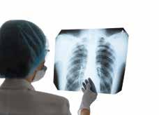
In 2011, results from the National Lung Screening Trial—to date the largest clinical trial of its kind in the U.S.—found that low-dose CT scans were effective at identifying small, early-stage lung tumors. That study led the National Comprehensive Cancer Network to issue new guidelines that encourage screening for those at risk of developing lung cancer because they are 50 years old or older and have a smoking history of a 20-pack year or greater. (See the sidebar on Page 38 for a definition of a 20-pack year and for information about screening eligibility.)
Centers for Disease Control
Photo courtesy of Shutterstock
30 million people in the U.S. smoke cigarettes. Centers for Disease Control and Prevention Image courtesy of Shutterstock
David Gierada, MD, Washington University radiologist at Siteman Cancer Center, served as a principal investigator for the National Lung Screening Trial . He notes: “It’s too early to tell what the significant impact of low-dose CT screening has been because it didn’t become recommended clinical practice until 2015. Though the proportion of eligible persons who get screened has been increasing, it’s still very low.”
Early findings from the trial, however, suggest that a CT scan can reduce mortality from lung cancer by 20%, simply because it can identify the disease at an early stage. Anne Stilinovic, RN, BSN, supervisor of the Lung Cancer Screening Program at Siteman Cancer Center, is one of the center’s three nurse navigators who guide people through the screening program and offer referrals to thoracic surgeons and pulmonologists, if needed. In 2021, the program
screened 2,880 people for lung cancer. Of those who were diagnosed with cancer, 70% were found to be in stage 1—the earliest stage of the disease.
To be most effective, lung cancer screening should be done annually for at least three years. “The test by itself takes one to two minutes,” says Stilinovic. “It’s non-invasive: no needles, no contrast dye, no prep work. Radiologists can detect lung nodules as small as 1 mm in size with a CT scan.”
The challenge, she says, is that 60% to 75% of people who undergo screening will have a lung nodule detected. For the person screened, that can mean a lot of anxiety and worry: Is the nodule benign or cancerous? Says Stilinovic: “Of 100 persons screened for the first time, about 15 have a false alarm. Lung nodules are common as people age; even those who have never
LUNG CANCER SCREENING: WHO’S ELIGIBLE?
Most private insurance companies and Medicare will cover the cost of lung cancer screenings in high-risk patients with a long history of smoking.
Early in 2022, Medicare expanded the eligibility age to get more people screened.
You qualify for a screening if you have a referral from your primary care physician and if you:
> are between the ages of 50 and 80 (Medicare covers age 50-77) AND
> have a smoking history of 20 pack-years* or more, OR
> are a former heavy smoker who quit less than 15 years ago
*To determine a 20-pack year: Multiply the average number of packs smoked per day times the years smoked:
> 1 pack per day for 20 years = a 20-pack year
> ¾ pack per day for 30 years = a 20-pack year
> ½ pack per day for 40 years = a 20-pack year

According to the American Cancer Society, this is what happens after someone quits smoking:
20 minutes after quitting
Heart rate and blood pressure drop.
A few days after quitting
Carbon monoxide level in the blood drops to normal.
2 weeks to 3 months after quitting
Circulation improves and lung function increases.
1 to 12 months after quitting
Coughing and shortness of breath decrease.
Tiny hair-like structures (called cilia) that move mucus out of the lungs start to regain normal function, increasing their ability to handle mucus, clean the lungs and reduce the risk of infection.
1 to 2 years after quitting
Risk of heart attack drops dramatically.
5 to 10 years after quitting
Risk of cancers of the mouth, throat and larynx is cut in half. Risk of stroke decreases.
10 years after quitting
Risk of lung cancer is about half that of a non-smoker (after 10 to 15 years). The risk of cancer of the bladder, esophagus, and kidney decreases.
15 years after quitting
Risk of coronary heart disease is close to that of a non-smoker.
smoked can develop them.” But, she adds, “a nodule also can be an early cancer. If something suspicious is detected, we make a referral for additional testing.”
For those requiring a follow-up visit with a surgeon, the recommendation may be watchful waiting followed by another appointment in six months. In some cases, a PET scan or tissue biopsy might be recommended.
It’s important to note that not all lung cancers are detected with low-dose CT screening, but it currently is the only screening tool shown to lower the risk of death from the disease. The screening may also identify coronary artery calcifications and other conditions in the neck or upper abdomen, which would prompt further testing.
Ongoing research is focused on identifying barriers that might prevent people from getting screened, as well as on opportunities to increase the number of people who choose screening. Other investigations are focused on developing better guidelines for
managing the different types of lung nodules found during screening. Washington University researchers at Siteman also are participating in a clinical trial called I-STEP—Increasing Screening through Engaging Primary Care Providers—aimed at improving awareness among primary care physicians of the benefits of screening.
Says Gierada: “The death rate from lung cancer has been decreasing for more than 20 years, even before the advent of screenings, and it parallels an earlier drop in smoking rates. Therefore, efforts to promote quitting and keep people from starting to smoke have paid off. I think expanding the screening eligibility criteria, broadening physician and public awareness and increasing access and availability of smoking cessation programs are critical to lowering the mortality rate even further.” Y
Find this story and others online: barnesjewish.org/curiosus
Photo courtesy of Shutterstock
Spina bifida is a condition that affects the spine during the growth and development of a fetus. As the brain and spinal column begin to form, a portion of the spine doesn’t close completely. The resulting opening in the spine leaves the spinal cord and nerves exposed. In the most severe form of spina bifida, called myelomeningocele, a portion of the spinal cord and its nerves protrude from this opening.
Spina bifida: making repairs before birth
BY STEPHANIE STEMMLER

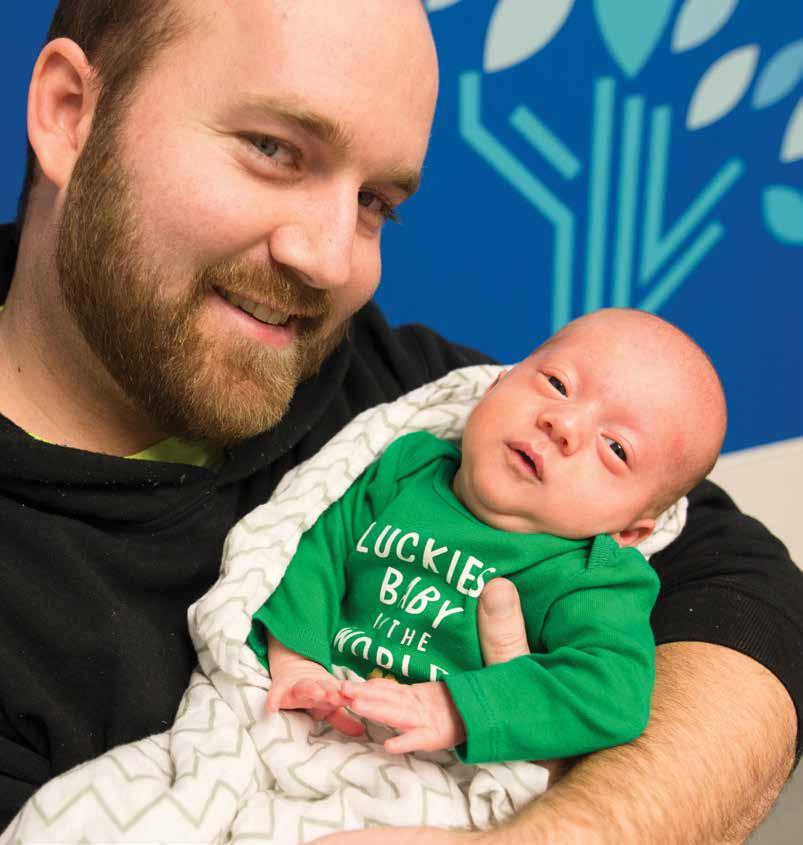
Photo by Gara Elizabeth Photography
JACKSON AND HIS DAD, CHRIS. JACKSON HAD PRENATAL SURGERY FOR SPINA BIFIDA AT THE FETAL CARE CENTER.
“
WHEN
WE USE PRENATAL SURGERY TO TREAT SEVERE SPINA BIFIDA, WE ARE WORKING TO CARE FOR THE UNBORN BABY WHILE PROTECTING THE HEALTH OF THE MOTHER.
— ANTHONY
ODIBO, MD, MATERNAL-FETAL MEDICINE SPECIALIST
”
As the fetus grows in the uterus, these fragile nerves are exposed to surrounding amniotic fluid. And that exposure can cause damage, leading to paralysis or other mobility problems after birth, as well as bladder and bowel complications. The opening’s location can determine the kinds of complications that arise; for those located higher on the spine, hydrocephalus, a build-up of excess fluid in the brain, is a possibility.
Before the 1990s, treatment for severe spina bifida involved postnatal surgery—a procedure performed on a newborn after delivery. Though it closed the spinal cord to help prevent infection—and could treat hydrocephalus with a shunt to drain fluid—the procedure could not repair already damaged nerves. Consequently, children born with spina bifida faced a number of complications as they grew.
A leap forward in treatment
In 1997, the first prenatal surgery to repair spina bifida complications—done while the fetus was in the uterus—took place at Vanderbilt University Medical Center. Similar surgeries were subsequently performed at a few, select medical institutions in the U.S.
In 2011, results of a major study sponsored by the National Institutes of Health (NIH) found that
prenatal repair of severe spina bifida resulted in better outcomes than surgery after birth. Specifically, the study found that prenatal surgery not only improved the chances that a child could walk independently, but it also showed that the need for a shunt to divert excess fluid from the brain was reduced by 50% when compared with those children who underwent surgery after birth. A follow-up study, published in 2020, noted the long-term physical and emotional advantages of prenatal surgery for the condition.
A delicate dance
“Severe spina bifida occurs in about one in 1,000 pregnancies,” says Anthony Odibo, MD, Washington University maternal-fetal medicine specialist at the Women & Infants Center at Barnes-Jewish Hospital, Washington University Physicians and St. Louis Children’s Hospital. “When we use prenatal surgery to treat the condition, we are working to care for the unborn baby while protecting the health of the mother.” Odibo also is interim co-director of the Fetal Care Center, part of the Women & Infants Center.
Traditionally, prenatal surgery for spina bifida is what’s known as an “open” procedure. At the Fetal Care Center, Washington University pediatric surgeon Jesse Vrecenak, MD, and Washington
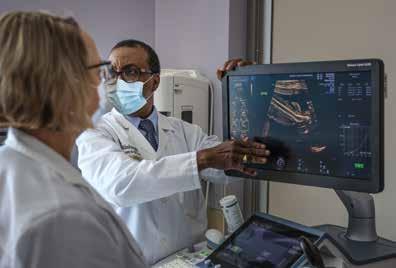
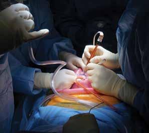
University pediatric neurosurgeon Jennifer Strahle, MD, work together to repair spina bifida complications. Vrecenak describes the procedure this way: “In an open surgery, we make a large incision across the mother’s abdomen, much like we do with a cesarean section. We then open the uterus, turn the baby so that the back is facing upward and lift the uterus partially out of the abdominal cavity. Dr. Strahle then places the protruding sac containing spinal cord and nerves into the spinal canal and closes the surrounding tissue and skin over the opening. Once that part of the procedure is completed, we then close the incisions in the mom’s uterus and abdomen.”
The procedure is much like a choreographed ballet performed inside an operating room, each specialist’s role carefully considered. The Fetal Care Center uses a dedicated operating room for fetal surgeries that is large enough to accommodate an anesthesia team for mom and fetus, a fetal cardiologist, pediatric surgeons, a pediatric neurosurgeon, maternalfetal medicine specialists and multiple nursing professionals. “We use a team of this size to ensure that we provide the highest level of care for both patients during a complex procedure that requires surgical, neurosurgical and newborn-medicine expertise,” notes Strahle.
She adds: “If we can surgically close the opening in the spine and put the spinal cord and nerves in their proper place before a baby is born, we can prevent or minimize the progressive nerve damage that would occur if we waited until after delivery.”
Open surgery for spina bifida is usually performed between 24 and 26 weeks of pregnancy. After discharge from the hospital several days later, the pregnant patient must limit activity to bed rest, which helps deter the risks of premature birth and uterine rupture. She also must reside with a support person and remain within 30 minutes of the hospital. “The benefit of open fetal surgery is proven in high-caliber studies funded by the NIH,” says Odibo. After such a procedure, however, Odibo notes, “the patient will need to deliver the baby and all future babies by cesarean section.”
The Fetal Care Center offers moms and babies excellent outcomes after open surgical repair. Preterm delivery rates and incidences of fluid leakage are lower than those reported in the national clinical trial. “We’re proud of the expertise we can offer our patients,” says Odibo.
ANTHONY ODIBO, MD, AND A SONOGRAPHER VIEW A SCAN OF A FETUS.
Photo by Werremeyer Creative
Photo by Tim Mudrovic
A FETAL CARE CENTER TEAM PERFORMS OPEN SURGERY TO REPAIR SPINA BIFIDA.
in depth | spina bifida: making repairs before birth
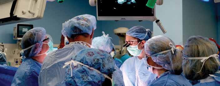
Further refinements
Surgeons at the Fetal Care Center now are able to perform a less invasive prenatal procedure for spina bifida. Called fetoscopic repair, it differs from open surgery in the number of incisions required and it offers additional benefits.
During fetoscopic surgery, a surgeon makes an incision in the abdomen similar to the one used in open surgery. But instead of using another large incision to open the uterus, two smaller incisions are made; they allow surgeons to insert specialized instruments inside the uterus. These tools provide
The Fetal Care Center uses prenatal surgery to treat the following conditions:
> Airway/trachea obstruction
> Amniotic band release
> Blocked urinary tract
> Lung lesions
> Lymphatic malformation
> Myelomeningocele (severe spina bifida)
> Neonatal tumors
> Pleural effusion (fluid in the lungs)
> Twin-to-twin transfusion syndrome
The center offers integrated care for both mother and baby in a single location. All birthing suites and mother-baby rooms are connected to a Level IV neonatal intensive care unit.
ultrasound images to the team of surgeons, offering precision guidance.
“Fetoscopic surgery allows some patients to preserve their ability to deliver vaginally,” says Vrecenak. “It also gives them the option of returning home for the duration of their pregnancy after surgery.”
Specialists at the Fetal Care Center work with families to determine the best surgical repair option. “We offer both open and fetoscopic,” Vrecenak says. Strahle notes: “Not every baby is a candidate for fetal surgery. We discuss all available options with our patients.”
After delivery, babies that have had prenatal surgery are cared for in the St. Louis Children’s Hospital newborn intensive care unit. Though there is no known cure for spina bifida, the team at the Fetal Care Center notes that various treatment options may prevent significant problems and can help manage the effects of the disease.
“Fetal surgery offers a once-in-a-lifetime opportunity to prevent some complications rather than simply treat the disease,” notes Vrecenak. “If we can prevent nerve damage while a baby is still in the womb, outcomes are better when that child is born.” Y
Find this story and others online: barnesjewish.org/curiosus
JESSE VRECENAK, MD, NEAR RIGHT, AND A FETAL CARE CENTER SURGERY TEAM
Photo by Timothy Mudrovic
One
In This Issue
RECURRENT UTIS AND THE GUT MICROBIOME
COVID-19 VACCINE BOOSTERS AND SOLID ORGAN TRANSPLANTS
RISKY DRIVING BEHAVIORS AND COMMON SLEEP DISORDERS
STUDY LOOKS AT DUAL NICOTINE USE
FIERCE ADVOCACY: DENTAL CARE FOR THE UNDERSERVED
A HISTORY OF HEART FAILURE— AND RECOVERY
BRIDGING COMMUNICATION AND CULTURAL GAPS
NEW DRUG, POSITIVE RESULTS FOR HYPERTROPHIC CARDIOMYOPATHY
A TWO-STEP INTERVENTION FOR LUNG CANCER
SPINA BIFIDA: MAKING REPAIRS BEFORE BIRTH
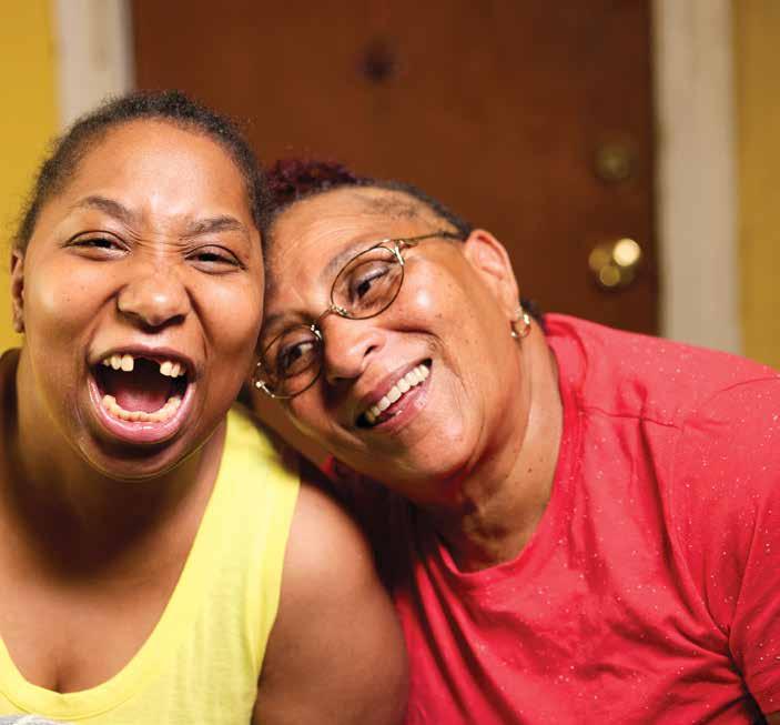
by
Photo
Matt Miller